Embed Size (px)
Citation preview
This is a 47-year-old male who has a symptomatic medial femoral condyle cartilage defect in the
setting of varus alignment.
This gentleman is a very active patient who plays multiple club sports including, soccer, lacrosse,
and downhill skiing. His symptomatic pain and swelling limits his activities, especially a half mile
into a run and 10-15 min into a club soccer match.
His examination demonstrated full knee range of motion, but a mild to moderate effusion. There
was slight inflammation and warmth to the joint, but no signs of infection. All of the ligaments
were stable including the ACL, PCL, MCL, LCL. His standing alignment demonstrated no varus
thrust. He had slight varus, left greater than right, alignment.
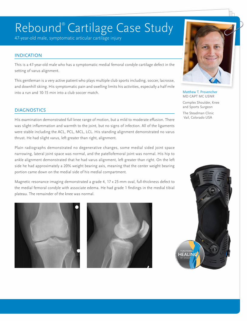
Plain radiographs demonstrated no degenerative changes, some medial sided joint space
narrowing, lateral joint space was normal, and the patellofemoral joint was normal. His hip to
ankle alignment demonstrated that he had varus alignment, left greater than right. On the left
side he had approximately a 20% weight bearing axis, meaning that the center weight bearing
portion came down on the medial side of his medial compartment.
Magnetic resonance imaging demonstrated a grade 4, 17 x 25-mm oval, full-thickness defect to
the medial femoral condyle with associate edema. He had grade 1 findings in the medial tibial
plateau. The remainder of the knee was normal.
INDICATION
DIAGNOSTICS
Rebound® Cartilage Case Study47-year-old male, symptomatic articular cartilage injury
Matthew T. Provencher MD CAPT MC USNR
Complex Shoulder, Knee and Sports Surgeon
The Steadman Clinic Vail, Colorado USA

The overall goal of the treatment was to restore the patient’s
function to a high level, to initiate early knee range motion, and
to protect his cartilage repair as well as the osteotomy. The high
tibial osteotomy and medial femoral condyle osteochondral
allograft, when done together, provide additional cartilage
protective effect. However, the post-operative bracing is
important to reinforce this effect.
The patient initially started with the Rebound® Cartilage Brace
as he had varus alignment in the setting of a symptomatic
medial femoral condyle defect. This provided him significant
relief and stated that his knee was approximately 70% better
while wearing the brace, however he desired additional stability,
as he was still able to feel crepitus inside of his knee.
The goal of the surgical treatment was to restore alignment,
as well as the cartilage, and this treatment was augmented by
also adequately protecting the construct post-operatively. Given
that the patient had an excellent response to Rebound Cartilage
bracing pre-operatively, it was felt that a combination high
tibial osteotomy, as well as fresh osteochondral allograft to the
medial femoral condyle would be the most favorable treatment
option for returning to a high level of activity.
TREATMENT OVERVIEW / TREATMENT GOAL
946.4 mm det. 953.0 mm det.

The patient initiated physical therapy on postoperative day one.
He started with early flexion, as well as continuous passive
motion given his cartilage procedure. In order to protect his
medial side, we reinstituted the use of his Rebound® Cartilage
Brace and unloaded the medial side. With proper fit he was able
to use this brace not only with the continuous passive motion
machine, but also with touch-down weight bearing for the first
6 weeks. After 6 weeks, the patient still maintained Rebound
Cartilage Brace use and increased to full motion, as he started
strengthening and full weight bearing. After 12 weeks, the patient
demonstrated continued incorporation of the allograft, as well
as healing of the high tibial osteotomy. By 4 months the patient
was released to full light jogging and light impact activities. The
patient was instructed to wear the Rebound Cartilage Brace
during all of his activities. At 5.5 months, he was released to full
activities, again using the Rebound Cartilage Brace.
The surgical procedure consisted of a high tibial osteotomy, as
well as a fresh osteochondral allograft to the medial femoral
condyle. This was a oval defect and a workstation and oblong
cutter were used to fashion the graft from an allograft donor
condyle according to the dimensions of the recipient defect.
The high tibial osteotomy was completed with an 12.5-mm
trapeziodal plate, which was placed in order to preserve the slope
of the knee and overall kinematics. The high tibial osteotomy
was bone grafted with a combination of osteoinductive donated
bone putty, as well as allograft cancellous bone chips.
After the high tibial osteotomy, a medial femoral condyle fresh
osteochondral allograft was performed using a 17 x 20 x 7-mm
single oval graft utilizing a press-fit technique. Since a press fit
technique was used, we wanted to make sure to protect this
adequately post-operative. The patient was placed in a range of
motion brace at the conclusion of the procedure.
SURGICAL TREATMENT
POST-SURGICAL REHABILITATION

The use of the Rebound Cartilage brace after OCA/OATS and a high tibial
osteotomy supports regeneration of the knee cartilage by maintaining joint
unloading in flexion for protection of the cartilage during the healing process.
It also unloads the affected knee compartment to facilitate osteotomy healing.
In other words, the load on the affected knee compartment is mediated by
distracting the involved compartment via external valgus force applied to the
knee. Thus, tibiofemoral alignment and kinematics are improved, and load is
shifted off the reconstructed compartment. Too much load may have a negative
effect on the repair tissue development, which has been shown to impede healing
after surgery. Cartilage, particularly, is the slowest tissue to repair and use of early
post-operative application of the Rebound Cartilage Brace facilitates an early
improvement of knee motion.
Overall the patient had an excellent outcome. He had full restoration of knee
motion. The patient had near resolution of his knee symptoms and rates his
knee, on a scale of 0 to 100, a 95, a significant improvement from his baseline
rating of 40. He was able to return to full activities at 5.5 months including, all
farming/ranching, impact activities, club soccer, and downhill skiing.
Patients with symptomatic knee articular cartilage injuries can be restored back to high levels of activity with modern anatomic-
based osteochondral graft reconstructions. It is important address the overall alignment, and given the patient’s varus knee at 20%
of overall alignment, the use of a high tibial osteotomy greatly facilitates the overall outcome. Finally, it is extremely important to
protect the knee post-operatively and unload the knee further to allow for full cartilage, as well as high tibial osteotomy, integration.
The Rebound Cartilage Brace is an ideal solution for this post-operative case.
REASONS TO USE THE REBOUND® CARTILAGE BRACE
CLINICAL OUTCOME
CONCLUSION
© ÖSSUR, 01.2018
USA (800) 233-6263WWW.OSSUR.COM
CANADA (800) 663-5982 WWW.OSSUR.CA
FOLLOW ÖSSUR ON