Embed Size (px)
Citation preview
Reanimating the Dead: Reconstruction of Expressive Faces from Skull Data
Kolja Kahler Jorg Haber Hans-Peter Seidel
MPI Informatik,Saarbrucken,Germany
(Siggraph2003)

Introduction
Introduction
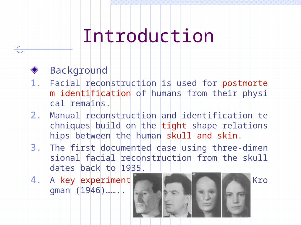
Background1. Facial reconstruction is used for postmortem identific
ation of humans from their physical remains.2. Manual reconstruction and identification techniques
build on the tight shape relationships between the human skull and skin.
3. The first documented case using three-dimensional facial reconstruction from the skull dates back to 1935.
4. A key experiment was later performed by Krogman (1946)……..

IntroductionThe Manual Reconstruction Process
The actual face reconstruction proceeds with one of two available approaches: the anatomical method and the tissue depth method.
The anatomical method attempts reconstruction by sculpting muscles, glands, and cartilage, fleshing out the skull layer by layer. This technique is more often used in the reconstruction of fossil faces, where no statistical population data exists.
Which is very time consuming, occupying “many hundreds of hours”. It also requires a great deal of detailed anatomical knowledge.
Introduction
The Manual Reconstruction ProcessTherefore, the tissue depth method has become th
e more popular reconstruction technique.Standard sets of statistical tissue thickness measur
ements at specific points on the face are used. Each measurement describes the total distance from skin surface to the skull, including fat and muscle layers.
The method is thus more rapid than the anatomical method and does not require as much anatomical knowledge.
Preparation of the Skull
Preparation of the SkullOur approach uses three-dimensional skull data acquired, for instance, from volume scans and extraction of the bone layers, or by range scanning a physical skull.To speed up processing, a triangle mesh of the skull model comprised of 50-250k polygons is produced by mesh decimation techniques.In general, the original data should be simplified as little as possible since minute details on the skull can give important clues for the reconstruction.

Preparation of the SkullIn the editor, the skull model is equipped with landmarks, which can then be moved around on the surface for fine positioning.Each landmark is associated with a vector in surface normal direction, corresponding to the typical direction of thickness measurements.The landmark vector is scaled to the local tissue thickness.
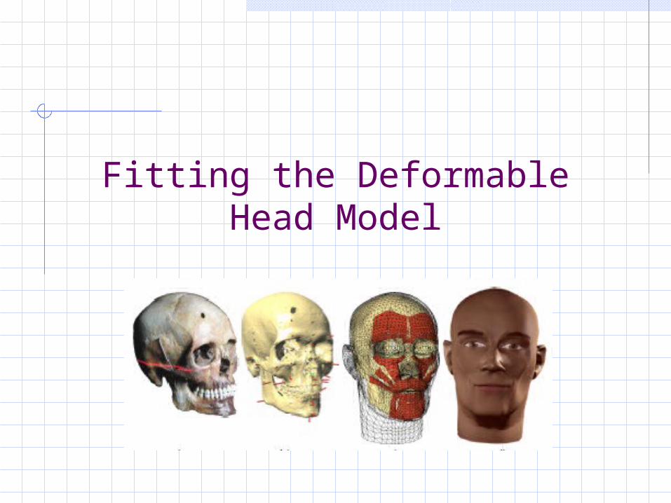
Fitting the Deformable Head Model
Head Model Structure
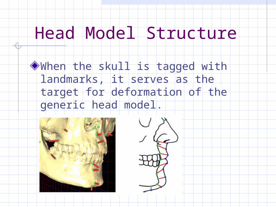
When the skull is tagged with landmarks, it serves as the target for deformation of the generic head model.
Head Model StructureSince the head model is used in a physics-based animation system, it does not only consist of the visible outer geometry. The encapsulated structure includes:
1. The skin surfaces: Our template head mesh consists of 8164 triangles.
2. Virtual muscles: To control the animation. Each muscle is specified by a grid laid out on the skin, the actual muscle shape being computed automatically to fit underneath the skin surface. Each muscle consists of an array of fibers, which can contract in a linear or circular fashion. Our model includes 24 facial muscles responsible for facial expressions.
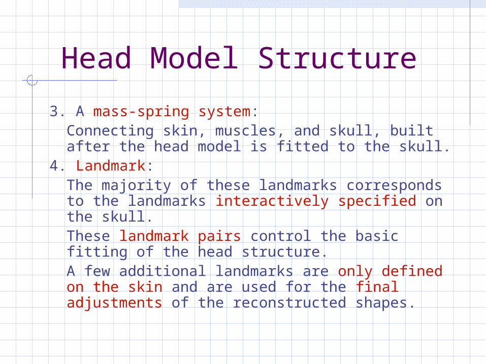
Head Model Structure3. A mass-spring system:
Connecting skin, muscles, and skull, built after the head model is fitted to the skull.
4. Landmark: The majority of these landmarks
corresponds to the landmarks interactively specified on the skull.
These landmark pairs control the basic fitting of the head structure.
A few additional landmarks are only defined on the skin and are used for the final adjustments of the reconstructed shapes.

Landmark-based RBF Deformation
pi: skin landmark, si: skull landmark, di: tissue depth vector qi: corresponding skin landmark
The problem can be treated as :we need to find function f that maps the pi to the qi
Landmark-based RBF Deformation
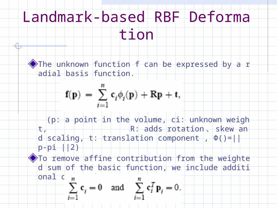
The unknown function f can be expressed by a radial basis function.
(p: a point in the volume, ci: unknown weight, R: adds rotation 、 skew and scaling, t: translation component , Ф()=|| p-pi ||2)To remove affine contribution from the weighted sum of the basic function, we include additional constraints:
Landmark-based RBF Deformation
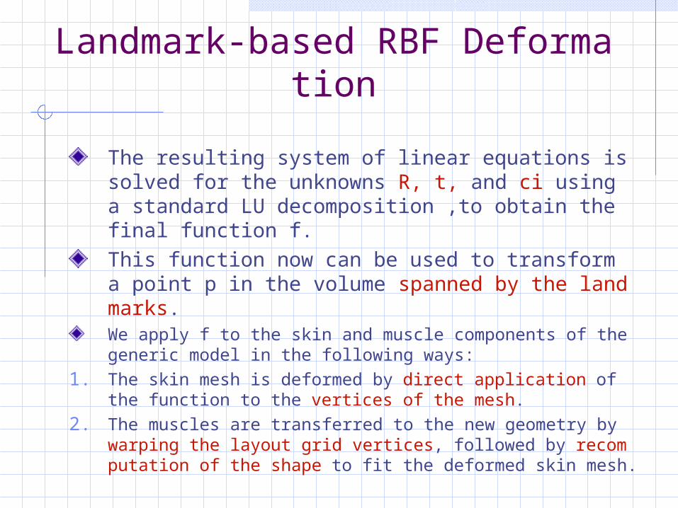
The resulting system of linear equations is solved for the unknowns R, t, and ci using a standard LU decomposition ,to obtain the final function f.This function now can be used to transform a point p in the volume spanned by the landmarks.We apply f to the skin and muscle components of the generic model in the following ways:
1. The skin mesh is deformed by direct application of the function to the vertices of the mesh.
2. The muscles are transferred to the new geometry by warping the layout grid vertices, followed by recomputation of the shape to fit the deformed skin mesh.

Additional Reconstruction Hints
Additional Reconstruction Hints
The tissue depth values at the marker positions define the basic shape of the reconstructed head, assuming depth measurements being always strictly orthogonal to the skull surface. As mentioned in ,but this assumption is not always valid.A number of rules are thus used in traditional facial reconstruction to help locate certain features of the face based on the skull shape, employing empirical knowledge about shape relations between skin and skull.
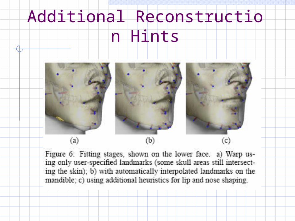
Additional Reconstruction Hints
Additional Reconstruction HintsTo keep the user interface uniform, most rules are expressed by the placement of vertical and horizontal guides in a frontal view of the skull. From this user input, the placement of a few landmarks on the skin is adjusted, resulting in a new target landmark configuration. The updated landmark set is used to compute another warp function, which deforms the pre-fitted head model in the adjusted regions.

Additional Reconstruction Hints
Additional Reconstruction HintsFive rules influence the shape of the nose and the shape of the mouth
1. The width of the nose wings corresponds to the width of the nasal aperture at its widest point, plus 5mm on either side in Caucasoids. In the editor, the user places two vertical guides to the left and right of the nasal aperture.
2. The position of the nose tip depends on the shape of the anterior nasal spine.
3. The width of the mouth is determined by measuring the front six teeth, placing the mouth angles horizontally at the junction between the canine and the first premolar in a frontal view.
Additional Reconstruction Hints
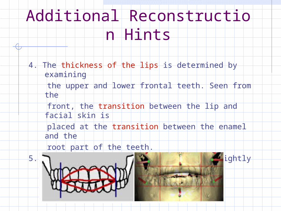
4. The thickness of the lips is determined by examining
the upper and lower frontal teeth. Seen from the front, the transition between the lip and facial skin
is
placed at the transition between the enamel and the
root part of the teeth.
5. The parting line between the lips is slightly above the
blades of the incisors.
Facial Expressions and Rendering
Facial Expressions and Rendering
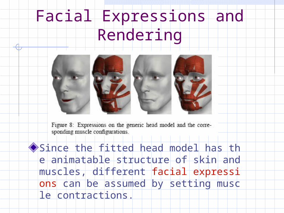
Since the fitted head model has the animatable structure of skin and muscles, different facial expressions can be assumed by setting muscle contractions.
Facial Expressions and Rendering
For a completely animatable head model, it is necessary to include a separately controllable mandible, a tongue, rotatable eyeballs, and eye lids into the head model. We have decidedly left them out of the reconstruction approach since these features are not particularly useful in this application: while a modest change of expression such as a smile or a frown might aid identification, rolling of eyes, blinking, and talking would probably not.

Results
Results
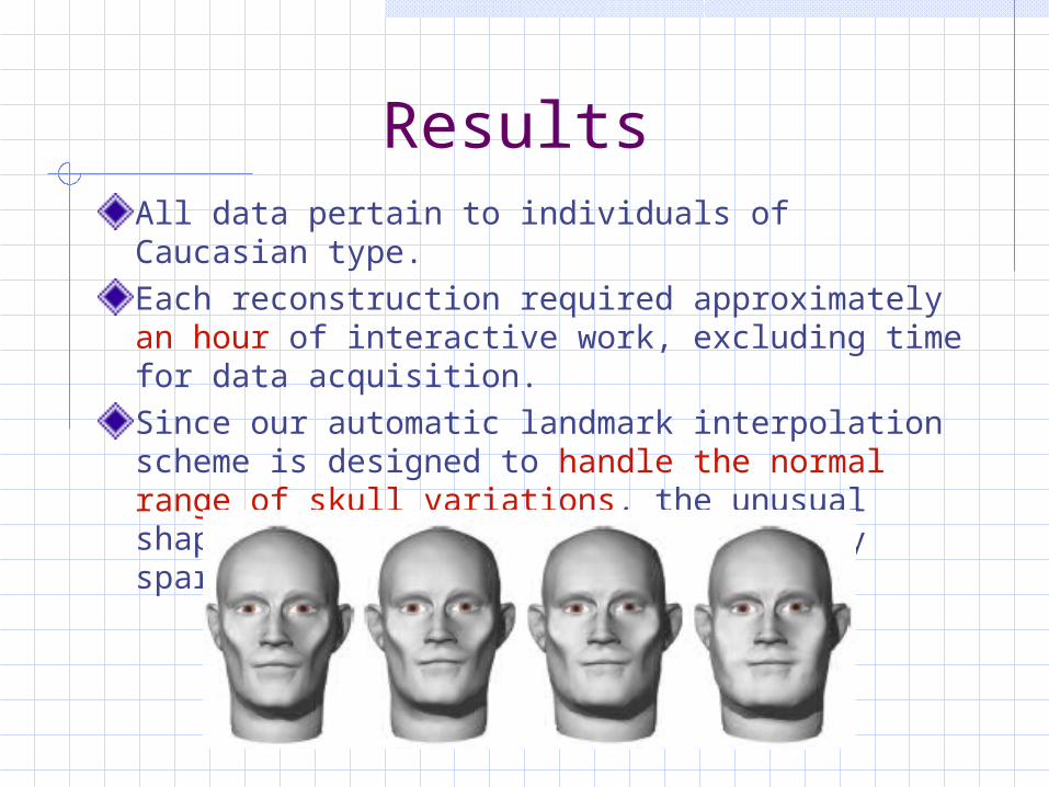
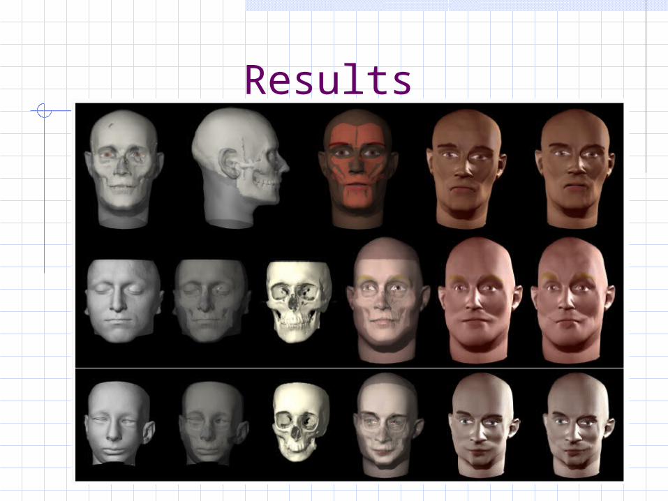
ResultsAll data pertain to individuals of Caucasian type.Each reconstruction required approximately an hour of interactive work, excluding time for data acquisition.Since our automatic landmark interpolation scheme is designed to handle the normal range of skull variations, the unusual shape of the mandible resulted in very sparse sampling of the chin area.

Conclusion and Future Work
Conclusion and Future Work
The face reconstruction approach presented in this paper mirrors the manual tissue depth method and thus has essentially the same prediction power. Our results show overall good reproduction of facial shape and proportions, and some surprisingly well-matched details. It should be noted that our examples were produced by computer scientists with no training in forensic reconstruction.The advantages of the computerized solution are evident: instead of weeks, it takes less than a day to create a reconstructed face model, including scanning of the skull.
Conclusion and Future WorkSince the virtual reconstruction is based on 3D scans, which can be acquired contact-free, the risk of damage to the original skull is reduced.On the other hand, the scanning process has inherent limitations: depending on the maximum resolution of the digital scanner, much of the finer detail on the skull is lost.The interactive system allows for an iterative reconstruction approach: a model is produced quickly from a given landmark configuration, so landmarks can be edited repeatedly until the desired result is obtained.
報告完畢


![COUNTER MEMORIALS Hoheisel [Berlin-Kassel-Buchenwald] Ullman [Berlin] Eisenman/Happold [Berlin] Gerz [Harburg/Saarbrucken]](https://img.pdfslide.us/doc/110x75/56649f135503460f94c27537/counter-memorials-hoheisel-berlin-kassel-buchenwald-ullman-berlin-eisenmanhappold.jpg)