Embed Size (px)
Citation preview
North Carolina Science Olympiad2010-2011
Rashmi Chandra, PhD
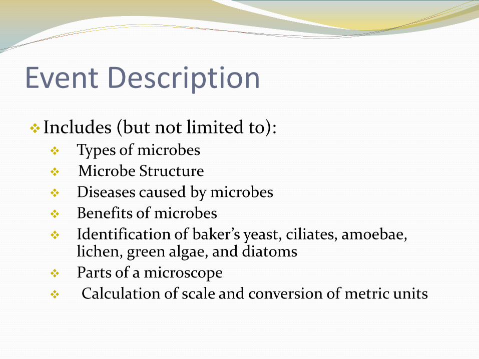
Event DescriptionIncludes (but not limited to): Types of microbes Microbe Structure Diseases caused by microbes Benefits of microbes Identification of baker’s yeast, ciliates, amoebae,
lichen, green algae, and diatoms Parts of a microscope Calculation of scale and conversion of metric units

Rules
Team of 2 students Things to Bring:One 8.5” X 11” sheet of two-sided notesNon-programmable calculator Z87 chemical splash goggles Pencil/Pen/Eraser/Ruler

Competition
Stations or written test Time 50 minutes Similar topics for Division B and C, but level of test
for Division B will be at the Middle school level.
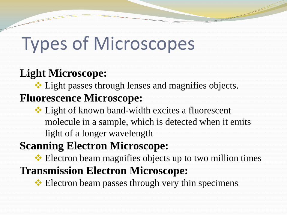
Types of MicroscopesLight Microscope: Light passes through lenses and magnifies objects.
Fluorescence Microscope: Light of known band-width excites a fluorescent
molecule in a sample, which is detected when it emits light of a longer wavelength
Scanning Electron Microscope: Electron beam magnifies objects up to two million times
Transmission Electron Microscope: Electron beam passes through very thin specimens
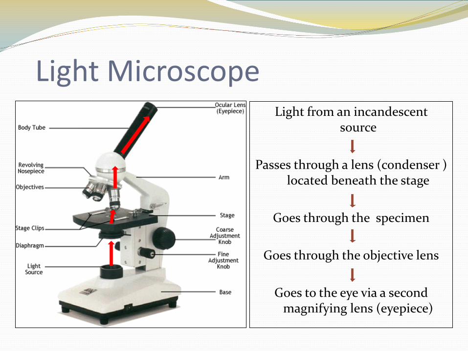
Light MicroscopeLight from an incandescent
source
Passes through a lens (condenser ) located beneath the stage
Goes through the specimen
Goes through the objective lens
Goes to the eye via a second magnifying lens (eyepiece)
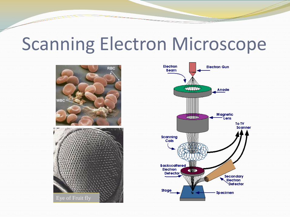
Scanning Electron Microscope
Eye of Fruit fly
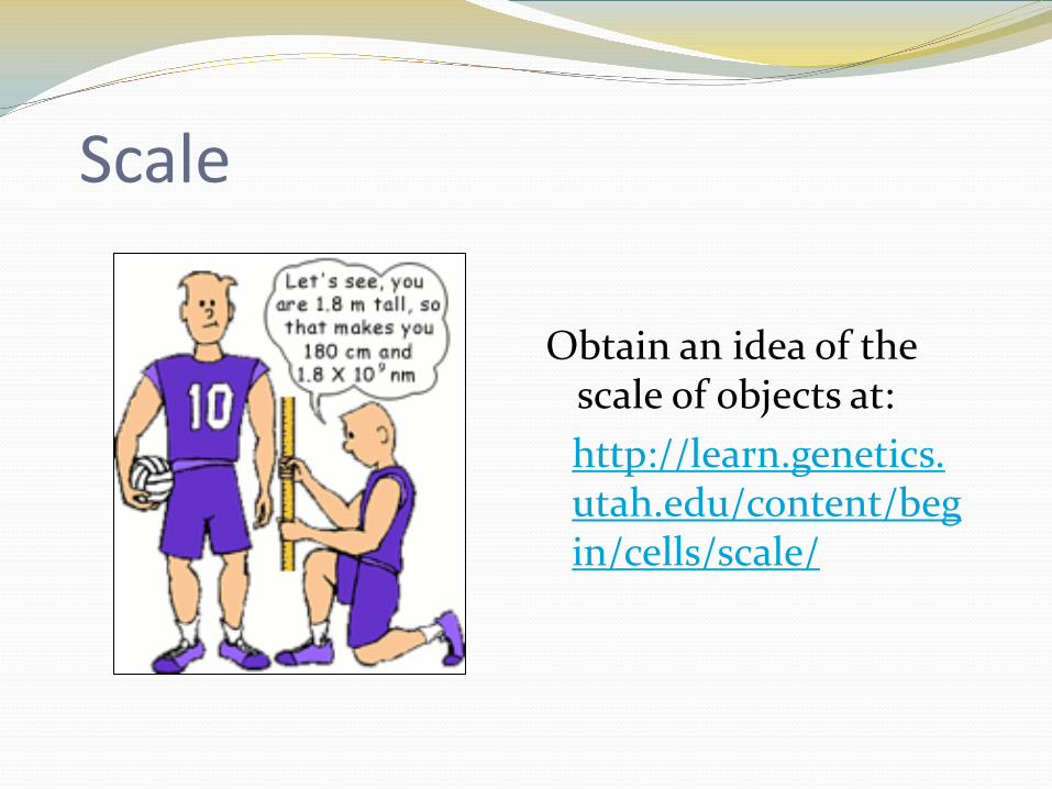
Scale
Obtain an idea of the scale of objects at:http://learn.genetics.utah.edu/content/begin/cells/scale/

Metric Conversion
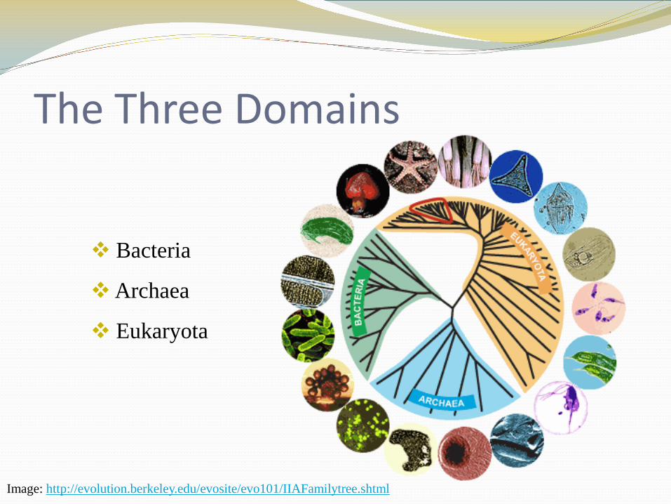
The Three Domains
Bacteria
Archaea
Eukaryota
Image: http://evolution.berkeley.edu/evosite/evo101/IIAFamilytree.shtml
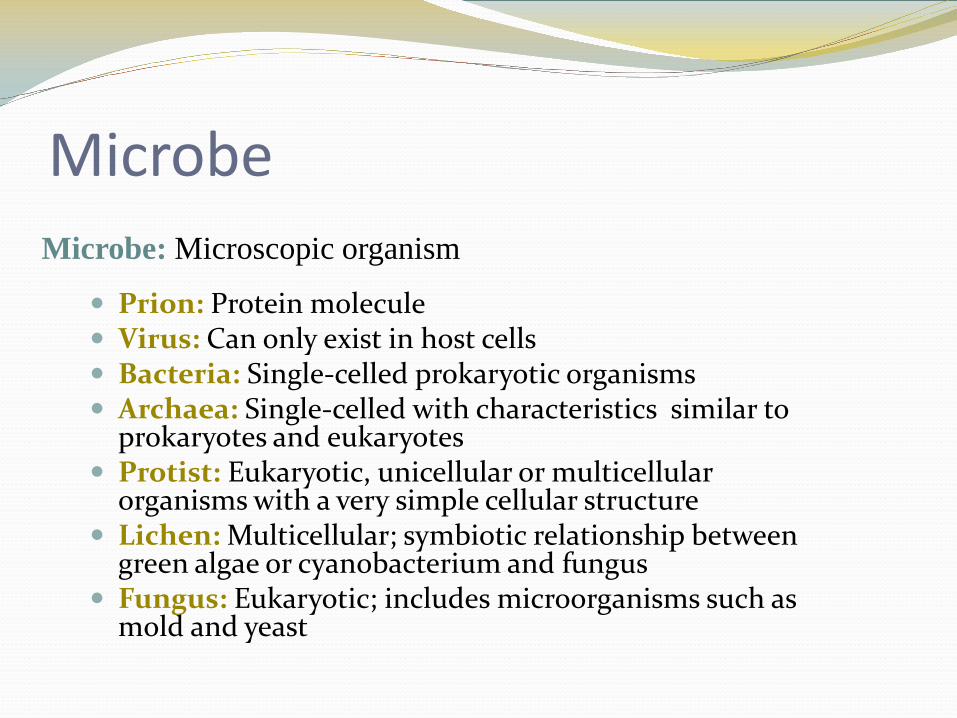
Microbe
Prion: Protein molecule Virus: Can only exist in host cells Bacteria: Single-celled prokaryotic organisms Archaea: Single-celled with characteristics similar to
prokaryotes and eukaryotes Protist: Eukaryotic, unicellular or multicellular
organisms with a very simple cellular structure Lichen: Multicellular; symbiotic relationship between
green algae or cyanobacterium and fungus Fungus: Eukaryotic; includes microorganisms such as
mold and yeast
Microbe: Microscopic organism
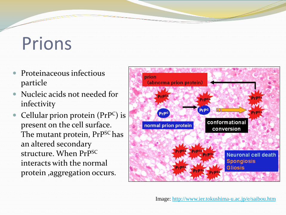
Prions Proteinaceous infectious
particle Nucleic acids not needed for
infectivity Cellular prion protein (PrPC) is
present on the cell surface. The mutant protein, PrPSC has an altered secondary structure. When PrPSC
interacts with the normal protein ,aggregation occurs.
Image: http://www.ier.tokushima-u.ac.jp/e/saibou.htm
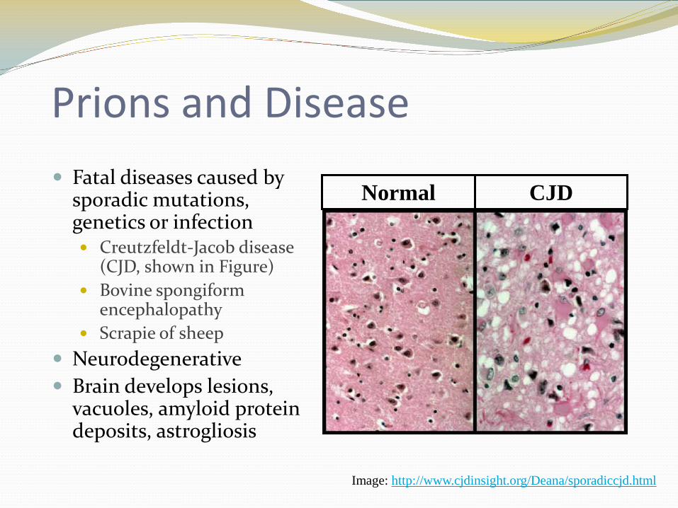
Prions and Disease Fatal diseases caused by
sporadic mutations, genetics or infection Creutzfeldt-Jacob disease
(CJD, shown in Figure) Bovine spongiform
encephalopathy Scrapie of sheep
Neurodegenerative Brain develops lesions,
vacuoles, amyloid protein deposits, astrogliosis
Image: http://www.cjdinsight.org/Deana/sporadiccjd.html
Normal CJD
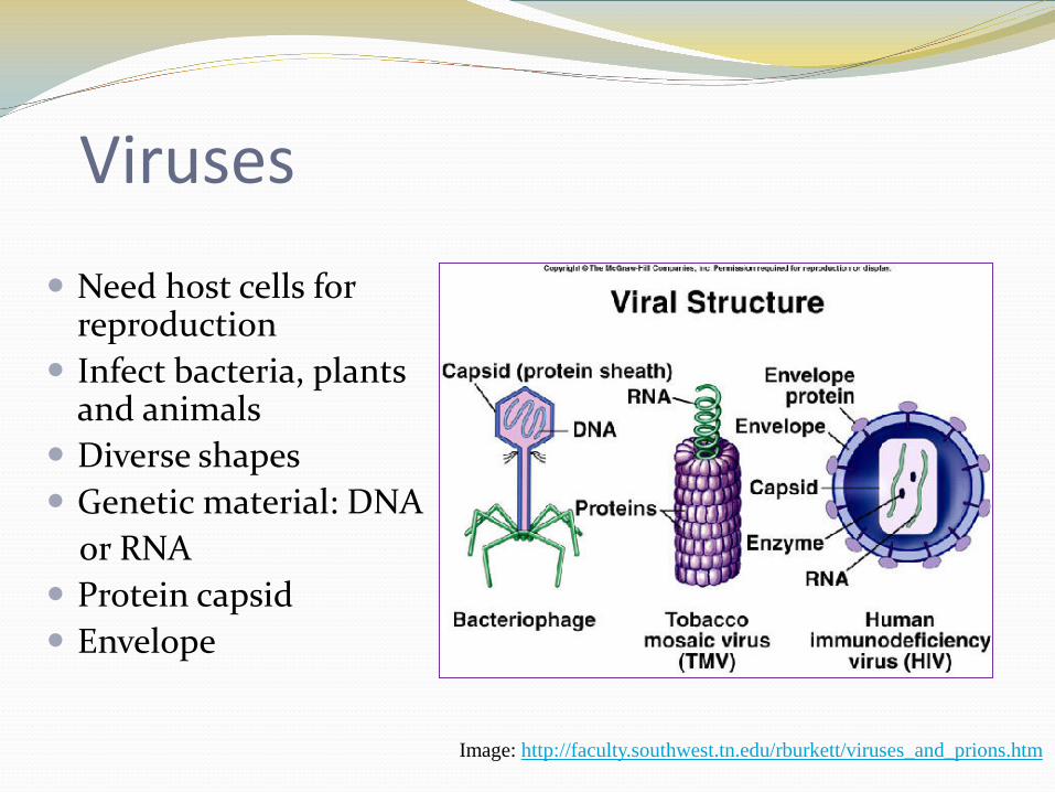
Viruses
Need host cells for reproduction
Infect bacteria, plants and animals
Diverse shapes Genetic material: DNA
or RNA Protein capsid Envelope
Image: http://faculty.southwest.tn.edu/rburkett/viruses_and_prions.htm
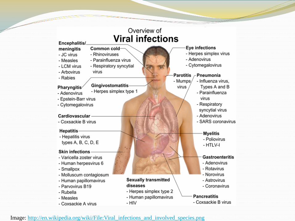
Image: http://en.wikipedia.org/wiki/File:Viral_infections_and_involved_species.png
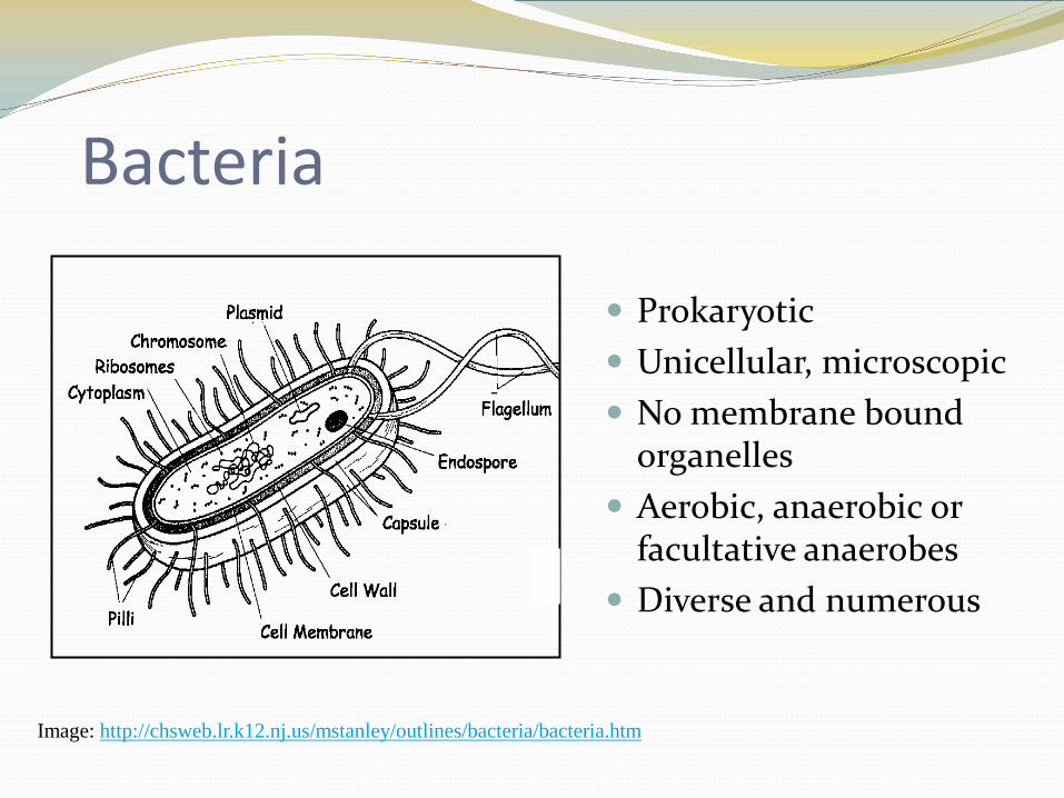
Bacteria
Prokaryotic Unicellular, microscopic No membrane bound
organelles Aerobic, anaerobic or
facultative anaerobes Diverse and numerous
Image: http://chsweb.lr.k12.nj.us/mstanley/outlines/bacteria/bacteria.htm
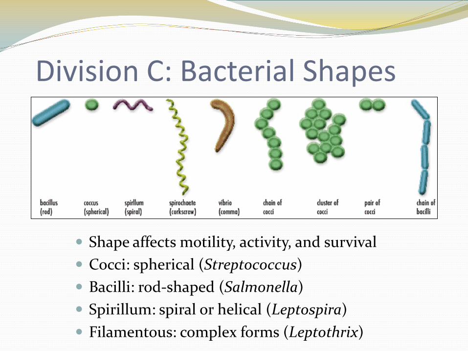
Division C: Bacterial Shapes
Shape affects motility, activity, and survival Cocci: spherical (Streptococcus) Bacilli: rod-shaped (Salmonella) Spirillum: spiral or helical (Leptospira) Filamentous: complex forms (Leptothrix)
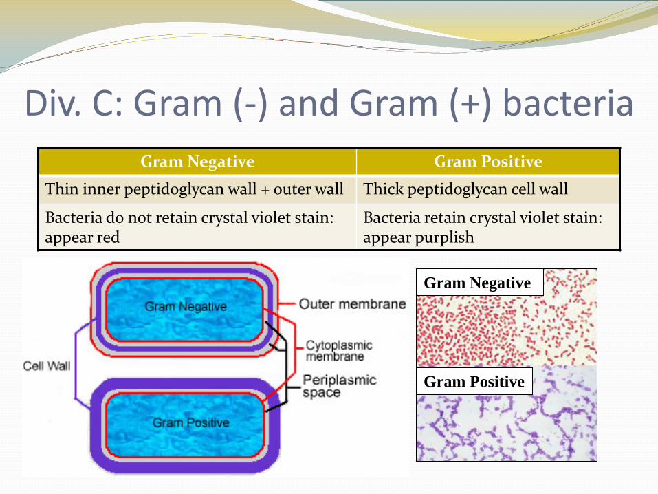
Div. C: Gram (-) and Gram (+) bacteria
Gram Positive
Gram Negative
Gram Negative Gram Positive
Thin inner peptidoglycan wall + outer wall Thick peptidoglycan cell wall
Bacteria do not retain crystal violet stain: appear red
Bacteria retain crystal violet stain:appear purplish
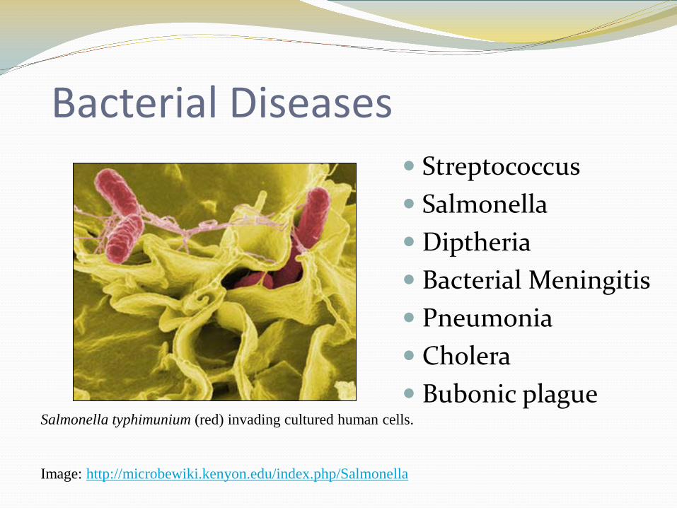
Bacterial Diseases Streptococcus Salmonella Diptheria Bacterial Meningitis Pneumonia Cholera Bubonic plague
Salmonella typhimunium (red) invading cultured human cells.
Image: http://microbewiki.kenyon.edu/index.php/Salmonella
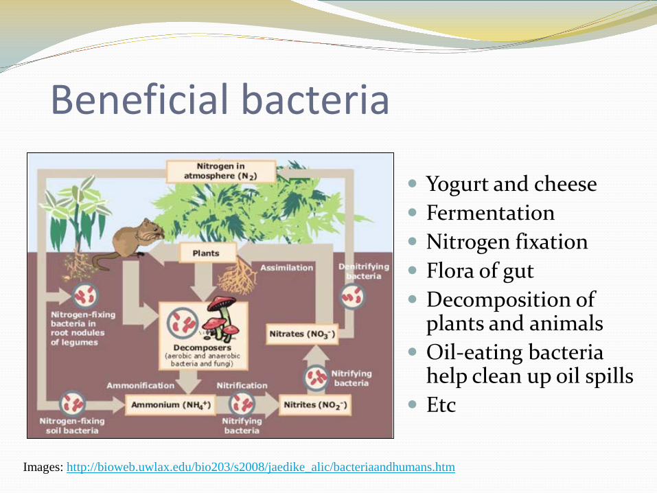
Beneficial bacteria
Yogurt and cheese Fermentation Nitrogen fixation Flora of gut Decomposition of
plants and animals Oil-eating bacteria
help clean up oil spills Etc
Images: http://bioweb.uwlax.edu/bio203/s2008/jaedike_alic/bacteriaandhumans.htm
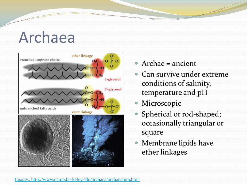
Archaea Archae = ancient Can survive under extreme
conditions of salinity, temperature and pH
Microscopic Spherical or rod-shaped;
occasionally triangular or square
Membrane lipids have ether linkages
Images: http://www.ucmp.berkeley.edu/archaea/archaeamm.html
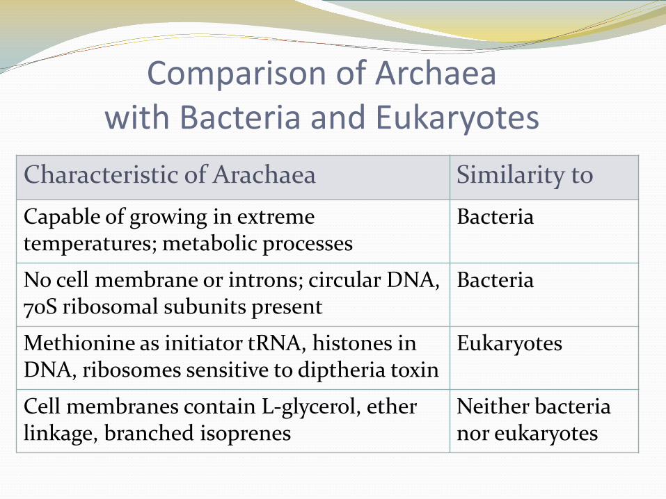
Comparison of Archaea with Bacteria and Eukaryotes
Characteristic of Arachaea Similarity toCapable of growing in extreme temperatures; metabolic processes
Bacteria
No cell membrane or introns; circular DNA, 70S ribosomal subunits present
Bacteria
Methionine as initiator tRNA, histones in DNA, ribosomes sensitive to diptheria toxin
Eukaryotes
Cell membranes contain L-glycerol, ether linkage, branched isoprenes
Neither bacteria nor eukaryotes
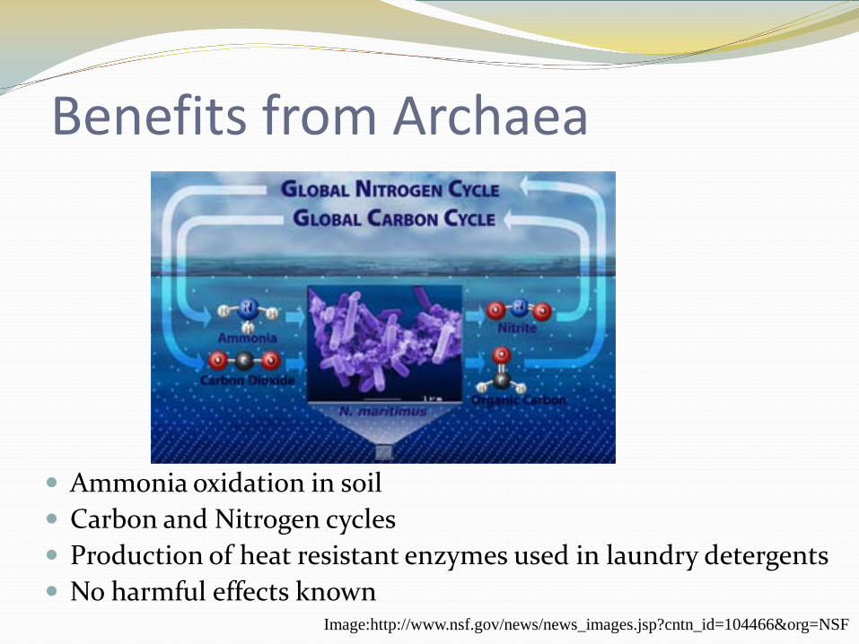
Benefits from Archaea
Ammonia oxidation in soil Carbon and Nitrogen cycles Production of heat resistant enzymes used in laundry detergents No harmful effects known
Image:http://www.nsf.gov/news/news_images.jsp?cntn_id=104466&org=NSF
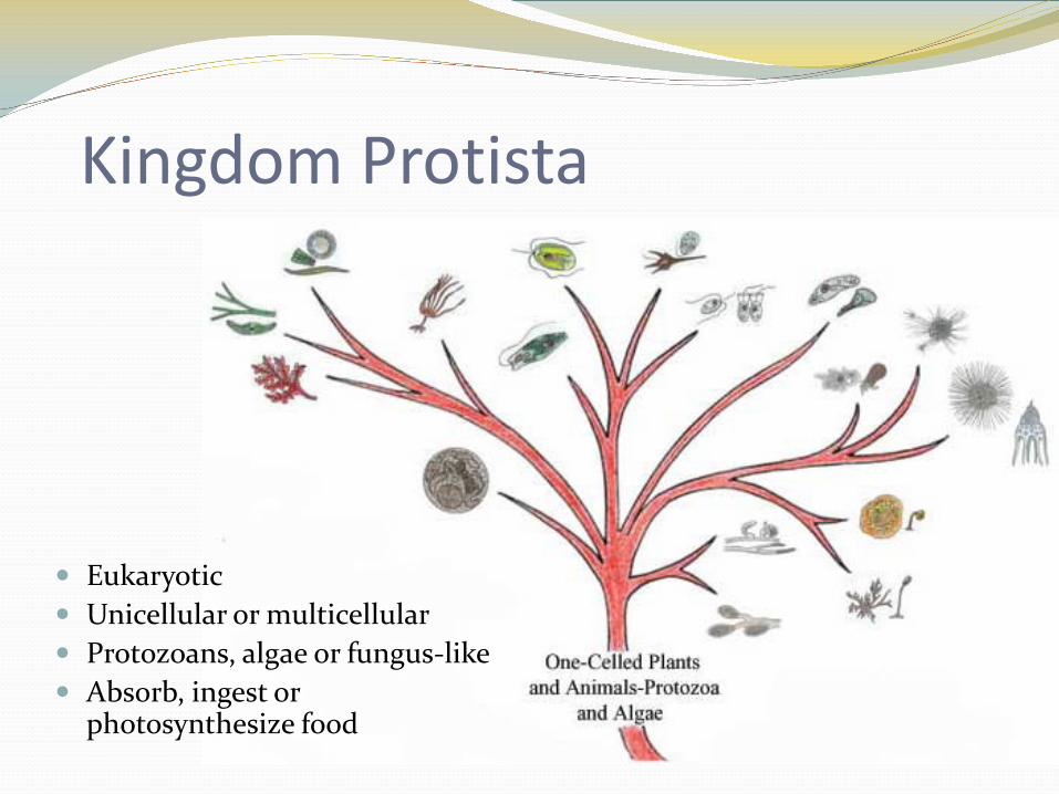
Kingdom Protista
Eukaryotic Unicellular or multicellular Protozoans, algae or fungus-like Absorb, ingest or
photosynthesize food
Protists Autotrophs and heterotrophs
Apicomplexans: Plasmodium Diplomonads: Giardia Gymnamoebas Parabasalids: Trichomonas Kinetoplastids: Trypanosoma Oomycetes: Water mold, white rust, mildew Plasmodial (acellular) Slime Molds Cellular Slime Molds Euglenoids: Euglena Dinoflagellates Ciliates: Paramecium Diatoms Foraminifera Radiolarians
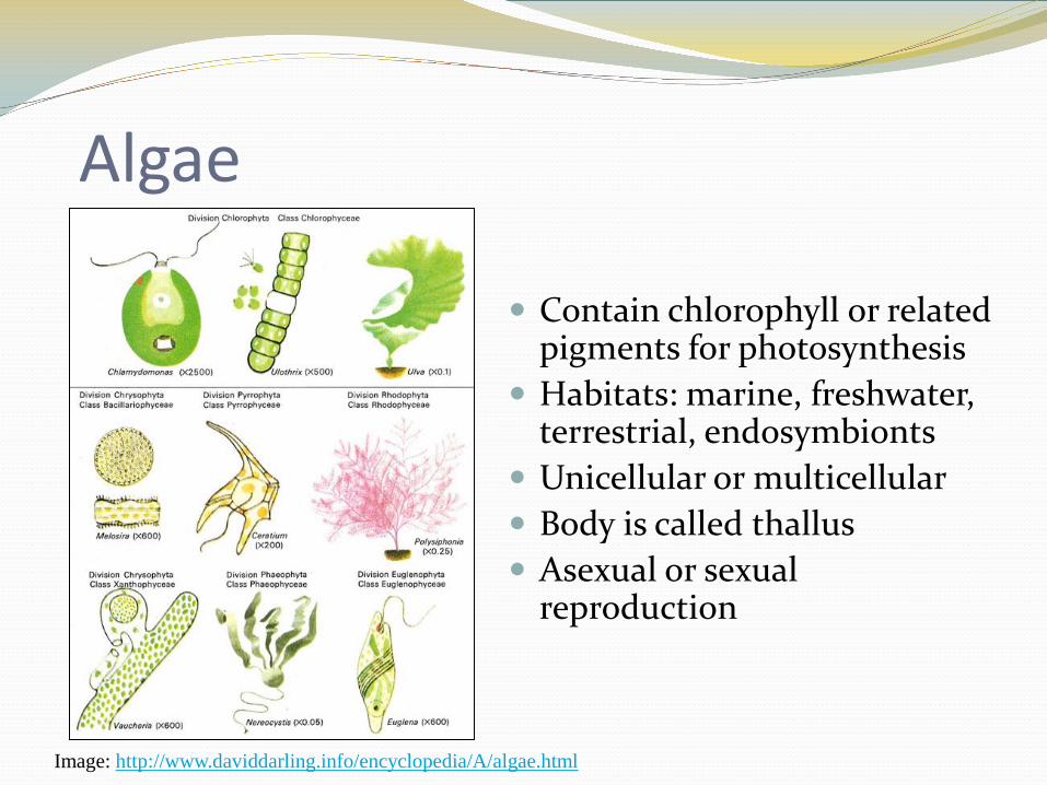
Algae
Contain chlorophyll or related pigments for photosynthesis
Habitats: marine, freshwater, terrestrial, endosymbionts
Unicellular or multicellular Body is called thallus Asexual or sexual
reproduction
Image: http://www.daviddarling.info/encyclopedia/A/algae.html
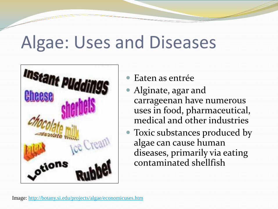
Algae: Uses and Diseases
Eaten as entrée Alginate, agar and
carrageenan have numerous uses in food, pharmaceutical, medical and other industries
Toxic substances produced by algae can cause human diseases, primarily via eating contaminated shellfish
Image: http://botany.si.edu/projects/algae/economicuses.htm
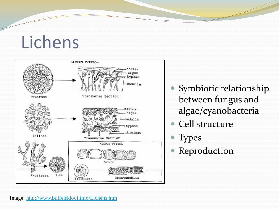
Lichens
Symbiotic relationship between fungus and algae/cyanobacteria
Cell structure Types Reproduction
Image: http://www.buffelskloof.info/Lichens.htm
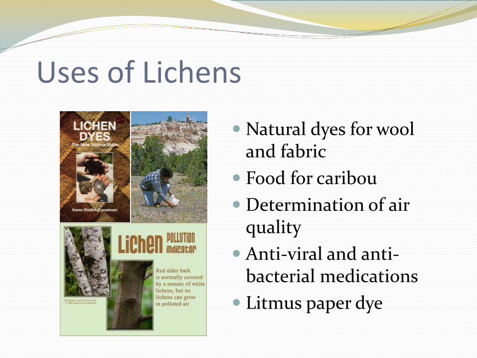
Uses of Lichens
Natural dyes for wool and fabric
Food for caribou Determination of air
quality Anti-viral and anti-
bacterial medications Litmus paper dye
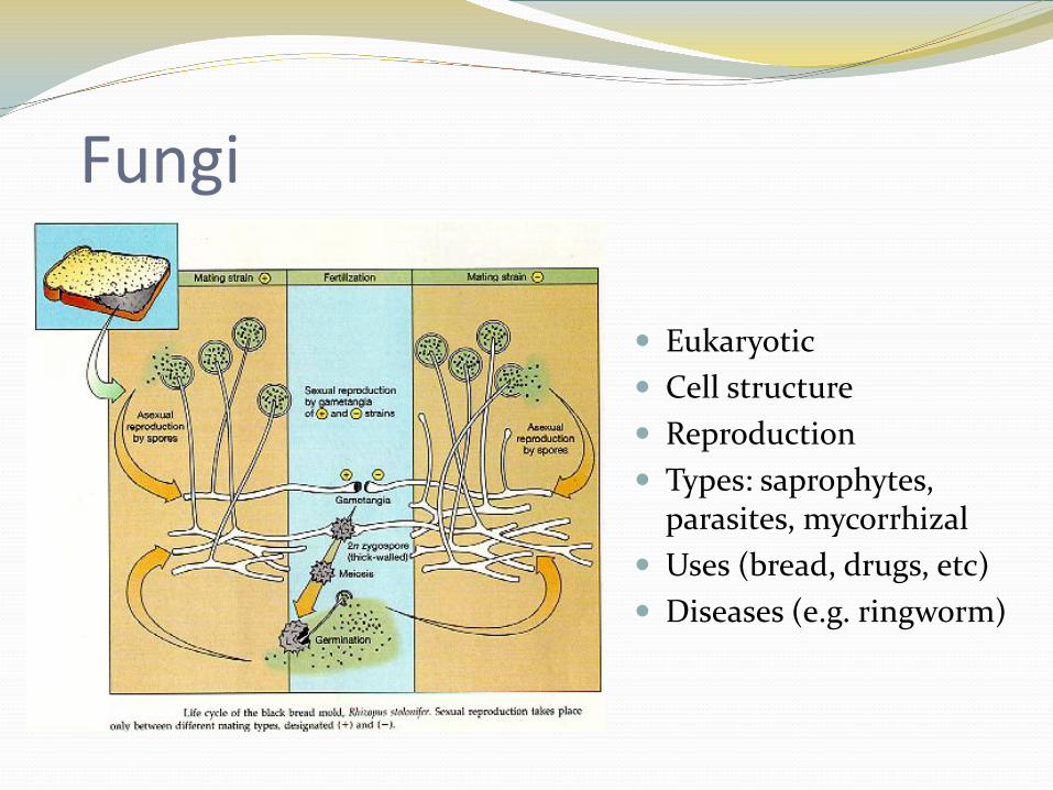
Fungi
Eukaryotic Cell structure Reproduction Types: saprophytes,
parasites, mycorrhizal Uses (bread, drugs, etc) Diseases (e.g. ringworm)
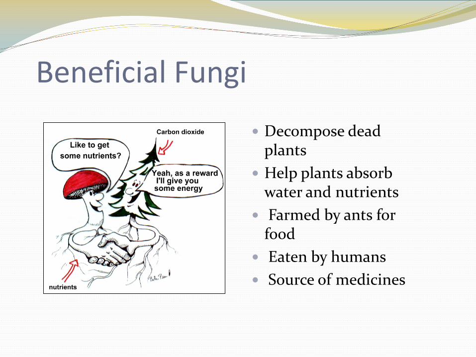
Beneficial Fungi
Decompose dead plants
Help plants absorb water and nutrients
Farmed by ants for food
Eaten by humans Source of medicines
Fungal Infections
• Superficial, localized skin conditions or deep tissue infections • Caused by exposure to spores• May or may not be transmitted• Categorized by:
• part of the body affected• how deeply the fungus penetrates the body • the organism causing the infection, • the form(s) that the fungi take
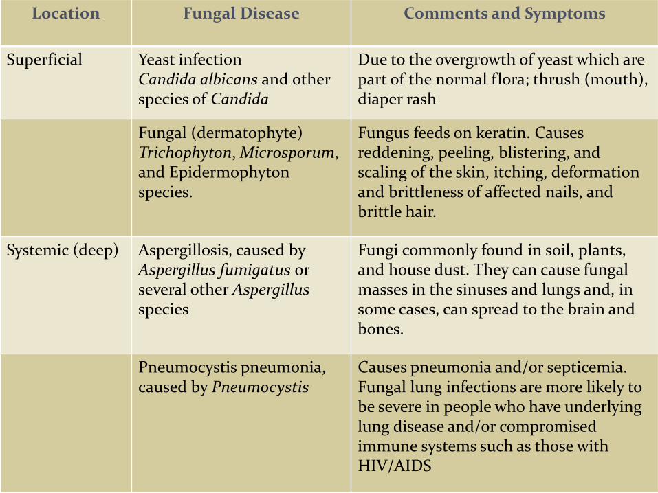
Location Fungal Disease Comments and Symptoms
Superficial Yeast infectionCandida albicans and other species of Candida
Due to the overgrowth of yeast which are part of the normal flora; thrush (mouth), diaper rash
Fungal (dermatophyte)Trichophyton, Microsporum, and Epidermophytonspecies.
Fungus feeds on keratin. Causes reddening, peeling, blistering, and scaling of the skin, itching, deformation and brittleness of affected nails, and brittle hair.
Systemic (deep) Aspergillosis, caused by Aspergillus fumigatus or several other Aspergillusspecies
Fungi commonly found in soil, plants, and house dust. They can cause fungal masses in the sinuses and lungs and, in some cases, can spread to the brain and bones.
Pneumocystis pneumonia, caused by Pneumocystis
Causes pneumonia and/or septicemia. Fungal lung infections are more likely to be severe in people who have underlying lung disease and/or compromised immune systems such as those with HIV/AIDS
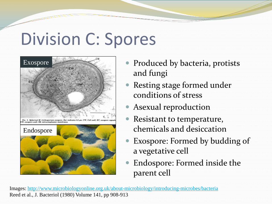
Division C: Spores Produced by bacteria, protists
and fungi Resting stage formed under
conditions of stress Asexual reproduction Resistant to temperature,
chemicals and desiccation Exospore: Formed by budding of
a vegetative cell Endospore: Formed inside the
parent cell
Images: http://www.microbiologyonline.org.uk/about-microbiology/introducing-microbes/bacteriaReed et al., J. Bacteriol (1980) Volume 141, pp 908-913
Endospore
Exospore
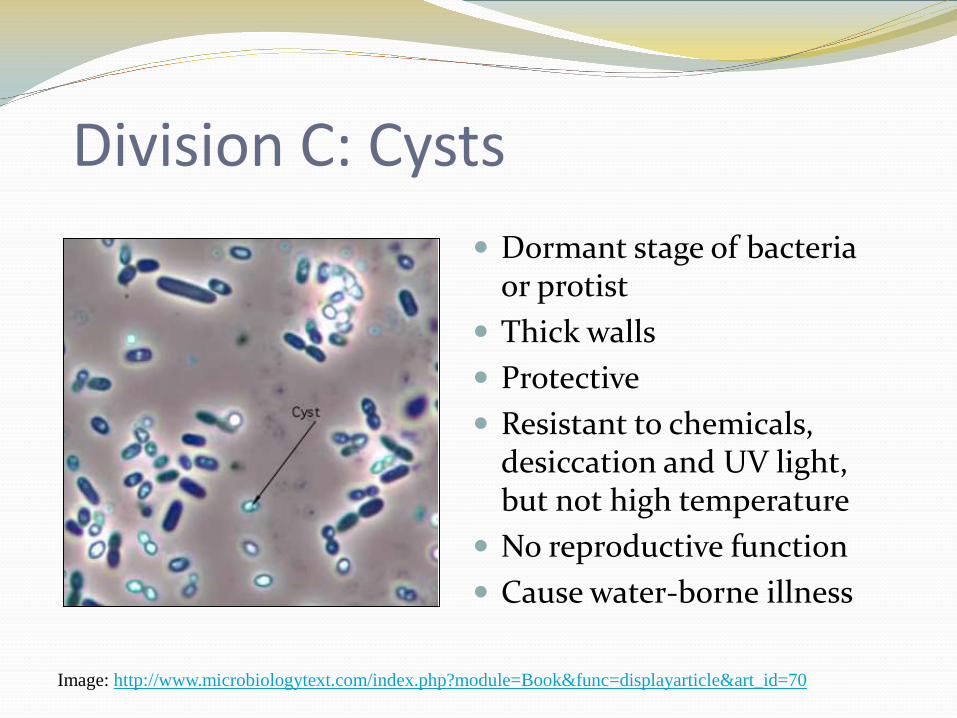
Division C: Cysts Dormant stage of bacteria
or protist Thick walls Protective Resistant to chemicals,
desiccation and UV light, but not high temperature
No reproductive function Cause water-borne illness
Image: http://www.microbiologytext.com/index.php?module=Book&func=displayarticle&art_id=70
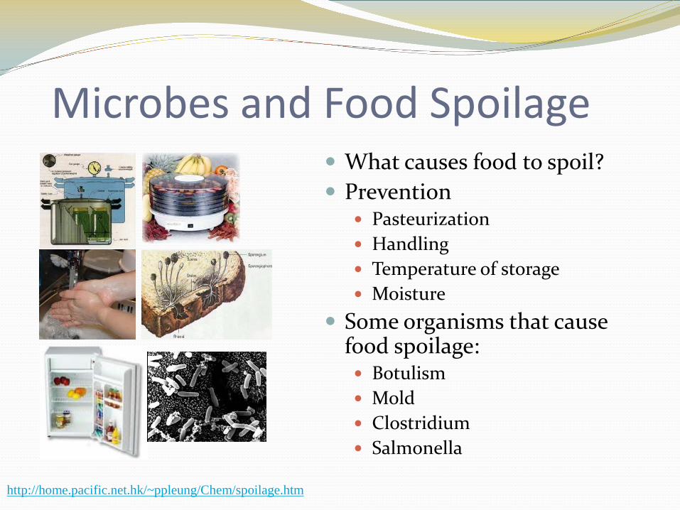
Microbes and Food Spoilage What causes food to spoil? Prevention
Pasteurization Handling Temperature of storage Moisture
Some organisms that cause food spoilage: Botulism Mold Clostridium Salmonella
http://home.pacific.net.hk/~ppleung/Chem/spoilage.htm
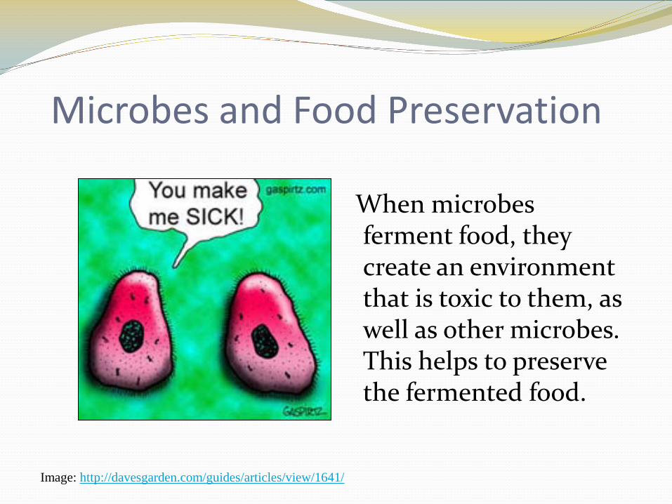
Microbes and Food Preservation
When microbes ferment food, they create an environment that is toxic to them, as well as other microbes. This helps to preserve the fermented food.
Image: http://davesgarden.com/guides/articles/view/1641/
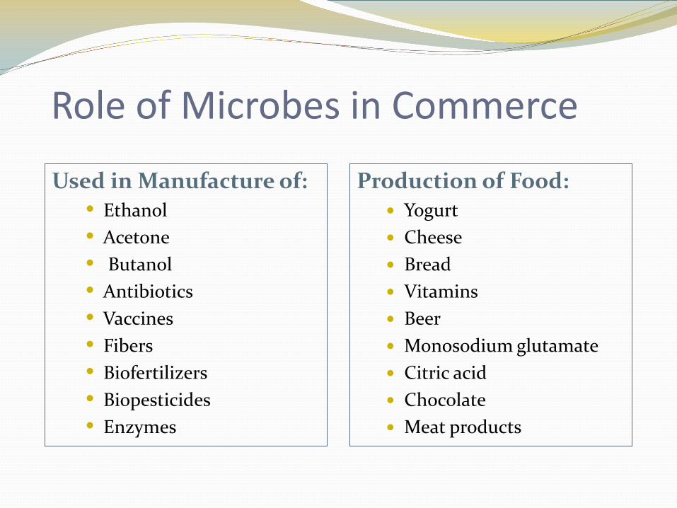
Role of Microbes in Commerce
Production of Food: Yogurt Cheese Bread Vitamins Beer Monosodium glutamate Citric acid Chocolate Meat products
Used in Manufacture of:• Ethanol• Acetone• Butanol• Antibiotics• Vaccines• Fibers• Biofertilizers• Biopesticides• Enzymes
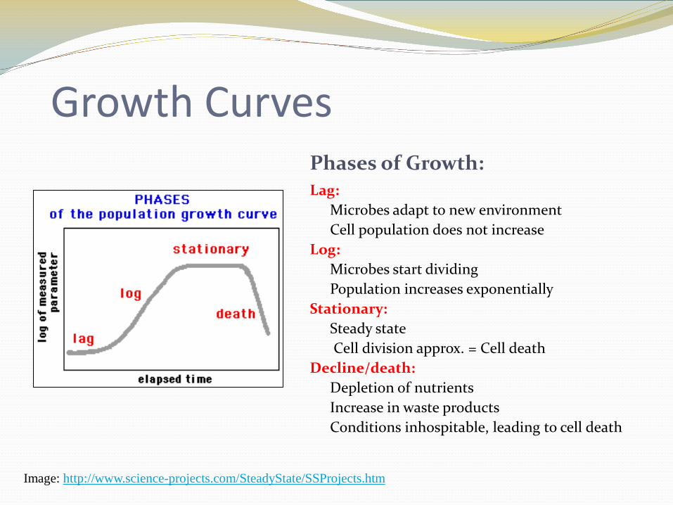
Growth CurvesPhases of Growth:Lag:
Microbes adapt to new environmentCell population does not increase
Log:Microbes start dividingPopulation increases exponentially
Stationary:Steady stateCell division approx. = Cell death
Decline/death:Depletion of nutrientsIncrease in waste productsConditions inhospitable, leading to cell death
Image: http://www.science-projects.com/SteadyState/SSProjects.htm
Resources http://www.edu.pe.ca/southernkings/microbe.ht
m http://www.microbeworld.org/ http://microbiologyprocedure.com/index.htm http://TomVolkFungi.net/ http://www.lichen.com/biology.html
Division B http://commtechlab.msu.edu/sites/dlc-me/


