Embed Size (px)
Citation preview
Edinburgh Research Explorer
Raf1 Is a DCAF for the Rik1 DDB1-Like Protein and HasSeparable Roles in siRNA Generation and ChromatinModification
Citation for published version:Buscaino, A, White, SA, Houston, DR, Lejeune, E, Simmer, F, de Lima Alves, F, Diyora, PT, Urano, T,Bayne, E, Rappsilber, J & Allshire, RC 2012, 'Raf1 Is a DCAF for the Rik1 DDB1-Like Protein and HasSeparable Roles in siRNA Generation and Chromatin Modification', PLoS Genetics, vol. 8, no. 2, e1002499.https://doi.org/10.1371/journal.pgen.1002499
Digital Object Identifier (DOI):10.1371/journal.pgen.1002499
Link:Link to publication record in Edinburgh Research Explorer
Document Version:Publisher's PDF, also known as Version of record
Published In:PLoS Genetics
Publisher Rights Statement:This is an open-access article distributed under the terms of the Creative Commons Attribution License, whichpermitsunrestricted use, distribution, and reproduction in any medium, provided the original author and source arecredited.
General rightsCopyright for the publications made accessible via the Edinburgh Research Explorer is retained by the author(s)and / or other copyright owners and it is a condition of accessing these publications that users recognise andabide by the legal requirements associated with these rights.
Take down policyThe University of Edinburgh has made every reasonable effort to ensure that Edinburgh Research Explorercontent complies with UK legislation. If you believe that the public display of this file breaches copyright pleasecontact [email protected] providing details, and we will remove access to the work immediately andinvestigate your claim.
Download date: 08. Feb. 2022
Raf1 Is a DCAF for the Rik1 DDB1-Like Protein and HasSeparable Roles in siRNA Generation and ChromatinModificationAlessia Buscaino1, Sharon A. White1, Douglas R. Houston1, Erwan Lejeune1, Femke Simmer1¤a, Flavia de
Lima Alves1, Piyush T. Diyora1, Takeshi Urano2, Elizabeth H. Bayne1¤b, Juri Rappsilber1, Robin C.
Allshire1*
1 Wellcome Trust Centre for Cell Biology, School of Biological Sciences, The University of Edinburgh, Edinburgh, United Kingdom, 2 Department of Biochemistry, Shimane
University Faculty of Medicine, Izumo, Japan
Abstract
Non-coding transcription can trigger histone post-translational modifications forming specialized chromatin. In fission yeast,heterochromatin formation requires RNAi and the histone H3K9 methyltransferase complex CLRC, composed of Clr4, Raf1,Raf2, Cul4, and Rik1. CLRC mediates H3K9 methylation and siRNA production; it also displays E3-ubiquitin ligase activity invitro. DCAFs act as substrate receptors for E3 ligases and may couple ubiquitination with histone methylation. Here,structural alignment and mutation of signature WDxR motifs in Raf1 indicate that it is a DCAF for CLRC. We demonstratethat Raf1 promotes H3K9 methylation and siRNA amplification via two distinct, separable functions. The association of theDCAF Raf1 with Cul4-Rik1 is critical for H3K9 methylation, but dispensable for processing of centromeric transcripts intosiRNAs. Thus the association of a DCAF, Raf1, with its adaptor, Rik1, is required for histone methylation and to allow RNAi tosignal to chromatin.
Citation: Buscaino A, White SA, Houston DR, Lejeune E, Simmer F, et al. (2012) Raf1 Is a DCAF for the Rik1 DDB1-Like Protein and Has Separable Roles in siRNAGeneration and Chromatin Modification. PLoS Genet 8(2): e1002499. doi:10.1371/journal.pgen.1002499
Editor: Sue Biggins, Fred Hutchinson Cancer Research Center, United States of America
Received July 20, 2011; Accepted December 9, 2011; Published February 2, 2012
Copyright: � 2012 Buscaino et al. This is an open-access article distributed under the terms of the Creative Commons Attribution License, which permitsunrestricted use, distribution, and reproduction in any medium, provided the original author and source are credited.
Funding: This research was supported by EMBO long-term fellowships to AB and FS, the Wellcome Trust (Senior Research Fellowship 084229/Z/07/Z to JR), andPrincipal Research Fellowship 065061/Z/01/A to RCA). The Centre for Cell Biology is supported by core funding from the Wellcome Trust (077707/Z/05/A and092076/Z/10/Z). The funders had no role in study design, data collection and analysis, decision to publish, or preparation of the manuscript.
Competing Interests: The authors have declared that no competing interests exist.
* E-mail: [email protected]
¤a Current address: Department of Molecular Biology, NCMLS, Radboud University Nijmegen, Nijmegen, The Netherlands¤b Current address: Wellcome Trust Centre for Gene Regulation and Expression, College of Life Sciences, University of Dundee, Dundee, United Kingdom
Introduction
Silencing mechanisms mediated by small RNAs occur in most
eukaryotes. RNA interference (RNAi) can reduce gene expression
post-transcriptionally by cleaving homologous transcripts or by
inhibiting their translation [1]. Small RNAs can also act in the
nucleus, inducing transcriptional gene silencing (TGS) [2,3].
Although siRNA-directed modification of homologous chromatin
is widespread in eukaryotes, the details of how siRNAs mediate
such events remain limited in most systems. [4,5]. In plants and
fungi, the link between siRNA-directed DNA/chromatin modifi-
cation and heterochromatin formation is well established [2,3].
The fission yeast, Schizosaccharomyces pombe, is a powerful system in
which to study RNAi-directed heterochromatin formation in part
because it contains single non-essential genes encoding each of the
key RNAi components.
In fission yeast, siRNAs are important for heterochromatin
formation on the centromeric outer repeats (composed of dg and dh
elements) and other chromosomal locations. Although marker
genes inserted at these loci are silenced [6], centromeric repeats
are bi-directionally transcribed by RNAPII, producing double-
stranded RNA (dsRNA) [2,3,7,8,9]. Dicer (Dcr1) cleaves these
non-coding transcripts into siRNAs that guide the Argonaute
(Ago1)-containing RITS complex to homologous nascent tran-
scripts by sequence complementarity. Chromatin-bound-RITS
recruits the histone methyltranferase Clr4SuVar3-9 (a CLRC
component; see below) to centromeric repeats via the linker
protein Stc1 [10]. Clr4 methylates lysine 9 of histone H3
(H3K9me), providing binding sites for the chromodomain proteins
Swi6, Chp1 (a RITS component) and Chp2 [11,12,13,14]. Two
different non-mutually exclusive models have been proposed to
explain how heterochromatin, once assembled, silences centro-
mere repeat transcription. In the ‘transcriptional gene silencing’
(TGS) model, heterochromatin factors directly repress transcrip-
tion [15]. In the ‘co-transcriptional gene silencing’ (CTGS) model
the repeats are continuously transcribed and silencing is due to the
efficient processing of transcripts to siRNA [16]. In wild-type cells,
siRNA production and H3K9 methylation are coupled processes:
deletion of genes encoding CLRC components results not only in
loss of H3K9 methylation but also in loss of siRNA [17,18,19].
Similarly, deletion of genes encoding RNAi components abrogates
siRNA production and reduces H3K9me [9]. However, cells
expressing only mutant histone H3 (H3K9R) produce some
detectable siRNAs, even though H3K9 can not be methylated
[20,21]. Moreover, tethering the Rik1 CLRC component to ura4
RNA triggers silencing of the ura4+ gene independently of other
PLoS Genetics | www.plosgenetics.org 1 February 2012 | Volume 8 | Issue 2 | e1002499
CLRC subunits, but requires RNAi [21]. Thus CLRC itself,
rather than its substrate H3K9, promotes siRNA production
independently of H3K9. However, it is not known how CLRC
integrates histone methylation with siRNA generation.
CLRC is composed of the histone methyltransferase Clr4, the b-
propeller protein Rik1, the cullin protein Cul4, the WD-40 protein
Raf1 (Clr8/Cmc1/Dos1) and Raf2 (Clr7/Cmc2/Dos2), which
contains a RFTS domain [17,22,23,24,25]. Although it is not
known whether CLRC acts as an E3-ubiquitin ligase in vivo,
CLRC purified from cells exhibits E3 ligase activity in vitro [24]. E3
ligases regulate a variety of biological processes by bridging the E2
ubiquitin conjugating enzyme to specific substrates, allowing their
ubiquitination (for review see [26]). Histone methylation is
frequently regulated by ubiquitination but the mechanistic details
remain unknown [27].
The Cullin-RING ligases (CRLs) are the largest family of multi-
subunit E3 ligases in eukaryotes and are formed by a neddylated
member of the cullin family scaffold (CUL1-CUL5), a RING
finger protein (RBX1 or RBX2) and an adaptor protein that
bridges the CUL/RING scaffold to substrate receptors [28].
Amongst CRLs, CRL4 is known to ubiquitinate histones [29,30].
In the CRL4 complex, DDB1 acts as the adaptor. Affinity
purification of DDB1 from human cells identified various WD-40
proteins as possible substrate receptors termed DCAFs (DDB1 and
CUL4 Associated Factors) [31,32,33,34]. Several DCAFs (WDR5,
RBP5 and EED) not only interact with DDB1 but are also
members of histone methyltransferase complexes [31,32,33,34].
Although it is not known whether the interaction between these
DCAFs and CUL4/DDB1 leads to ubiquitination of substrates, it
might have important functional consequences since the knock-
down of CUL4 or DDB1 reduces histone methylation [31]. Many
DCAFs contain a specific WDxR motif that is important for their
association with DDB1 [31,32,33,34].
In S. pombe, Cul4 associates with the canonical adaptor Ddb1
[35] and with Rik1 (which shares similarity with Ddb1; [36]) in
CLRC [17,22,23,24]. Both Cul4-Ddb1 complex and CLRC affect
heterochromatin integrity and neddylation of Cul4 is required for
H3K9 methylation of heterochromatic regions [22,37]. This
suggests that E3 ligase activity is involved in heterochromatin
formation. However, heterochromatin defects observed in neddy-
lation-defective Cul4 (cul4-K680R) could arise either from
impaired Cul4-Ddb1 activity or from defective CLRC function.
Furthermore, although Rik1 exhibits some similarity to Ddb1 and
thus might act as an adaptor, recent analyses suggest that Rik1 is
primarily an RNA binding protein which associates with
centromeric transcripts via its CPSF-A domain [21]. Thus, it
remains to be determined how CLRC contributes to heterochro-
matin integrity and whether CLRC acts as a DDB1-related E3
ligase which promotes heterochromatin formation in vivo.
Here we demonstrate that the CLRC components Rik1 and
Raf1 can be structurally aligned with the human adaptor DDB1
and its DCAF DDB2, respectively. Our analyses are consistent
with Raf1 acting as a DCAF for CLRC that contributes to siRNA
amplification and H3K9 methylation by two distinct and
separable routes. Our findings provide mechanistic insights into
how siRNA production is integrated with chromatin modification
via CLRC.
Results
Raf1 is a chromatin associated component of the CLRCcomplex
Components involved in heterochromatin formation are
typically localised in distinct chromatin-associated foci [14,38].
In contrast, previous studies overexpressing GFP-Raf1 from a
strong promoter showed that it is a nuclear protein with no
obvious chromatin localisation [23], however, this might not
reflect its true subcellular localisation. Indeed, the genome-wide
distribution of Raf1 suggests that it interacts predominantly with
heterochromatic loci [13]. To reassess Raf1 localisation, we
examined cells expressing GFP-Raf1 from its native promoter and
found that it decorates several distinct chromatin-associated foci,
the largest of which lies adjacent to the clustered kinetochores
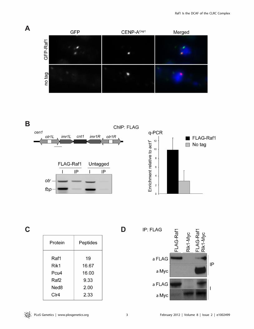
(Figure 1A). ChIP confirmed that FLAG-Raf1 associates with
centromeric repeats (Figure 1B). We conclude that Raf1 is a
chromatin-associated protein concentrated at heterochromatin.
Biochemical purification of CLRC components Rik1, Raf2 and
Clr4 identified Raf1 as an interacting partner [17,22,24]. In
contrast, two-step purification of TAP-Raf1 identified the histone
demethylase Lid2 plus Cul4 and Rik1, but apparently not Raf2
and Clr4, known CLRC subunits [39]. This raises the possibility
that Raf1 is a component of two distinct complexes: CLRC (Cul4/
Rik1/Raf1/Raf2/Clr4) and an alternative Lid2/Cul4/Rik1/Raf1
complex. To further investigate this, we affinity selected FLAG-
Raf1 from cell lysates and identified associated proteins by mass
spectrometry. Silencing assays indicate that FLAG-tagged Raf1 is
functional (Figure S1A). Single step FLAG affinity purification is
less stringent than the two-step TAP affinity purification and was
performed in mild conditions to identify as many Raf1 interacting
proteins as possible. Although we detect all CLRC components,
Lid2 peptides were absent (Figure 1C). Moreover, while Rik1
coimmunoprecipitated with Raf1 (co-IP; Figure 1D), Rik1 could
not be detected in Lid2-TAP IPs (Figure S1B). We conclude that
Raf1 mainly associates with the known CLRC components.
Raf1 is a DDB2-like WDxR proteinIn CRL4 complexes DDB1 is the adaptor that recruits WD-40-
containing DCAF proteins [28]. In CLRC, Rik1 is a DDB1-
related protein that interacts with Raf1 [23]. Thus, Rik1 and Raf1
Author Summary
Heterochromatin is a specialized form of chromatin whichis frequently assembled on DNA sequences with little or nocoding potential. Heterochromatin formation involvesspecific post-translational modifications of histone tails(e.g. methylation of histone H3 on lysine 9). In fission yeast,Schizosaccharomyces pombe, heterochromatin is found atcentromeres, telomeres, and the mating type locus.Heterochromatin integrity at centromeres is importantfor normal chromosome segregation. The heterochromatinassociated repeats at fission yeast centromeres are knownto be transcribed, and these non-coding transcripts areprocessed into siRNAs. siRNA production is required forestablishment and maintenance of H3K9 methylation. ButH3K9 methylation itself is required for siRNA production. Itis not known how these two processes are coupled. In thisstudy we use structural modelling and genetic analyses todemonstrate that the heterochromatin component Raf1plays an essential role in coupling H3K9 methylation andsiRNA production. Our analyses show that the heterochro-matin factors Rik1 and Raf1 can be structurally alignedwith Cul4-E3 ubiquitin ligase components DDB1 andDDB2, respectively. We show that specific mutationsimpair the association of Raf1 with Rik1 and preventH3K9 methylation but not siRNA production. Thesefunctional studies provide mechanistic insights into howsiRNA production and chromatin modification are inte-grated.
Raf1 Is the DCAF of the CLRC Complex
PLoS Genetics | www.plosgenetics.org 2 February 2012 | Volume 8 | Issue 2 | e1002499
Raf1 Is the DCAF of the CLRC Complex
PLoS Genetics | www.plosgenetics.org 3 February 2012 | Volume 8 | Issue 2 | e1002499
might represent the adaptor and the DCAF, respectively, for
CLRC. To test these possibilities, we performed structural
alignments of Rik1 and Raf1 and found that they can be modelled
on DDB1 and on the DCAF DDB2 (Rik1: DOPE score 211385;
Raf1: GA431 score 0.6 respectively) (Figure 2A and 2B). This
model reveals that, like human DDB1, Rik1 is composed of three
WD-40 b-propeller domains (BPA, BPB and BPC) composed of
216WD-40 repeats and a C-terminal helical domain (Figure 2B
and S2A). Raf1, like DDB2, is predicted to contain an N-terminal
helical domain and a 7-bladed WD-40 b-propeller domain
forming a ring (Figure 2A and 2B). Unlike DDB2, the helical
and WD-40 domains of Raf1 are separated by an additional
domain of unknown function (Figure S2B). As in the DDB1/
DDB2 complex, our model predicts that the Raf1 helical domain
can be inserted into the Rik1 BPA-BPC cleft and the ‘top’ of the b-
propeller ring interacts with the ‘bottom’ surface of Rik1
(Figure 2B).
Alignment of the Raf1 WD-40 repeats with those of other
known DCAFs revealed that Raf1 contains two WDxR motifs (1:
aa515–518 and 2: aa573–576) (Figure S3A). WDxR motifs
represent a ‘DCAF signature’ and are important for docking
DCAFs to DDB1 [31,32,33,34]. Our Raf1 model indicates that
these two WDxR motifs are located on the surface of the b-
propeller ring and might provide specific sites for interactions with
other factors (Figure S3E). To test the importance of Raf1 WDxR
motifs, we mutated the endogenous raf1 gene in S. pombe to express
FLAG-Raf1-R518A or FLAG-Raf1-R576A. Western analysis
indicated that both mutant proteins are expressed at levels similar
to wild-type FLAG-Raf1 (Figure S3B). All components of CLRC
are required for heterochromatin integrity and consequently for
the transcriptional silencing of marker genes placed within
centromeric heterochromatin, and centromere function [6]. Like
clr4D cells, heterochromatin-mediated silencing of cen1:ade6+ is
disrupted in raf1-R518A and raf1-R576A mutants, as indicated by
white/expressing rather than red/silent colonies (Figure 2C).
Moreover, both mutations impair heterochromatin dependent
silencing at the mating type locus (Figure S3D).
Centromeric heterochromatin mediates robust sister-centro-
mere cohesion and is therefore required for accurate chromosome
segregation during mitosis [40,41]. Defective heterochromatin
causes a quantifiable increase in the frequency of lagging
chromosomes on late anaphase spindles [42]. Cells bearing the
raf1-R518A or raf1-R576A mutation exhibit a frequency of lagging
chromosomes equivalent to clr4D cells, indicating that this
centromeric function of heterochromatin is disrupted (Figure 2D).
Heterochromatin formation requires processing of centromeric
transcripts into siRNA and methylation of H3K9 (H3K9me).
Deletion of any gene encoding a CLRC component results in loss
of H3K9me, accumulation of centromeric transcripts and a
dramatic reduction of siRNA levels [17,18,19]. ChIP analyses
indicate that H3K9me2 is reduced to background levels in raf1-
R518A and raf1-R576A cells (Figure 2E). Importantly, Clr4 levels
are equivalent to wild-type in both mutants (Figure S3C).
Consistent with this loss of H3K9me2 and as observed in clr4Dcells, high levels of centromeric transcripts also accumulate
(Figure 2F). This is due to increased transcription given that
higher levels of RNAPII are detected on centromeric repeats in
both mutants compared to wild-type cells (Figure S5E). However,
raf1-R576A cells contain low levels of centromeric siRNAs whereas
raf1-R518A cells retain high levels of these siRNAs (Figure 2G and
S3F). Our structural model predicts that R518 resides on the
surface of Raf1 that interacts with Rik1 and predicts that the
R518A mutation may specifically impair Raf1-Rik1 interactions
(Figure S3G). In contrast, R576 is more distantly located from
Rik1 and may have more profound structural effects.
Together our findings demonstrate that the CLRC complex is
likely to adopt a canonical Cul4-E3 ligase architecture and that
Raf1 is a DCAF for CLRC. Moreover, we show that WDxR
motifs are required for heterochromatin integrity and that the raf1-
R518A mutation separates the function of CLRC in methylating
H3K9 from its role in generating siRNAs.
The raf1-1 mutation conditionally disruptsheterochromatin without affecting siRNA generation
To dissect mechanisms governing heterochromatin assembly
and maintenance, we screened for temperature-sensitive mutants
that disrupt heterochromatin at the restrictive temperature (36uC)
but not the permissive temperature (25uC) and isolated the raf1-1
mutation. raf1-1 produces a protein with a missense mutation
(T495I) in the third WD-40 repeat; this residue is conserved in
fungi suggesting that it is important for Raf1 function (Figure
S3A). Similar levels of mutant FLAG-Raf1-1 and wild-type FLAG-
Raf1 proteins are detected at 36uC thus phenotypes are not due to
Raf1-1 degradation (Figure S4A). Marker gene silencing within
centromeric repeats (cen1:ade6+ or cen1:ura4+), the mating type locus
(mat3-M:ura4+) and at telomeres (tel1L:his3+) is alleviated in raf1-1
cells at 36uC, but not 25uC (Figure 3A and Figure S4B, S4C).
Consistent with defective centromeric heterochromatin integrity, a
high frequency of lagging chromosomes is observed in late
anaphase raf1-1 cells at 36uC (Figure 3B). Moreover, H3K9me2
levels on centromeric repeats in raf1-1 cells are similar to wild-type
cells at 25uC but negligible at 36uC (Figure 3C). Consistent with
reduced H3K9me2, Swi6 localisation and unprocessed centro-
mere repeat transcript accumulation is temperature dependent in
raf1-1 cells (Figure 4A and 4B). Importantly, Clr4 levels are
unaffected by the raf1-1 mutation (Figure S4B).
Although we detect no H3K9me2 on centromere repeats in
raf1-1 at 36uC, it is possible that residual levels of H3K9me2
remain. To determine if heterochromatin is erased at 36uC, a
heterochromatin establishment assay was performed. At the
mating type locus, active RNAi is required to establish
heterochromatin, but once assembled, RNAi is not required to
maintain heterochromatin [43]. Hence, loss of dcr1/RNAi has no
impact on mating type locus silencing (i.e. mat3-M:ura4+).
However, when presented with a naive template, dcr1D cells fail
to assemble heterochromatin de novo and are unable to silence
mat3-M:ura4+. To test whether the raf1-1 mutant erases hetero-
chromatin structures, a raf1-1 dcr1D double mutant was generated
at 36uC and mat3-M:ura4+ silencing assessed after shifting cells to
25uC. Silencing of mat3-M:ura4+ could not be established in this
double mutant following a shift down from 36u to 25uC, but
remained intact in the raf1-1 dcr1D double mutant generated at
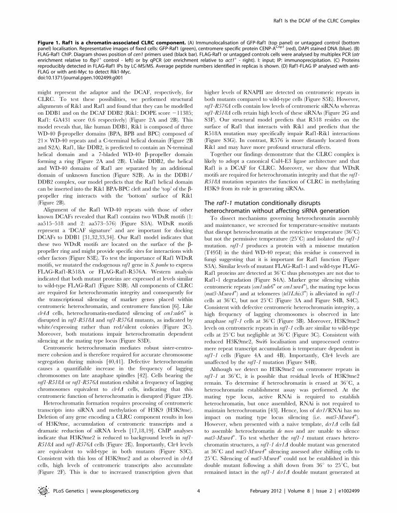
Figure 1. Raf1 is a chromatin-associated CLRC component. (A) Immunolocalisation of GFP-Raf1 (top panel) or untagged control (bottompanel) localisation. Representative images of fixed cells: GFP-Raf1 (green), centromere specific protein CENP-ACnp1 (red), DAPI stained DNA (blue). (B)FLAG-Raf1 ChIP. Diagram shows position of cen1 primers used (black bar). FLAG-Raf1 or untagged controls cells were analysed by multiplex PCR (otrenrichment relative to fbp1+ control - left) or by qPCR (otr enrichment relative to act1+ - right). I: input; IP: immunoprecipitation. (C) Proteinsreproducibly detected in FLAG-Raf1 IPs by LC-MS/MS. Average peptide numbers identified in replicas is shown. (D) Raf1-FLAG IP analysed with anti-FLAG or with anti-Myc to detect Rik1-Myc.doi:10.1371/journal.pgen.1002499.g001
Raf1 Is the DCAF of the CLRC Complex
PLoS Genetics | www.plosgenetics.org 4 February 2012 | Volume 8 | Issue 2 | e1002499
Raf1 Is the DCAF of the CLRC Complex
PLoS Genetics | www.plosgenetics.org 5 February 2012 | Volume 8 | Issue 2 | e1002499
25uC (Figure 4C). This demonstrates that raf1-1 cells are unable to
form heterochromatin without RNAi and indicates that hetero-
chromatin is completely erased by the raf1-1 mutation at 36uC.
Surprisingly, unlike clr4D and raf1D mutants, centromeric
siRNAs are produced at wild-type levels in raf1-1 cells at both
temperatures (Figure 4D, Figure S4E and S5D). It is possible that
siRNA levels remain high because raf1-1 inhibits the degradation
of pre-existing siRNAs. However, high siRNA levels remain in
raf1-1 cells which have undergone 144 divisions at 36uC; pre-
existing siRNA would be diluted out (Figure S4E). Thus, the
continual synthesis of siRNAs from centromere repeat transcripts
must be unaffected by the defect in raf1-1. Moreover, these
siRNAs are loaded into Ago1/RITS as indicated by their
association with FLAG-Ago1 (Figure 4E). We conclude that as
with raf1-R518A, the raf1-1 mutation uncouples siRNA production
from H3K9 methylation.
Methylation of H3K9, but not siRNA production, dependson an intact CLRC complex
The interaction between the adaptor DDB1 and its DCAFs is
known to require intact WDxR motifs in the DCAF [32,33,34].
Our structural alignment predicts that the raf1-1 T495I mutation
is located on the ‘top’ surface of the WD-40 ring that interacts with
Rik1 (Figure S5A). It is therefore possible that our specific raf1
mutations impair the ability of Raf1 to associate with Rik1 and
that this interaction is essential for establishing and maintaining
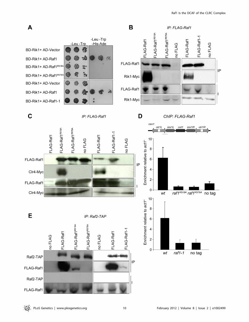
H3K9 methylation on heterochromatic loci. Indeed two-hybrid
assays reveal that the binding of Raf1 to Rik1 [23] is disrupted in
both WDxR motif mutants and the raf1-1 mutation (Figure 5A).
All three mutations also impair co-immunoprecipitation of Rik1
with Raf1 from S. pombe extracts (Figure 5B). In addition, the
association of the Clr4 H3K9 methyltransferase with Raf1 and
Rik1 is lost in raf1-R518A, raf1-R576A and raf1-1 mutants
(Figure 5C and Figure S5B). Recently we identified Stc1 as
protein that links the RNAi machinery to CLRC [10]. Consistent
with this, these same three specific raf1 mutations abolish the
association of the Rik1 CLRC component with Stc1 (Figure S5C).
Furthermore, ChIP analyses indicate that FLAG-Raf1-R518A,
FLAG-Raf1-R576A and FLAG-Raf1-1 do not associate with
centromeric repeats (Figure 5D). We conclude that binding of
Raf1 to Rik1 is required to allow Raf1 to associate with
centromeric repeats. This interaction is required to assemble an
active CLRC complex in order to establish and maintain H3K9
methylation and hence functional heterochromatin.
The coupling of H3K9 methylation and siRNA production
could be achieved by bringing the distinct activities required for
both processes together in the same complex. Since the raf1D and
raf1-R576A mutations results in loss of siRNAs, Raf1 is clearly
required for siRNA production (Figure S5D and Figure 2G).
However, the raf1-R518A and raf1-1 mutants lose H3K9
methylation without affecting siRNA synthesis (Figure 2E, 2G;
Figure 3C; Figure 4D; Figures S4E and S5D). This suggests that
specific interactions between Raf1 and individual proteins in
CLRC and/or other unknown factors are sufficient to allow
siRNA generation in the absence of H3K9 methylation. An intact
CLRC complex may not be required. Indeed, co-immunoprecip-
itation experiments show that a weak but detectable interaction
between Raf1 and Raf2 remains in raf1-1 and raf1-R518A, but not
raf1-R576A cells (Figure 5E). We conclude that although an intact
CLRC complex is dispensable for siRNA synthesis, a minimal
Raf1-Raf2 interaction might be sufficient to allow the processing
of centromere repeat transcripts to siRNAs.
Discussion
The interaction between the E3 ligase adaptor Rik1 andthe DCAF Raf1 correlates with methylation of lysine 9 onhistone H3
The CLRC complex, harbouring the histone methyltransferase
Clr4Suvar3-9, performs an essential role in the establishment and
maintenance of heterochromatic structures. In addition to Clr4,
the complex contains the components Cul4, Rik1, Raf1 and Raf2.
Rik1 is a WD-40 repeat protein that shares homology with the
E3 ligase adaptor DDB1 [36]. DDB1 contains 21 WD-40 repeats
that form the seven blades of three b-propellers that mediate
association with Cul4 and DCAFs. This supports the possibility
that Cul4-Rik1 form the core of an E3 ligase [24]. However, the
C-terminus of Rik1 also shows homology with the b-propeller
domain of cleavage and polyadenylation factor CSPF-A a well-
known RNA binding protein [18]. Based on this CPSF-A
homology, it has been suggested that Rik1 might bind RNA
through this domain [21]. Although we cannot exclude that Rik1
is a bifunctional protein, our manual alignment detects 21 WD-40
repeats within Rik1 allowing it to be structurally aligned along its
entire length with DDB1 (Figure 2B and Figure S2A). In our
model, the CSPF-A homology domain corresponds to the b-
propeller BPC involved in the interaction with the DCAF Raf1
(Figure 2B and Figure S2A). Moreover, we also show that Raf1
can be structurally aligned to the DCAF DDB2, the partner of
DDB1 (Figure 2A, 2B and Figure S2B). As expected for a bona fide
DCAF, Raf1 associates directly with the putative substrate adaptor
Rik1 and contains two signature WDxR motifs which we show are
required for the association of Raf1 with Rik1 and for H3K9
methylation and heterochromatin formation (Figure 2B and
Figure S3). This suggests that the CLRC complex can adopt the
architecture of a Cul4 E3 ligase in which Rik1 is the adaptor
protein bridging the interaction between Cul4 and the DCAF
Raf1 (Figure 6A). In addition, we show that the association of the
DCAF Raf1 with Rik1 within an intact CLRC complex is critical
for H3K9 methylation but it is dispensable for the processing of
centromeric transcripts into siRNAs (Figure 2, Figure 4, and
Figure 5).
We envisage two alternative scenarios that can explain how the
Cul4-Rik1-Raf1 complex might mediate H3K9 methylation
(Figure 6A). First, it is possible that the CLRC is an active E3
ligase and that mono- or poly-ubiquitination of specific factors
must occur to allow methylation of H3K9. Thus the DCAF Raf1
would be essential for the recognition and ubiquitination of key
specific substrate(s) (Figure 6A top panel). In other systems, Cul4-
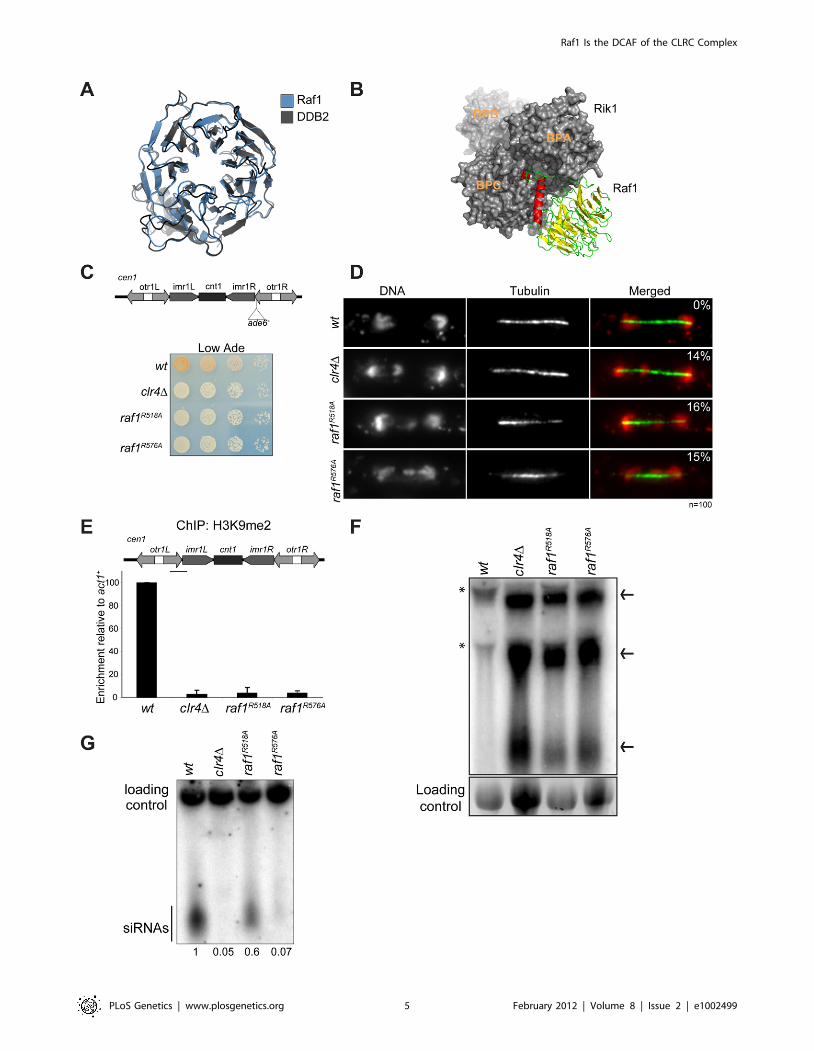
Figure 2. Raf1 is the DCAF for CLRC with WDxR motifs essential for heterochromatin formation. (A) Structural alignment of Raf1 (blue)with human DCAF DDB2 (grey). (B) Structural model of Rik1 (based on DDB1: grey) associated with Raf1 (red, green and yellow). In Raf1, the N-terminal helix (red) and the b-propeller (green and yellow) make specific interaction with Rik1. (C) Centromere silencing assay. Position of ade6+
marker gene in cen1. Wild-type cells with silenced cen1:ade6+ form red colonies; loss of silencing causes white colonies. (D) Lagging chromosomes inanaphase. Representative images of fixed cells stained with DAPI (red) and anti-tubulin (green) and % anaphase cells with lagging chromosomes. (E)H3K9me2 ChIP; levels associated with cen-dg relative to act1+, normalised to wild-type. Error bars: standard deviation (SD). (F) Northern: unprocessedotr transcripts (arrows) in wt, clr4D, raf1-R518A and raf1-R576A cells. Loading control: rRNA. (*): rRNA background. (G) Northern: centromeric siRNAs inwt, clr4D, raf1-R518A and raf1-R576A cells. Loading control: snoRNA58.doi:10.1371/journal.pgen.1002499.g002
Raf1 Is the DCAF of the CLRC Complex
PLoS Genetics | www.plosgenetics.org 6 February 2012 | Volume 8 | Issue 2 | e1002499
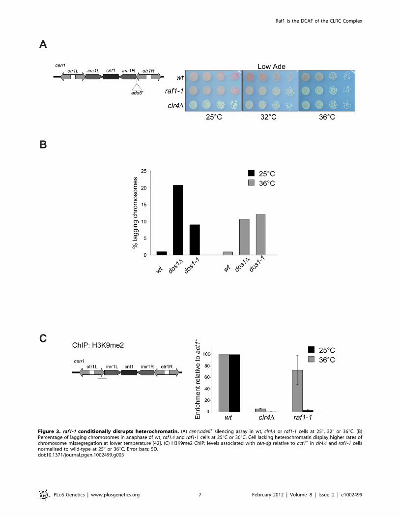
Figure 3. raf1-1 conditionally disrupts heterochromatin. (A) cen1:ade6+ silencing assay in wt, clr4D or raf1-1 cells at 25u, 32u or 36uC. (B)Percentage of lagging chromosomes in anaphase of wt, raf1D and raf1-1 cells at 25uC or 36uC. Cell lacking heterochromatin display higher rates ofchromosome missegregation at lower temperature [42]. (C) H3K9me2 ChIP: levels associated with cen-dg relative to act1+ in clr4D and raf1-1 cellsnormalised to wild-type at 25u or 36uC. Error bars: SD.doi:10.1371/journal.pgen.1002499.g003
Raf1 Is the DCAF of the CLRC Complex
PLoS Genetics | www.plosgenetics.org 7 February 2012 | Volume 8 | Issue 2 | e1002499
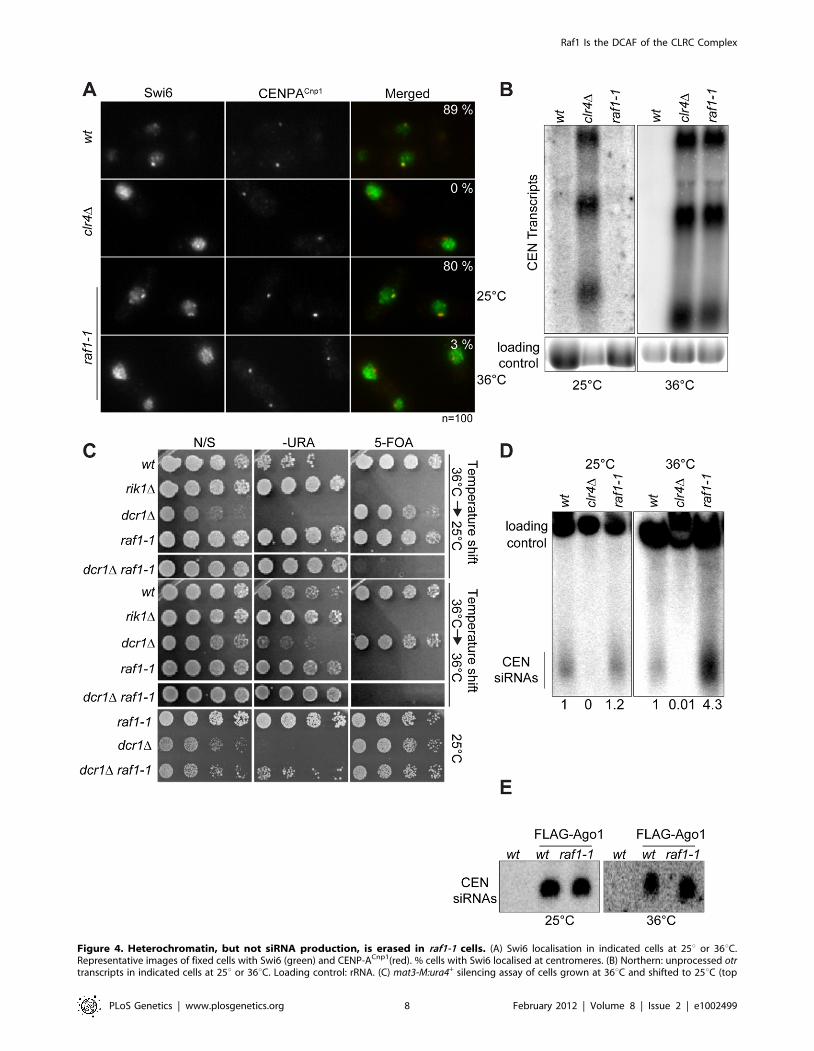
Figure 4. Heterochromatin, but not siRNA production, is erased in raf1-1 cells. (A) Swi6 localisation in indicated cells at 25u or 36uC.Representative images of fixed cells with Swi6 (green) and CENP-ACnp1(red). % cells with Swi6 localised at centromeres. (B) Northern: unprocessed otrtranscripts in indicated cells at 25u or 36uC. Loading control: rRNA. (C) mat3-M:ura4+ silencing assay of cells grown at 36uC and shifted to 25uC (top
Raf1 Is the DCAF of the CLRC Complex
PLoS Genetics | www.plosgenetics.org 8 February 2012 | Volume 8 | Issue 2 | e1002499
E3 ligases have been shown to ubiquitinate histones [29,30], it is
possible that a specific histone residue needs to be ubiquitinated to
allow methylation of H3K9. E3 ligase activity has been shown to
associate with affinity selected CLRC in vitro [24], however, we
have been unable to detect this activity in similar experiments.
This suggests that CLRC E3 ligase activity may be particularly
inefficient and/or the ubiquitination events that it mediates are
transient.
The alternative scenario is that the CLRC complex is not an
active E3 ligase in vivo and that Cul4, Rik1, Raf1 and Raf2 just
form a protein scaffold that acts to target Clr4 methyltransferase to
heterochromatic repeats, independently of ubiquitination
(Figure 6A bottom panel). In this case the specific mutations
which impair the Rik1-Raf1 interaction may just disrupt the
scaffold so that Raf1 is not targeted to the heterochromatic repeats
and Clr4 no longer associates with Rik1 and Raf1. Interestingly,
the DCAF DDB2 has been shown to bind DNA with the ‘bottom’
surface of its WD-40 ring [44]. Similarly, the WD-40 ring of the
DCAF WDR5 specifically binds the histone H3 N-terminal tail
methylated on lysine 4 [45,46,47]. Thus, it is possible that the
WD-40 ring of the DCAF Raf1 also binds DNA or histones to
allow the correct targeting of the histone methyltransferase Clr4 to
heterochromatic repeats.
Raf1 function in H3K9 methylation and RNAi areseparable
CLRC plays a dual role in heterochromatin formation: it
harbours the histone methyltransferase Clr4 and hence it is
responsible for H3K9 methylation, it also mediates siRNA
production since cells lacking any single CLRC subunit have
low centromeric siRNA levels (Figure S5D and [17,18]).
Therefore, in wild-type fission yeast H3K9 methylation and
siRNAs synthesis are coupled. This ensures formation of
heterochromatin at centromeres, telomeres, and the mating type
locus, and prevents promiscuous silencing at other chromosomal
regions. However, cells expressing only mutant histone H3
(H3K9R) have been shown to produce some detectable siRNAs,
even though H3K9 can not be methylated [20,21]. Such analyses
implicate CLRC itself, rather than its known substrate H3K9, in
promoting siRNA production independently of H3K9.
Here we have isolated two mutations in the raf1 gene (raf1-1 and
raf1R518A) that destroy CLRC integrity without affecting siRNA
levels. Although we cannot exclude that a minimal CLRC
complex is still present in these mutants, our results indicate that
individual CLRC components can stimulate processing of
centromeric transcripts into siRNAs independently of CLRC
complex integrity (Figure 6B). It remains to be determined how
CLRC components promote this H3K9 methylation-independent
siRNA production. Our analyses of specific raf1 mutants suggests
that the putative E3 ligase activity of the CLRC complex is not
required for siRNA synthesis given that in raf1-1 and raf1-R518A
mutants (predicted to impair the E3 ligase activity of the complex)
siRNA levels remain high (Figure 2G and Figure 4D). One
possibility is that Clr4 can methylate specific substrates indepen-
dently of CLRC integrity and that this is a key event in siRNA
production [21]. In accordance with this, Clr4 was recently shown
to methylate the RNA processing factor Mlo3 and this methylation
correlates with high siRNA levels [48]. However, Clr4-mediated
methylation of specific substrates cannot be the only event
required to trigger siRNA production since the loss of any CLRC
component results in dramatic reduction of siRNA levels. We find
that high siRNA levels correlate with a low but detectable Raf1-
Raf2 interaction. Complete disruption of this Raf1-Raf2 interac-
tion (as observed in raf1-R576A) cuts siRNA production to
undetectable levels. Interestingly, centromeric repeats are tran-
scribed preferentially in S-phase and Raf2 has been recently
shown to interact with Cdc20 (the catalytic subunit of DNA-
polymerase e) [49,50,51]. Coordination of DNA replication,
siRNA generation and methylation of specific substrates may be
essential for heterochromatin establishment and maintenance of
heterochromatic structures.
Our analyses suggest that integration of DNA replication,
siRNA production and methylation of H3K9 is achieved by
bringing the distinct activities required for these processes together
in the same protein complex (CLRC). This ensures the assembly of
robust heterochromatin structures.
Cotranscriptional gene silencing plays a subsidiary role inrepression of heterochromatic repeats
Two different models have been proposed to explain how
heterochromatin could silence centromeric repeats. In the ‘TGS’
model, heterochromatin factors have been proposed to repress
transcription at centromeres [15] whereas the ‘CTGS’ model
suggests that centromere repeats are continuously transcribed and
silencing is caused by the efficient cleavage of centromeric
transcripts by RNAi into siRNA [16]. Our analyses demonstrate
that the RNAi machinery is active in raf1-1 cells, so that
centromeric transcripts are processed into siRNAs independently
of heterochromatin integrity. raf1-1 cells produce siRNAs at levels
similar to wild-type cells and they are loaded into Ago1. However,
despite this, high levels of unprocessed centromeric transcripts
persist, indicating that RNAi fails to destroy them. The fact that
heterochromatin is absent in raf1-1 and raf1-R518A cells and
transcripts remain high, even though high levels of homologous
Ago1-associated centromeric siRNAs are present, is more
compatible with a TGS model where heterochromatin directly
represses RNAPII transcription of centromere repeats.
In agreement with this model, RNAPII ChIP clearly shows an
increase of RNAPII occupancy in the raf1 mutants compared to
wild-type cells (Figure S5E). This observation is also consistent
with the fact that tethered Clr4 can generate heterochromatin and
silence marker genes independently of RNAi [52].
The coupling of non-coding transcription with chromatin
modification to establish and maintain distinct epigenetic states
is a general mechanism of gene regulation in eukaryotes. Links
between Cul4-dependent ubiquitination and histone methylation
are continuing to emerge in different systems. Further analyses will
determine how these activities are integrated to regulate specific
chromatin states.
Materials and Methods
Strain and plasmid constructionStandard procedures were used for bacterial and fission yeast
growth and genetic manipulations [53]. S. pombe strains used in this
panel) or kept at 36uC (bottom panel). non-selective (N/S), –URA or 5-FOA plates indicated. Silencing (wt, dcr1D and raf1-1 at 25uC) allows littlegrowth on -URA but good growth on 5-FOA. Loss of silencing (rik1D, raf1-1 at 36uC and raf1-1 dcr1D at 25u or 36uC) results in good growth on –URAand 5-FOA sensitivity. (D) Northern: centromeric siRNA in indicated cells at 25u or 36uC. Loading control: snoRNA58. (E) Northern: FLAG-Ago1-associated siRNA from indicated cells at 25u or 36uC.doi:10.1371/journal.pgen.1002499.g004
Raf1 Is the DCAF of the CLRC Complex
PLoS Genetics | www.plosgenetics.org 9 February 2012 | Volume 8 | Issue 2 | e1002499
Raf1 Is the DCAF of the CLRC Complex
PLoS Genetics | www.plosgenetics.org 10 February 2012 | Volume 8 | Issue 2 | e1002499
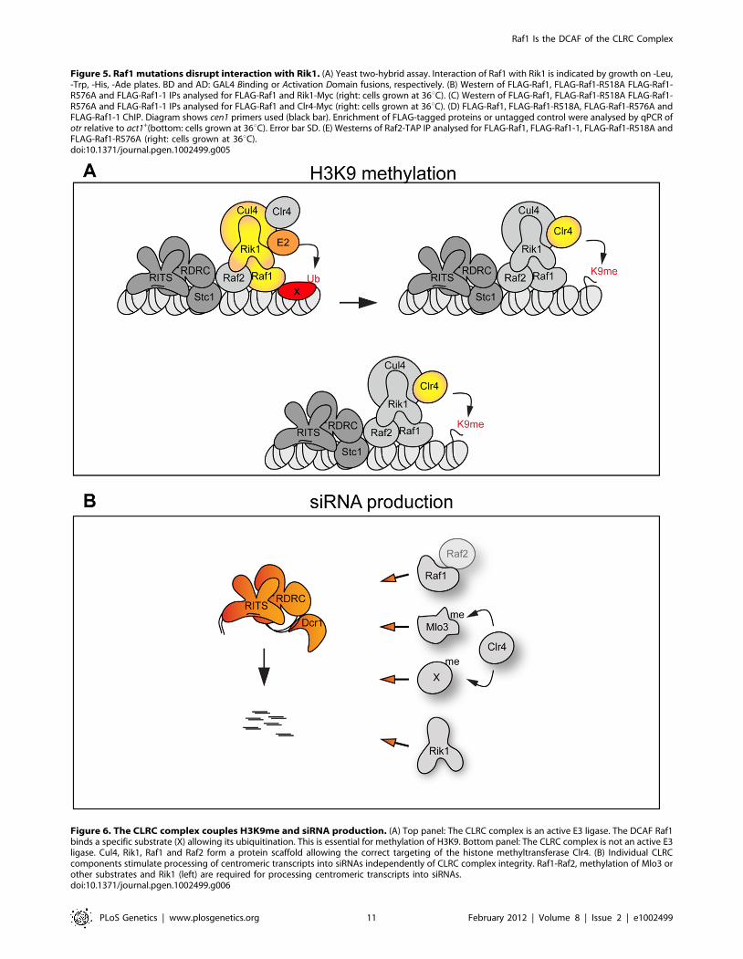
Figure 5. Raf1 mutations disrupt interaction with Rik1. (A) Yeast two-hybrid assay. Interaction of Raf1 with Rik1 is indicated by growth on -Leu,-Trp, -His, -Ade plates. BD and AD: GAL4 Binding or Activation Domain fusions, respectively. (B) Western of FLAG-Raf1, FLAG-Raf1-R518A FLAG-Raf1-R576A and FLAG-Raf1-1 IPs analysed for FLAG-Raf1 and Rik1-Myc (right: cells grown at 36uC). (C) Western of FLAG-Raf1, FLAG-Raf1-R518A FLAG-Raf1-R576A and FLAG-Raf1-1 IPs analysed for FLAG-Raf1 and Clr4-Myc (right: cells grown at 36uC). (D) FLAG-Raf1, FLAG-Raf1-R518A, FLAG-Raf1-R576A andFLAG-Raf1-1 ChIP. Diagram shows cen1 primers used (black bar). Enrichment of FLAG-tagged proteins or untagged control were analysed by qPCR ofotr relative to act1+(bottom: cells grown at 36uC). Error bar SD. (E) Westerns of Raf2-TAP IP analysed for FLAG-Raf1, FLAG-Raf1-1, FLAG-Raf1-R518A andFLAG-Raf1-R576A (right: cells grown at 36uC).doi:10.1371/journal.pgen.1002499.g005
Figure 6. The CLRC complex couples H3K9me and siRNA production. (A) Top panel: The CLRC complex is an active E3 ligase. The DCAF Raf1binds a specific substrate (X) allowing its ubiquitination. This is essential for methylation of H3K9. Bottom panel: The CLRC complex is not an active E3ligase. Cul4, Rik1, Raf1 and Raf2 form a protein scaffold allowing the correct targeting of the histone methyltransferase Clr4. (B) Individual CLRCcomponents stimulate processing of centromeric transcripts into siRNAs independently of CLRC complex integrity. Raf1-Raf2, methylation of Mlo3 orother substrates and Rik1 (left) are required for processing centromeric transcripts into siRNAs.doi:10.1371/journal.pgen.1002499.g006
Raf1 Is the DCAF of the CLRC Complex
PLoS Genetics | www.plosgenetics.org 11 February 2012 | Volume 8 | Issue 2 | e1002499
study are described in Table S1. Primer sequences are listed in
Table S2.
Structural modellingThe homology model of Raf1 was created with the program
Modeller (9v8) using the structure of DDB2 (PDB code 3EI4) [44].
Iterative rounds of alignment adjustment of the two protein
sequences and model building were attempted until the Modeller
scores (diagnostic of model quality) were optimised. The homology
model of Rik1 was created with the program Modeller (9v8) using
5 templates (PDB code: 2B5L; 317N; 3189; 318C; 3E0C). The
generated model (85.2) had the following model scores: RMSD:
1.536; MolPDF: 453.98; DOPE: 211385.
Chromatin immunoprecipitationChromatin immunoprecipitation (ChIP) was performed as
described [54] with the following modifications. Cells were fixed
in 1% PFA/15 min for H3K9me2 ChIP or in 1% PFA/20 min
for RNA Polymerase II ChIP. For ChIP analyses of raf1-R518A
and raf1-R576A strains, cells were grown at 32uC. For ChIP
analyses of raf1-1 strain, cells were kept at the indicated
temperature (25uC, 32uC or 36uC) for at least 96 hours. One
microliter of monoclonal H3K9me2 antibody (m5.1.1), two
microliter of anti-FLAG M2 monoclonal antibody (Sigma,
F1804) or five microliters of RNA Polymerase II 8WG16
antibody (COVANCE, MMS-126R) was used per ChIP. Duplex
PCR was performed to analyse ChIP samples using oligonucle-
otides specific to the regions of interest and to the control gene
fbp1 (Table S2). Real-time PCR (qPCR) was performed using the
LightCycler 480 SYBR Green I Master (Roche) on a LightCycler
480 Instrument (Roche). qPCR analysis primers are in Table S2.
Relative enrichments were calculated as the ratio of product of
interest to control product (act1+ or tRNA) in IP over input.
Histograms represent data from three biological replicates
analysed in parallel.
Immunoaffinity purificationImmunoaffinity purifications (IP) for LC-MS/MS analysis were
performed as described [55], with the following modifications: 5 g
of cells were resuspended in ice-cold-lysis buffer (50 mM Hepes
pH7.5, 150 mM KCl, 0.1% NP40). Immunoprecipitation was
performed using proteinG Dynabeads resin (Life Technologies)
coupled to anti-FLAG M2 antibody (Sigma, F1804) for 15 min.
The IP’d material was treated with 500 U Benzonase, washed,
subjected to on-bead Tryptic digestion, and prepared for LC-MS/
MS analysis as described previously [56].
Co-IPs for Western analysis were performed on 2 g of cells as
above but for 1 hr. IP’d material was washed four times with ice-
cold lysis buffer (50 mM Hepes pH7.5, 150 mM KCl, 0.1%
NP40), resuspended in SDS sample buffer and analysed by SDS-
PAGE. For tandem affinity purification (TAP)-tagged strains,
Dynabeads coupled to IgG were used (gift from K. Hardwick). For
Western analysis the following antibodies were used: anti-FLAG
M2 (Sigma, F1804), anti-HA 12CA5 (gift from K. Samejima) and
anti-myc (A14) (Santa Cruz sc-789), all at 1:1000. Further details
of IP protocol are provided in Text S1.
RNA analysisNorthern analysis of long non-coding centromeric transcripts
and centromeric siRNAs were performed as described previously
[56]. RNA probes are listed in Table S2.
Note: Further details of all experimental procedures are
provided in Text S1.
Supporting Information
Figure S1 Lid2-TAP is not pulled down with FLAG-Rik1. (A)
Centromere silencing assay. Top: Position of ade6+ marker gene in
cen1. Bottom: Wild-type cells with silenced cen1:ade6+ form red
colonies. Cells expressing FLAG-Rik1 form red colonies. Loss of
silencing results in white colonies, as observed in raf1D cells. (B)
Lid2-TAP IP followed by western with FLAG antibody to detect
Rik1-FLAG. I: 5% of input; IP: immunoprecipitation.
(TIF)
Figure S2 Rik1 contains 21 WD-40 repeats and Raf1 can adopt
a DDB2-like structure. (A) Detection and comparison of WD-40
repeats in human DDB1 and Rik1. Alignment of DDB1 and Rik1
along with predicted structure. The 21 WD-40 repeats are
highlighted in blue which were identified manually in Rik1 by
comparison of secondary structures using PSIPRED. (B) Com-
parison of DDB2 structure (left) with Raf1 model (right). Both
proteins contain an N-terminal H-T-H (red) and a 7 bladed b-
propeller (green). Unlike DDB2, Raf1 contains an additional
domain (blue) of unknown function.
(TIF)
Figure S3 Raf1 contains two WDxR motifs important for
protein function. (A) Representative multiple sequence alignment
of the C-terminal WD-40 repeats of Raf1 in homologous proteins
from fungi. Mutated residues mentioned in the text (T495I,
R518A, R576A) are highlighted. (B) Western analyses of untagged,
FLAG-Raf1, FLAG-Raf1R518A and FLAG-Raf1R576A from whole
cell extracts. Wild-type and mutant Raf1 proteins (arrow) are
expressed at similar levels. Asterisk *: indicates cross-reacting band
which serves as a loading control. (C) Western analyses of
untagged and Clr4-Myc in wild-type, raf1-R518A and raf1-R576A
whole cell extracts. Loading control: Bip1. (D) Assay for silencing
at mat3-M:ura4+. Plates are non-selective (N/S), lacking uracil (-
URA) or supplemented with 5-FOA. Loss of silencing results in
growth on -URA and loss of resistance to 5-FOA. (E) Top view of
Raf1 b-propeller ring. Residues R518 and R576 are highlighted.
(F) Northern: centromeric siRNAs in wild-type, clr4D, raf1-R518A
and raf1-R576A cells. Loading control: snoRNA58. (G) Rik1-Raf1
model showing the position of residues R518 and R576.
(TIF)
Figure S4 raf1-1 impairs heterochromatin formation. (A)
Western analyses of untagged, FLAG-Raf1 and FLAG-Raf1-1
from whole cell extracts. (B) Western analyses of Clr4-Myc in wild-
type and raf1-1 whole cell extracts. A strain not expressing Clr4-
Myc (no Myc) is included as a control. Loading control: Bip1. Cells
were grown at 36uC. (C) Assay for silencing at mat3-M:ura4+. Plates
are non-selective (N/S), lacking uracil (-URA) or supplemented
with 5-FOA. Loss of silencing results in growth on -URA and loss
of resistance to 5-FOA. (D) Assay for silencing at tel1L:his3+. Plates
are non-selective (N/S) and lacking histidine (-HIS). Loss of
silencing results in growth on –HIS. (E) Lagging chromosomes in
anaphase wild-type and raf1-1 cells at 25uC or 36uC. Represen-
tative images of fixed cells with DAPI (red) and anti-tubulin
(green). (F) Northern: centromeric siRNAs in wild-type, clr4D, raf1-
1 cells. Cells were shifted from 25uC to 36uC for the indicated
period of time (3, 6 and 12 days resulted in 36, 72 and 144
divisions, respectively, at the restrictive temperature). Loading
control: snoRNA58.
(TIF)
Figure S5 raf1-1, raf1-R518A and raf1-R576A disrupts CLRC
and heterochromatin integrity. (A) Rik1-Raf1 model showing the
position of the T495I mutatedresidue in raf1-1. (B) Westerns of
Rik1-FLAG IP analysed for Clr4-Myc in wild-type, raf1-R518A,
Raf1 Is the DCAF of the CLRC Complex
PLoS Genetics | www.plosgenetics.org 12 February 2012 | Volume 8 | Issue 2 | e1002499
raf1-R576A and raf1-1 cells. Right: cells grown at 36uC. (C)
Westerns of Stc1-FLAG IP analysed for Rik1-Myc in wild-type,
raf1-R518A, raf1-R576A and raf1-1 cells. Right: cells grown at
36uC. (D) Northern: centromeric siRNAs in wild-type, raf1-1,
clr4D, and raf1D cells. Cells grown at 36uC. Loading control:
snoRNA58. (E) RNAPII ChIP in wild-type, clr4D, raf1-R518A and
raf1-R576A cells. Diagram (left) indicates position of cen1 primers
used (grey bar). RNAPII enrichment (right) was analysed by qPCR
relative to tRNA gene primers. Error bar SD.
(TIF)
Table S1 List of strains used in this study.
(DOC)
Table S2 List of primers used in this study.
(DOC)
Text S1 Supplementary experimental procedures.
(DOC)
Acknowledgments
We are grateful to the following for strains and reagents: S. I. Grewal, D.
Moazed, W. Z. Cande, K. Hardwick. We thank A. Pidoux and members of
the Allshire lab for discussion and comments.
Author Contributions
Conceived and designed the experiments: AB SAW FS RCA. Performed
the experiments: AB SAW DRH EL FS FdLA PTD EHB. Analyzed the
data: AB SAW DRH EL FS FdLA JR RCA. Contributed reagents/
materials/analysis tools: TU DRH JR. Wrote the paper: AB RCA.
References
1. Farazi TA, Juranek SA, Tuschl T (2008) The growing catalog of small RNAs
and their association with distinct Argonaute/Piwi family members. Develop-
ment 135: 1201–1214.
2. Grewal SI (2010) RNAi-dependent formation of heterochromatin and its diverse
functions. Curr Opin Genet Dev 20: 134–141.
3. Moazed D (2009) Small RNAs in transcriptional gene silencing and genome
defence. Nature 457: 413–420.
4. Guang S, Bochner AF, Burkhart KB, Burton N, Pavelec DM, et al. (2010) Small
regulatory RNAs inhibit RNA polymerase II during the elongation phase of
transcription. Nature 465: 1097–1101.
5. Kuramochi-Miyagawa S, Watanabe T, Gotoh K, Totoki Y, Toyoda A, et al.
(2008) DNA methylation of retrotransposon genes is regulated by Piwi family
members MILI and MIWI2 in murine fetal testes. Genes Dev 22: 908–917.
6. Allshire RC, Javerzat JP, Redhead NJ, Cranston G (1994) Position effect
variegation at fission yeast centromeres. Cell 76: 157–169.
7. Djupedal I, Portoso M, Spahr H, Bonilla C, Gustafsson CM, et al. (2005) RNA
Pol II subunit Rpb7 promotes centromeric transcription and RNAi-directed
chromatin silencing. Genes Dev 19: 2301–2306.
8. Kato H, Goto DB, Martienssen RA, Urano T, Furukawa K, et al. (2005) RNA
polymerase II is required for RNAi-dependent heterochromatin assembly.
Science 309: 467–469.
9. Volpe TA, Kidner C, Hall IM, Teng G, Grewal SI, et al. (2002) Regulation of
heterochromatic silencing and histone H3 lysine-9 methylation by RNAi.
Science 297: 1833–1837.
10. Bayne EH, White SA, Kagansky A, Bijos DA, Sanchez-Pulido L, et al. (2010)
Stc1: a critical link between RNAi and chromatin modification required for
heterochromatin integrity. Cell 140: 666–677.
11. Sadaie M, Iida T, Urano T, Nakayama J (2004) A chromodomain protein,
Chp1, is required for the establishment of heterochromatin in fission yeast.
Embo J 23: 3825–3835.
12. Bannister AJ, Zegerman P, Partridge JF, Miska EA, Thomas JO, et al. (2001)
Selective recognition of methylated lysine 9 on histone H3 by the HP1 chromo
domain. Nature 410: 120–124.
13. Zhang K, Mosch K, Fischle W, Grewal SI (2008) Roles of the Clr4
methyltransferase complex in nucleation, spreading and maintenance of
heterochromatin. Nat Struct Mol Biol 15: 381–388.
14. Petrie VJ, Wuitschick JD, Givens CD, Kosinski AM, Partridge JF (2005) RNA
interference (RNAi)-dependent and RNAi-independent association of the Chp1
chromodomain protein with distinct heterochromatic loci in fission yeast. Mol
Cell Biol 25: 2331–2346.
15. Sugiyama T, Cam HP, Sugiyama R, Noma K, Zofall M, et al. (2007) SHREC,
an effector complex for heterochromatic transcriptional silencing. Cell 128:
491–504.
16. Buhler M, Verdel A, Moazed D (2006) Tethering RITS to a nascent transcript
initiates RNAi- and heterochromatin-dependent gene silencing. Cell 125:
873–886.
17. Hong EJ, Villen J, Gerace EL, Gygi SP, Moazed D (2005) A cullin E3 ubiquitin
ligase complex associates with Rik1 and the Clr4 histone H3-K9 methyltrans-
ferase and is required for RNAi-mediated heterochromatin formation. RNA Biol
2: 106–111.
18. Motamedi MR, Verdel A, Colmenares SU, Gerber SA, Gygi SP, et al. (2004)
Two RNAi complexes, RITS and RDRC, physically interact and localise to
noncoding centromeric RNAs. Cell 119: 789–802.
19. Noma K, Sugiyama T, Cam H, Verdel A, Zofall M, et al. (2004) RITS acts in cis
to promote RNA interference-mediated transcriptional and post-transcriptional
silencing. Nat Genet 36: 1174–1180.
20. Djupedal I, Kos-Braun IC, Mosher RA, Soderholm N, Simmer F, et al. (2009)
Analysis of small RNA in fission yeast; centromeric siRNAs are potentially
generated through a structured RNA. EMBO J 28: 3832–3844.
21. Gerace EL, Halic M, Moazed D (2010) The methyltransferase activity of
Clr4Suv39h triggers RNAi independently of histone H3K9 methylation. Mol
Cell 39: 360–372.
22. Jia S, Kobayashi R, Grewal SI (2005) Ubiquitin ligase component Cul4
associates with Clr4 histone methyltransferase to assemble heterochromatin. Nat
Cell Biol 7: 1007–1013.
23. Li F, Goto DB, Zaratiegui M, Tang X, Martienssen R, et al. (2005) Two novel
proteins, dos1 and dos2, interact with rik1 to regulate heterochromatic RNA
interference and histone modification. Curr Biol 15: 1448–1457.
24. Horn PJ, Bastie JN, Peterson CL (2005) A Rik1-associated, cullin-dependent E3
ubiquitin ligase is essential for heterochromatin formation. Genes Dev 19:
1705–1714.
25. Thon G, Hansen KR, Altes SP, Sidhu D, Singh G, et al. (2005) The Clr7 and Clr8
directionality factors and the Pcu4 cullin mediate heterochromatin formation in
the fission yeast Schizosaccharomyces pombe. Genetics 171: 1583–1595.
26. Jackson S, Xiong Y (2009) CRL4s: the CUL4-RING E3 ubiquitin ligases.
Trends Biochem Sci 34: 562–570.
27. Shilatifard A (2006) Chromatin modifications by methylation and ubiquitina-
tion: implications in the regulation of gene expression. Annu Rev Biochem 75:
243–269.
28. Petroski MD, Deshaies RJ (2005) Function and regulation of cullin-RING
ubiquitin ligases. Nat Rev Mol Cell Biol 6: 9–20.
29. Kapetanaki MG, Guerrero-Santoro J, Bisi DC, Hsieh CL, Rapic-Otrin V, et al.
(2006) The DDB1-CUL4ADDB2 ubiquitin ligase is deficient in xeroderma
pigmentosum group E and targets histone H2A at UV-damaged DNA sites. Proc
Natl Acad Sci U S A 103: 2588–2593.
30. Wang H, Zhai L, Xu J, Joo HY, Jackson S, et al. (2006) Histone H3 and H4
ubiquitylation by the CUL4-DDB-ROC1 ubiquitin ligase facilitates cellular
response to DNA damage. Mol Cell 22: 383–394.
31. Higa LA, Wu M, Ye T, Kobayashi R, Sun H, et al. (2006) CUL4-DDB1
ubiquitin ligase interacts with multiple WD-40-repeat proteins and regulates
histone methylation. Nat Cell Biol 8: 1277–1283.
32. Jin J, Arias EE, Chen J, Harper JW, Walter JC (2006) A family of diverse Cul4-
Ddb1-interacting proteins includes Cdt2, which is required for S phase
destruction of the replication factor Cdt1. Mol Cell 23: 709–721.
33. Angers S, Li T, Yi X, MacCoss MJ, Moon RT, et al. (2006) Molecular
architecture and assembly of the DDB1-CUL4A ubiquitin ligase machinery.
Nature 443: 590–593.
34. He YJ, McCall CM, Hu J, Zeng Y, Xiong Y (2006) DDB1 functions as a linker
to recruit receptor WD-40 proteins to CUL4-ROC1 ubiquitin ligases. Genes
Dev 20: 2949–2954.
35. Liu C, Powell KA, Mundt K, Wu L, Carr AM, et al. (2003) Cop9/signalosome
subunits and Pcu4 regulate ribonucleotide reductase by both checkpoint-
dependent and -independent mechanisms. Genes Dev 17: 1130–1140.
36. Neuwald AF, Poleksic A (2000) PSI-BLAST searches using hidden markov
models of structural repeats: prediction of an unusual sliding DNA clamp and of
beta-propellers in UV-damaged DNA-binding protein. Nucleic Acids Res 28:
3570–3580.
37. Braun S, Garcia JF, Rowley M, Rougemaille M, Shankar S, et al. (2011) The
cul4-ddb1(cdt2) ubiquitin ligase inhibits invasion of a boundary-associated
antisilencing factor into heterochromatin. Cell 144: 41–54.
38. Pidoux AL, Uzawa S, Perry PE, Cande WZ, Allshire RC (2000) Live analysis of
lagging chromosomes during anaphase and their effect on spindle elongation
rate in fission yeast. J Cell Sci 113 Pt 23: 4177–4191.
39. Li F, Huarte M, Zaratiegui M, Vaughn MW, Shi Y, et al. (2008) Lid2 is required
for coordinating H3K4 and H3K9 methylation of heterochromatin and
euchromatin. Cell 135: 272–283.
40. Bernard P, Maure JF, Partridge JF, Genier S, Javerzat JP, et al. (2001) Requirement
of heterochromatin for cohesion at centromeres. Science 294: 2539–2542.
Raf1 Is the DCAF of the CLRC Complex
PLoS Genetics | www.plosgenetics.org 13 February 2012 | Volume 8 | Issue 2 | e1002499
41. Nonaka N, Kitajima T, Yokobayashi S, Xiao G, Yamamoto M, et al. (2002)
Recruitment of cohesin to heterochromatic regions by Swi6/HP1 in fissionyeast. Nat Cell Biol 4: 89–93.
42. Ekwall K, Javerzat JP, Lorentz A, Schmidt H, Cranston G, et al. (1995) The
chromodomain protein Swi6: a key component at fission yeast centromeres.Science 269: 1429–1431.
43. Hall IM, Shankaranarayana GD, Noma K, Ayoub N, Cohen A, et al. (2002)Establishment and maintenance of a heterochromatin domain. Science 297:
2232–2237.
44. Scrima A, Konickova R, Czyzewski BK, Kawasaki Y, Jeffrey PD, et al. (2008)Structural basis of UV DNA-damage recognition by the DDB1-DDB2 complex.
Cell 135: 1213–1223.45. Han Z, Guo L, Wang H, Shen Y, Deng XW, et al. (2006) Structural basis for the
specific recognition of methylated histone H3 lysine 4 by the WD-40 proteinWDR5. Mol Cell 22: 137–144.
46. Couture JF, Collazo E, Trievel RC (2006) Molecular recognition of histone H3
by the WD-40 protein WDR5. Nat Struct Mol Biol 13: 698–703.47. Schuetz A, Allali-Hassani A, Martin F, Loppnau P, Vedadi M, et al. (2006)
Structural basis for molecular recognition and presentation of histone H3 byWDR5. EMBO J 25: 4245–4252.
48. Zhang K, Fischer T, Porter RL, Dhakshnamoorthy J, Zofall M, et al. (2011)
Clr4/Suv39 and RNA quality control factors cooperate to trigger RNAi andsuppress antisense RNA. Science 331: 1624–1627.
49. Li F, Martienssen R, Cande WZ (2011) Coordination of DNA replication and
histone modification by the Rik1-Dos2 complex. Nature 475: 244–248.
50. Chen ES, Zhang K, Nicolas E, Cam HP, Zofall M, et al. (2008) Cell cycle
control of centromeric repeat transcription and heterochromatin assembly.
Nature 451: 734–737.
51. Kloc A, Zaratiegui M, Nora E, Martienssen R (2008) RNA interference guides
histone modification during the S phase of chromosomal replication. Curr Biol
18: 490–495.
52. Kagansky A, Folco HD, Almeida R, Pidoux AL, Boukaba A, et al. (2009)
Synthetic heterochromatin bypasses RNAi and centromeric repeats to establish
functional centromeres. Science 324: 1716–1719.
53. Moreno S, Klar A, Nurse P (1991) Molecular genetic analysis of fission yeast
Schizosaccharomyces pombe. Methods Enzymol 194: 795–823.
54. Pidoux A, Mellone B, Allshire R (2004) Analysis of chromatin in fission yeast.
Methods 33: 252–259.
55. Oeffinger M, Wei KE, Rogers R, DeGrasse JA, Chait BT, et al. (2007)
Comprehensive analysis of diverse ribonucleoprotein complexes. Nat Methods 4:
951–956.
56. Bayne EH, Portoso M, Kagansky A, Kos-Braun IC, Urano T, et al. (2008)
Splicing factors facilitate RNAi-directed silencing in fission yeast. Science 322:
602–606.
Raf1 Is the DCAF of the CLRC Complex
PLoS Genetics | www.plosgenetics.org 14 February 2012 | Volume 8 | Issue 2 | e1002499


![10 Hanggi 2003 Making Sense of Security Sector Governance [Dcaf]](https://img.pdfslide.us/doc/110x75/5447faccb1af9f5b618b4705/10-hanggi-2003-making-sense-of-security-sector-governance-dcaf.jpg)


![WIM Nepal DCAF Strategic Concept Low[1]](https://img.pdfslide.us/doc/110x75/577d234e1a28ab4e1e9977a0/wim-nepal-dcaf-strategic-concept-low1.jpg)