Embed Size (px)
Citation preview
Radiotherapy
Marta Anguiano Millan
Departamento de Fısica Atomica, Molecular y Nuclear
Facultad de Ciencias. Universidad de Granada
Marta Anguiano Millan Radiotherapy
Overview
Introduction
Marta Anguiano Millan Radiotherapy

Overview
Introduction
Brachytherapy
Radioisotopes in contact with the tumor
Marta Anguiano Millan Radiotherapy
Overview
Introduction
Brachytherapy
Radioisotopes in contact with the tumor
External Radiotherapy
X-ray emitting equipment
γ-ray equipment
LINACS
Applications
Marta Anguiano Millan Radiotherapy
Introduction
Goal of the radiotherapy: to get a compromise between theadministration of a high dose to the tumour and a veryreduced dose to the surrounding healthy tissues.
Marta Anguiano Millan Radiotherapy
Introduction
Goal of the radiotherapy: to get a compromise between theadministration of a high dose to the tumour and a veryreduced dose to the surrounding healthy tissues.
Very high dose → Rate of problems grows.
Marta Anguiano Millan Radiotherapy
Introduction
Goal of the radiotherapy: to get a compromise between theadministration of a high dose to the tumour and a veryreduced dose to the surrounding healthy tissues.
Very high dose → Rate of problems grows.
Very low dose → The TCP is reduced.
Marta Anguiano Millan Radiotherapy
Introduction
Goal of the radiotherapy: to get a compromise between theadministration of a high dose to the tumour and a veryreduced dose to the surrounding healthy tissues.
Very high dose → Rate of problems grows.
Very low dose → The TCP is reduced.
High doses (40-70 Gy) in delimited areas, with curativepurposes
Marta Anguiano Millan Radiotherapy
Introduction
Goal of the radiotherapy: to get a compromise between theadministration of a high dose to the tumour and a veryreduced dose to the surrounding healthy tissues.
Very high dose → Rate of problems grows.
Very low dose → The TCP is reduced.
High doses (40-70 Gy) in delimited areas, with curativepurposes.
Different sources and energies, depending on1 Type of tumour.2 Location of tumour.3 Depth of tumor.
Marta Anguiano Millan Radiotherapy
Brachytheraphy
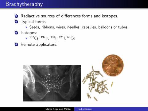
1 Radiactive sources of differences forms and isotopes.2 Typical forms:
Seeds, ribbons, wires, needles, capsules, balloons or tubes.3 Isotopes:
137Cs, 192Ir, 131I, 125I, 60Co
4 Remote applicators.
Marta Anguiano Millan Radiotherapy
Brachytherapy
Radioisotopes in brachytherapy
Marta Anguiano Millan Radiotherapy
Brachytherapy
Radioisotopes in brachytherapy
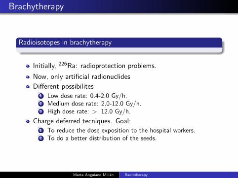
Initially, 226Ra: radioprotection problems.
Marta Anguiano Millan Radiotherapy
Brachytherapy
Radioisotopes in brachytherapy
Initially, 226Ra: radioprotection problems.
Now, only artificial radionuclides
Marta Anguiano Millan Radiotherapy
Brachytherapy
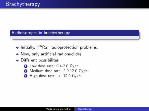
Radioisotopes in brachytherapy
Initially, 226Ra: radioprotection problems.
Now, only artificial radionuclides
Different possibilites1 Low dose rate: 0.4-2.0 Gy/h.2 Medium dose rate: 2.0-12.0 Gy/h.3 High dose rate: > 12.0 Gy/h.
Marta Anguiano Millan Radiotherapy
Brachytherapy
Radioisotopes in brachytherapy
Initially, 226Ra: radioprotection problems.
Now, only artificial radionuclides
Different possibilites1 Low dose rate: 0.4-2.0 Gy/h.2 Medium dose rate: 2.0-12.0 Gy/h.3 High dose rate: > 12.0 Gy/h.
Charge deferred tecniques. Goal:1 To reduce the dose exposition to the hospital workers.2 To do a better distribution of the seeds.
Marta Anguiano Millan Radiotherapy
Brachytherapy
Radioisotopes in brachytherapy
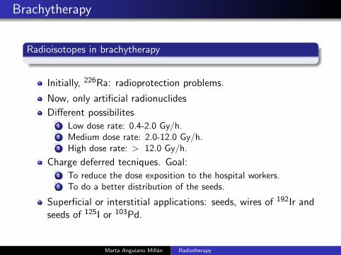
Initially, 226Ra: radioprotection problems.
Now, only artificial radionuclides
Different possibilites1 Low dose rate: 0.4-2.0 Gy/h.2 Medium dose rate: 2.0-12.0 Gy/h.3 High dose rate: > 12.0 Gy/h.
Charge deferred tecniques. Goal:1 To reduce the dose exposition to the hospital workers.2 To do a better distribution of the seeds.
Superficial or interstitial applications: seeds, wires of 192Ir andseeds of 125I or 103Pd.
Marta Anguiano Millan Radiotherapy
Brachytherapy
Radioisotopes in brachytherapy
Initially, 226Ra: radioprotection problems.Now, only artificial radionuclidesDifferent possibilites
1 Low dose rate: 0.4-2.0 Gy/h.2 Medium dose rate: 2.0-12.0 Gy/h.3 High dose rate: > 12.0 Gy/h.
Charge deferred tecniques. Goal:1 To reduce the dose exposition to the hospital workers.2 To do a better distribution of the seeds.
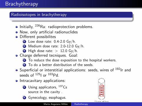
Superficial or interstitial applications: seeds, wires of 192Ir andseeds of 125I or 103Pd.Intracavitary applications:
1 Using applicators, 137Cssource in the cavity .
2 Gynecology, esophagus.
Marta Anguiano Millan Radiotherapy
Brachytherapy
Radioisotopes in brachytherapy
Marta Anguiano Millan Radiotherapy
Brachytherapy
Radioisotopes in brachytherapy
Specific applications:1 Eyes tumours: plaques of 125I, applied directly on the tumour.
100 Gy, between 5 and 12 days.2 Intravascular brachytherapy: Coronary diseases. Lessions
around mm. Radionuclide emitting low energy photons: 125Iand 103Pd.
Marta Anguiano Millan Radiotherapy
Brachytherapy
Radioisotopes in brachytherapy
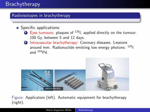
Specific applications:1 Eyes tumours: plaques of 125I, applied directly on the tumour.
100 Gy, between 5 and 12 days.2 Intravascular brachytherapy: Coronary diseases. Lessions
around mm. Radionuclide emitting low energy photons: 125Iand 103Pd.
Figure: Applicators (left). Automatic equipment for brachytherapy(right).
Marta Anguiano Millan Radiotherapy
External Radiotherapy (ER)
Types of equipment in ER
Marta Anguiano Millan Radiotherapy
External Radiotherapy (ER)
Types of equipment in ER
Purpose of ER: to administrate in a number of sesions(Treatment), a specific quantity of energy in the targetvolume, without affecting the healthy structures.
Marta Anguiano Millan Radiotherapy
External Radiotherapy (ER)
Types of equipment in ER
Purpose of ER: to administrate in a number of sesions(Treatment), a specific quantity of energy in the targetvolume, without affecting the healthy structures.
Dose/tumour/session ≈ 1.8-2.0 Gy, in very short times.
The ER process implies:1 Location of the tumour.2 Simulation of the treatment.3 Anatomical data collection of the patient.4 Placement of the target volume to radiate and critical organs.5 Calculation of the dose.6 Elaboration of individual protections.7 Positioning of the beams.8 Treatment.
Marta Anguiano Millan Radiotherapy
External Radiotherapy (ER)
Types of equipment in ER
Purpose of ER: to administrate in a number of sesions(Treatment), a specific quantity of energy in the targetvolume, without affecting the healthy structures.
Dose/tumour/session ≈ 1.8-2.0 Gy, in very short times.The ER process implies:
1 Location of the tumour.2 Simulation of the treatment.3 Anatomical data collection of the patient.4 Placement of the target volume to radiate and critical organs.5 Calculation of the dose.6 Elaboration of individual protections.7 Positioning of the beams.8 Treatment.
Types of equipment: X-ray equipment, γ-ray equipment,LINACS,..
Marta Anguiano Millan Radiotherapy
External Radiotherapy (ER)
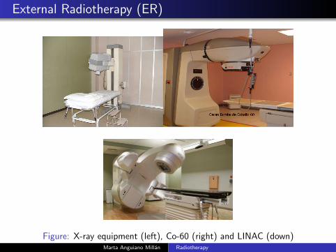
Figure: X-ray equipment (left), Co-60 (right) and LINAC (down)Marta Anguiano Millan Radiotherapy
External Radiotherapy (ER)
X-ray emitting equipment
Marta Anguiano Millan Radiotherapy
External Radiotherapy (ER)
X-ray emitting equipment
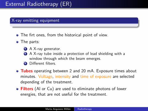
The firt ones, from the historical point of view.
Marta Anguiano Millan Radiotherapy
External Radiotherapy (ER)
X-ray emitting equipment
The firt ones, from the historical point of view.
The parts:1 A X-ray generator.2 A X-ray tube inside a protection of lead shielding with a
window through which the beam emerges.3 Different filters.
Marta Anguiano Millan Radiotherapy
External Radiotherapy (ER)
X-ray emitting equipment
The firt ones, from the historical point of view.
The parts:
1 A X-ray generator.2 A X-ray tube inside a protection of lead shielding with a
window through which the beam emerges.3 Different filters.
Tubes operating between 2 and 20 mA. Exposure times aboutminutes. Voltage, intensity an time of exposure are selecteddepending of the treatment.
Marta Anguiano Millan Radiotherapy
External Radiotherapy (ER)
X-ray emitting equipment
The firt ones, from the historical point of view.
The parts:
1 A X-ray generator.2 A X-ray tube inside a protection of lead shielding with a
window through which the beam emerges.3 Different filters.
Tubes operating between 2 and 20 mA. Exposure times aboutminutes. Voltage, intensity and time of exposure are selecteddepending of the treatment.
Filters (Al or Cu) are used to eliminate photons of lowerenergies, that are not useful for the treatment.
Marta Anguiano Millan Radiotherapy
External Radiotherapy (RE)
X-ray equipment
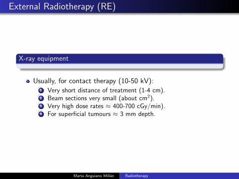
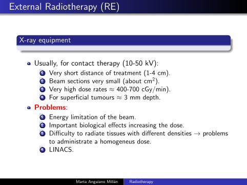
Usually, for contact therapy (10-50 kV):1 Very short distance of treatment (1-4 cm).2 Beam sections very small (about cm2).3 Very high dose rates ≈ 400-700 cGy/min).4 For superficial tumours ≈ 3 mm depth.
Marta Anguiano Millan Radiotherapy
External Radiotherapy (RE)
X-ray equipment
Usually, for contact therapy (10-50 kV):1 Very short distance of treatment (1-4 cm).2 Beam sections very small (about cm2).3 Very high dose rates ≈ 400-700 cGy/min).4 For superficial tumours ≈ 3 mm depth.
Problems:1 Energy limitation of the beam.2 Important biological effects increasing the dose.3 Difficulty to radiate tissues with different densities → problems
to administrate a homogeneus dose.4 LINACS.
Marta Anguiano Millan Radiotherapy
External Radiotherapy (RE)
X-ray equipment
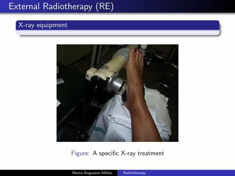
Figure: A specific X-ray treatment
Marta Anguiano Millan Radiotherapy
External Radiotherapy (ER)
γ ray equipment
Marta Anguiano Millan Radiotherapy
External Radiotherapy (ER)
γ ray equipment
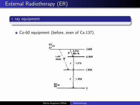
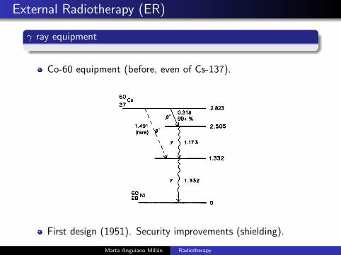
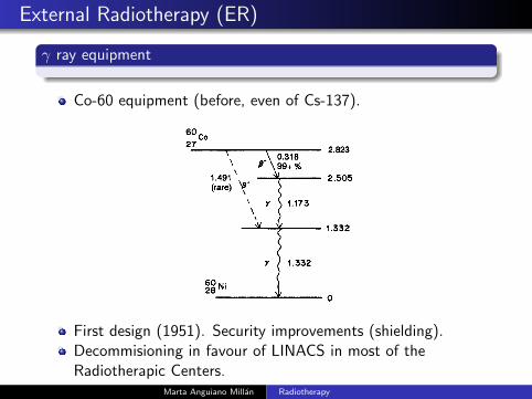
Co-60 equipment (before, even of Cs-137).
Marta Anguiano Millan Radiotherapy
External Radiotherapy (ER)
γ ray equipment
Co-60 equipment (before, even of Cs-137).
Marta Anguiano Millan Radiotherapy
External Radiotherapy (ER)
γ ray equipment
Co-60 equipment (before, even of Cs-137).
First design (1951). Security improvements (shielding).
Marta Anguiano Millan Radiotherapy
External Radiotherapy (ER)
γ ray equipment
Co-60 equipment (before, even of Cs-137).
First design (1951). Security improvements (shielding).Decommisioning in favour of LINACS in most of theRadiotherapic Centers.
Marta Anguiano Millan Radiotherapy
External Radiotherapy (ER)
γ ray equipment
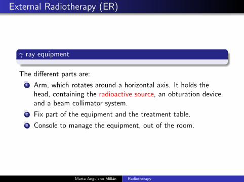
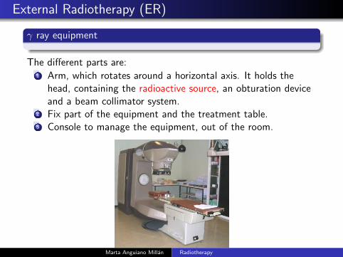
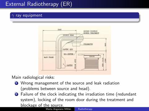
The different parts are:
1 Arm, which rotates around a horizontal axis. It holds thehead, containing the radioactive source, an obturation deviceand a beam collimator system.
2 Fix part of the equipment and the treatment table.
3 Console to manage the equipment, out of the room.
Marta Anguiano Millan Radiotherapy
External Radiotherapy (ER)
γ ray equipment
The different parts are:1 Arm, which rotates around a horizontal axis. It holds the
head, containing the radioactive source, an obturation deviceand a beam collimator system.
2 Fix part of the equipment and the treatment table.3 Console to manage the equipment, out of the room.
Marta Anguiano Millan Radiotherapy
External Radiotherapy (ER)
γ ray equipment
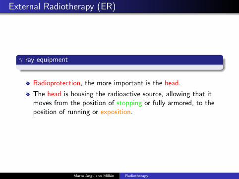
Radioprotection, the more important is the head.
Marta Anguiano Millan Radiotherapy
External Radiotherapy (ER)
γ ray equipment
Radioprotection, the more important is the head.
The head is housing the radioactive source, allowing that itmoves from the position of stopping or fully armored, to theposition of running or exposition.
Marta Anguiano Millan Radiotherapy
External Radiotherapy (ER)
γ ray equipment
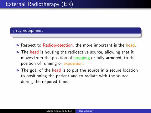
Respect to Radioprotection, the more important is the head.
The head is housing the radioactive source, allowing that itmoves from the position of stopping or fully armored, to theposition of running or exposition.
The goal of the head is to put the source in a secure locationto positioning the patient and to radiate with the sourceduring the required time.
Marta Anguiano Millan Radiotherapy
External Radiotherapy (ER)
γ ray equipment
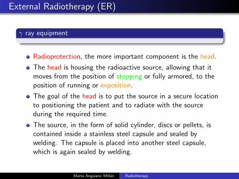
Radioprotection, the more important component is the head.
The head is housing the radioactive source, allowing that itmoves from the position of stopping or fully armored, to theposition of running or exposition.
The goal of the head is to put the source in a secure locationto positioning the patient and to radiate with the sourceduring the required time.
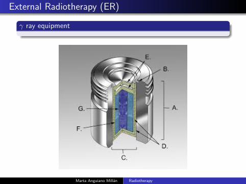
The source, in the form of solid cylinder, discs or pellets, iscontained inside a stainless steel capsule and sealed bywelding. The capsule is placed into another steel capsule,which is again sealed by welding.
Marta Anguiano Millan Radiotherapy
External Radiotherapy (ER)
γ ray equipment
Marta Anguiano Millan Radiotherapy
Radioterapia externa (RE)
γ ray equipment
60Co decays β−. Electrons are slow down by the stainlesssteel capsule. 1.17 and 1.33 MeV photons are used fortreatment. T = 5.27 y → Activity decreases 1% per month.
Marta Anguiano Millan Radiotherapy
Radioterapia externa (RE)
γ ray equipment
60Co decays β−. Electrons are slow down by the stainlesssteel capsule. 1.17 and 1.33 MeV photons are used fortreatment. T = 5.27 y → Activity decreases 1% per month.
Head design must ensure that the radiation leaks areaccording to Radioprotection limits:
1 Workers can go inside the before and after the radiationsessions.
2 Ensure that the patient receives no radiation outside the areato be treated.
Most of the heads are made of cast steel lead filled: primaryshielding.
Isocentric setup
Marta Anguiano Millan Radiotherapy
External Radiotherapy (ER)
γ ray equipment
Main radiological risks:1 Wrong management of the source and leak radiation
(problems between source and head).2 Failure of the clock indicating the irradiation time (redundant
system), locking of the room door during the treatment andblockage of the source.
Marta Anguiano Millan Radiotherapy
External Radiotherapy (ER)
Linear accelerators (LINACS)
Marta Anguiano Millan Radiotherapy
External Radiotherapy (ER)
Linear accelerators (LINACS)
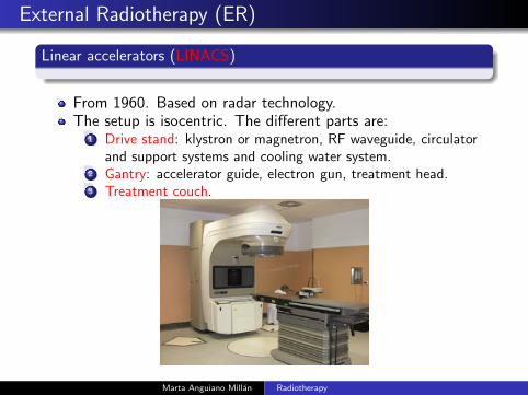
From 1960. Based on radar technology.
Marta Anguiano Millan Radiotherapy
External Radiotherapy (ER)
Linear accelerators (LINACS)
From 1960. Based on radar technology.
The setup is isocentric. The different parts are:1 Drive stand: klystron or magnetron, RF waveguide , circulator
and cooling water system.2 Gantry: accelerator guide, electron gun, bending magnet,
treatment head.3 Treatment couch.
Marta Anguiano Millan Radiotherapy
External Radiotherapy (ER)
Linear accelerators (LINACS)
From 1960. Based on radar technology.The setup is isocentric. The different parts are:
1 Drive stand: klystron or magnetron, RF waveguide, circulatorand support systems and cooling water system.
2 Gantry: accelerator guide, electron gun, treatment head.3 Treatment couch.
Marta Anguiano Millan Radiotherapy
External Radiotherapy (ER)
(LINACS: The modulator cabinet)
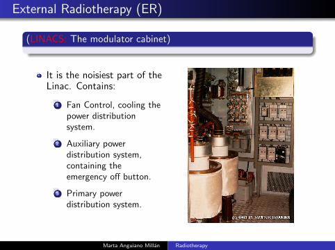
It is the noisiest part of theLinac. Contains:
1 Fan Control, cooling thepower distributionsystem.
2 Auxiliary powerdistribution system,containing theemergency off button.
3 Primary powerdistribution system.
Marta Anguiano Millan Radiotherapy
External Radiotherapy (ER)
Linear accelerators (LINACS: The klystron)
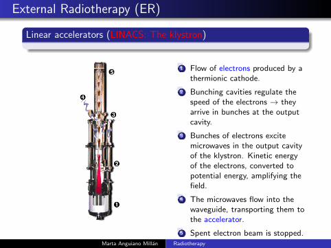
1 Flow of electrons produced by athermionic cathode.
2 Bunching cavities regulate thespeed of the electrons → theyarrive in bunches at the outputcavity.
3 Bunches of electrons excitemicrowaves in the output cavityof the klystron. Kinetic energyof the electrons, converted topotential energy, amplifying thefield.
4 The microwaves flow into thewaveguide, transporting them tothe accelerator.
5 Spent electron beam is stopped.Marta Anguiano Millan Radiotherapy
Radioterapia externa (RE)
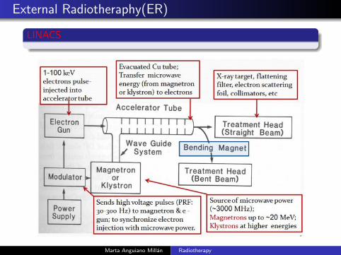
LINACS
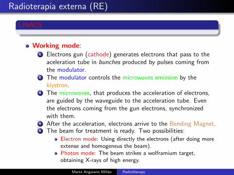
Working mode:1 Electrons gun (cathode) generates electrons that pass to the
aceleration tube in bunches produced by pulses coming fromthe modulator.
2 The modulator controls the microwaves emission by theklystron.
3 The microwaves, that produces the acceleration of electrons,are guided by the waveguide to the acceleration tube. Eventhe electrons coming from the gun electrons, synchronizedwith them.
4 After the acceleration, electrons arrive to the Bending Magnet.5 The beam for treatment is ready. Two possibilities:
Electron mode: Using directly the electrons (after doing moreextense and homogeneus the beam).Photon mode: The beam strikes a wolframium target,obtaining X-rays of high energy.
Marta Anguiano Millan Radiotherapy
External Radiotheraphy(ER)
LINACS
Marta Anguiano Millan Radiotherapy
External radiotherapy (ER)
LINACS
Radiological risks:1 Fail in the interceptation of the beam.2 Fail in the monitorization of the dose.3 Beam not uniform.4 Fail in choosing the modality (electrons or photons).5 Fail in choosing the value of the energy.6 Radiation leak from the head.7 Production of neutrons (LINACS 10 MeV).
Marta Anguiano Millan Radiotherapy
External radiotherapy (ER)
LINACS
Radiological risks:1 Fail in the interceptation of the beam2 Fail in the monitorization of the dose.3 Beam not uniform.4 Fail in choosing the modality (electrons or photons).5 Fail in choosing the value of the energy.6 Radiation leak from the head.7 Production of neutrons (LINACS 10 MeV).
Applications:1 IMRT: Photons beams with modulated intensity → Variable
flow → protection of the healthy tissue.2 Stereotactic radiosurgery: very narrow beams to irradiate
intracraneal structures. 20 Gy.3 Intraoperative radiotheraphy: Electrons applicators.
Marta Anguiano Millan Radiotherapy
External Radiotheraphy(ER)
LINACS
Marta Anguiano Millan Radiotherapy
External Radiotheraphy (ER)
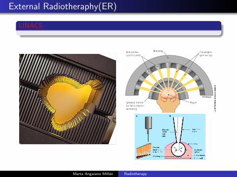
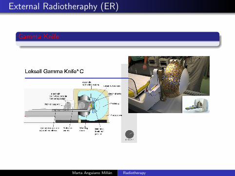
Gamma Knife
Marta Anguiano Millan Radiotherapy
External Radiotheraphy (ER)
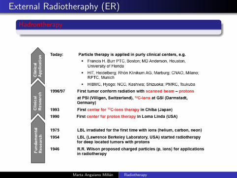
Hadrontherapy
Marta Anguiano Millan Radiotherapy
External Radiotheraphy (ER)
Hadrontherapy
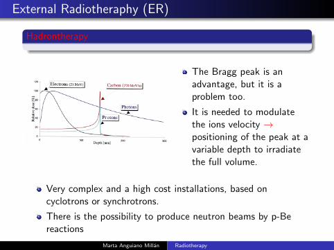
The Bragg peak is anadvantage, but it is aproblem too.
It is needed to modulatethe ions velocity →positioning of the peak at avariable depth to irradiatethe full volume.
Very complex and a high cost installations, based oncyclotrons or synchrotrons.
There is the possibility to produce neutron beams by p-Bereactions
Marta Anguiano Millan Radiotherapy
External Radiotheraphy (ER)
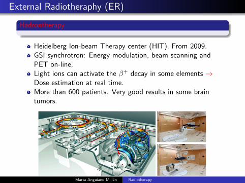
Hadrontherapy
Heidelberg Ion-beam Therapy center (HIT). From 2009.GSI synchrotron: Energy modulation, beam scanning andPET on-line.Light ions can activate the β+ decay in some elements →Dose estimation at real time.More than 600 patients. Very good results in some braintumors.
Marta Anguiano Millan Radiotherapy
External Radiotheraphy (ER)
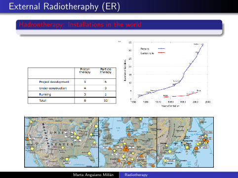
Hadrontherapy: Installations in the world
Marta Anguiano Millan Radiotherapy