Embed Size (px)
Citation preview

F R A N C I S J . C U R R Y N A T I O N A L T U B E R C U L O S I S C E N T E R
CHAPTER TWO
Introduction
In Chapter One, we learned the basic principles of chest radiography and how to read and interpret a chest radiograph using standard terminology.
In Chapter Two, we review the radiographic manifestations of pulmonary tuberculosis and use the terminology we learned in the previous chapterto describe the radiographic findings. By the end of this chapter, you willbe familiar with the various radiographic manifestations of tuberculosis.
Let’s begin with an overview of the pathogenesis of tuberculosis to betterunderstand the radiographic manifestations that you will encounter inyour practice.
Radiographic Manifestations of Tuberculosis
R A D I O G R A P H I C M A N I F E S T A T I O N S O F T U B E R C U L O S I S / 2 - 1

Overview of the Pathogenesis of Tuberculosis
When tubercle bacilli are inhaled into the lung, the bacilli are deposited in the airways and alveoli inmore ventilated areas of the lung—typically in the middle to lower regions. The initial inflammatoryreaction in the lung is referred to as a primary or Ghon focus.
During this early stage of infection, organisms can spread via lymphatics to the draining lymph nodesin the chest and result in enlargement of hilar and mediastinal lymph nodes. Bacilli can also enter theblood stream where they spread hematogenously throughout the body. Disease presenting at thisstage is referred to as primary tuberculosis and is associated with certain radiographic findings, whichwe will review shortly.
After several weeks, the host develops cell-mediated immunity and delayed-type hypersensitivity that,in most cases, result in control of the infection. However, the healed lesions often contain viable bacillithat can progress to disease in the future. Such progression causes post-primary or reactivationtuberculosis. Post-primary disease is also associated with certain radiographic findings related to thefact that the host has now developed cell-mediated immunity and delayed-type hypersensitivity. It is notsurprising that this entire pathogenetic sequence represents a continuum and many of the radiograph-ic manifestations of primary and post-primary tuberculosis overlap.
The pathogenetic sequence described above becomes even less distinct in patients with underlyinghuman immunodeficiency virus (HIV) infection. The radiographic presentation of tuberculosis in HIV-infected patients includes both primary and post-primary disease. In patients with advanced HIV disease, post-primary disease may present radiographically as primary tuberculosis.
This chapter reviews the radiographic manifestations of tuberculosis by dividing them into two categories:
� Primary disease� Post-primary disease
It is important to point out that the distinction between primary and post-primary tuberculosis has littleclinical relevance. Active tuberculosis disease should be treated regardless of whether it is primary orpost-primary in nature. Patients who are suspected of having tuberculosis should be evaluated for dis-ease regardless of the appearance of the chest radiograph.
2 - 2 / R A D I O G R A P H I C M A N I F E S T A T I O N S O F T U B E R C U L O S I S
F R A N C I S J . C U R R Y N A T I O N A L T U B E R C U L O S I S C E N T E R

Primary Tuberculosis
Primary tuberculosis occurs soon after infection with M. tuberculosis, in some instances before cell-mediated immunity and delayed-type hypersensitivity have developed.
After inhalation of the tubercle bacillus, an early inflammatory response develops at the site of infectionthat is referred to as the primary focus or Ghon focus. The Ghon focus may be visualized on the chestradiograph as an airspace opacity and is commonly associated with a radiographically evident enlarge-ment of the ipsilateral hilar or paratracheal lymph nodes. The combination of the Ghon focus and ipsilat-eral lymphadenopathy is called the primary complex or Ranke complex.
In order to review the radiographic manifestations of primary tuberculosis we will divide the findingsinto the following categories:
� Distribution of parenchymal disease� Patterns of disease� Tracheobronchial disease � Hilar and mediastinal lymphadenopathy� Pleural disease
Distribution of Parenchymal Disease
Although primary tuberculosis can affect any segment of the lung parenchyma, the lower lobes are characteristically involved more often in primary tuberculosis than in post-primary disease. However,this predilection varies with age. In children, it appears that the upper and lower lobes are involvedwith equal frequency, whereas in adults, there is a slight predilection for lower lobe involvement.
The following are examples of the parenchymal distribution of primary tuberculosis in children and adults.
R A D I O G R A P H I C M A N I F E S T A T I O N S O F T U B E R C U L O S I S / 2 - 3
F R A N C I S J . C U R R Y N A T I O N A L T U B E R C U L O S I S C E N T E R

Primary Tuberculosis in a Child
Figure 2.1: Primary Tuberculosis in a Child
Figure 2.1 demonstrates a peripheral airspace opacity (small arrows) in the right lower lobe and righthilar lymphadenopathy (large arrows). This is an example of the primary complex (Ghon focus andipsilateral hilar lymphadenopathy) that is typical of primary tuberculosis in a child.
2 - 4 / R A D I O G R A P H I C M A N I F E S T A T I O N S O F T U B E R C U L O S I S
F R A N C I S J . C U R R Y N A T I O N A L T U B E R C U L O S I S C E N T E R

Figure 2.2: Primary Tuberculosis in a Child
Figure 2.2 demonstrates a left upper lobe airspace opacity in a 4-year-old child with tuberculosis. Note the silhouette sign (absence of a distinct left heart border).
� The upper and lower lobes are affected equally in children.� Radiographically, the primary complex consists of a parenchymal opacity and
enlargement of ipsilateral thoracic lymph nodes.� Involvement of the anterior segment of the upper lobes can occur in primary disease
but is uncommon in reactivation disease in adults.� There is a slight predilection for right-sided involvement.
R A D I O G R A P H I C M A N I F E S T A T I O N S O F T U B E R C U L O S I S / 2 - 5
F R A N C I S J . C U R R Y N A T I O N A L T U B E R C U L O S I S C E N T E R

Primary Tuberculosis in an Adult
Figure 2.3: Primary Tuberculosis in an Adult
Figure 2.3 demonstrates a left lower lobe airspace opacity and a homogeneous opacity extendingup the left lateral chest wall (arrows). These findings are consistent with consolidation and a pleuraleffusion, which are characteristic of primary tuberculosis in an adult. Note that the left hemidi-aphragm is not visible (silhouette sign).
� The lower lobes are affected more often in adults with primary disease than the upper lobes.
� Anterior segment involvement can occur, which is unusual in post-primary disease.� Cavitation, though unusual, can occur in adults with progressive primary tuberculosis.
2 - 6 / R A D I O G R A P H I C M A N I F E S T A T I O N S O F T U B E R C U L O S I S
F R A N C I S J . C U R R Y N A T I O N A L T U B E R C U L O S I S C E N T E R

Patterns of Disease
In the setting of primary tuberculosis, parenchymal opacities may be airspace or interstitial in nature. Airspace consolidation is the most common radiographic pattern in primary disease. The most commoninterstitial pattern of primary disease is that of miliary (or disseminated) tuberculosis. Other primarymanifestations of tuberculosis include tracheobronchial disease, hilar and mediastinal lymphadenopathyand pleural disease.
Airspace Consolidation
Figure 2.4: Primary Tuberculosis in a Child with Airspace Consolidation
Figure 2.4 demonstrates a right upper lobe consolidation and right hilar adenopathy in a young child.Note the absence of aerated lung in the right upper lobe.
R A D I O G R A P H I C M A N I F E S T A T I O N S O F T U B E R C U L O S I S / 2 - 7
F R A N C I S J . C U R R Y N A T I O N A L T U B E R C U L O S I S C E N T E R

Figure 2.5: Primary Tuberculosis in a Young Adult with Airspace Consolidation
Figure 2.5 demonstrates right lower lobe airspace consolidation with air bronchograms (arrows) and left mid-lung airspace opacities. Note the difficulty in seeing the right hemidiaphragm becauseconsolidated lung is adjacent to the tissue density of the diaphragm (silhouette sign). The patient was a young college student with primary tuberculosis.
� Airspace consolidation is the typical appearance of primary disease in an adult.� The consolidation is usually homogeneous in density.� Air bronchograms may be visualized in the area of consolidation.� Cavitation is unusual.
2 - 8 / R A D I O G R A P H I C M A N I F E S T A T I O N S O F T U B E R C U L O S I S
F R A N C I S J . C U R R Y N A T I O N A L T U B E R C U L O S I S C E N T E R

Airspace Consolidation with Cavitation
Figure 2.6: Primary Tuberculosis with Cavitation
Figure 2.6 demonstrates right lower and middle lobe airspace consolidation with multiple cystic areas (pneumatoceles) and cavities. The patient was a 29-year-old woman who developed tuberculosissoon after exposure to a homeless man with tuberculosis. Note the large cystic area (arrows) that was confirmed by CT scan.
� Cavitation is relatively uncommon in primary disease, particularly in young children.� Cavitation can occur with progressive primary disease.� Pneumatocele formation is uncommon but can develop in the setting of progressive
primary disease.
R A D I O G R A P H I C M A N I F E S T A T I O N S O F T U B E R C U L O S I S / 2 - 9
F R A N C I S J . C U R R Y N A T I O N A L T U B E R C U L O S I S C E N T E R

Interstitial Pattern (Miliary)
Figure 2.7: Miliary Pattern
Figure 2.7 demonstrates bilateral diffuse small nodules (2–3 mm in diameter) consistent with a miliary pattern. The patient was a 5-year-old girl with disseminated tuberculosis.
� Miliary disease can occur as a consequence of primary or post-primary disease.� A miliary pattern results from hematogenous dissemination of tubercle bacilli that leads to
many nodules of variable size, initially present in the interstitium and ultimately involving the airspaces.
� Most of the nodules in miliary tuberculosis are 2 mm in diameter.� Because miliary nodules result from hematogenous dissemination, more are usually
present in the lower lung zones because of greater blood flow to the bases comparedwith the apices of the lungs.
2 - 1 0 / R A D I O G R A P H I C M A N I F E S T A T I O N S O F T U B E R C U L O S I S
F R A N C I S J . C U R R Y N A T I O N A L T U B E R C U L O S I S C E N T E R

R A D I O G R A P H I C M A N I F E S T A T I O N S O F T U B E R C U L O S I S / 2 - 1 1
F R A N C I S J . C U R R Y N A T I O N A L T U B E R C U L O S I S C E N T E R
Tracheobronchial Disease
Volume loss (atelectasis) can be caused by fibrotic scarring, endobronchial obstruction, or extrinsiccompression of airways by enlarged lymph nodes. Extrinsic compression of airways is particularlycommon in children because they have compressible airways. In primary tuberculosis, endobronchiallesions and extrinsic compression by enlarged lymph nodes are the most common reasons for volumeloss.
Figure 2.8: Airspace Consolidation with Atelectasis
Figure 2.8 demonstrates left upper lobe airspace opacification with atelectasis. The inferior margin of theairspace consolidation is straight and well visualized (arrows) against the air-containing lower lobe. Thisrepresents the major fissure separating the upper and lower lobes.
� Atelectasis caused by tuberculosis may result from obstruction of an airway from endobronchial disease or from extrinsic compression due to enlarged lymph nodes.
� The anterior segment of the upper lobe or the medial segment of the middle lobe are most often involved.
� Although less common in adults, segmental collapse is most likely to affect the anterior segment of the upper lobes.

Hilar and Mediastinal Lymphadenopathy
Early in the pathogenesis of tuberculosis, tubercle bacilli spread via lymphatics to draining lymphnodes in the hilar areas and mediastinum. Enlargement of these lymph nodes can sometimes be visu-alized on the chest radiograph. Adenopathy is particularly common in children with primary tuberculo-sis and adults with HIV infection.
Figure 2.9a: Lymphadenopathy Figure 2.9b: Lymphadenopathy
Figure 2.9a, a radiograph of a 4-year-old child, demonstrates right hilar (large arrows) and paratra-cheal (smaller arrow) lymphadenopathy. The lateral radiograph, Figure 2.9b, also demonstrates hilaradenopathy (arrows).
� Adenopathy is common among children and persons with HIV infection.� There is a predilection for the right side, especially in the paratracheal and hilar areas. � The younger the child, the more commonly adenopathy is present and the more often it is
seen without parenchymal disease.� Enlarged lymph nodes may cause compression of airways leading to atelectasis.� A lateral chest radiograph is often necessary to confirm the presence of hilar adenopathy in
young children.
2 - 1 2 / R A D I O G R A P H I C M A N I F E S T A T I O N S O F T U B E R C U L O S I S
F R A N C I S J . C U R R Y N A T I O N A L T U B E R C U L O S I S C E N T E R

R A D I O G R A P H I C M A N I F E S T A T I O N S O F T U B E R C U L O S I S / 2 - 1 3
F R A N C I S J . C U R R Y N A T I O N A L T U B E R C U L O S I S C E N T E R
Figure 2.10: Lymphadenopathy
Figure 2.10, a radiograph of a 10-year-old child with tuberculosis, shows thickening of the right paratracheal stripe (arrow) due to adenopathy.

Figure 2.11: Lymphadenopathy
Figure 2.11 is notable for probable right hilar adenopathy (large arrows), right mid-lung airspace opacity, and blunting of the right costophrenic angle (small arrow) consistent with a small pleural effusion. This HIV-negative patient had culture-confirmed primary tuberculosis.
2 - 1 4 / R A D I O G R A P H I C M A N I F E S T A T I O N S O F T U B E R C U L O S I S
F R A N C I S J . C U R R Y N A T I O N A L T U B E R C U L O S I S C E N T E R

R A D I O G R A P H I C M A N I F E S T A T I O N S O F T U B E R C U L O S I S / 2 - 1 5
F R A N C I S J . C U R R Y N A T I O N A L T U B E R C U L O S I S C E N T E R
Pleural Disease
Pleural effusions that develop in the setting of primary disease are usually due to a delayed-type hypersensitivity reaction. These effusions can vary in size from small to large, sometimes occupying anentire hemithorax. In many cases, no parenchymal abnormality can be visualized on plain radiographsalthough CT scans and autopsy studies have documented underlying parenchymal disease in mostcases. Recognition of a pleural effusion is important so that pleural fluid can be aspirated for diagnosticstudies.
Figure 2.12: Pleural Effusion
Figure 2.12 demonstrates a large left-sided pleural effusion (arrows). Note that the diaphragmatic border cannot be seen because the pleural liquid is adjacent to the diaphragm (silhouette sign).
� Pleural effusions are uncommon in children (10%).� Pleural effusions are very common in adults with primary tuberculosis (40%). � Pleural effusions may represent the only manifestation of primary tuberculosis, particularly in
adolescents and young adults.� Pleural effusions are usually unilateral and may vary in size.

2 - 1 6 / R A D I O G R A P H I C M A N I F E S T A T I O N S O F T U B E R C U L O S I S
F R A N C I S J . C U R R Y N A T I O N A L T U B E R C U L O S I S C E N T E R
This page left intentionally blank.

Post-primary (Reactivation) Tuberculosis
Post-primary tuberculosis is the most common form of disease in adults and occurs in individuals whohave developed cell-mediated immunity and delayed-type hypersensitivity to M. tuberculosis. In mostindividuals with latent tuberculous infection, the immune system is able to control the infection. Insome individuals however, the organism is able to reactivate and proliferate, leading to post-primarytuberculosis.
Although the radiographic manifestations of post-primary tuberculosis overlap with those of primarydisease, there are several distinguishing features:
� Predilection for upper lobes� Lack of lymphadenopathy� Propensity for cavitation
Cavitation is an important characteristic of post-primary tuberculosis. In tuberculosis, cavities occur as the result of caseous necrosis and usually contain the highest concentration of mycobacteria of any tuberculous lesion. Hilar and mediastinal adenopathy will not be discussed here because they are unusual in the setting of post-primary tuberculosis. As with our previous discussion of primary disease, we will examine the radiographic manifestations of post-primary tuberculosis using the following categories:
� Distribution of parenchymal disease� Patterns of disease
R A D I O G R A P H I C M A N I F E S T A T I O N S O F T U B E R C U L O S I S / 2 - 1 7
F R A N C I S J . C U R R Y N A T I O N A L T U B E R C U L O S I S C E N T E R

Distribution of Parenchymal Disease
As with primary tuberculosis, any lung segment can be involved with tuberculosis. However, post-primary tuberculosis typically involves apical and posterior segments of the upper lobe. If the lowerlobe is involved, the superior segment is the most common site of disease. Isolated anterior segmentinvolvement, without other segmental disease, is very unusual in post-primary tuberculosis. The pre-dilection for the upper lobes is thought to be due to decreased lymph flow in the upper regions of thelung. Historically, an alternative explanation is the presence of higher oxygen tension in that region.
Figure 2.13a: Post-primary Tuberculosis
Figure 2.13a demonstrates bilateral upper lobe apicoposterior segment consolidation characteristic of post-primary tuberculosis. Note the superior retraction of the hilar structuresdenoting volume loss.
2 - 1 8 / R A D I O G R A P H I C M A N I F E S T A T I O N S O F T U B E R C U L O S I S
F R A N C I S J . C U R R Y N A T I O N A L T U B E R C U L O S I S C E N T E R

Figure 2.13b: Post-primary Tuberculosis, Lateral View
In Figure 2.13b, a lateral view of the same patient in Figure 2.13a, the typical location ofthe apicoposterior segment is outlined by arrows.
� Post-primary tuberculosis characteristically involves the apical and posterior segments of the upper lobes or the superior segment of the lower lobes.
� Decreased lymph flow in the upper regions of the lung is thought to be the causeof the predilection for the upper lobes.
� This upper lobe apical and posterior distribution is so typical that involvement of the anterior segment of the upper lobe without apical or posterior infiltrates makes the diagnosis of post-primary tuberculosis very unlikely.
� In most cases, more than one pulmonary segment is involved.
R A D I O G R A P H I C M A N I F E S T A T I O N S O F T U B E R C U L O S I S / 2 - 1 9
F R A N C I S J . C U R R Y N A T I O N A L T U B E R C U L O S I S C E N T E R

Patterns of Disease
Airspace consolidation is the most common pattern of disease, as in primary tuberculosis. In mostcases, however, there is a mixture of radiographic patterns. For example, a mixture of linear, reticular,and nodular opacities is often called “fibronodular” or “fibroproductive.” Although these terms have fallen out of favor with radiologists, you are likely to see them used by clinicians. It is important tokeep in mind that disease activity cannot be determined based on the pattern of parenchymalinvolvement.
Airspace Consolidation
Figure 2.14: Extensive Airspace Consolidation with Cavitation
Figure 2.14 demonstrates extensive airspace consolidation (large arrows) in the right upper lobe withareas of cavitation (small arrows).
� Airspace consolidation is the most common parenchymal pattern in post-primary disease.� Consolidation may be patchy or confluent.� Air bronchograms may be present within the area of consolidation.� Cavitation is commonly seen within the consolidated lung.
2 - 2 0 / R A D I O G R A P H I C M A N I F E S T A T I O N S O F T U B E R C U L O S I S
F R A N C I S J . C U R R Y N A T I O N A L T U B E R C U L O S I S C E N T E R

Airspace Consolidation With Cavitation
Figure 2.15: Airspace Consolidation with Cavitation
Figure 2.15 demonstrates bilateral airspace consolidation with multiple areas of cavitation.
� Important radiographic features of cavities include the thickness of the cavity wall (walls of cavities are thicker than those of cysts), the presence of fluid, and whether lesions are solitary or multiple.
� In tuberculosis, cavities are the result of caseous necrosis and usually contain the highest concentrations of mycobacteria of any tuberculous lesion.
� Cavitation on chest radiographs is present in more than half of post-primary cases.� Air-fluid levels within the cavity are uncommon but do occur.
R A D I O G R A P H I C M A N I F E S T A T I O N S O F T U B E R C U L O S I S / 2 - 2 1
F R A N C I S J . C U R R Y N A T I O N A L T U B E R C U L O S I S C E N T E R

Airspace Consolidation with Bronchogenic Spread
Figure 2.16: Airspace Consolidation with Bronchogenic Spread
Figure 2.16 demonstrates bilateral (right>left) upper lobe airspace consolidation. There is a large cavity in the right upper lobe (arrows). Note the nodular airspace opacities in the left upper lobe and right middle lobe that represent bronchogenic spread of tuberculosis from the right upper lobe.
� In tuberculosis, bronchogenic spread results from the communication of infectious material within the bronchial tree, leading to new foci of infection in other bronchopulmonary segments, manifested as airspace nodules.
� Airspace nodules are 4 to 10 mm in diameter. They have poorly defined borders and multi- ple small radiolucencies within their confines caused by air within bronchioles and alveoli.
� These nodules are best seen with high-resolution CT.
2 - 2 2 / R A D I O G R A P H I C M A N I F E S T A T I O N S O F T U B E R C U L O S I S
F R A N C I S J . C U R R Y N A T I O N A L T U B E R C U L O S I S C E N T E R

Airspace Consolidation with Volume Loss
Volume loss (atelectasis) can be caused by fibrotic scarring, endobronchial obstruction, or extrinsic com-pression of airways by enlarged lymph nodes. In the setting of post-primary tuberculosis, volume loss isusually due to fibrosis. In some cases, fibrosis leads to narrowing of an airway (bronchostenosis), whichcan result in segmental or lobar collapse.
Figure 2.17: Atelectasis
Figure 2.17 demonstrates airspace opacities and volume loss in the left upper lobe. The shift of themediastinum and the left hilar elevation (small arrow) are signs of volume loss or atelectasis. Note the nodular opacity along the left heart border, which represents bronchogenic spread of tuberculosis(larger arrow).
� Post-primary tuberculosis is often associated with significant fibrosis. The resultant scarring can cause volume loss of the involved lung or lobe.
� Fibrotic lesions are often sharply defined and irregular in contour.� These lesions are much more common in the upper lobes.� Fibrotic lesions may be indicative of either active or prior tuberculosis, a distinction that can
only be made by clinical and bacteriological evaluation.
R A D I O G R A P H I C M A N I F E S T A T I O N S O F T U B E R C U L O S I S / 2 - 2 3
F R A N C I S J . C U R R Y N A T I O N A L T U B E R C U L O S I S C E N T E R
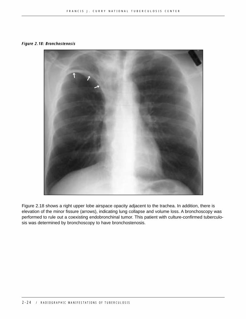
Figure 2.18: Bronchostenosis
Figure 2.18 shows a right upper lobe airspace opacity adjacent to the trachea. In addition, there is elevation of the minor fissure (arrows), indicating lung collapse and volume loss. A bronchoscopy wasperformed to rule out a coexisting endobronchinal tumor. This patient with culture-confirmed tuberculo-sis was determined by bronchoscopy to have bronchostenosis.
2 - 2 4 / R A D I O G R A P H I C M A N I F E S T A T I O N S O F T U B E R C U L O S I S
F R A N C I S J . C U R R Y N A T I O N A L T U B E R C U L O S I S C E N T E R

Interstitial Opacities (Miliary)
Figure 2.19: Miliary Pattern
Figure 2.19 demonstrates bilateral diffuse small nodules characteristic of a miliary pattern.
� A miliary pattern results from hematogenous dissemination of tubercle bacilli.� This dissemination leads to many nodules of variable size, initially present in the
interstitium and ultimately involving the airspaces.� Most of the nodules in miliary tuberculosis are 2–3 mm in diameter.� Because miliary nodules result from hematogenous dissemination, more are present
in the lower lung zones, due to greater blood flow to the bases compared with the apices of the lungs.
R A D I O G R A P H I C M A N I F E S T A T I O N S O F T U B E R C U L O S I S / 2 - 2 5
F R A N C I S J . C U R R Y N A T I O N A L T U B E R C U L O S I S C E N T E R

Tuberculoma
Tuberculomas are round or oval opacities, 1–5 cm in diameter, and usually found in the upper lobes.The pathophysiology of tuberculomas is unclear. Most experts believe that a tuberculoma represents a primary infection that has healed. Although they may remain stable for many years they can enlargevery slowly and eventually develop cavitation.
Figure 2.20: Tuberculoma
Figure 2-20 demonstrates a well-circumscribed nodule in the left lower lobe (arrows). Note the densecalcification in the center of the nodule. Radiographically, tuberculomas can simulate a bronchogeniccarcinoma.
� Tuberculomas are round or oval opacities, usually 1–5 cm in diameter, and usually found in the upper lobe.
� Tuberculomas are normally smooth and sharply defined.� Satellite lesions, which are small, discrete nodules surrounding the tuberculoma, occur
in 80% of cases and are clues to the diagnosis. However, they may only be visible on high-resolution CT.
2 - 2 6 / R A D I O G R A P H I C M A N I F E S T A T I O N S O F T U B E R C U L O S I S
F R A N C I S J . C U R R Y N A T I O N A L T U B E R C U L O S I S C E N T E R

Pleural Disease
Pleural effusions can be a manifestation of primary or post-primary tuberculosis. However, in post-primary disease, the effusion is more likely to be associated with radiographically visible parenchymalabnormalities. Rarely, the effusion is a frank tuberculous empyema. You saw examples of simpletuberculous pleural effusions earlier in Figures 2.3 and 2.12.
Figure 2.21: Tuberculous Empyema
Figure 2.21 shows an example of a tuberculous empyema that developed when a cavitary tuberculous pneumonia ruptured into the pleural space, creating a bronchopleural fistula. This case demonstratesa left pleural effusion with air-fluid levels (arrows) consistent with a hydroneumothorax caused by thebronchopleural fistula.
� Diagnosis of hydropneumothorax is based on the presence of a pleural effusion accompanied by an air-fluid level within the pleural space.
� The term hydropneumothorax signifies communication of the pleural space with the bronchial tree. Hydropneumothorax is often due to a necrotizing pneumonia such as tuberculosis.
R A D I O G R A P H I C M A N I F E S T A T I O N S O F T U B E R C U L O S I S / 2 - 2 7
F R A N C I S J . C U R R Y N A T I O N A L T U B E R C U L O S I S C E N T E R

Tuberculosis and HIV Infection
The radiographic manifestations of HIV-related tuberculosis vary depending on the degree of immunosuppression. In an HIV-infected patient whose immune system is relatively intact (i.e., >200CD4 cells/µL), the radiographic manifestations of tuberculosis represent those seen in post-primarydisease.
� The opacities occur in the upper lobe. � Cavitation may be present.� Thoracic adenopathy is uncommon.
As the CD4 lymphocyte count declines, the radiographic findings look more like those seen in primary disease.
� The radiographic opacities may be in the lower lung zones and multilobar in nature.� Thoracic adenopathy is more common.
Following are three examples of unusual (atypical) radiographic manifestations of HIV-related tuberculosis.
Figure 2.22: Bilateral Diffuse Opacities
Figure 2.22 demonstrates bilateral diffuse opacities, primarily of the airspaces, with bilateral hilaradenopathy. The patient had AFB smear-positive tuberculosis.
2 - 2 8 / R A D I O G R A P H I C M A N I F E S T A T I O N S O F T U B E R C U L O S I S
F R A N C I S J . C U R R Y N A T I O N A L T U B E R C U L O S I S C E N T E R

Figure 2.23: Large Paratracheal Adenopathy
Figure 2.23 demonstrates large bilateral paratracheal adenopathy, causing widening of the medi-astinum (arrows) with right middle and lower lung zone airspace and linear opacities. Note loss of the normal aortopulmonary window contour. Despite radiographically limited parenchymal disease, the patient was AFB smear-positive.
R A D I O G R A P H I C M A N I F E S T A T I O N S O F T U B E R C U L O S I S / 2 - 2 9
F R A N C I S J . C U R R Y N A T I O N A L T U B E R C U L O S I S C E N T E R
2 - 3 0 / R A D I O G R A P H I C M A N I F E S T A T I O N S O F T U B E R C U L O S I S
F R A N C I S J . C U R R Y N A T I O N A L T U B E R C U L O S I S C E N T E R
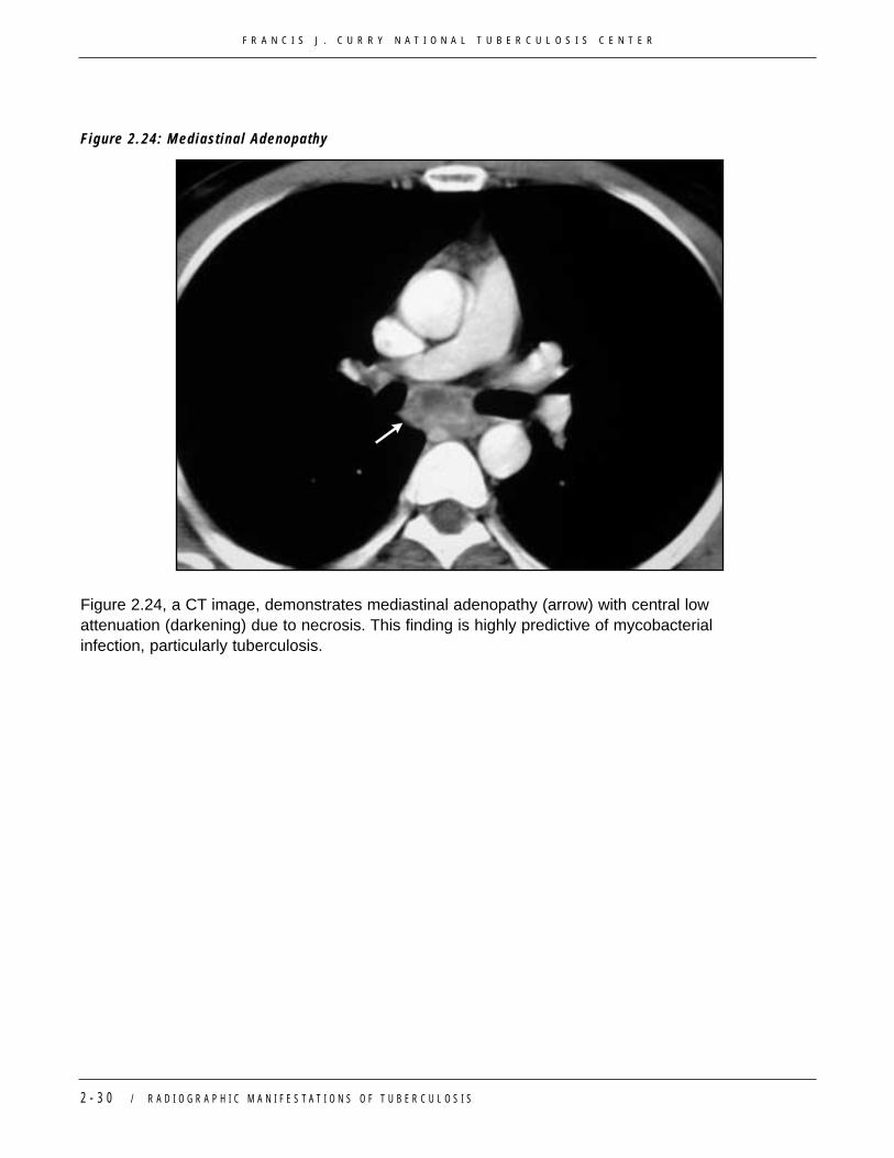
Figure 2.24: Mediastinal Adenopathy
Figure 2.24, a CT image, demonstrates mediastinal adenopathy (arrow) with central low attenuation (darkening) due to necrosis. This finding is highly predictive of mycobacterial infection, particularly tuberculosis.

R A D I O G R A P H I C M A N I F E S T A T I O N S O F T U B E R C U L O S I S / 2 - 3 1
F R A N C I S J . C U R R Y N A T I O N A L T U B E R C U L O S I S C E N T E R
Resolution of Radiographic Abnormalities and HealedTuberculosisThe chest abnormalities seen by radiography in tuberculosis are slow to resolve. In many cases,parenchymal opacities and thoracic adenopathy actually worsen before improving. For this reason,the chest radiograph is not the best way to follow the response to antituberculosis therapy. Instead,a clinical assessment should be performed and a bacteriological response to therapy should bemonitored in order to determine if the patient is or is not improving with treatment.
Primary Tuberculosis
Healing of the primary complex, with or without therapy, can result in fibrosis and calcification of the Ghon focus. The Ghon focus is represented radiographically as a calcified nodular opacity on thechest radiograph (e.g., calcified granuloma). The Ghon focus, in combination with a calcified ipsilat-eral hilar or mediastinal calcification, are the radiographic manifestations of the Ranke Complex.
Primary tuberculosis typically resolves with minimal fibrosis and volume loss. However, patients who develop progressive primary disease with cavitation may suffer significant fibrosis and maydevelop bronchiectasis, similar to post-primary disease. Lymphadenopathy may take months toresolve and, in some cases, there may be prolonged enlargement of lymph nodes, particularly inchildren.
Post-primary Tuberculosis
The degree of fibrosis and scarring varies considerably with post-primary tuberculosis. In general, the more extensive the disease and the worse the cavitation, the more likely it is that there will befibrosis with associated volume loss. It is important to note that fibrosis and volume loss can occurin the presence of active tuberculosis, so these findings should not be used to dismiss a diagnosisof active disease.
Following are examples of healed primary and post-primary tuberculosis.

Figure 2.25: Ranke Complex
Figure 2.25 demonstrates a calcified peripheral nodular opacity (large arrow) consistent with a Ghonlesion. There is also a calcified right hilar node (small arrow). Together, these lesions are referred toas a Ranke complex.
� A Ghon lesion represents a calcified granuloma in the lung parenchyma.� A Ranke complex is the combination of a Ghon lesion and an ipsilateral calcified hilar
lymph node.� Neither a Ghon lesion nor Ranke complex represent active tuberculosis.� Isolated calcified granulomas are not associated with an increased risk of progression to
active disease in people with latent tuberculosis infection.
2 - 3 2 / R A D I O G R A P H I C M A N I F E S T A T I O N S O F T U B E R C U L O S I S
F R A N C I S J . C U R R Y N A T I O N A L T U B E R C U L O S I S C E N T E R

Figure 2.26a: Previously Treated Pulmonary Tuberculosis
Figure 2.26a depicts a patient who had been treated previously for pulmonary tuberculosis. Thepatient has a calcified nodule (large arrow) consistent with a calcified granuloma. In addition, there isbilateral apical pleural thickening (small arrows). See also Figure 2.26b.
R A D I O G R A P H I C M A N I F E S T A T I O N S O F T U B E R C U L O S I S / 2 - 3 3
F R A N C I S J . C U R R Y N A T I O N A L T U B E R C U L O S I S C E N T E R

Figure 2.26b: Apical Pleural Thickening
Figure 2.26b depicts a close-up view of the left apex seen in Figure 2.26a, demonstrating apical pleuralthickening (arrows).
� Apical pleural thickening may be seen with or without surrounding apical parenchymal opacities.
� Apical pleural thickening is not associated with active tuberculosis unless there are also accompanying parenchymal opacities such as airspace consolidation, nodules, or fibrosis.
� Isolated pleural thickening is not associated with an increased risk of progression to activedisease in people with latent tuberculosis infection.
2 - 3 4 / R A D I O G R A P H I C M A N I F E S T A T I O N S O F T U B E R C U L O S I S
F R A N C I S J . C U R R Y N A T I O N A L T U B E R C U L O S I S C E N T E R

Figure 2.27: Fibrotic Scarring
Figure 2.27 demonstrates right upper lobe linear opacities, apical pleural thickening, and volume loss.Note the elevation of the right hilum and hemidiaphragm. This patient was asymptomatic and had negative AFB smears and cultures.
� Post-primary tuberculosis is often associated with significant fibrosis. The resultant scarring can cause volume loss of the involved lung or lobe.
� Fibrotic lesions may indicate either active or prior tuberculosis. This distinction can only be made by clinical and microbiological evaluation.
� The presence of parenchymal opacities—representing old healed tuberculosis—increasesthe risk of progression to tuberculosis in individuals who have received inadequate prior treatment for tuberculosis or latent tuberculosis infection.
R A D I O G R A P H I C M A N I F E S T A T I O N S O F T U B E R C U L O S I S / 2 - 3 5
F R A N C I S J . C U R R Y N A T I O N A L T U B E R C U L O S I S C E N T E R

Self-Check Three
The following self-check has three chest radiographs to analyze and three multiple choice questions.After completing the self-check, look at the answers beginning on page 2-41. Review the material inthe previous pages to clarify any answers you have missed.
1. Describe the chest radiograph below in Figure 2.28.
Figure 2.28
Description:
2 - 3 6 / R A D I O G R A P H I C M A N I F E S T A T I O N S O F T U B E R C U L O S I S
F R A N C I S J . C U R R Y N A T I O N A L T U B E R C U L O S I S C E N T E R

2. Describe the chest radiograph below in Figure 2.29.
Figure 2.29
Description:
R A D I O G R A P H I C M A N I F E S T A T I O N S O F T U B E R C U L O S I S / 2 - 3 7
F R A N C I S J . C U R R Y N A T I O N A L T U B E R C U L O S I S C E N T E R

Self-Check Three (continued)
3. Describe the chest radiograph in Figure 2.30.
Figure 2.30
Description:
2 - 3 8 / R A D I O G R A P H I C M A N I F E S T A T I O N S O F T U B E R C U L O S I S
F R A N C I S J . C U R R Y N A T I O N A L T U B E R C U L O S I S C E N T E R

4. Which of the following radiographic manifestations is most consistent with active primary tuberculosis in an adult?A. Upper lobe, posterior segment involvement
B. Bronchopleural fistula
C. Calcified granuloma
D. Pleural effusion
5. Which of the following radiographic manifestations of tuberculosis is more common in
HIV-infected adults than in HIV-uninfected adults?A. Hilar adenopathy
B. Pleural effusion
C. Upper lobe opacities
D. Calcified granuloma
6. The most common parenchymal pattern of disease of both primary and post-primary
(reactivation) tuberculosis is:A. Miliary pattern
B. Pleural effusion
C. Airspace consolidation
D. Cavitation
R A D I O G R A P H I C M A N I F E S T A T I O N S O F T U B E R C U L O S I S / 2 - 3 9
F R A N C I S J . C U R R Y N A T I O N A L T U B E R C U L O S I S C E N T E R

Conclusion
Now that you have completed this chapter, you should be able to identify the various radiographicmanifestations of tuberculosis and use the terminology learned in Chapter One to describe your findings.
In the next chapter, you will review several clinical cases and use the knowledge and skills that youhave developed in the first two chapters to read chest radiographs and make clinical decisionsbased on your interpretation.
2 - 4 0 / R A D I O G R A P H I C M A N I F E S T A T I O N S O F T U B E R C U L O S I S
F R A N C I S J . C U R R Y N A T I O N A L T U B E R C U L O S I S C E N T E R

Self-Check Three Answers
1. Description:There is a right paratracheal opacity behind the right clavicle in Figure 2.31 (see arrows). This patient had culture-confirmed tuberculosis.
Figure 2.31
R A D I O G R A P H I C M A N I F E S T A T I O N S O F T U B E R C U L O S I S / 2 - 4 1
F R A N C I S J . C U R R Y N A T I O N A L T U B E R C U L O S I S C E N T E R

Self-Check Three Answers (continued)
2. Description:There is a nodular density overlying the left first rib in Figure 2.29. This patient radiograph was taken in the apical lordotic view, allowing demonstration of the left upper lobe nodular density (arrows).
Figure 2.32
2 - 4 2 / R A D I O G R A P H I C M A N I F E S T A T I O N S O F T U B E R C U L O S I S
F R A N C I S J . C U R R Y N A T I O N A L T U B E R C U L O S I S C E N T E R

3. Description:Figure 2.33 demonstrates a right upper lobe airspace opacity with cavitation. Note the large cavity (small arrow). There is also a large right hydropneumothorax with anair-fluid level (large arrow). This patient had smear-positive pulmonary tuberculosis and a tuberculous empyema.
Figure 2.33
4. D5. A6. C
R A D I O G R A P H I C M A N I F E S T A T I O N S O F T U B E R C U L O S I S / 2 - 4 3
F R A N C I S J . C U R R Y N A T I O N A L T U B E R C U L O S I S C E N T E R

References
Fraser RS, Pare JAP, Fraser RG, Pare PD. Infectious diseases of the lungs. In: Fraser RS, Pare JAP,Fraser RG, Pare PD, eds. Synopsis of Diseases of the Chest. 2nd ed. Philadelphia, PA: WB Saunders;1994:315-332.
Jones BE, Ryu R, Yang Z, et al. Chest radiographic findings in patients with tuberculosis with recentor remote infection. Am J Respir Crit Care Med. 1997;156:1270-1273.
Leung AN. Pulmonary tuberculosis: the essentials. Radiology. 1999;210:307-322.
McAdams HP, Erasmus J, Winter JA. Radiologic manifestations of pulmonary tuberculosis. Radiol ClinNorth Am. 1995;33:655-678.
Palmer PE. Pulmonary tuberculosis--usual and unusual radiographic presentations. SeminRoentgenol. 1979;14:204-43.
Rich AR. The pathogenesis of pulmonary tuberculosis. In: Rich AR, ed. The Pathogenesis ofPulmonary Tuberculosis. Springfield, Ill: Charles C. Thomas Publisher; 1944.
Woodring JH, Vandiviere HM, Fried AM, Dillon ML, Williams TD, Melvin IG. Update: the radiographicfeatures of pulmonary tuberculosis. Am J Roentgenol. 1986;146:497-506.
2 - 4 4 / R A D I O G R A P H I C M A N I F E S T A T I O N S O F T U B E R C U L O S I S
F R A N C I S J . C U R R Y N A T I O N A L T U B E R C U L O S I S C E N T E R


![Standardized radiographic interpretation of thoracic ... · culosis depends largely on chest imaging [1 ]. Pulmonary tuber-culosis has been classically classified as primary tuberculosis](https://img.pdfslide.us/doc/110x75/607490a4c43e164edd032250/standardized-radiographic-interpretation-of-thoracic-culosis-depends-largely.jpg)