Embed Size (px)
Citation preview
William G. Armington1
H. Ric Harnsberger1
Anne G. Osborn1 Alan R. Seay2
Received April 25 . 1986; accepted after revision September 13. 1986.
Presented in part at the annual meeting of the American Society of Neurology, New Orleans, February 1985.
, Department of Radiology, University of Utah Medical Center, 50 North Medical Drive, Salt Lake City, UT 84132. Address reprint requests to H. R. Harnsberger.
2 Department of Neurology, University of Utah Medical Center, 50 North Medical Drive, Salt Lake City, UT 84132.
AJNR 8:361-367, March/April 1987 0195- 6108/87/0802-0361 © American Society of Neuroradiology
Radiographic Evaluation of Brachial Plexopathy
361
Neurologic signs and symptoms of brachial plexopathy may be subtle or confusing, making clinical localization of disease along the length of the brachial plexus difficult. To determine the most direct radiographic approach to diagnosing and anatomically delineating the cause of brachial plexopathy, we reviewed the clinical and radiographic records of 43 patients presenting with signs and symptoms referable to the brachial plexus who received CT and/or myelography as part of their radiographic evaluation. The study population was divided into two groups, those with and those without trauma, Significant deficiencies were detected in the radiographic evaluation of the nontraumatic group, with 35% of these patients having an incomplete or inappropriate CT examination that failed to visualize the full extent of the brachial plexus, In four patients, this led to a significant (greater than 6 months) delay in diagnosis, It was concluded that trauma patients presenting with brachial plexus symptoms should have cervical myelography first, rather than CT, Patients without a history of trauma should be classified on the basis of clinical findings as having central (cord, epidural space, neural foramen) or peripheral (retroclavicular space, axillary apex) disease, If the abnormality is central, myelography should be the first technique used; if peripheral disease is present, CT should be the first study, If the disease extends beyond the confines of the anatomic compartment suggested clinically, the other technique should be used for further evaluation, CT scan protocols for brachial plexus evaluation should employ bolus/drip contrast enhancement to distinguish vascular structures from masses. Sequential demagnification of CT should be done to ensure comprehensive imaging of the brachial plexus from its origin in the spinal cord to its distal ramifications in the apex of the axilla.
Neurologic symptoms and signs of brachial plexopathy may be subtle or confusing, making clinical localization of disease along the length of the brachial plexus difficult when no mass is palpable. To date, reports focus on the use of CT as the sole imaging technique useful in evaluating this problem [1-8]. However, depending on the suspected location of the leSion, cervical myelography, CT, or MR imaging of the cervical-thoracic junction may be used to image the brachial plexus, Often , these studies are done poorly owing to an insufficient understanding of the anatomic, technical , or clinical aspects of the brachial plexus.
We reviewed the clinical and radiographic records of 43 patients presenting with signs and symptoms referable to the brachial plexus who had CT and/or myelography as part of their radiographic evaluation. From this experience, a diagnostic decision tree was designed to provide a step-wise approach to the diagnosis of brachial plexus abnormalities. A specific CT technique protocol is described to ensure comprehensive imaging of the brachial plexus from its origin in the cervical spinal cord to its distal ramifications in the apex of the axilla. The anatomy necessary to understand the images is presented, and technical and interpretive pitfalls encountered are described.
Materials and Methods
A retrospective analysis was performed of 43 patients presenting with signs and symptoms
362 ARMINGTON ET AL AJNR:8, March/April 1987
of brachial plexopathy who underwent radiographic evaluation . Clinical data assessed included the specific clinical complaint signaling the presence of brachial plexopathy, the findings on neurologic examination, and the results of electromyography. When possible, a
TABLE 1: Clinical Information: Patients Presenting with Symptoms of Brachial Plexopathy (n = 43)
Nontraumatic (n = 32) Age range: 14-78 Gender ratio: 16M/16F Final diagnosis:
Benign tumor Schwannoma
Malignant tumor Non-Hodgkins lymphoma Hodgkins lymphoma Acute lymphatic leukemia
(epidural) Metastases
Breast Lung Esophagus Squamous cell , head and
neck Miscellaneous
Miscellaneous Syrinx Brachial neuritis Iatrogenic (postsurgical)
Traumatic (n = 11) Age range: 9- 67 Gender ratio: 7M/4F Final diagnosis:
Nerve root avulsion Dural tear
6 3 2
2 4
No. of
Cases
8
2
5 2
1 17
2 2 1
2
25
5
10 1
final pathologic diagnosis was determined for each patient. All radiologic techniques performed-including cervical plain films,
cervical myelograms, and CT scans-were reviewed. The time interval between the onset of symptoms and the diagnosis of the lesion was recorded. Cases of delayed or misdiagnosis were identified. CT examinations were evaluated to determine if the abnormal portion of the brachial plexus was visualized completely or if the true extent of disease was identified.
Results
Among the 43 patients evaluated, 10 had trauma (Table 1). In the 32 patients without trauma, 25 had malignant tumors (Table 1 and Fig. 1). Other diagnoses were benign tumor (two) (Fig. 2), syrinx (two), brachial neuritis (two), and postsurgical brachial neuropathy (one).
Of the 32 cases of nontraumatic brachial plexopathy, the clinical record showed 26 lesions that were either palpable or nonpalpable. Of these, 14 lesions were palpable and 12 were nonpalpable. All 12 of the non palpable lesions were found on CT to be in clinically "silent" areas, including the axillary apexretrociavicular region, the paravertebral gutter, and the neural foramina-epidural space (Fig. 3). Ten of the 12 nonpalpable lesions were infiltrating with only two displaying a focal mass on CT.
The radiographic evaluations of the 32 nontraumatic brachial plexopathies included six patients who had both CT and cervical myelography (Fig. 4), 25 patients who had CT only, and one patient who had myelography only. Of those receiving CT, 20 had a complete CT evaluation while 11 were incompletely examined. In three cases, the disease process responsible for the symptoms was missed as a result of incomplete CT evaluation . Four patients experienced a significant delay in diagnosis (> 6 months) as a result of inadequate CT examination.
Key for Figure 1 White arrows = Perineural tumor along
brachial plexus within axillary apex Black arrows = Enhancing perineural tu
mor extending into epidural space Arrowheads = Perineural tumor along
course of brachial plexus a = Anterior scalene
m = Middle scalene p = Phrenic nerve v = Subclavian vein • = Subclavian artery
Fig. 1.-72-year-old woman with breast carcinoma who presented with left arm and hand paresthesia 10 years after left mastectomy. Exploration of left axillary /supraclavicular region showed no evidence of tumor infiltration. Axial CT scans through level of thyroid bed (A) and axillary apex (8) show enhancing infiltrating tumor within left epidural space and extending along structures of brachial plexus within neural foramen, between anterior and middle scalene mUSCles, and in region of phrenic nerve (A) . In addition, elements of brachial plexus within axillary apex (8) are thickened and enhancing, suggesting perineural tumor spread with development of diffuse atrophy of muscles served by brachial plexus on ipsilateral side. Although adequate demagnification to show entire axillary apex was not done, abnormality is apparent.
AJNR:8, March/April 1987 BRACHIAL PLEXOPATHY 363
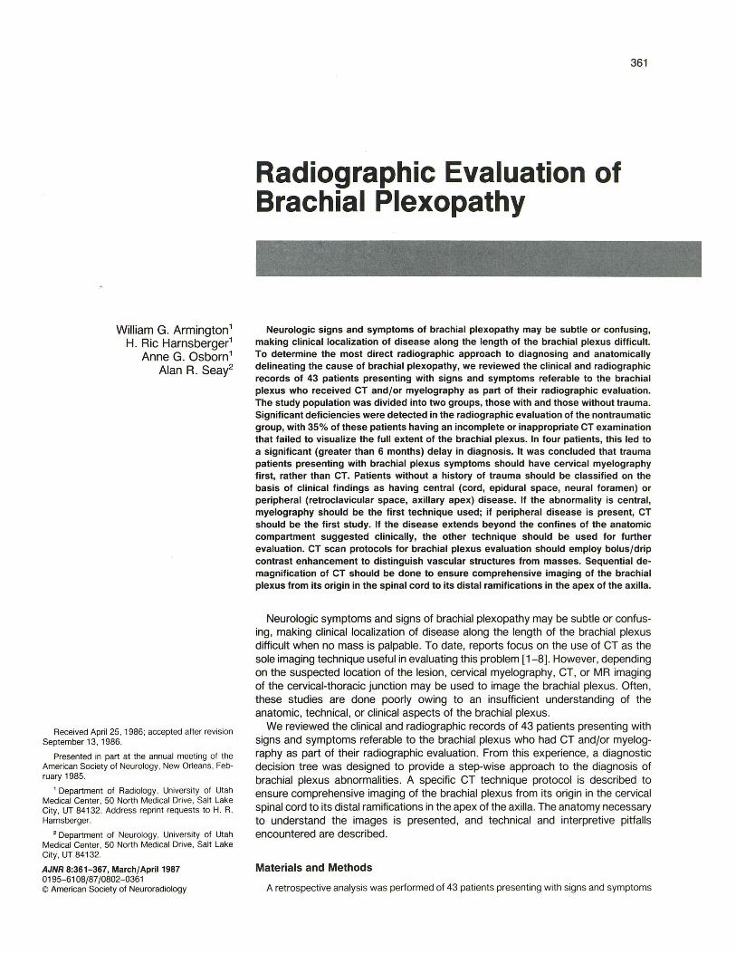
Fig. 2.-60-year-old woman with lett posterior triangle mass, lett arm and shoulder pain, and paresthesia. Axial CT scans at level of high (A) and low (B) thyroid bed show well-circumscribed mixed-density mass causing widening of lettsided neural foramen (S) with extension laterally along course of brachial plexus to a pOSition between anterior and middle scalene muscles (arrowheads). Surgical exploration revealed a schwannoma.
A
A B
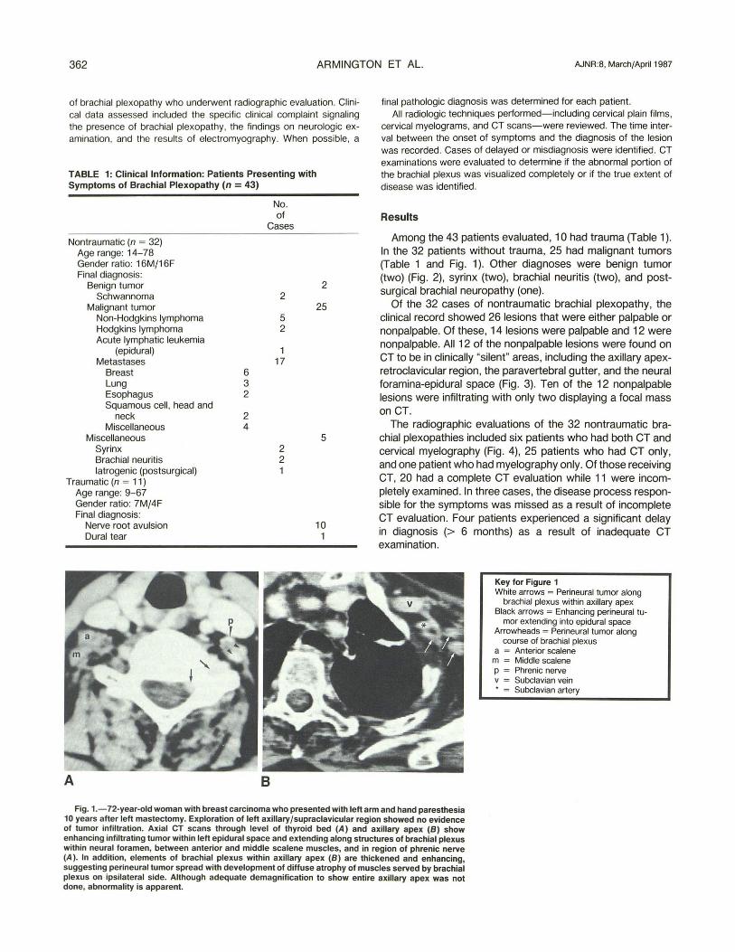
B Fig. 3.-Axial CT scan shows
right axillary apex mass adjacent to brachial plexus in a patient with non-Hodgkins lymphoma who presented with right-sided brachial plexopathy. The mass was not palpable. Peripheral enhancement of mass with central low attenuation suggested enlarged lymph nodes with central necrosis, confirmed at biopsy.
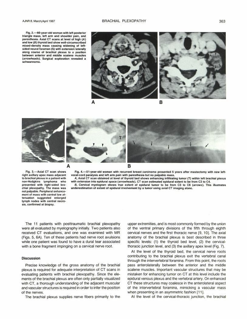
Fig. 4.-51-year-old woman with recurrent breast carcinoma presented 5 years atter mastectomy with new lettvocal-cord paralysis and lett arm pain with paresthesia but no palpable mass.
A, Axial CT scan obtained at level of thyroid bed shows enhancing infiltrating tumor (T) within lett brachial plexus with extension into epidural space (arrowheads). CT scan estimated epidural extent to be from C3 to C4.
B, Cervical myelogram shows true extent of epidural tumor to be from C3 to C6 (arrows). This illustrates underestimation of extent of epidural involvement by a tumor using axial CT imaging alone.
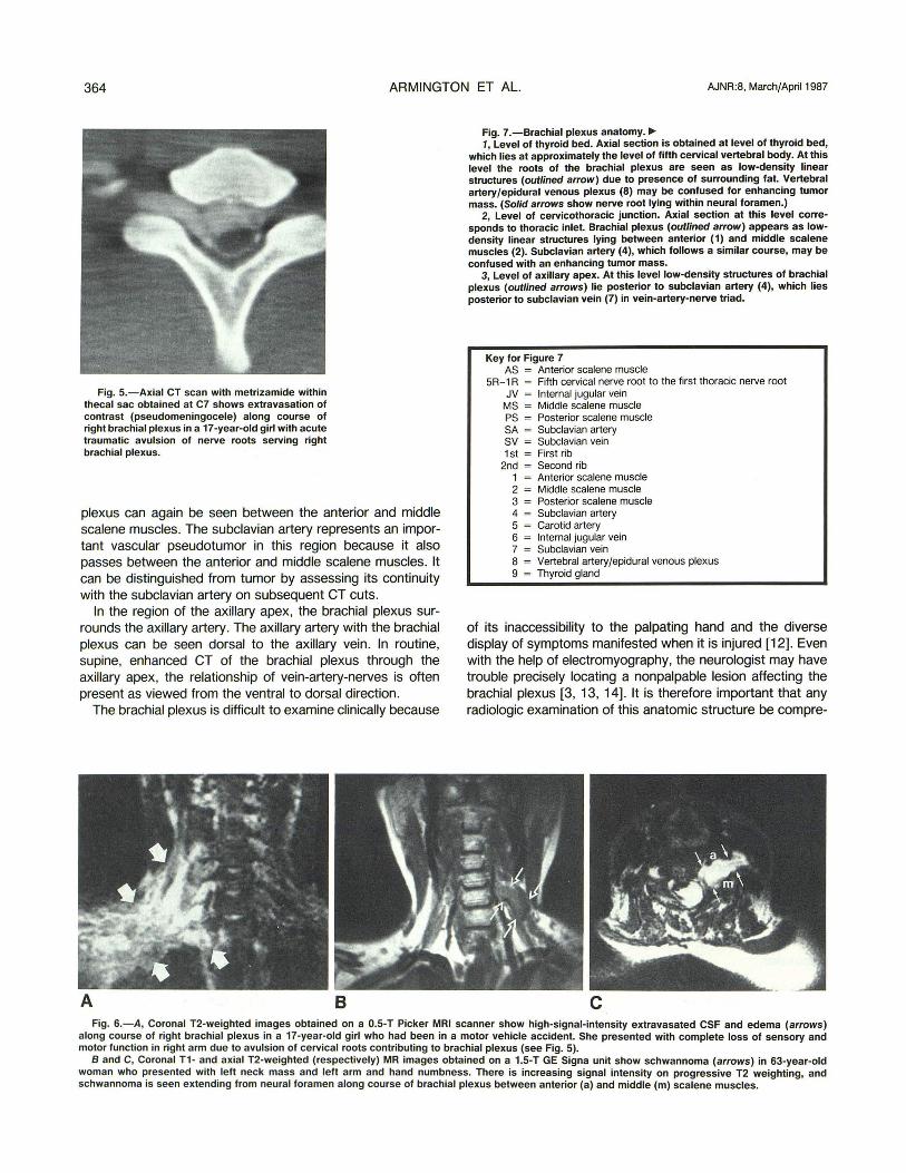
The 11 patients with posttraumatic brachial plexopathy were all evaluated by myelography initially. Two patients also received CT evaluations, and one was examined with MR (Figs. 5, 6A). Ten of these patients had nerve root avulsions while one patient was found to have a dural tear associated with a bone fragment impinging on a cervical nerve root.
Discussion
Precise knowledge of the gross anatomy of the brachial plexus is required for adequate interpretation of CT scans in evaluating patients with brachial plexopathy. Since the elements of the brachial plexus are often only partially visualized with CT, a thorough understanding of the adjacent muscular and vascular structures is required in order to infer the position of the nerves.
The brachial plexus supplies nerve fibers primarily to the
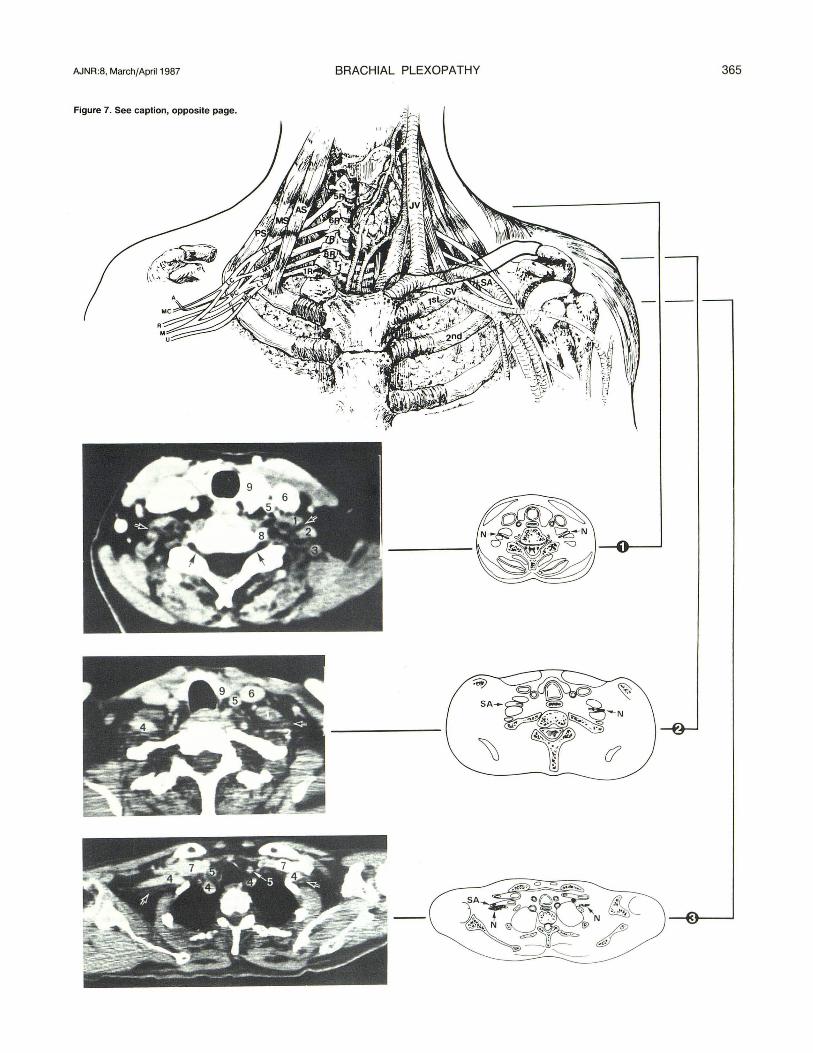
upper extremities, and is most commonly formed by the union of the ventral primary divisions of the fifth through eighth cervical nerves and the first thoracic nerve [9, 10]. The axial anatomy of the brachial plexus is best described in three specific levels: (1) the thyroid bed level, (2) the cervicalthoracic junction level, and (3) the axillary apex level (Fig. 7).
At the level of the thyroid bed, the cervical nerve roots contributing to the brachial plexus exit the vertebral canal through the intervertebral foramina. From this point, the roots pass anterolaterally between the anterior and the middle scalene muscles. Important vascular structures that may be mistaken for enhancing tumor on CT at this level include the epidural venous plexus and the vertebral artery. On enhanced CT these structures may coalesce in the anterolateral aspect of the intervertebral foramina, mimicking a vascular mass when presenting in an asymmetriC fashion [11] .
At the level of the cervical-thoracic junction, the brachial
364 ARMINGTON ET AL. AJNR:8, March/April 1987
Fig. 5.-Axial CT scan with metrizamide within thecal sac obtained at C7 shows extravasation of contrast (pseudomeningocele) along course of right brachial plexus in a 17 -year-old girl with acute traumatic avulsion of nerve roots serving right brachial plexus.
plexus can again be seen between the anterior and middle scalene muscles. The subclavian artery represents an important vascular pseudotumor in this region because it also passes between the anterior and middle scalene muscles. It can be distinguished from tumor by assessing its continuity with the subclavian artery on subsequent CT cuts.
In the region of the axillary apex, the brachial plexus surrounds the axillary artery. The axillary artery with the brachial plexus can be seen dorsal to the axillary vein. In routine, supine, enhanced CT of the brachial plexus through the axillary apex, the relationship of vein-artery-nerves is often present as viewed from the ventral to dorsal direction.
The brachial plexus is difficult to examine clinically because
A B
Fig. 7.-Brachial plexus anatomy. ~ 1, Level of thyroid bed. Axial section is obtained at level of thyroid bed,
which lies at approximately the level of fifth cervical vertebral body. At this level the roots of the brachial plexus are seen as low-density linear structures (outlined arrow) due to presence of surrounding fat. Vertebral artery/epidural venous plexus (8) may be confused for enhancing tumor mass. (Solid arrows show nerve root lying within neural foramen.)
2, Level of cervicothoracic junction. Axial section at this level corresponds to thoracic inlet. Brachial plexus (outlined arrow) appears as lowdensity linear structures lying between anterior (1) and middle scalene muscles (2). Subclavian artery (4), which follows a similar course, may be confused with an enhancing tumor mass.
3, Level of axillary apex. At this level lOW-density structures of brachial plexus (outlined arrows) lie posterior to subclavian artery (4), which lies posterior to subclavian vein (7) in vein-artery-nerve triad.
Key for Figure 7 AS Anterior scalene muscle
5R-1 R Fifth cervical nerve root to the first thoracic nerve root JV Internal jugular vein
MS Middle scalene muscle PS Posterior scalene muscle SA Subclavian artery SV Subclavian vein 1st
2nd 1 2 3 4 5 6 7 8 = 9 =
First rib Second rib Anterior scalene muscle Middle scalene muscle Posterior scalene muscle Subclavian artery Carotid artery Internal jugular vein Subclavian vein Vertebral artery/epidural venous plexus Thyroid gland
of its inaccessibility to the palpating hand and the diverse display of symptoms manifested when it is injured [12]. Even with the help of electromyography, the neurologist may have trouble precisely locating a non palpable lesion affecting the brachial plexus [3,13,14]. It is therefore important that any radiologic examination of this anatomic structure be compre-
c Fig. 6.-A, Coronal T2-weighted images obtained on a O.5-T Picker MRI scanner show high-signal-intensity extravasated CSF and edema (arrows)
along course of right brachial plexus in a 17-year-old girl who had been in a motor vehicle accident. She presented with complete loss of sensory and motor function in right arm due to avulsion of cervical roots contributing to brachial plexus (see Fig. 5).
Band C, Coronal T1 - and axial T2-weighted (respectively) MR images obtained on a 1.5-T GE Signa unit show schwan noma (arrows) in 63-year-old woman who presented with left neck mass and left arm and hand numbness. There is increasing signal intensity on progressive T2 weighting, and schwannoma is seen extending from neural foramen along course of brachial plexus between anterior (a) and middle (m) scalene muscles.
AJNR:8, March/April 1987 BRACHIAL PLEXOPATHY 365
Figure 7. See caption, opposite page.
366 ARMINGTON ET AL. AJNR:8, March/April1987
hensive in order to avoid misdiagnosis and diagnostic delays. Lesions affecting the brachial plexus may be divided into
two main groups: traumatic and nontraumatic. The traumatic group is generally a straightforward clinical-radiographic problem with myelography playing the central role in diagnosing nerve root avulsion [15]. It is the nontraumatic group that presents the most challenging imaging problems.
Although clinical assessment of patients with nontraumatic brachial plexopathy is difficult, a non palpable lesion can usually be labeled as central (epidural space, neural foramen, paravertebral gutter) or peripheral (retroclavicular space, axillary apex) [12, 14]. When symptoms and signs are bilateral or associated with cord-compression symptoms, the designation of central disease is particularly easy. It becomes more difficult when central disease causes lateralized symptoms. Contralateral clinical findings may be subtle or absent. In this study, 20 of 24 patients for whom data were available were correctly identified as having lesions of the central or peripheral brachial plexus. Clinical assessment serves as a best first approximation of disease location.
This division of lesion location , especially when the disease is nonpalpable, serves to direct the radiologist to the best first imaging tool. Suspected central lesions are first evaluated with cervical myelography; suspected peripheral lesions are first evaluated with CT. When either study is negative, the opposite study may be undertaken when there is strong clinical suspicion of disease. Also, when CT shows the lesion tracking from peripheral to central along the brachial plexus, myelography is recommended to assess epidural extension.
TABLE 2: CT Technique for Complete Brachial Plexus Evaluation
Patient position: supine, arms down Slice thickness/increment: ::;5 mm/contiguous Scan extent: C4 vertebral body to axillary apex Computer settings: high mAs; maximum magnification with sequen
tial demagnification to visualize axilla Contrast: bolus/drip technique. Intravenous bolus of 50 ml of 60%
iodinated contrast followed by rapid-drip infusion of 300 ml of 30% iodinated contrast
B
Conversely, if myelography is done for central symptoms but the patient has palpable disease in the neck (peripheral mass), CT is indicated to assess the extent of deep-tissue involvement. Additionally, if epidural disease is diagnosed with cervical myelography, a CT scan of the region performed while intrathecal contrast is present is helpful to assess the relationship between the disease and the thecal sac.
Diseases affecting the brachial plexus are often clinically occult (nonpalpable) as a result of their location or their infiltrating nature. In 11 of 25 (44%) patients for whom data were available, the lesion was nonpalpable. The areas "silent" to palpation were found to be the epidural space, the neural foramina, and the paravertebral gutter for central lesions and the retroclavicular space and high axillary apex for peripheral lesions. Infiltrating malignancies that invade a focal area of the brachial plexus or perineural malignancy along the course of the brachial plexus are non palpable lesions of the brachial plexus that require a tailored CT technique and careful image interpretation to avoid mistaken and/or delayed diagnosis.
CT examination of the entire brachial plexus is frustrating from the outset because an axial imaging tool is being applied to a longitudinal anatomic structure. Technical considerations for focused CT are summarized in Table 2. Salient features of this CT protocol include the need for thin section (:s 5 mm), contiguous scans, and gradual on-line demagnification as the scan progresses from the neck into the chest to avoid incomplete imaging of the retroclavicular and axillary regions. In 11 of the 30 patients evaluated in our series for nontraumatic brachial plexopathy, disease was initially missed because of inadequate visualization at the axillary apex region. Intense intravascular contrast, as proposed in the bolus/drip strategy, prevents misassessment of vessels as masses.
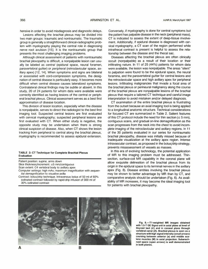
In this era of evolving technology, the potential application of MR to this imaging problem must be addressed . Thinsection, surface-coil MR capability in the coronal plane will allow exquisite delineation of the brachial plexus from its origin in the epidural space to its terminal nerves in the axillary apex (Fig. 8) . Disease entities involving the brachial plexus may be shown to better advantage by MR than by CT, and comparative analysis should be undertaken (Fig. 6). As availability of MR increases, it may become the ideal imaging tool for patients with brachial plexopathy.
Fig. 8.- T1-weighted MR images obtained with 1.5-T GE Signa unit in axial plane at level of thryroid bed (A) and in coronal plane through vertebral canal (8). Brachial plexus is seen as a structure with high signal intensity (small arrows) coursing between anterior (a) and middle scalene muscles (M) in axial projection. Subarachnoid space (open arrows) is well demonstrated in both planes.

AJNR:8, March/April 1987 BRACHIAL PLEXOPATHY 367
REFERENCES
1. Gebarski KS, Glazer GM, Gebarski SS. Brachial plexus: anatomic radiologic, and pathologic correlation using computed tomography. J Comput Assist Tomogr 1982;6 : 1058-1 063
2. Takasagi JE, Godwin JD, Halvorsen RE, Williford ME, Silverman PM, Putman CT. Computed tomographic evaluation of lesions in the thoracic apex. Invest Radio/1985 ;20 :260- 266
3. Cascino TL, Kori S, Krol G, Foley KM. CT of the brachial plexus in patients with cancer. Neurology 1983;33: 1553-1557
4. Vock P, Owens A. Computed tomography of the normal and pathologic thoracic inlet. Eur J Radio/1982;2 :187-193
5. Powers SK, Norman D, Edwards MSB. Computerized tomography of peripheral nerve lesions. J Neurosurg 1983;59: 131 - 136
6. Kaiser MC, Capesius P, Petti M. Thoracic inlet mass due to cervical root avulsion diagnosed by CT-scanning. Fortschr Roentgenstr 1983;138:505-506
7. Usselman JA. Vint VC. Waltz TA. CT demonstration of a brachial plexus neuroma. AJNR 1980;1 :346-347
8. Stewart JD, Schmidt B, Wee R. Computed tomography in the evaluation of plexopathies and proximal neuropathies. Can J Neurol Sci 1983;1 0: 244-247
9. Clemente CD . ed . Gray 's anatomy. Philadelphia: Lea and Febiger, 1985:1205-1212
10. Thompson GE, Rorie DK. Functional anatomy of the brachial plexus sheaths . Anesthesiology 1983;59: 117 -122
11 . Heinz ER , Yeates A, Burger P, Drayer BP, Osborne D, Hill R. Opacification of epidural venous plexus and dura in evaluation of cervical nerve roots: CT technique. AJNR 1984 ;5 :621-624
12. Adams RD, Victor M, eds. Principles of neurology . New York: McGrawHill , 1981 :917-922
13. Synek VM , Cowan JC. Somatosensory evoked potentials in patients with metastatic involvement of the brachial plexus. Electromyogr Clin Neurophysio/ 1983;23 :545-551
14. Stohr M, Riffel B, Buettner US. Somatosewsible Evozierte potentiale in der Diagnostik von Armplexuslasionen . EEG EMG 1981 ;12: 195- 197
15. Virapongse C, Kier EL. Trauma to the spinal cord and nerve roots. In: Shapiro R. Myelography. Chicago: Yearbook , 1984:247-281