Embed Size (px)
Citation preview

RADIOGRAPHIC
APPEARANCE OF
COMMON DENTAL
DISEASE
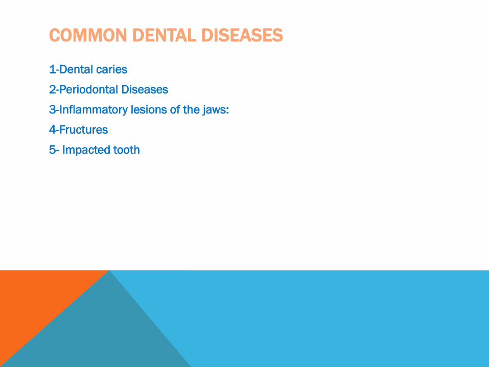
COMMON DENTAL DISEASES
1-Dental caries
2-Periodontal Diseases
3-Inflammatory lesions of the jaws:
4-Fructures
5- Impacted tooth

1-Dental caries: dental carise is the common infectious disease
strongly influenced by diet, affecting 95% of population.
Radiography is useful for detecting dental caries because the
carious process causes tooth demineralization.
The carious lesion (the demineralized area of the tooth that
allows greater infiltration of x-rays) is darker (i.e.,
more radiolucent) than the unaffected portion and may be
detected on radiographs. An early carious lesion may
not have yet caused sufficient demineralization to be detected
radiographically. Intraoral radiography can reveal carious lesions
that otherwise might go undetected during a thorough clinical
examination. A number of studies have shown the value of dental
radiographs by repeatedly demonstrating that approximately half
of all proximal surface lesions cannot be seen clinically and may
be detected only with radiographs.

Interpretation of Incipient Occlusal Lesions
Radiographs are usually not effective for the detection of an
occlusal carious lesion until it reaches the dentin.
I nterpretation of Moderate Occlusal Lesions
The moderate occlusal lesion is usually the first to induce
specific radiographic changes, prompting a definitive
decision regarding the presence of caries. The classic
radiographic change is a broad-based, thin radiolucent
zone in the dentin with little or no changes apparent in the
enamel.


PROXIMAL CARIES
Radiographic detection of carious lesions on the proximal
surfaces of teeth depends on loss of enough mineral to
result in a detectable change in radiographic density.
Because the proximal surfaces of posterior teeth are often
broad, the loss of small amounts of mineral from incipient
lesions or the advancing front of more advanced lesions is
often difficult to detect on a radiograph. For this reason, the
actual depth of penetration of a carious lesion is deeper
than may be detected radiographically.
Approximately 40% demineralization is required for
radiographic detection of a lesion

Facial, buccal, and lingual caries
Facial, buccal, and lingual carious lesions occur in enamel
pits and fissures of teeth. When small, these lesions are
usually round; as they enlarge, they become elliptic or
semilunar. They demonstrate sharp, well-defined borders. It
is difficult to differentiate between buccal and lingual caries
on a radiograph.
ROOT SURFACE CARIES
Root surface caries (also called cemental caries) involves
both cementum and dentin. Its prevalence is
approximately40% to 70% in an aged population. The tooth
surfaces most frequently affected are, in order, buccal,
lingual, and proximal.

RECURRENT CARIES
Recurrent caries is that occurring immediately next to a restoration.
It may result from poor adaptation of a restoration, which allows for
marginal leakage, or from inadequate extension of a restoration.
In addition, caries may remain if the original lesion is not
completely evacuated, which later may appear as residual or
recurrent caries.
Approximately 16% of restored tooth surfaces have recurrent
caries.
The radiographic appearance of recurrent caries depends on the
amount of decalcification present and whether a restoration is
obscuring the lesion.

2-Periodontal Diseasesseveral distinct yet related disorders of the periodontium are collectively
known as periodontal disease. The most common of these are gingivitis and
periodontitis. Gingivitis is a sequela of infection. It is limited to the marginal
gingiva and usually is seen as a common, nonspecific form of the disease.
Periodontitis is also the result of infection, but it differs from gingivitis in that
loss of alveolar bone also occurs. The various types of periodontal disease
are caused by different specific infections, which are classified according to
their distinctive clinical manifestations. Some examples are **localized and
generalized forms of prepubertal periodontitis (patients 1 to 12 years),
**localized and generalized forms of juvenile periodontitis (patients 13 to
20), **a rapidly progressing periodontitis, and **localized and generalized
forms of adult periodontitis (usually occurring after 30 years). These various
periodontal diseases all are caused by an infection and all result in
deleterious changes in the supporting tissues of the dentition. They differ
with respect to cause, pathogenesis, progression, natural history, and
response to treatment.

Assessment of Periodontal Disease, contributions of radiographs
Radiographs play an integral role in the assessment of periodontal
disease. They provide unique information about the status of the
periodontium and a permanent record of the condition of the bone
throughout the course of the disease. Radiographs aid the clinician in
identifying the extent of destruction of alveolar bone, local
contributing factors, and features of the periodontium that influence
the prognosis. It is important to emphasize that the clinical and
radiographic examinations are complementary. The clinical
examination should include periodontal probing, a gingival index,
mobility charting, and an evaluation of the amount of attached
gingiva. Features that are not well delineated by the radiograph are
most apparent clinically, and those that the radiograph best
demonstrates are difficult to identify and evaluate clinically.
Radiographs are an adjunct to the diagnostic process. Although a
radiograph demonstrates advanced periodontal lesions well, other
equally important changes in the periodontium may not be seen
radiographically.
Therefore a complete diagnosis of periodontal disease requires

Radiographic features of healthy periodontium
A healthy periodontium can be regarded as periodontal tissue
exhibiting no evidence of disease. Unfortunately, health
cannot be ascertained from radiographs alone, clinical
information is also required. However, to be able to interpret
radiographs successfully clinicians need to know the usual
radiographic features of healthy tissues where there has been
no bone loss.
The only reliable radiographic feature is the relationship
between the crestal bone margin and the cemento-enamel
junction (CEJ). If this distance is within normal limits (2-3 mm)
and there are no clinical signs of loss of attachment., then it
can be said that there has been no periodontitis.


Radiographic features of periodontal disease and the
assessment of bone loss and furcation involvement
-Acute and chronic gingivitis.:
Radiographs provide no direct evidence of the soft tissue
involvement in gingivitis. However, in severe cases of acute
ulcerative gingivitis (AUG) where there has been extensive
cratering of the interdental papilla, inflammatory
destruction of the underlying crystal bone may be
observed.

-Periodontitis:
Periodontitis is the name given to periodontal disease when the
superficial inflammation in the gingival tissues extends into the
underlying alveolar bone and there has been loss of attachment.
The destruction of the bone can be either localized affecting a few
areas of the mouth, or generalized affecting all
areas.
The rate of this progression and subsequent bone destruction is
usually slow and continues intermittently over many years or it
may be rapid.
The radiographic features of the different forms of periodontitis
are similar; it is the distribution and the rate of bone destruction
that varies.
.

The terms used to describe the various appearances of bone
destruction include:
• Horizontal bone loss
• Vertical bone loss
• Furcation involvements.
The terms horizontal and vertical have been used traditionally to
describe the direction or pattern of bone loss using the line joining
two adjacent teeth at their cemento-enamel junctions as a line of
reference. The amount of bone loss is then assessed as mild,
moderate or severe.
Severe vertical bone loss, extending from the alveolar crest and
involving the tooth apex, in which necrosis of pulp tissue is also
believed to be a contributory factor, is described as a perio-endo
lesion.

The term furcation involvement describes the radiographic
appearance of bone loss in the furcation area of the roots
which is evidence of advanced disease in this zone.
Although central furcation involvements are seen more
readily in mandibular molars, they can also be seen in
maxillary molars despite the superimposed shadow of the
overlying palatal root. In addition, early maxillary molar
furcation involvement between the mesiobuccal or
distobuccal roots and the palatal.


3-Inflammatory lesions of the jaws:
inflammatory lesions are by far the most common pathologic
condition of the jaws.
The jaws are unique from other bones of the body in that the
presence of teeth creates a direct pathway for infectious and
inflammatory agents to invade bone by means of caries and
periodontal disease.
When the initial source of inflammation is a necrotic pulp and the
bony lesion is restricted to the region of the tooth, the condition is
called a periapical inflammatory lesion.

When the infection spreads in the bone marrow and is no
longer contained, it is called osteomyelitis.
Another type of inflammatory lesion in bone is characterized by
extension of inflammation into the overlying soft tissues; this
type of lesion includes periodontal lesions and, an
inflammation that arises in the tissues surrounding the crown
of a partially erupted tooth.
It must be emphasized that the names of the various
inflammatory lesions tend to describe their clinical and
radiologic presentations and behavior; however, all have the
same underlying disease mechanism.


Radiographic Features
The radiographic features of periapical inflammatory lesions vary
depending on the time course of the lesion. Because very early lesions
may not show any radiographic changes. More chronic lesions may show
lytic or sclerotic changes, or both.

Normal radiographic appearances
A reminder of the complex three-dimensional anatomy of the
hard tissues surrounding the teeth in the maxilla and
mandible, which contribute to the two-dimensional periapical
radiographicimage.
The appearances of normal, healthy, periapical tissues vary
from one patient to another, from one area of the mouth to
another and at different stages in the development of the
dentition.
Radiographic Features of periapical lesion
The radiographic features of periapical inflammatory lesions
vary depending on the time course of the lesion.

A-Early lesion
Early periapical inflammatory lesions may show no
radiographic change in the normal bone pattern. The
earliest detectable change is loss of bone density,
which usually results in widening of the periodontal
ligament space at the apex of the tooth and later
involves a larger diameter of surrounding bone. At
this early stage no evidence may be seen of a sclerotic
bone reaction.
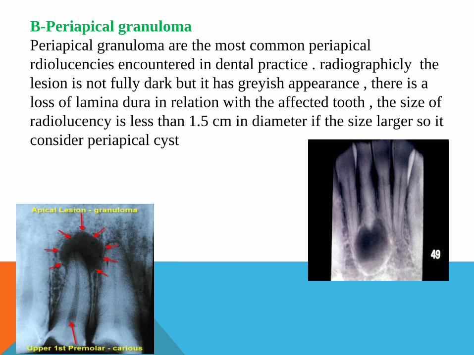
B-Periapical granuloma
Periapical granuloma are the most common periapical
rdiolucencies encountered in dental practice . radiographicly the
lesion is not fully dark but it has greyish appearance , there is a
loss of lamina dura in relation with the affected tooth , the size of
radiolucency is less than 1.5 cm in diameter if the size larger so it
consider periapical cyst

C-Chronic periapical abscess
Radiographic appearance of the lesion may be quite variable, the
lesion may have radiolucent appearance with ill-defined borders
and in this time it impossible to differentiate from granuloma or
cyst.
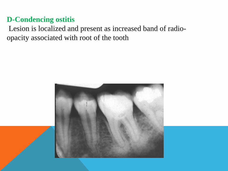
D-Condencing ostitis
Lesion is localized and present as increased band of radio-
opacity associated with root of the tooth
4-FRUCTURES:
Radiographic signs of fractures
The following are general signs that may indicate the presence
of a fracture of bone or tooth:
1. The presence of a radiolucent line (usually sharply defined) w
2. A change in the normal anatomic outline or shape of the
structure.
3. A defect in the outer cortical boundary, which may appear as
a deviation in the smooth outline.
4. An increase in the density of the bone, which may be caused
by the overlapping of two fragments of bone

Tooth Fracture
Intraoral periapical films (a minimum of two) should be taken at
differing horizontal angulations of the x-ray beam. A panoramic
film may serve as a survey film, but it may not have the image
detail to reveal a no displaced
root fracture

5- Impacted tooth is one that fails to erupt into the dental arch
within the expected developmental window. Because impacted
teeth do not erupt, they are retained throughout the individual's
lifetime unless extracted or exposed surgically. Mandibular third
molars are more commonly impacted than their maxillary
counterparts. As a general rule, all impacted teeth must be
removed, except canine teeth; canines do not need surgery and
may just remain buried and give no further problems.
Accurate diagnostic imaging is an essential requirement to derive
the correct diagnosis and optimal treatment plan, as well as
monitor and document the treatment progress and final outcome.
Intra oral periapical and occlusal films can provide this.


THANCK YOU