Embed Size (px)
Citation preview
Radioactive Material License Guide
Medical Use
Louisiana Department of Environmental Quality Emergency & Radiological Services Division
Licensing & Registrations Section P. O. Box 4312
Baton Rouge, Louisiana 70821-4312 602 N. Fifth Street
Baton Rouge, LA 70802 Telephone (225) 219-3041
Fax (225) 219-3154 (Rev. 1/06)
2
INTRODUCTION General: This guide describes the type and extent of information needed by the Registrations and Certifications staff to evaluate an application for a specific license for the possession and use of radioactive material. This type of license is provided for under LAC 33:XV.324 and 325. The applicant should carefully study the regulations and this guide and submit all information requested. Please remember that any necessary information that is not submitted will delay completion of the review of your application. The Registration and Certification Section usually issues a single radioactive material license to cover an institution's entire radioisotope program. Separate licenses are not normally issued to different departments of a medical institution nor are they issued to individuals associated with the institution. A separate license may be issued for teletherapy if it is necessary. Purpose of Guide: This guide is designed to describe the type and extent of information needed by the Department to evaluate an application for a medical use license and to describe the medical use byproduct material regulations. This guide does not apply to academic programs that do not use byproduct material for medical use. Purpose of Appendices to Guide: The regulations require that the licensee develop and implement procedures that will ensure compliance with the regulations. Appendices A through S to this guide describe model radiation safety procedures. Each applicant should carefully read the applicable regulations and model procedures and then decide if the model procedures are appropriate for its specific radiation safety needs. Applicable Regulations: The following chapters of LAC 33:XV apply to medical programs and should be used in conjunction with this guide. The applicant should carefully study the regulations and this guide and submit all the information requested. This guide does not substitute for understanding the requirements of the regulations. A. Chapter 1 - General Provisions B. Chapter 3 - Licensing of Radioactive Material
3
C. Chapter 4 - Standards for Protection Against Radiation D. Chapter 7 - Use of Radionuclides in the Healing Arts E. Chapter 10 - Notices, Instructions and Reports to Workers; Inspections Please note that this guide is intended only for general guidance in preparation of the license application and should not be considered a substitute for the applicant's safety evaluation of the proposed use of radioactive material. The applicant must insure that the application correctly and adequately describes the radiation safety measures and procedures to be followed in order to provide adequate protection. AS LOW AS REASONABLY ACHIEVABLE: (ALARA) The applicant should, in addition to complying with the requirements set forth in LAC 33:XV, make every reasonable effort to maintain radiation exposures and releases of radioactive materials in effluents to unrestricted areas as low as reasonably achievable (ALARA). The term "as low as in reasonably achievable" means as low as is reasonably achievable taking into account the state of technology, and the economics of improvements in relation to benefits to the public health and safety, and other societal and socio-economic considerations, and in relation to the utilization of ionizing radiation in the public interest. ALARA in Medical Institutions: Each medical licensee must have a formal ALARA program (see LAC 33:XV.705). The success of an ALARA program depends on the cooperation of each person who works at the licensee's facility. Management should make a formal policy commitment to the ALARA philosophy and implement that commitment with adequate resources. A Radiation Safety Committee composed of individuals who have special expertise in the safe use of byproduct material is required by LAC 33:XV.707 to review uses for safety and ALARA considerations. The Committee, the RSO and management should audit the byproduct material program to ensure the continued safe use of byproduct material. In addition to being a member of the Committee, the RSO serves as a technical consultant to the Committee and is also responsible for the day-to-day operation of the radiation safety program. A model ALARA management program is contained in Appendix G to this guide. Applicants should consider the ALARA philosophy in the development of plans for work with radioactive materials. Types of Medical Licenses:
4
A specific license may be issued to institutions detailing each radioisotope and clinical use performed by physicians named on the institution's license. If the physicians have met the training and experience criteria applicable for the intended program and the proposed radiation safety program is acceptable, a license will be issued. Specific licenses of broad scope for medical use, such as licenses authorizing multiple quantities and types of radioactive material for unspecified uses, are issued to institutions that 1) have had previous experience operating under a specific institutional license of limited scope; and 2) are engaged in medical research as well as routine diagnostic and therapy procedures using radioisotopes. Such programs operate under the supervision of a medical isotopes committee. Individuals users are not named on the license nor are radioisotopes limited to specified uses. Individual users and procedures are approved by the institution's medical isotope committee. Physicians may obtain basic and clinical radioisotope training and experience in the use of radiopharmaceuticals in such programs. This type of license is not appropriate for most institutions using radioactive material in medical programs. Chapter 7 divides byproduct material for medical use into different types of use. You may indicate only the types of use you want or if you do not want all the material listed in Chapter 7, you must identify the material you do want from that section. Radioactive aerosols and gases may be used only if specific application is made to and approved by the Department. License Fees: A fee is required for all initial applications and for licenses that are required to be reissued. The applicant should refer to the Department’s fee schedule LAC 33:XV.Chapter 25 to determine the amount of the fee that must accompany the application. Review of the application will not begin until the proper fee is received by the Department. If you have any questions concerning the fee or the amount to submit, please do not hesitate to contact the Department. Filing an Application: A license application for radioactive material should be submitted on Form DRC-11, "Application for Radioactive Material License". The applicant should complete all items on the application form in sufficient detail for the Department to determine that the
5
applicant's equipment, facilities, personnel training and qualifications, and radiation protection program are adequate to protect health and to minimize danger to property. Only a single copy of the application and all attachments needs to be submitted to the Department. The applicant should retain one copy, since the license will require as a condition that the institution follow the statements and representations set forth in the application and any supplements. It is also a requirement of LAC 33:XV.1011 to make a copy of your operating procedures available to employees. Since the space on Forms DRC-11 and 13 is usually not sufficient to contain all of the required information, additional sheets should be appended. A heading indicating the appropriate item number should identify each separate sheet or document submitted with the application. When completed, Form DRC-11 should be signed and dated by a representative of the institution's management. CONTENTS OF APPLICATION This portion of the guide explains, item by item, the information requested on Form DRC-11. The appendices to this guide serve several different purposes, i.e., to provide additional information on certain subject areas, to provide a model procedure the licensee may adopt in response to an item on the application form, or to provide an outline the applicant may use to develop a procedure for review the Department staff. Form DRC-11 Item 1 - Enter the name of the applicant, the telephone number and mailing address to which correspondence should be directed. If a physician is requesting use of radioactive material at his office, then he is named as the applicant. If radioactive material is to be stored and used at an institution, the institution is named as the applicant. Item 2 - Indicate whether this is an application for a new license, an amendment, or a renewal. Item 3 - If the mailing address in Item 1 is a post office box or if different from the location where medical radioisotopes will be used and/or stored, then enter the street address where medical radioisotopes will be primarily used. A post office box address is not acceptable. If appropriate, specify the department or location within the institution where medical radioisotopes will be used and/or stored. If radioactive material is to be used at more than one location, you must give the specific address of each location. Item 4 - The Radiation Safety Officer (RSO) is the person designated responsible for the day-to-day radiation safety program. The RSO maintains all records required by the
6
Louisiana Radiation Regulations and is the primary contact with the Department on matters pertaining to the license and the use of radioactive materials. The RSO does not have to be a physician, but his training and experience with the types and quantities of radioactive material for which a license is being requested must be submitted. (See Appendix S) List the names of physicians who will use, supervise, or direct the use of radioactive material. Acceptable training and experience for physicians is specified in Appendix S of this guide. This list should include the physicians who supervise other physicians in training and/or who direct technologists or other paramedical personnel who use radioactive material for human or non-human use. Non-physicians may only be authorized to use radioactive material for non-human use, e.g., instrument calibration or in vitro procedures. Authorized physician users have the following responsibilities: A. approving of procedures involving the administration of radiopharmaceuticals to
patients or the application of radiation from radioisotope sources to patients; B. prescribing the radiopharmaceuticals or source of radiation and the amount of or
dosage to be administered; C. determining the route of administration; and D. interpreting the results of diagnostic procedures in which radiopharmaceuticals are
administered. Items A through D may be delegated to physicians who are in training under the supervision of authorized physician users. Supervision means that the physician user has adequately instructed the physician in training in the specific human use of radioactive materials and has also ascertained that safe use of these materials in humans has been properly emphasized to the physician in training. It also means that the physician user periodically reviews the work of those supervised and assures him that proper medical records are made of each use. It is required that the authorized user be able to be physically present and available to the supervised individual on one hour's notice (the supervising authorized user need not be present for each use of radioactive material). Properly trained technicians, technologists, or other paramedical personnel under an authorized user's direction may be delegated to the following activities: A. the preparation and quality control testing of radiopharmaceuticals and sources of
radiation; B. the assay of radiopharmaceutical dosages prior to administration;
7
C. the use of appropriate instrumentation for the collection of data used by the physician; and
D. the administration of radiopharmaceuticals or radiation from radioisotope sources to
patients if permitted under applicable federal, state or local laws. This delegation should be made in writing.
A qualified individual must be designated the responsibility for radiation protection (radiation safety officer). Such an individual may be a physician user or other qualified person. The name and title of the person designated by and responsible to the institution's management for the coordination of the institution's radiation safety program should be included under radiation program personnel. If the radiation safety officer is assisted by a consultant or part time employee, state the individual's name and describe his duties and responsibilities. If the radiation safety officer is not one of the physician users, submit a complete description of his training and experience. The names of any technicians directly associated with the medical radioactive material program should be listed, and their qualifications and training should be given on the back of Form DRC-13, under radiological training and qualifications. For more details concerning training, please refer to Appendix A. Item 5 - Personnel Monitoring: A. Chapter 4 of LAC 33:XV requires licensees to provide radiation monitoring for
occupationally exposed individuals who might receive a dose in excess of 10 percent of the limits in LAC 33:XV.410.
B. Personnel monitoring is required for all physicians and technicians associated with
medical radioisotope programs utilizing either radioactive drugs or sealed sources, such as cesium, radium, cobalt, or iridium. In a sealed source program, there is the possibility that extremely high radiation exposures may occur in a short period of time; therefore, the use of pocket dosimeters or pocket chambers is encouraged as a means of an immediate indication of exposure. If pocket chambers or dosimeters are to be used, then complete the requested information in Item 5.B of Form DRC-11.
C. It should be understood that if a Cobalt-60 teletherapy unit is utilized in the medical
radioisotope program, all personnel working with the unit shall wear film or TLD badges at all times while in the work area.
D. If the medical radioisotope program involves the use of generators or the preparation
of kits, the program's personnel shall wear wrist or ring badges. Also, programs utilizing sealed sources for medical implant should use extremity monitoring.
E. If the isotope program is strictly limited to the use of beta eye applicators, personnel
8
monitoring is not required but is recommended. F. The need for bioassays should be thoroughly considered if the chemical or physical
form or if procedures and equipment used make it likely that radioactive material will be ingested, inhaled, or absorbed through the skin, e.g., therapeutic doses of liquid iodine-131. If applicable, describe your procedure for performing bioassays, i.e., schedule, action levels, actions to be taken, equipment to be used, calibration of equipment, count standard in lucite neck phantom, and conversion of count rate to activity units.
Please describe your personnel occupational exposure monitor program. See APPENDIX D of this guide. Item 6 - Contamination Surveys and Area Surveys: Submit your survey procedures. SEE APPENDIX N. Item 7 - Leak Test: Indicate the company performing leak testing and at what intervals. If you will performing your own analysis, see Appendix H. Item 8 - Waste Disposal: Submit your procedures for waste disposal. SEE APPENDIX R. Item 9 - Health Physics Program: a. Radiation Safety Committee/Radiation Safety Officer Describe your Radiation Safety Committee Charter and Radiation Safety Officer delegation
of authority. A Radiation Safety Committee must be established by each medical institution licensee. If you are not an institution, you only need to submit the Radiation Safety Officer delegation of authority. SEE APPENDIX F.
b. ALARA Program Submit your ALARA program. Each medical licensee must have an ALARA program.
SEE APPENDIX G. c. Training Program
9
Describe your training program for individuals who work with or in the vicinity of
radioactive material. SEE APPENDIX A of this guide. d. Leak Test Submit your procedures for leak-testing sealed sources. SEE APPENDIX H. e. Safe Use of Radiopharmaceuticals Submit a copy of your rules for the safe use of radiopharmaceuticals. SEE APPENDIX I. f. Spill Procedures Submit a copy of your spill procedures. SEE APPENDIX J. g. Ordering and Receiving Submit a copy of your procedure for ordering and receiving radioactive material. SEE
APPENDIX K. h. Opening Packages Submit your procedure for opening packages that contain radioactive material. SEE
APPENDIX L. i. Unit Dosage Records Submit your procedure for keeping records of unit dosage use. SEE APPENDIX M.1. j. Multidose Vial Records Submit your procedure for keeping records of multidose vial use. SEE APPENDIX M.2. k. Molybdenum Concentration Records Submit your procedure for measuring and recording molybdenum concentration. SEE
APPENDIX M.3. l. Implant Source Use Records Submit your procedure for keeping an inventory of implant sources. SEE APPENDIX M.4. m. Air Concentration Control
10
Submit your procedure for calculating spilled gas clearance times. SEE APPENDIX O. n. Radiopharmaceutical Therapy Submit your procedure for radiation safety during radiopharmaceutical therapy. See
Appendix P. o. Implant Therapy Submit your procedure for radiation safety during implant therapy. See Appendix Q. p. Physical Facilities Submit a drawing of the room or rooms and adjacent areas where radioactive material will
be used. Note the following: A. Room numbers and principal use of each room or area (for example, in vitro, hot lab,
waiting, examining, imaging, reading, office, file, fresh materials storage, radioactive waste storage, film processor, toilet, closet, hallway).
B. Any shielding available. C. Additional safety equipment (for example, fume hoods, L-blocks, or fixed area
monitors. D. The diagram should show the proximity of radiation sources to unrestricted areas. q. Quality Management Program This section is applicable for any licensee administering: any teletherapy radiation dose; any
gamma sterotactic radiosurgery radiation dose; any brachytherapy radiation dose; any administration of quantities greater than 30 microcuries of either sodium iodide I-125 or I-131; or any therapeutic administration of a radiopharmaceutical, other than sodium iodide I-125 or I-131. Each applicant shall submit to the Department a quality management program as part of the application for a license and implement the program upon issuance of the license by the Department. Please refer to LAC 33:XV.777 for guidance.
Item 10 - Health Physics Instrumentation: List all radiation monitoring or measuring instruments that will be available. List the manufacturer's name and model number of each instrument, number of instruments available, the type of radiation detected (e.g., beta, gamma) the range (mR/hour, counts per minute), window thickness if
11
applicable, and how used (measuring or surveying). Describe the method, frequency, and standard used for the maintenance and calibration of radiation detection and survey instruments. These procedures should be included even if the services of an individual or company is employed. SEE APPENDIX B for model procedures. Item 11 - General Instrumentation: List the manufacturer and model number of all instruments used in conjunction with the requested procedures. This would include rectilinear scanners, gamma cameras, and dose calibrators. Submit your procedure for calibrating the dose calibrator. SEE APPENDIX C for requirements. Item 12 - Medical Supplements: See Appendices P and Q for additional information. Attach approvals from hospitals where radioactive materials are used and hospitals that admit patients containing radioactive materials. Item 13 - Industrial Radiography Supplement Not applicable. Item 14 - List the name and company affiliation of any individuals who assisted in the completion of the license application. THE APPLICATION MUST BE DATED AND SIGNED BY THE INDIVIDUAL AUTHORIZED TO SIGN ON BEHALF OF THE INSTITUTION. SUBMIT ONLY THE ORIGINAL. FORM DRC-13 Sealed Sources: In the space provided or on a separate attachment, give the proposed inventory of calibration, reference, and medical radioisotopes in the form of sealed sources. Indicate if the sources will be owned or leased either on a long term or case-by-case basis. If the model numbers of the sealed sources are not available, then indicate the type of sources such as plaques, needles, seeds, tubes, or wire. Radiological Qualifications and Training: Complete the required information for all technologists listed under Item 4 of Form DRC-11. This information may be submitted on a separate attachment if desired, but the attachment should be clearly referenced. Schedule of Radioactive Materials:
12
For routine human use, if you have met the minimum training requirement for diagnostic procedures, you may state only the section number of Chapter 7 of LAC 33:XV (LAC 33:XV.729 and/or 731) for which you are requesting licensure. Indicate specific therapy procedures that you wish to perform and are qualified to perform in accordance with the submitted preceptor statements. Evidence of board eligibility or certification provides qualification for the use of sealed sources in therapy procedures. For routine human use not listed in LAC 33:XV.729 and 731, and for non-human use, list each radionuclide to be used. Provide the chemical and physical form and the maximum quantity in millicuries and/or microcuries to be possessed. List the manufacturer, model number, and quantity for all sealed sources. Describe the intended use of each isotope listed above. For additional requirements for the use of radioactive gases or aerosols, please refer to Appendix O. Also submit the following information to support a request to use Xenon-133. 1. Describe the procedures to be followed for routine uses of Xe-133. If you plan to use a
special apparatus for administration and collection of Xe-133, such as charcoal traps, please specify the manufacturer's name and model number and include a description of its design characteristics. Inclusion of a brochure will be helpful.
2. Describe any special procedures that you plan to employ to reduce leakage. Example: use
of nose clamps or special enclosures. 3. Describe the emergency procedures to be used in case of an accidental release of Xe-133.
This should include such considerations as temporary evacuation of the area or increasing the ventilation of the area.
4. Please describe the method of disposal for Xe-133 that will be employed. One method for
disposal is release to the atmosphere through an air exhaust system. Licensees are required to perform surveys to ensure that they are in compliance with regulatory requirements for release of radioactive material to unrestricted areas.
The disposal method of choice is absorption on charcoal traps. The traps are then stored for
decay. If a charcoal trap is used, please describe how you will handle the problem of leakage from such trapping devices and how you will handle saturated filters.
Your discussion should include a description of the area, available shielding and ventilation.
13
APPENDIX A Model Training Program The following guidance may be used to develop a training program. It may not be assumed that safety instruction has been adequately covered by prior occupational training, board certification etc. Site-specific training should be provided for all workers. Ancillary personnel (e.g., nursing, clerical, housekeeping, security) whose duties may require them to work in the vicinity of radioactive material (whether escorted or not) need to be informed about radiation hazards and appropriate precautions. All training should be tailored to meet the needs of the individuals in attendance. A training program that provides necessary instruction should be written and implemented. Model Program Personnel will be instructed: 1. Before assuming duties with, or in the vicinity of, radioactive materials. 2. During annual refresher training. 3. Whenever there is a significant change in duties, regulations, or the terms of the license. Instruction for individuals in attendance will include the following subjects: 1. Applicable regulations and license conditions. 2. Areas where radioactive material is used or stored. 3. Potential hazards associated with radioactive material in each area where the employees will
work. 4. Appropriate radiation safety procedures. 5. Licensee's in-house work rules. 6. Each individual's obligation to report unsafe conditions to the Radiation Safety Officer. 7. Appropriate response to emergencies or unsafe conditions. 8. Worker's right to be informed of occupational radiation exposure and bioassay results. 9. Locations where the licensee has posted or made available notices, copies of pertinent
regulations, and copies of pertinent licenses and license conditions. 10. Question and answer period.
14
APPENDIX B Model Procedure for Calibrating Survey Instruments Radiation survey meters should be calibrated with a radioactive source. Electronic calibrations alone are not acceptable. Survey meters must be calibrated at least annually and after servicing. (Battery changes are not considered "servicing.") Model Procedure 1. The source must be approximately a point source. 2. Either the apparent source activity or the exposure rate at a given distance must be traceable
by documented measurements to a standard certified within 5 percent accuracy by the National Bureau of Standards.
3. A source that has approximately the same photon energy as the environment in which the
calibrated device will be employed should be used for the calibration. 4. The source should be of sufficient strength to give an exposure rate of about 30 mR/hr at 100
cm. Minimum activities of typical sources are 85 mCi of Cs-137 or 21 mCi of Co-60. 5. The inverse square law and the radioactive decay law must be used to correct for change in
exposure rate due to changes in distance or source decay. 6. A record must be made of each survey meter calibration. 7. A single point on a survey meter scale may be considered satisfactorily calibrated if the
indicated exposure rate differs from the calculated exposure rate by less than 10 percent. 8. Three kinds of scales are frequently used on survey meters: a. Meters on which the user selects a linear scale must be calibrated at no less than two
points on each scale. The points should be at approximately 1/3 and 2/3 of full scale.
b. Meters that have a multidecade logarithmic scale must be calibrated at no less than one point on each decade and no less than two points on one of the decades. Those points should be at approximately 1/3 and 2/3 of the decade.
c. Meters that have an automatically ranging digital display device for indicating rates
must be calibrated at no less than one point on each decade and at no less than two points on one of the decades. Those points should be approximately 1/3 and 2/3 of the decade.
9. Readings above 1,000 mR/hr need not be calibrated. However, such scales should be
15
checked for operation and approximately correct response. 10. At the time of calibration, the apparent exposure rate from a built-in or owner-supplied check
source must be determined and recorded. 11. The report of a survey meter calibration should indicate the procedure used and the data
obtained. The description of the calibration will include: a. The owner or user of the instrument; b. A description of the instrument that includes manufacturer, model number, serial
number and type of detector; c. A description of the calibration source, including exposure rate at a specified
distance on a specified date, and the calibration procedure; d. For each calibration point, the calculated exposure rate, the indicated exposure rate,
the deduced correction factor (the calculated exposure rate divided by the indicated exposure rate), and the scale selected on the instrument;
e. The reading indicated with the instrument in the "battery check" mode (if available
on the instrument); f. The angle between the radiation flux field and the detector (for external cylindrical
GM or ionization-type detectors, this will usually be "parallel" or "perpendicular" indicating photons traveling either parallel with or perpendicular to the central axis of the detector; for instruments with internal detectors, this should be the angle between the flux field and a specified surface of the instrument);
g. For detectors with removable shielding, an indication of whether the shielding was in
place or removed during the calibration procedure;
h. The apparent exposure rate from the check source; and
i. The name of the person who performed the calibration and the date on which the calibration was performed.
16
APPENDIX C Model Procedure for Calibrating Dose Calibrator Model Procedure 1. Test for the following at the indicated frequency. Consider repair, replacement, or arithmetic
correction if the dose calibrator falls outside the suggested tolerances. (These recommended tolerances are more restrictive than those in the regulations to ensure that corrective action will be taken before the dose calibrator is outside permissible tolerances.)
a. Constancy at least once each day prior to assay of patient dosages (±5 percent). b. Linearity at installation and at least quarterly thereafter (±5 percent). c. Geometry dependence at installation (±5 percent). d. Accuracy at installation and at least annually thereafter (±5 percent). 2. After repair, adjustment, or relocation of the dose calibrator, repeat the above tests as
appropriate. 3. Constancy means reproducibility in measuring a constant source over a longer period of
time. Assay at least one relatively long-lived source such as Cs-137, Co-60, Co-57, or Ra-226 using a reproducible geometry each day before using the calibrator. Consider the use of two or more sources with different photon energies and activities. Use the following procedure:
a. Assay each reference source using the appropriate dose calibrator setting (i.e., use the
Cs-137 setting to assay Cs-137). b. Measure background at the same setting, and subtract or confirm the proper
operation of the automatic background subtract circuit if it is used. c. For each source used, either plot on graph paper or log in a book the background
level for each setting checked and the net activity of each constancy source. d. Using one of the sources, repeat the above procedure for all commonly used
radioisotope settings. Plot or log the results.
17
e. Establish an action level or tolerance for each recorded measurement at which the
individual performing the test will automatically notify the chief technician or authorized user of suspected malfunction of the calibrator. These action levels should be written in the log book or posted on the calibrator. The regulation requires repair or replacement if the error exceeds 10 percent.
4. Inspect the instrument on a quarterly basis to ascertain that the measurement chamber liner is
in place and that the instrument is zeroed according to the manufacturer's instructions. 5. Linearity means that the calibrator is able to indicate the correct activity over the range of
use of that calibrator. This test is done using a vial or syringe of Tc-99m whose activity is at least as large as the maximum activity normally assayed in a prepared radiopharmaceutical kit, in a unit dosage syringe, or in a radiopharmaceutical therapy, whichever is largest.
Decay Method a. Assay the Tc-99m syringe or vial in the dose calibrator, and subtract background to
obtain the net activity in millicuries. Record the date, time to the nearest minute, and net activity on the Dose Calibrator Linearity Test Form. This first assay should be done in the morning at a regular time, for example, 8 a.m.
b. Repeat the assay at about noon, and again at about 4 p.m. Continue on subsequent
days until the assayed activity is less than 10 microcuries. For dose calibrators on which you select a range with a switch, select the range you would normally use for the measurement.
c. Convert the time and date information you recorded to hours elapsed since the first
assay. d. On a sheet of semilog graph paper, label the logarithmic vertical axis in millicuries
and label the linear horizontal axis in hours elapsed. At the top of the graph, note the date and the manufacturer, model number, and serial number of the dose calibrator. Then plot the data.
e. Draw a "best fit" straight line through the data points. For the point farthest from the
line, calculate its deviation from the value on the line (A-observed - A-line)/(A-line) = deviation.
18
f. If the worst deviation is more than ±0.05, the dose calibrator should be repaired or
adjusted. If this cannot be done, it will be necessary to make a correction table or graph that will allow you to convert from activity indicated by the dose calibrator to "true activity."
g. Put a sticker on the dose calibrator that says when the next linearity test is due. Shield Method If you decide to use a set of "sleeves" of various thicknesses to test for linearity, it will first be necessary to calibrate them. a. Begin the linearity test as described in the decay method described above. After
making the first assay, the sleeves can be calibrated as follows. Steps b through d below must be completed within 6 minutes.
b. Put the base and sleeve 1 in the dose calibrator with the vial. Record the sleeve
number and indicated activity. c. Remove sleeve 1 and put in sleeve 2. Record the sleeve number and indicated
activity. d. Continue for all sleeves. e. Complete the decay method linearity test steps b through g above. f. From the graph made in step d of the decay method, find the decay time associated
with the activity indicated with sleeve 1 in place. This is the "equivalent decay time" for sleeve 1. Record that time with the data recorded in step b.
g. Find the decay time associated with the activity indicated with sleeve 2 in place.
This is the "equivalent decay time" for sleeve 2. Record that time with the data recorded in step c.
h. Continue for all sleeves. i. The table of sleeve numbers and equivalent decay times constitutes the calibration of
the sleeve set. The sleeve set may now be used to test dose calibrators for linearity. a. Assay the Tc-99m syringe or vial in the dose calibrator, and subtract background to
19
obtain the net activity in millicuries. Record the net activity. b. Steps c through e below must be completed within 6 minutes. c. Put the base and sleeve 1 in the dose calibrator with the vial. Record the sleeve
number and indicated activity. d. Remove sleeve 1 and put in sleeve 2. Record the sleeve number and indicated
activity. e. Continue for all sleeves. f. On a sheet of semilog graph, label the logarithmic vertical axis in millicuries, and
label the linear horizontal axis in hours elapsed. At the top of the graph, note the date and the model number and serial number of the dose calibrator.
g. Plot the data using the equivalent decay time associated with each sleeve. h. Draw a "best fit" straight line through the data points. For the point farthest from the
line, calculate its deviation from the value on the line (A-observed - A-line)/A-line = deviation.
i. If the worst deviation is more than ±0.05, the dose calibrator should be repaired or
adjusted. If this cannot be done, it will be necessary to make a correction table or graph that will allow you to convert from activity indicated by the dose calibrator to "true activity."
j. Put a sticker on the dose calibrator that says when the next linearity test is due. 6. Geometry independence means that the indicated activity does not change with volume or
configuration. This test should be done using a syringe that is normally used for injections. Licensees who use generators and radiopharmaceuticals kits should also do the test using a vial similar in size, shape, and construction to the radiopharmaceutical kit vials normally used. The following test assumes injections are done with 3-cc plastic syringes and that radiopharmaceutical kits are made in 30-cc glass vials. If you do not use these, change the procedure so that your syringes and vials are tested throughout the range of volumes commonly used.
a. In a small beaker or vial, mix 2 cc of a solution of Tc-99m with an activity
concentration between 1 and 10 mCi/ml. Set out a second small beaker or vial with nonradioactive saline. You may also use tap water.
b. Draw 0.5 cc of the Tc-99m solution into the syringe and assay it. Record the volume
20
and millicuries indicated on a Dose Calibrator Geometry and Accuracy Form. c. Remove the syringe from the calibrator, draw an additional 0.5 cc of nonradioactive
saline or tap water, and assay again. Record the volume and millicuries indicated. d. Repeat the process until you have assayed a 2.0-cc volume. e. Select as a standard the volume closest to that normally used for injections. For all
the other volumes, divide the standard millicuries by the millicuries indicated for each volume. The quotient is a volume correction factor. Alternatively, you may graph the data and draw horizontal 5 percent error lines above and below the chosen "standard volume."
f. If any correction factors are greater than 1.05 or less than 0.95, or if any data points
lie outside the 5 percent error lines, it will be necessary to make a correction table or graph that will allow you to convert from "indicated activity" to "true activity." If this is necessary, be sure to label the table or graph "syringe geometry dependence," and note the date of the test and the model number and serial number of the calibrator.
g. To test the geometry dependence for 3 30-cc glass vial, draw 1.0 cc of the Tc-99m
solution into a syringe and then inject it into the vial. Assay the vial. Record the volume and millicuries indicated.
h. Remove the vial from the calibrator and, using a clean syringe, inject 2.0 cc of
nonradioactive saline or tap water, and assay again. Record the volume the millicuries indicated.
i. Repeat the process until you have assayed a 19.0-cc volume. The entire process
must be completed within 10 minutes. j. Select as a standard the volume closest to that normally used for mixing
radiopharmaceutical kits. For all the other volumes, divide the standard millicuries by the millicuries indicated for each volume. The quotient is a volume correction factor. Alternatively, you may graph the data and draw horizonal 5 percent error lines above and below the chosen "standard volume."
k. If any correction factors are greater than 1.05 or less than 0.95 or if any data points
lie outside the 5 percent error lines, it will be necessary to make a correction table or graph that will allow you to convert from "indicated activity" to "true activity." If this is necessary, be sure to label the table or graph "vial geometry dependence," and note the date of the test and the model number and serial number of the calibrator.
21
7. Accuracy means that, for a given calibrated reference source, the indicated millicurie value is equal to the millicurie value determined by the National Bureau of Standards (NBS) or by the supplier who has compared that source to a source that was calibrated by the NBS. Certified sources are available from the NBS and from many radioisotope suppliers. At least two sources with different principal photon energies (such as Co-57, Co-60 or Cs-137) should be used. The regulations require that one must have a principal photon energy between 100 keV and 500 keV. The regulations also require that, if a Ra-226 source is used, it must be at least 10 microcuries; other sources must be at least 50 microcuries. Consider using at least one reference source whose activity is within the range of activities normally assayed.
a. Assay a calibrated reference source at the appropriate setting (i.e., use the Co-57
setting to assay Co-57), and then remove the source and measure background. Subtract background from the indicated activity to obtain the net activity. Record this measurement on a Dose Calibrator Geometry and Accuracy Form. Repeat for a total of three determinations.
b. Average the three determinations. The average value should be within 5 percent of
the certified activity of the reference source, mathematically corrected for decay. c. Repeat the procedure for other calibrated reference sources. d. If the average value does not agree within 5 percent with the certified value of the
reference source, the dose calibrator may need to be repaired or adjusted. The Regulation requires repair or replacement if the error exceeds 10 percent.
e. At the same time the accuracy test is done, assay the source that will be used for the
daily constancy test (it need not be a certified reference source) on all commonly used radioisotope settings. Record the settings and indicated millicurie values with the accuracy data.
f. Put a sticker on the dose calibrator that says when the next accuracy test is due. 8. The RSO will review and sign the records of all geometry, linearity and accuracy tests.
22
APPENDIX D Model Personnel External Exposure Monitoring Program Model Program 1. The RSO will promptly review all exposure reports to look for workers or groups of workers
whose exposure is unexpectedly high or low. This procedure does not apply to backup monitor records, for example, pocket ionization chambers, when the monitor of record is a film or thermoluminescence dosimeter (TLD).
2. All individuals who are occupationally exposed to ionizing photon radiation on a regular
basis will be issued a film or TLD whole body monitor that will be processed by a contract service on a monthly basis.
3. All individuals who, on a regular basis, handle radioactive material that emits ionizing
photons will be issued a film or TLD finger monitor that will be processed by a contract service on a monthly basis.
4. All individuals who are occupationally exposed to radiation on an occasional basis, such as
nurses caring for radiopharmaceutical therapy or implant patients, will be issued a whole body monitor when caring for such patients.
5. Other individuals who are exposed to radiation on an occasional basis such as security
personnel who deliver packages, secretarial personnel who work in the nuclear medicine clinic but do not work with patients, and nurses who occasionally care for patients who have received diagnostic dosages will not normally be issued exposure monitors.
23
APPENDIX E Model Procedure for Checking Equipment Used in Mobile Nuclear Medicine Service The Department normally limits its review of equipment quality assurance programs to those programs developed for radiation safety equipment. However, when delicate imaging equipment is transported from one location of use to another, e.g., by a mobile nuclear medicine service, it is reasonable to assume that it may suffer damage in transit. Therefore, the Department requires that mobile nuclear medicine services have an imaging equipment quality assurance program to ensure that the use of radioactive material will not be inimical to the public health and safety. Mobile nuclear medicine services should also check ventilation equipment if gases will be used. Model Procedure Survey Meter Check the survey meter with the dedicated check source at each location of use. Material may not be used if the survey meter is not working. There is no need to keep a record of these checks. Camera 1. Perform the following checks daily at each location of use before administering radioactive
material: a. Peak each camera according to the manufacturer's instructions. b. Using either Tc-99m or Co-57, perform an extrinsic flood field with a frequently
used collimator in place, or perform an intrinsic flood field test. Accumulate at least 1,000,000 counts for small-field-of-view cameras and 3,000,000 counts for large-field-of-view cameras. Process the image as if it were an image of a patient.
c. Do not administer material until an authorized user or a designated technologist
approves the camera for use. d. You do not have to make a permanent record of these daily checks. 2. Perform the following checks weekly: a. With the same frequently used collimator in place, image a flood source and either a
parallel-line-equal-space (PLES), bar, orthogonal-hole (OH) or resolution-quadrant phantom with the flood field as a source.
24
b. If a PLES or bar phantom is used, rotate it 90 degrees so that the camera is tested for both vertical and horizontal geometric linearity.
c. If a resolution-quadrant phantom is used, rotate it so that each quadrant is imaged in
each quadrant of the crystal. Then turn it over and again image it four more times. This procedure will check both resolution and horizontal and vertical geometric linearity in each quadrant of the crystal.
d. Process the images as if they were images of a patient. Mark them clearly to indicate
image orientation, source activity, and date. e. Retain the images for 2 years. 3. Perform the following safety checks after repairs and quarterly: a. Check the motion interlocks by activating the emergency-off switches on the camera.
With the camera in motion, activation of the emergency-off switch should stop the motion. If this might jeopardize imaging components in the system, perform only the checks described in paragraph 3.b.
b. Check the motion switches. Put the camera in motion and first release just the
direction switch to stop the motion. Then put the camera back in motion and release just the dead-man switch. Test all motion switches and all directions in this manner. Release of either the motion switch or the dead-man switch alone should disable the camera motion. If this is not the case, repair the camera before clinical use.
4. Set the equipment in the same manner each time checks are run. Make a record of all these
checks. Keep a separate file or ring binder for each camera. Retain the record for 2 years. Ventilation - If gases or aerosols will be used, check the ventilation supply, exhaust vents, and collection devices for operation with tissue paper or a velometer. There is no need to keep a record of these checks.
25
APPENDIX F Model Radiation Safety Committee Charter and Radiation Safety Officer Delegation of Authority MODEL CHARTER Charge. The Committee shall: 1. Ensure that licensed material will be used safely. This includes review, as necessary of
training programs, equipment, facility, supplies, and procedures; 2. Ensure that licensed material is used in compliance with the Louisiana Radiation Regulations
and the institutional license; 3. Ensure that the use of licensed material is consistent with the ALARA philosophy and
program; 4. Establish a table of investigational levels for individual occupational radiation exposures;
and 5. Identify program problems and solutions. Responsibilities. The Committee shall: 1. Be familiar with all pertinent regulations, the license application, the license, and
amendments; 2. Review the training and experience of the proposed authorized users, the Radiation Safety
Officer (RSO), and the authorized medical physicist/teletherapy physicist to determine that their qualifications are sufficient to enable the individuals to perform their duties safely and are in accordance with the regulations and the license;
3. Review on the basis of safety and approve or deny, consistent with the limitations of the
regulations, the license, and the ALARA philosophy, all requests for authorization to use radioactive material within the institution;
4. Prescribe special conditions that will be required during a proposed method of use of
radioactive material such as requirements for bioassay, physical examinations of users, and special monitoring procedures;
5. Review quarterly the RSO's summary report of the occupational radiation exposure records
of all personnel, giving attention to individuals or groups of workers whose occupational exposure appears excessive;
26
6. Establish a program to ensure that all persons whose duties may require them to work in or frequent areas where radioactive materials are used (e.g., nursing, security, housekeeping, physical plant) are appropriately instructed as required.
7. Review at least annually the RSO's summary report of the entire radiation safety program to
determine that all activities are being conducted safely, in accordance with radiation regulations and the conditions of the license, and consistent with the ALARA program and philosophy. The review must include an examination of records, reports from the RSO, results of the Department’s inspections, written safety procedures, and the adequacy of the management control system;
8. Recommend remedial action to correct any deficiencies identified in the radiation safety
program; 9. Maintain written minutes of all Committee meetings, including members in attendance and
members absent, discussions, actions, recommendations, decisions, and numerical results of all votes taken; and
10. Ensure that the radioactive material license is amended if required prior to any changes in
facilities, equipment, policies, procedures, and personnel. Administrative Information 1. The Committee shall meet as often as necessary to conduct its business but not less than
once in each calendar quarter.
2. Membership must include one authorized user for each type of use authorized by the license, the RSO, a representative of the nursing service, and a representative of management who is neither an authorized user nor an RSO. Management may appoint alternate members to participate in meetings in the case of absence of principal members and should consider appointing as adjunct members representatives from security, physical plant, housekeeping, and other departments. (Adjunct members should abstain from balloting on radiation safety technical questions such as Items 2 through 5 in the "Responsibilities" section above.)
3. To establish a quorum, one-half of the Committee's membership, including the RSO and the
management representative, must be present. 4. To the extent that they do not interfere with the mission of the Committee, management may
assign other responsibilities such as X-ray radiation safety, quality assurance oversight, and research project review and approval.
Model Delegation of Authority
27
Memo to: All Employees From: Chief Executive Officer Subject: Delegation of Authority _________________________ has been appointed Radiation Safety Officer and is responsible for ensuring the safe use of radiation. The Radiation Safety Officer is responsible for managing the radiation safety program; identifying radiation safety problems; initiating, recommending, or providing corrective actions; verifying implementation of corrective actions; and ensuring compliance with regulations. The Radiation Safety Officer is hereby delegated the authority necessary to meet those responsibilities. The Radiation Safety Officer is also responsible for assisting the Radiation Safety Committee in the performance of its duties and serving as its secretary.
28
APPENDIX G Model Program for Maintaining Occupational Radiation Exposure at Medical Institutions ALARA ALARA Program __________________________________ (Licensee's Name) __________________________________ (Date) 1. Management Commitment a. We, the management of this (medical facility, hospital, etc.) are committed to the
program described hereby for keeping individual and collective doses as low as is reasonably achievable (ALARA). In accord with this commitment, we hereby describe an administrative organization for radiation safety and will develop the necessary written policy, procedures, and instructions to foster the ALARA concept within our institution. The organization will include a Radiation Safety Committee (RSC) and a Radiation Safety Officer (RSO).
b. We will perform a formal annual review of the radiation safety program, including
ALARA considerations. This will include reviews of operating procedures and past dose records, inspections, etc., and consultations with the radiation safety staff or outside consultants.
c. Modifications to operating and maintenance procedures and to equipment and
facilities will be made if they will reduce exposures unless the cost, in our judgment, is considered to be unjustified. We will be able to demonstrate, if necessary, that improvements have been sought, that modifications have been considered, and they have been implemented when reasonable. If modifications have been recommended but not implemented, we will be prepared to describe the reasons for not implementing them.
d. In addition to maintaining doses to individuals as far below the limits as is
reasonably achievable, the sum of the doses received by all exposed individuals will also be maintained at the lowest practicable level. It would not be desirable, for example, to hold the highest doses to individuals to some fraction of the applicable limit if this involved exposing additional people and significantly increasing the sum of radiation doses received by all involved individuals.

29
2. Radiation Safety Committee a. Review of Proposed Users and Uses (1) The RSC will thoroughly review the qualifications of each applicant with
respect to the types and quantities of materials and methods of use for which application has been made to ensure that the applicant will be able to take appropriate measures to maintain exposure ALARA.
(2) When considering a new use of radioactive material, the RSC will review the
efforts of the applicant to maintain exposure ALARA. (3) The RSC will ensure that the users justify their procedures and that
individual and collective doses will be ALARA. b. Delegation of Authority (The judicious delegation of RSC authority is essential to the enforcement of an
ALARA program.) (1) The RSC will delegate authority to the RSO for enforcement of the ALARA
concept. (2) The RSC will support the RSO when it is necessary for the RSO to assert
authority. If the RSC has overruled the RSO, it will record the basis for its action in the minutes of the quarterly meeting.
c. Review of ALARA Program (1) The RSC will encourage all users to review current procedures and develop
new procedures as appropriate to implement the ALARA concept. (2) The RSO will perform a quarterly review of occupational radiation exposure
with particular attention to instances in which the investigational levels in Table 1 are exceeded. The principal purpose of this review is to assess trends in occupational exposure as an index of the ALARA program quality and to decide if action is warranted when investigational levels are exceeded (see Section 6 below for a discussion of investigational levels).
30
Table 1 Investigational Levels Investigational Levels (mRems per calendar quarter) ________________________________ Level I Level II ______________________________________________________________________ 1. Whole body; head and trunk 125 375 active blood-forming organs; lens of eyes; or gonads 2. Hands and forearms; feet 375 1125 and ankles 3. Skin of whole body* 1250 3750 _____________________________________________________________________ *Not normally applicable to medical use operations except those using significant quantities of beta-emitting isotopes ______________________________________________________________________ (3) The RSC will evaluate our institution's overall efforts for maintaining doses
ALARA on an annual basis. This review will include the efforts of the RSO, authorized users, and workers as well as those of management.
3. Radiation Safety Officer a. Annual and Quarterly Review (1) Annual review of the radiation safety program. The RSO will perform an
annual review of the radiation safety program for adherence to ALARA concepts. Reviews of specific methods of use may be conducted on a more frequent basis.
(2) Quarterly review of occupational exposures. The RSO will review at least
quarterly the external radiation doses of authorized users and workers to determine that their doses are ALARA in accordance with the provisions of Section 6 of this program and will prepare a summary report for the RSC.
31
(3) Quarterly review of records of radiation surveys. The RSO will review radiation surveys in unrestricted and restricted areas to determine that dose rates and amounts of contamination were at ALARA levels during the previous quarter and will prepare a summary report for the RSC.
b. Education Responsibilities for ALARA Program (1) The RSO will schedule briefings and educational sessions to inform workers
of ALARA program efforts. (2) The RSO will ensure that authorized users, workers, and ancillary personnel
who may be exposed to radiation will be instructed in the ALARA philosophy and informed that management, the RSC, and the RSO are committed to implementing the ALARA concept.
c. Cooperative Efforts for Development of ALARA Procedures Radiation workers will be given opportunities to participate in formulating the
procedures that they will be required to follow. (1) The RSO will be in close contact with all users and workers in order to
develop ALARA procedures for working with radioactive materials.
(2) The RSO will establish procedures for receiving and evaluating the suggestions of individual workers for improving health physics practices and will encourage the use of those procedures.
d. Reviewing Instances of Deviation from Good ALARA Practices The RSO will investigate all known instances of deviation from good ALARA
practices and, if possible, will determine the causes. When the cause is known, the RSO will implement changes in the program to maintain doses ALARA.
4. Authorized Users a. New Methods of Use Involving Potential Radiation Doses (1) The authorized user will consult with the RSO and/or RSC during the
planning stage before using radioactive materials for new uses. (2) The authorized user will review each planned use of radioactive materials to
ensure that doses will be kept ALARA. Trial runs may be helpful.
32
b. Authorized User's Responsibility to Supervised Individuals (1) The authorized user will explain the ALARA concept and the need to
maintain exposures ALARA to all supervised individuals. (2) The authorized user will ensure that supervised individuals who are subject
to occupational radiation exposure are trained and educated in good health physics practices and in maintaining exposures ALARA.
5. Individuals Who Receive Occupational Radiation Doses a. Workers will be instructed in the ALARA concept and its relationship to work
procedures and work conditions. b. Workers will be instructed in recourses available if they feel that ALARA is not
being promoted on the job. 6. Establishment of Investigational Levels in Order to Monitor Individual Occupational
External Radiation Doses This institution hereby establishes investigational levels for occupational external radiation
doses which, when exceeded, will initiate review or investigation by the RSC and/or the RSO. The investigational levels that we have adopted are listed in Table 1. These levels apply to the exposure of individual workers.
The RSO will review and record on Form DRC-5, "Current Occupational External Radiation
Exposures," or an equivalent form (e.g., dosimeter processor's report) results of personnel monitoring not less than once in any calendar quarter. The following actions will be taken at the investigational levels as stated in Table 1:
a. Personnel dose less than Investigational Level I. Except when deemed appropriate by the RSO, no further action will be taken in
those cases where an individual's dose is less than Table 1 values for the Investigational Level I.
b. Personnel dose equal to or greater than Investigational Level I but less than
Investigational Level II. The RSO will review the dose of each individual whose quarterly dose equals or
exceeds Investigational Level I and will report the results of the reviews at the first RSC meeting following the quarter when the dose was recorded. If the dose does not equal or exceed Investigational Level II, no action related specifically to the
33
exposure is required unless deemed appropriate by the Committee. The Committee will, however, review each such dose in comparison with those of others performing similar tasks as an index of ALARA program quality and will record the review in the Committee minutes.
c. Personnel dose equal to or greater than Investigational Level II. The RSO will investigate in a timely manner the causes of all personnel doses
equaling or exceeding Investigational Level II, and, if warranted, will take action. A report of the investigation, any actions taken, and a copy of the individual's Form DRC-5, or its equivalent will be presented to the RSC at its first meeting following completion of the investigation. The details of these reports will be included in the RSC minutes.
d. Reestablishment of investigational levels to levels above those listed in Table 1. In cases where a worker's or a group of workers' doses need to exceed an
investigational level, a new, higher investigational level may be established for that individual or group on the basis that it is consistent with good ALARA practices. Justification for new investigational levels will be documented.
The RSC will review the justification for and must approve or disapprove all
revisions of investigational levels. 7. Signature of Certifying Official (The person who is authorized to make commitments for the
administration of the institution (e.g., hospital administrator). I hereby certify that this institution has implemented the ALARA Program set forth above. _____________________________________ Signature _____________________________________ Name and Title (print or type)
34
APPENDIX H Model Procedure for Leak-Testing Sealed Sources Model Procedure 1. Make a list of all sources to be tested. This should include at least the isotope, the activity on
a specified date, and the physical form. 2. If you will be testing sources stronger than a few millicuries, set out a survey meter,
preferably with a speaker, so you can monitor your exposure rate. 3. Prepare a separate wipe sample for each source. A cotton swab, injection prep pad, filter
paper, or tissue paper is suitable. Number each wipe so you will know for which source it is to be used. Samples should be taken as follows:
a. For small sealed sources, it may be easier to wipe the entire accessible surface area.
Pay particular attention to seams and joints. However, do not wipe the port of beta applicators.
b. For larger sealed sources and devices (survey meter calibrator, bone mineral analyzer
source), take the wipe near the radiation port and on the activating mechanism. c. For teletherapy machines, take the wipe with the source in the off position. Wipe the
area near the shutter mechanism, taking care to touch neither field light and mirror nor crosshairs. Also wipe the primary and secondary collimators and trimmers.
d. If you are testing radium sources at the same time you are testing NRC-licensed
sources, they should also be checked for radon leakage. This can be done by submerging the source in a vial of fine-grained charcoal or cotton for a day. Then remove the source and analyze the absorbent sample as described below. A survey should be done to be sure the sources are adequately shielded during the leak-test period.
4. The samples will be analyzed as follows: a. Select an instrument that is sufficiently sensitive to detect 0.005 microcurie. For beta
sources, a proportional flow counter, liquid scintillation counter, or thin-end-window GM survey meter may be appropriate. For gamma sources, a crystal with a ratemeter or scaler or a GM survey meter may be appropriate. Dose calibrators used in nuclear medicine are not sufficiently sensitive.
35
b. To estimate the detection efficiency of the analyzer used to assay the wipe samples, assay a check source that has the same isotope as the sealed source and whose activity is certified by the supplier. If one is not available, it will be necessary to use a certified check source with a different isotope that has a similar spectrum. If calculations demonstrate that the instrument is not sufficiently sensitive to detect 0.005 microcurie, a different instrument must be used.
c. Assay the wipe sample. It must be in the same geometry relative to the detector as
was the certified check source. d. Record the wipe sample counts per minute. Then calculate and record the estimated
activity in microcuries on the wipe sample. e. Continue the same analysis procedure for all wipe samples. f. If the wipe sample activity is 0.005 microcurie or greater, notify the RSO. The
source must be withdrawn from use to be repaired or discarded. The Department must be notified.
g. Sign and date the list of sources, data, and calculations.
36
APPENDIX I Model Rules for Safe Use of Radiopharmaceuticals Model Rules: 1. Wear laboratory coats or other protective clothing at all times in areas where radioactive
materials are used. 2. Wear disposable gloves at all times while handling radioactive materials. 3. Either after each procedure or before leaving the area, monitor your hands for contamination
in a low-background area with a crystal probe or camera. 4. Use syringe shields for routine preparation of multi-dose vials and administration of
radiopharmaceuticals to patients, except in those circumstances in which their use is contraindicated (e.g., recessed veins, infants). In these exceptional cases, consider the use of other protective methods such as remote delivery of the dose (e.g., through use of a butterfly valve).
5. Do not eat, drink, smoke, or apply cosmetics in any area where radioactive material is stored
or used. 6. Do not store food, drink, or personal effects in areas where radioactive material is stored or
used. 7. Wear personnel monitoring devices at all times while in areas where radioactive materials
are used or stored. These devices should be worn as prescribed by the Radiation Safety Officer. When not being worn to monitor occupational exposures, personnel monitoring devices should be stored in the work place in a designated low-background area.
8. Wear a finger exposure monitor during the elution of generators; during the preparation,
assay, and injection of radiopharmaceuticals; and when holding patients during procedures. 9. Dispose of radioactive waste only in designated, labeled, and properly shielded receptacles. 10. Never pipette by mouth. 11. Wipe-test radioactive material storage, preparation, and administration areas weekly for
contamination. If necessary, decontaminate or secure the area for decay.
37
12. With a radiation detection survey meter, survey the generator storage, kit preparation, and
injection areas daily for contamination. If necessary, decontaminate or secure the area for decay as appropriate.
13. Confine radioactive solutions in shielded containers that are clearly labeled.
Radiopharmaceutical multidose diagnostic vials and therapy vials should be labeled with the isotope, the name of the compound, and the date and time of receipt or preparation. A log book should be used to record the preceding information and total prepared activity, specific activity as mCi/cc at a specified time, total volume prepared, total volume remaining, the measured activity of each patient dosage, and any other appropriate information. Syringes and unit dosages should be labeled with the radiopharmaceutical name or abbreviation, type of study, or the patient's name.
14. Assay each patient dosage in the dose calibrator before administering it. Do not use a dosage
if it is more than 10 percent off from the prescribed dosage, except for prescribed dosages of less than 10 microcuries. When measuring the dosage, you need not consider the radioactivity that adheres to the syringe wall or remains in the needle. Check the patient's name and identification number and the prescribed radionuclide, chemical form, and dosage before administering.
15. Always keep flood sources, syringes, waste, and other radioactive material in shielded
containers. 16. Because even sources with small amounts of radioactivity exhibit a high dose rate on
contact, you should use a cart or wheelchair to move flood sources, waste, and other radioactive material.
38
APPENDIX J Model Spill Procedures Model Procedures Minor Spills of Liquids and Solids 1. Notify persons in the area that a spill has occurred. 2. Prevent the spread of contamination by covering the spill with absorbent paper. 3. Clean up the spill using disposable gloves and absorbent paper. Carefully fold the absorbent
paper with the clean side out and place in a plastic bag for transfer to a radioactive waste container. Also put contaminated gloves and any other contaminated disposal material in the bag.
4. Survey the area with a low-range radiation detector survey meter. Check the area around the
spill. Also check your hands, clothing, and shoes for contamination. 5. Report the incident to the Radiation Safety Officer (RSO). 6. The RSO will follow up on the cleanup of the spill and will complete a Radioactive Spill
Report and Radioactive Spill Contamination Survey. Major Spills of Liquids and Solids 1. Clear the area. Notify all persons not involved in the spill to vacate the room. 2. Prevent the spread of contamination by covering the spill with absorbent paper, but do not
attempt to clean it up. To prevent the spread of contamination, limit the movement of all personnel who may be contaminated.
3. Shield the source if possible. This should be done only if it can be done without further
contamination or a significant increase in radiation exposure. 4. Close the room and lock or otherwise secure the area to prevent entry. 5. Notify the RSO immediately. 6. Decontaminate personnel by removing contaminated clothing and flushing contaminated
skin with lukewarm water and then washing with mild soap. If
39
contamination remains, induce perspiration by covering the area with plastic. Then wash the affected area again to remove any contamination that was released by the perspiration.
7. The RSO will supervise the cleanup of the spill and will complete a Radioactive Spill Report
and a Radioactive Spill Contamination Survey. The following is not part of the model spill procedure; Major Spills and Minor Spills The decision to implement a major spill procedure instead of a minor spill procedure depends on many incident-specific variables such as the number of individuals affected, other hazards present, likelihood of spread of contamination, and types of surfaces contaminated as well as the radiotoxicity of the spilled material. For some spills of short-lived radionuclides the best spill procedure may be restricted access pending complete decay. Table J-1, which may be used as general guidance to determine whether a major spill procedure or a minor spill procedure should be implemented. Table J-1 may need to be modified before being used for guidance in a specific area of use.
40
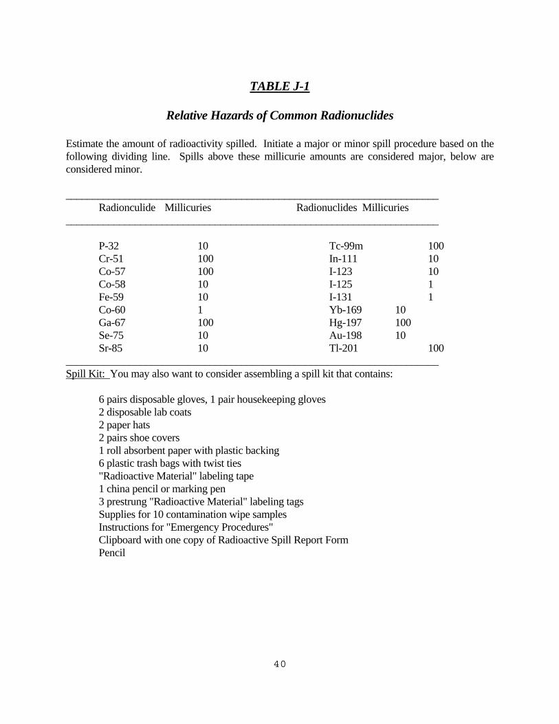
TABLE J-1 Relative Hazards of Common Radionuclides Estimate the amount of radioactivity spilled. Initiate a major or minor spill procedure based on the following dividing line. Spills above these millicurie amounts are considered major, below are considered minor. ______________________________________________________________________ Radionculide Millicuries Radionuclides Millicuries ______________________________________________________________________ P-32 10 Tc-99m 100 Cr-51 100 In-111 10 Co-57 100 I-123 10 Co-58 10 I-125 1 Fe-59 10 I-131 1 Co-60 1 Yb-169 10 Ga-67 100 Hg-197 100 Se-75 10 Au-198 10 Sr-85 10 Tl-201 100 ______________________________________________________________________ Spill Kit: You may also want to consider assembling a spill kit that contains: 6 pairs disposable gloves, 1 pair housekeeping gloves 2 disposable lab coats 2 paper hats 2 pairs shoe covers 1 roll absorbent paper with plastic backing 6 plastic trash bags with twist ties "Radioactive Material" labeling tape 1 china pencil or marking pen 3 prestrung "Radioactive Material" labeling tags Supplies for 10 contamination wipe samples Instructions for "Emergency Procedures" Clipboard with one copy of Radioactive Spill Report Form Pencil
41
APPENDIX K Model Guidance for Ordering and Receiving Radioactive Material Model Guidance 1. The Radiation Safety Officer (RSO) or a designee must authorize each order for radioactive
materials and ensure that the requested materials and quantities are authorized by the license for use by the requesting authorized user and that possession limits are not exceeded.
2. The RSO will establish and maintain a system for ordering and receiving radioactive
material. The system must contain the following information: a. For routinely used materials (1) Written records that identify the authorized user or department, isotope,
chemical form, activity, and supplier will be made. (2) The above records will be checked to confirm that material received was
ordered through proper channels. b. For occasionally used materials (e.g., therapeutic dosages) (1) The authorized user who will perform the procedure will make a written
request that indicates the isotope, radiopharmaceutical, activity, and supplier. (2) The person who receives the material will check the physician's written
request to confirm that the material received is what was ordered. 3. For deliveries during normal working hours, the RSO will tell carriers to deliver radioactive
packages directly to a specified area. 4. For deliveries during off-duty hours, the RSO will tell security personnel or other designated
persons to accept delivery of radioactive packages in accordance with procedures outlined in the sample memorandum below.
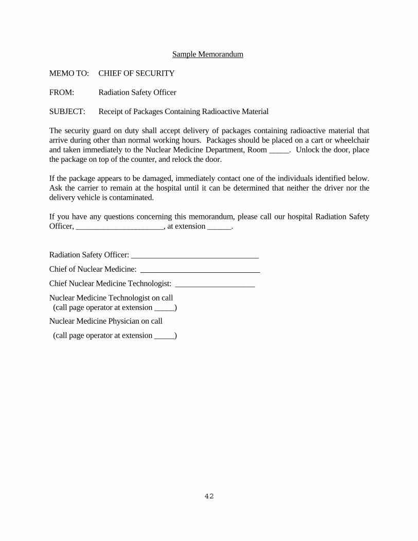
42
Sample Memorandum MEMO TO: CHIEF OF SECURITY FROM: Radiation Safety Officer SUBJECT: Receipt of Packages Containing Radioactive Material The security guard on duty shall accept delivery of packages containing radioactive material that arrive during other than normal working hours. Packages should be placed on a cart or wheelchair and taken immediately to the Nuclear Medicine Department, Room _____. Unlock the door, place the package on top of the counter, and relock the door. If the package appears to be damaged, immediately contact one of the individuals identified below. Ask the carrier to remain at the hospital until it can be determined that neither the driver nor the delivery vehicle is contaminated. If you have any questions concerning this memorandum, please call our hospital Radiation Safety Officer, ______________________, at extension ______. Radiation Safety Officer: ________________________________
Chief of Nuclear Medicine: ______________________________
Chief Nuclear Medicine Technologist: ____________________
Nuclear Medicine Technologist on call (call page operator at extension _____)
Nuclear Medicine Physician on call
(call page operator at extension _____)
43
APPENDIX L Model Procedure for Safely Opening Packages Containing Radioactive Material Model Procedure
1. Special requirements must be followed for packages containing quantities of radioactive material in excess of the Type A quantity limits, as defined in Section 1503.
The licensee shall make arrangements to receive: a. the package when the carrier offers it for delivery; or b. the notification of the arrival of the package at the carrier's terminal
and to take possession of the package expeditiously. Such packages must be monitored for external radiation levels and surface
contamination within 3 hours after receipt if received during working hours or within 18 hours if received after working hours. The Department must be notified if removable contamination exceed 0.01 microcurie (22,000 dpm)/100 cm2.
2. For packages received under the specific license, the following procedure for opening each
package will be followed: a. Put on gloves to prevent hand contamination. b. Visually inspect the package for any sign of damage (e.g., wet or crushed). If
damage is noted, stop the procedure and notify the Radiation Safety Officer (RSO). c. Measure the exposure rate from the package at 1 meter and at the package surface. If
it is higher than expected, stop and notify the RSO. (The "transport index" noted on packages with "Yellow II" or "Yellow III labels is the approximate dose rate, in millirem per hour, at 1 meter from the package surface; the surface dose rate for such packages should not exceed 200 millirem per hour. The dose rate from packages with "White I" labels should be less than 0.5 millirem per hour at the package surface.
d. Open the package with the following precautionary steps: (1) Remove the packing slip. (2) Open the outer package following the supplier's instructions, if provided.
44
(3) Open the inner package and verify that the contents agree with the packing slip.
(4) Check the integrity of the final source container. Look for broken seals or
vials, loss of liquid, condensation, or discoloration of the packing material. (5) If anything is other than expected, stop and notify the RSO. e. If there is any reason to suspect contamination, wipe the external surface of the final
source container and remove the wipe sample to a low-background area. Assay the wipe sample to determine if there is any removable radioactivity. (The licensee should specify in the procedure manual which instrument, for example, a thin-end-window GM survey meter, a NaI (Tl) crystal and ratemeter, a liquid scintillation counter, or a proportional flow counter, should be used for these assays. The detection efficiency must be determined to convert wipe sample counts per minute to disintegrations per minute. Note that a dose calibrator is not sufficiently sensitive for this measurement). Take precautions against the potential spread of contamination.
f. Check the user request to ensure that the material received is the material that was
ordered. g. Monitor the packing material and the empty packages for contamination with a
radiation detection survey meter before discarding. (1) If contaminated, treat this material as radioactive waste. (2) If not contaminated, remove or obliterate the radiation labels before
discarding in-house trash. h. Make a record of the receipt.
45
APPENDIX M
Records of Radioactive Material Use General Many suppliers include pressure-sensitive stickers or forms that have much of the information required by the regulations. You may use these in your records and need not duplicate the information on them. Be sure to write down whatever additional information is required but is not cued or printed on them. Information does not have to be recorded in the order given in these procedures. Also, you do not have to replicate entries. For example, if you prepare a multidose vial for use one day, you do not have to record the date each time you draw a dosage from it; if you take 30 Ir-192 seeds that are each 0.5 millicuries, you do not have to list each seed individually. M.1 Records of Unit Dosage Use You may use the following model procedure to keep a record of unit dosage use. Model Procedure For each unit dosage received from a supplier, make a record of the: 1. Radionuclide; 2. Generic name or its abbreviation or trade name; 3. Date of receipt; 4. Supplier; 5. Lot number or control number, if assigned; 6. Activity in millicuries or microcuries as recorded on the unit dosage or packing slip and its
associated time; 7. Date of administration or disposal; 8. If administered, a. Prescribed dosage (unless already recorded in clinical procedure manual), b. Measured activity in millicuries or microcuries and date and time of measurement,
46
c. Patient name and identification number if one has been assigned; 9. If discarded, the date and method of disposal; and 10. Initials of the individual who made the record. M.2. Records of Multidose Vial Use You may use the following model procedure to keep a record of multidose vial use. Model Procedure For each multidose vial that you receive from a supplier or that you prepare, make a record of the: 1. Radionuclide; 2. Generic name or its abbreviation or trade name; 3. Date of receipt or preparation; 4. Date and time of initial assay and amount in both millicuries and cubic centimeters (cc) or
milliliters (ml); 5. Supplier or kit manufacturer; 6. If administered, a. Prescribed dosage (unless already recorded in clinical procedure manual), b. Date and time dosage was drawn and measured, c. Calculated volume that is needed for the prescribed dosage, d. Measured activity in millicuries or microcuries,
e. Patient name and identification number if one has been assigned;
7. If discarded, the method of disposal and date; and 8. Initials of the individual who made the record.
47
M.3. Measuring and Recording Molybdenum Concentration The regulations require that each licensee who uses a technetium generator to prepare radiopharmaceuticals must test each elution or extraction for its molybdenum concentration and alumina contamination. (This does not have to be done when using radiopharmaceuticals obtained from a distributor.) This measurement is usually made with a dose calibrator. Licensees may not administer radiopharmaceuticals that contain more than 0.15 microcurie of Mo-99 per millicurie of Tc-99m at the time of administration. If an elution or extraction has a higher concentration, there may be a manufacturing defect that should be reported. The model procedure for measuring molybdenum concentration is based on the use of a "molybdenum breakthrough pig." Your dose calibrator manufacturer will usually supply, as an option, a molybdenum breakthrough pig made of lead. The pig is usually thick enough to shield all the technetium photons but only a fraction of the molybdenum photons. The manufacturer will specify the Mo-99 correction factor to convert from measured Mo-99 to total Mo-99. The following model procedure may be used to measure the molybdenum concentration in Mo-99/Tc-99m generator elution. Model Procedure Each time a generator is eluted, make a record of the: 1. Date the generator was received; 2. Date and time of elution; 3. Measured Mo-99 activity in microcuries; 4. Product of the measured Mo-99 activity and the correction factor noted by the molybdenum
breakthrough pig manufacturer; 5. Measured Tc-99m activity in millicuries; 6. Ratio of the total Mo-99 microcuries per millicurie of Tc-99m and checkmark that the ratio
is less than 0.07 microcurie of Mo-99 per millicurie of Tc-99m. (If it isn't, stop and notify the RSO. The licensee must notify the Department if a leaking generator is detected). (The 0.07 action level allows for the quicker decay of the Tc through the day of use. It is assumed that the material will be used within 6 hours, at which time the concentration of Mo-99 to Tc-99m would have doubled.)
7. Initials of the person who made the record.
48
M.4 Keeping an Inventory of Implant Sources Model Procedure 1. Use a locking installed cabinet or safe to store all implant sources. 2. Make a list of names of those individuals you allow to handle implant sources and have them
initial beside their names. 3. For long-lived sources, draw a map of the storage drawer and indicate the activity of the
source at each storage point. For short-lived sources that you store in the manufacturer's shipping container, indicate the area in the safe where you put the container. Also, be sure to add the sources to the inventory log.
4. Post the map and the list of individuals whom you permit to handle the sources in the storage
area or on the inventory log. 5. Each time you remove a source, make a record of the number and activity of sources
removed, the room number of use or patient's name, and the time and date they were removed from storage; initial the record.
6. Each time you return sources to storage, immediately count them to ensure that every source
removed has been returned. Then make a record of the number and activity of sources returned, the room number of use or patient's name, and the time and date they were returned to storage; initial the record.
7. If you ever perceive a discrepancy between the record and the number of sources in use and
in storage, notify the RSO immediately.
49
APPENDIX N Model Procedure for Area Surveys You may use the following model procedure to perform area surveys. MODEL PROCEDURE Ambient Dose Rate Surveys 1. Survey Areas a. In radiopharmaceutical elution, preparation, and administration areas,
survey at the end of each day of use with a radiation detection survey meter. If diagnostic administrations are occasionally made in patients' rooms and special care is taken to remove all paraphernalia, those rooms need not be surveyed.
b. In laboratory areas where only small quantities of gamma-emitting
radioactive material are processed (less than 200 microcuries at a time), survey monthly with a radiation detection survey meter.
c, In radiopharmaceutical storage and radiopharmaceutical waste
storage areas, survey weekly with a radiation detection survey meter. d. In sealed source and brachytherapy storage areas, survey quarterly
with a radiation measurement survey meter. 2. Immediately notify the RSO if you find unexpectedly high or low levels. Removable Contamination Surveys 1. Survey Areas a. In radiopharmaceutical elution, preparation, and administration areas,
survey weekly for removable contamination. If diagnostic administrations are occasionally made in patients' rooms and special care is taken to remove all paraphernalia, those rooms need not be surveyed.
b. In laboratory areas where only small quantities of photon-emitting radioactive material are processed (less than 200 microcuries at a time), survey monthly for removable contamination.
50
c. In radiopharmaceutical storage and radiopharmaceutical waste storage areas, survey weekly for removable contamination.
2. The wipe sample assay procedure should be sufficiently sensitive to detect the
presence of 2000 dpm/100 cm2 of removable contamination (200 dpm/100 cm2 for isotopes of iodine). You must use a radioactive source with a known amount of activity to convert sample measurements (usually in counts per minute or cpm) to disintegrations per minute or dpm.
3. Immediately notify the RSO if you find unexpectedly high levels. Records 1. Keep a record of dose rate and contamination survey results. It must include the
following information: a. The date, area surveyed, and equipment used, serial number, and
calibration date. b. The name or initials of the person who made the survey. c. A drawing of the areas surveyed with contamination and dose rate
action levels as established by the RSO. d. Measured dose rates in mR/hr or contamination levels in dpm/100
cm2 as appropriate. e. Actions taken in the case of excessive dose rates or contamination
and followup survey information. 2. The RSO will review and initial the record at least monthly and also promptly in
those cases in which action levels were exceeded.
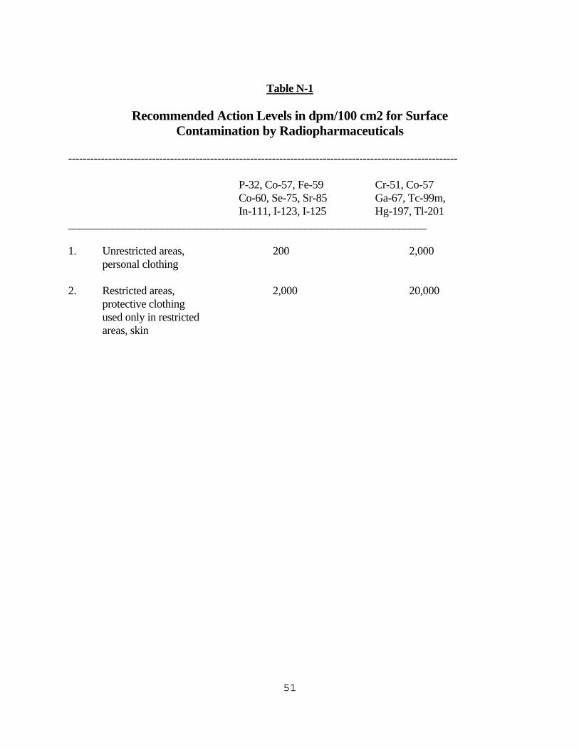
51
Table N-1 Recommended Action Levels in dpm/100 cm2 for Surface Contamination by Radiopharmaceuticals ----------------------------------------------------------------------------------------------------------- P-32, Co-57, Fe-59 Cr-51, Co-57 Co-60, Se-75, Sr-85 Ga-67, Tc-99m, In-111, I-123, I-125 Hg-197, Tl-201 _________________________________________________________________ 1. Unrestricted areas, 200 2,000 personal clothing 2. Restricted areas, 2,000 20,000 protective clothing used only in restricted areas, skin
52
APPENDIX O Model Procedure for Monitoring, Calculating, and Controlling Air Concentrations WORKER DOSE FROM NOBLE GASES Noble gases such as xenon in the air present an external source of radiation exposure that must be calculated. Many commercially available dosimeters and survey instruments are not capable of accurately measuring worker doses from immersion in noble gases. If you will collect spent gas in a shielded trap with an effluent air contamination monitor and will follow the monitor manufacturer's instructions for checking its accuracy and constancy, you may respond by saying, "We will collect spent noble gas in a shielded trap and monitor the trap effluent with an air contamination monitor that we will check regularly according to the manufacturer's instructions." If you will collect spent gas in a shielded trap and will follow the model procedure checking trap effluent, you may respond by saying, "We will collect spent noble gas in a shielded container and will establish and implement the model procedure for checking trap effluent that was published in Appendix 0.3 to the Guide. If you are not monitoring trap effluent or if you exhaust spent gas to the atmosphere, you must estimate worker dose by calculation. WORKER DOSE FROM AEROSOLS If you collect spent aerosol in a shielded trap, will use an air contamination monitor for reusable traps, and will follow the monitor manufacturer's instructions for checking for accuracy and constancy, you may respond by saying, "We will collect spent aerosol in a shielded trap and, for reusable traps, monitor the trap effluent with an air contamination monitor that we will check regularly according to the manufacturer's instructions." You do not have to monitor the trap effluent of single-use devices. If you are not monitoring reusable trap effluent or if you are exhausting spent aerosol to the atmosphere, you must estimate worker dose by calculation.
53
0.1 MODEL PROCEDURE FOR CALCULATING WORKER DOSE FROM
CONCENTRATIONS OF GASES AND AEROSOLS IN WORK AREAS 1. Collect the following data: a. Estimated number of studies per week; b. Activity to be administered per study; c. Estimated activity lost to the work areas per study (you may assume
20 percent loss); d. Measured airflow supplied by each vent in the imaging room (if
different during heating and cooling seasons, use the lesser value); e. Measured airflow exhausted by each vent in the imaging room (the
exhaust should be vented and not recirculated within the facility);
f. Measured airflow exhaust at the storage site (e.g., a fume hood); and g. Maximum permissible air concentrations in restricted and
unrestricted areas. For Xe-133, the maximum permissible values are 1 x 10⁻⁵ µCi/ml in restricted areas and 5 x 10⁻⁷ µCi/ml in unrestricted areas. For soluble Tc-99m, the maximum permissible values are 6 x 10⁻⁵ µCi/ml in restricted areas and 2 x E-7 µCi/ml in unrestricted areas.
2. The following calculations must be made: a. The sum of all measured exhaust rates and the sum of all measured
supply rates. If the former is larger than the latter, this ensures that the imaging room is at negative pressure.
b. The estimated average concentration in restricted areas. (1) The total activity released to the restricted area (activity used each
week multiplied by estimated fractional loss per study) divided by the total air exhausted (sum of all exhaust rates multiplied by the length of the work week) must be less than the applicable maximum permissible value for a restricted area.
54
(2) If this is not the case, plan for fewer studies. (An increase in the ventilation rate will not significantly reduce the downwind effluent concentration because it is primarily a function of the natural dispersion in the atmosphere.)
0.2 MODEL PROCEDURE FOR CALCULATING AIRBORNE EFFLUENT
CONCENTRATION 1. Divide the total activity released to an unrestricted area (activity used each week that
is released in an exhaust system) by the total volume of air exhausted over the week ("on" time multiplied by measured airflow rate). The quotient must be less than the applicable maximum permissible value for an unrestricted area.
2. If this is not the case, plan for fewer studies and do the calculation again.
Alternatively, you may consider collection and decay-in-storage for waste, or restriction of access to the release point and calculation of concentration at the boundary of the restricted area.
0.3 MODEL PROCEDURE FOR MONITORING OR CHECKING TRAP
EFFLUENT Charcoal traps can significantly reduce air contamination. They can also become saturated or be spoiled by improper use, humidity, chemicals, or inadequate maintenance. 1. If the trap effluent is monitored by a radiation detector designed to monitor effluent
gas, check the detector according to the manufacturer's instructions and keep a record of the checks.
2. If you do not monitor the trap effluent, check it on receipt and once each month.
Collect the effluent from the trap during one patient study in a plastic bag and then monitor the activity in the bag by holding the bag against a camera, with the camera adjusted to detect the noble gas, and compare its counts per minute (cpm) to background cpm with no other radioactivity in the area. Keep a record of the date, background cpm, and bag cpm.
3. The RSO will establish an action level based on cpm or a multiple of background
cpm. If you measure a significant increase in the bag cpm, the trap is breaking down and must be replaced.
4. Follow the trap manufacturer's instructions for replacing the trap.
55
SPILLED GAS CLEARANCE TIME Because normal room ventilation is usually not sufficient to ensure timely clearance of spilled gas, the calculations described in Appendix 0.4 should be done to determine for how long a room should be cleared in case of a gas spill. This clearance time should be posted in the room. 0.4 MODEL PROCEDURE FOR CALCULATING SPILLED GAS
CLEARANCE TIME 1. Collect the following data: a. A, the highest activity of gas in a single container, in microcuries; b. Measured airflow supply from each vent in the room (if different
during heating and cooling seasons, use the lesser value), in milliliters per minute;
c. Q, the total room air exhaust determined by measuring, in milliliters
per minute, the airflow to each exhaust vent in the room (the exhaust should be vented and not recirculated within the facility); this may be either the normal air exhaust or a specially installed gas exhaust system;
d. C, the maximum permissible air concentrations in restricted and
unrestricted areas. For Xe-133, the maximum permissible values are 1 x 10⁻⁵ µCi/ml in restricted areas and 3 x 10⁻⁷ µCi/ml in unrestricted areas.
e. V, the volume of the room in milliliters. 2. For each room make the following calculations: a. The airflow supply should be less than the airflow exhaust to ensure
the room is at negative pressure. b. The evacuation time t = -V x 1n (C x V/A). Q
56
APPENDIX P Model Procedure for Radiation Safety During Iodine Therapy Over 30 Millicuries You may use the following procedure for reducing worker and public dose during radiopharmaceutical therapy. MODEL PROCEDURE 1. The patient's room will be as far away from the nursing station and heavy traffic hallways as
is consistent with good medical care. It will be a private room with private sanitary facilities and should be without carpet.
2. Prepare the room for the procedure as follows: a. Use leak-proof absorbent paper to cover large surfaces (the bed, chairs, and the floor
around the toilet) that are likely to be contaminated. Small items (telephone, door knobs, bed remote control, television control, and nurse call cord) may be covered with absorbent paper or plastic bags.
b. Prepare separate boxes for linen, disposable waste, and nondisposable contaminated
items. Place a single large reclosable plastic bag in each box, or supply several small plastic bags.
c. Determine whether urine will be discarded by release to the sanitary sewer or
collected. If urine will be collected, prepare collection containers. (1) Containers should be unbreakable and closable. (2) If there is no need for assay or volumetric determination and urine will be
decayed in storage, add to each container an absorbent such as vermiculite. (3) To avoid room contamination in the case of a spill, place containers in a box
or deep tray that has been lined with a plastic bag and absorbent paper or vermiculite.
(4) Supply a few half-value layers of shielding for each container. (For I-131,
one half-value layer is approximately 3 mm of lead.) (5) Supply a wide-mouth antisplash funnel.
57
d. Stock additional disposable gloves, absorbent paper, and radioactive waste labels in
the room for use as necessary by nursing, nuclear medicine, and radiation safety personnel.
3. Order disposable table service for the duration of the patient's stay. Inform the Housekeeping
Office that personnel should stay out of the room until otherwise notified. 4. Supply the nurses with film badges, TLDs, or pocket ionization chambers. 5. Brief the nurses on radiation safety precautions. Use the sample form, "Nursing Instructions
for Patients Treated with Iodine-131, P-32, or Gold-198", or your own nursing instruction form as an outline. Allow time for questions and answers during the briefing. Leave a written copy of the radiation safety precautions in the patient's chart or at the nurses' station.
6. Brief the patient on radiation safety procedures for the dosage administration, visitor control,
urine collection, radioactive waste, and other items as applicable. 7. Only those persons needed for medical, safety, or training purposes should be present during
the administration. 8. Mark a visitors' "safe line" on the floor with tape as far from the patient as possible. 9. Following administration of the dosage, measure the exposure rate in mR/hr at bedside, at 1
meter from bedside, at the visitors' "safe line", and in the surrounding hallways and rooms. Record this and any other necessary information on the nursing instructions form or the nurses' dosimeter signout form. Post the room with a "Radioactive Materials" sign.
10. For patients treated with liquid or gelatin-capsulated I-131, 1 day after the dosage
administration, measure the thyroid burden of all personnel who were present for the administration. Also consider a thyroid burden assay for patient care personnel 2 days after the administration. Make a record of the worker's name, amount of I-131 activity in a thyroid phantom in microcuries and associated counts per minute, the counts per minute from the worker's thyroid, the calculated thyroid burden, and date.
11. As the therapy proceeds, pick up waste for transfer to a decay-in-storage or decontamination
area. 12. A licensee may authorize the release from its control of any individual who has been
administered radiopharmaceuticals containing radioactive material if the total effective dose equivalent to any other individual from exposure to the released individual is not likely to exceed 500 mRem. If you use the exposure rate standard as the release criterion, measure it with a radiation measurement survey meter at a distance of 1 meter from the umbilicus while
58
the patient is standing or, if the patient is not ambulatory, 1 meter from the bedside with the patient supine.
13. Before using the room for general occupancy, it must be decontaminated and released to the
Admitting Office. a. Remove all absorbent paper, and place it in the appropriate container. b. Transfer all containers to a decay-in-storage or decontamination area. c. Use a radiation detection survey meter to check for room contamination. Clean
contaminated areas until removable contamination is less than 200 dpm/100 cm2/ d. Call the Housekeeping Office to remove the cleaning restriction and call the
Admitting Office to return the room to the vacant list.
59
APPENDIX Q Model Procedure for Radiation Safety During Implant Therapy You may use the following procedure to reduce worker and public dose during implant therapy. Model Procedure 1. The patient's room will be as far away from the nursing station and heavy traffic
hallways as is consistent with good medical care. It should be a private room. 2. Supply the nurses with film badges, TLDs, or pocket ionization chambers. 3. Brief the nurses on radiation safety precautions. Use the sample form, "Nursing Instructions
for Patients Treated with Temporary Implant Sources," or your own nursing instruction form as an outline. Allow time for questions an answers during the briefing.
4. Brief the patient on radiation safety procedures for confinement to bed, visitor control, and
other items as applicable consistent with good medical care. 5. Only those persons needed for medical, safety, or training purposes should be present during
the implant procedure. 6. Mark a visitors "safe line" on the floor with tape as far from the patient as possible. 7. Following the implant, measure the exposure rate in mR/hr at bedside, at 1 meter from
bedside, at the visitors' "safe line," and in the surrounding hallways and rooms. Record this and any other necessary information on the nursing instruction form or the nurses' dosimeter signout form. Post the room with a "Radioactive Materials" sign.
8. Do not release any patient who has received a temporary implant from the hospital until both
a radiation survey of the patient and a count of implant sources, trains, or ribbons confirms that all sources have been removed from the patient are accounted for. Perform this check immediately after the removal of the sources. Keep a record confirming the source count and radiation survey on the implant source running inventory form. For low-
60
activity seeds (less than 1 millicurie), use an individual seed to check the survey meter to be sure it will easily detect a seed that has not been removed or has been lost.
9. Do not release any patient who has received a permanent implant from the hospital until the
exposure rate from the patient is less than 5 mR/hr at 1 meter. Measure this exposure rate with a radiation measurement survey meter at a distance of 1 meter from the umbilicus with the patient standing.
61
APPENDIX R Model Procedure for Waste Disposal The following general guidance and procedure may be used for disposal of radioactive waste. Overview There are four commonly used methods of waste disposal: release to the environment through the sanitary sewer or by evaporative release; decay-in-storage (DIS); transfer to a burial site or back to the manufacturer' and release to in-house waste. With the exception of the patient excreta and generally licensed in vitro kit exemptions, nothing in these guidelines relieves the licensee from maintaining records of the disposal of licensed material General Guidance 1. All radioactivity labels must be defaced or removed from containers and packages prior to
disposal in in-house waste. If waste is compacted, all labels that are visible in the compacted mass must be defaced or removed.
2. Remind employees that nonradioactive waste such as leftover reagents, boxes, and
packaging material should not be mixed with radioactive waste. 3. Occasionally monitor all procedures to ensure that radioactive waste is not created
unnecessarily. Review all new procedures to ensure that waste is handled in a manner consistent with established procedures.
4. In all cases, consider the entire impact of various available disposal routes. Consider
occupational and public exposure to radiation, other hazards associated with the material and routes of disposal (e.g., toxicity, carcinogenicity, pathogenicity, flammability), and expense.
MODEL PROCEDURE FOR DISPOSAL OF LIQUIDS AND GASES Liquids may be disposed of by release to the sanitary sewer or evaporative release to the atmosphere. This does not relieve licensees from complying with other regulations regarding toxic or hazardous properties of these materials. 1. Regulations for disposal in the sanitary sewer appear in Section 462. Material must be
readily water soluble or readily dispersible biological material in water. There are monthly limits based on the total sanitary sewerage release of your facility. (Excreta from patients undergoing medical diagnosis or therapy is exempt from all the above limitations). Make a record of the date, radionuclide, estimated activity that was released (in millicuries or microcuries), and of the sink or toilet at which the material was released.
62
2. Limits on permissible concentrations in effluents to unrestricted areas are enumerated in Appendix B of Chapter 4. This limits apply at the boundary of the restricted area. Make a record of the date, radionuclide, estimated activity that was released (in millicuries or microcuries) and estimated concentration, and of the vent site at which the material was released.
MODEL PROCEDURE FOR DISPOSAL BY DECAY-IN-STORAGE (DIS) Short-lived material (physical half-life less than 65 days) may be disposed of by DIS. If you use this procedure, keep material separated according to half-life. 1. Consider using separate containers for different types of waste, e.g., capped needles and
syringes in one container, other injection paraphernalia such as swabs and gauze in another, and unused dosages in a third container. Smaller departments may find it easier to use just one container for all DIS waste. Because the waste will be surveyed with all shielding removed, the containers in which waste will be disposed of must not provide any radiation shielding for the material.
2. When the container is full, seal it with string or tape and attach an identification tag that
includes the date sealed, the longest-lived radioisotope in the container, and the initials of the person sealing the container. The container may then be transferred to the DIS area.
3. Decay the material for at least 10 half-lives. 4. Prior to disposal as in-house waste, monitor each container as follows: a. Check your radiation detection survey meter for proper operation; b. Plan to monitor in a low-level (less than 0.05 millirem per hour) area;
c. Remove any shielding from around the container;
d. Monitor all surfaces of each individual container; e. Discard as in-house waste only those containers that cannot be distinguished from
background. Record the date on which the container was sealed, the disposal date, and type of material (e.g., paraphernalia, unused dosages). Check to be sure no radiation labels are visible.
f. Containers that can be distinguished from background radiation levels must be
returned to the storage area for further decay or transferred for burial. 5. If possible, Mo-99/Tc-99m generators should be held 60 days before being dismantled
63
because of the occasional presence of a long-lived contaminant. When dismantling generators, keep a radiation detection survey meter (preferable with a speaker) at the work area. Dismantle the oldest generator first, then work forward chronologically. Hold each individual column in contact with the radiation detection survey meter in a low-background (less than 0.06 mR/hr) area. Log the generator date and disposal date for your waste disposal records. Remove or deface the radiation labels on the generator shield.
MODEL PROCEDURE FOR TRANSFER FOR BURIAL Except for material suitable for DIS and some animal carcasses, solids must be transferred to a burial site. Follow the packaging instructions you received from the transfer agent, and the burial site operator. For your records of disposal, keep the consignment sheet that the transfer agent gave you. MODEL PROCEDURE FOR RETURNING GENERATORS TO THE MANUFACTURER Used Mo-99/Tc-99m generators may be returned to the manufacturer. This permission does not relieve licensees from the requirement to comply with Department of Transportation (DOT) regulations. 1. Retain the records needed to demonstrate that the package qualifies as a DOT Specification
7A container (see DOT Regulations, Paragraph 173.415(a) of 49 CFR Part 173). 2. Assemble the package in accordance with the manufacturer's instructions. 3. Perform the dose rate and removable contamination measurements. 4. Label the package and complete the shipping papers in accordance with the manufacturer's
instructions.
64
APPENDIX S Acceptable Training and Experience for Medical Uses of Radioactive Material Radiation Safety Officer Except as provided in LAC 33:XV.764, an individual fulfilling the responsibilities of the radiation safety officer as provided in LAC 33:XV.706 shall: 1. Be certified by the A. American Board of Health Physics in Comprehensive Health Physics; B. American Board of Radiology in Radiological Physics, Therapeutic
Radiological Physics, or Medical Nuclear Physics; C. American Board of Nuclear Medicine; D. American Board of Science in Nuclear Medicine; or E. Board of Pharmaceutical Specialties in Nuclear Pharmacy or Science; or 2. have had 200 hours of classroom and laboratory training as follows: A. radiation physics and instrumentation; B. radiation protection; C. mathematics pertaining to the use and measurement of radioactivity; D. radiation biology; E. radiopharmaceutical chemistry; and F. one year of full-time experience in radiation safety at a medical institution
under the supervision of the individual identified as the radiation safety officer on a Department, agreement state, licensing state, or U. S. Nuclear Regulatory Commission license that authorizes the medical use of radioactive material; or
65
3. be an authorized user for those radioactive material users that come within the radiation safety officer's responsibilities.
Training for Experienced Radiation Safety Officer An individual identified as a radiation safety officer on an agreement state, licensing state, the Radiation Control Section or U. S. Nuclear Regulatory Commission license on February 20, 1991, who oversees only the use of radioactive material for which the licensee was authorized on that date need not comply with the training requirements of LAC 33:XV.763. Training for Uptake, Dilution, or Excretion Studies Except as provided in LAC 33:XV.773 and 774, the licensee shall require the authorized user of a radiopharmaceutical listed in LAC 33:XV.729 to be a physician who: 1. is certified in: A. nuclear medicine by the American Board of Nuclear Medicine; B. diagnostic radiology by the American Board of Radiology; C. diagnostic radiology or radiology within the previous five years by the
American Osteopathic Board of Radiology; or D. nuclear medicine by the American Osteopathic Board of Nuclear Medicine;
or 2. has completed 40 hours of instruction in basic radionuclide handling techniques
applicable to the use of prepared radiopharmaceuticals, and 20 hours of supervised clinical experience.
A. To satisfy the basic instruction requirement, 40 hours of classroom and
laboratory instruction shall include: (1) radiation physics and instrumentation; (2) radiation protection; (3) mathematics pertaining to the use and measurement of radioactivity; (4) radiation biology; and (5) radiopharmaceutical chemistry.
66
B. To satisfy the requirement for 20 hours of supervised clinical experience, training must be under the supervision of an authorized user at a medical institution and shall include:
(1) examining patients and reviewing their case histories to determine
their suitability for radionuclide diagnosis, limitations, or contraindications;
(2) selecting the suitable radiopharmaceuticals and calculating and
measuring the dosages; (3) administering dosages to patients and using syringe radiation shields; (4) collaborating with the authorized user in the interpretation of
radionuclide test results and (5) patient follow-up; or 3. has successfully completed a six-month training program in nuclear medicine as part
of a training program that has been approved by the Accreditation Council for Graduate Medical Education and that included classroom and laboratory training, work experience, and supervised clinical experience in all the topics identified in LAC 33:XV.765.A.2.
Training for Imaging and Localization Studies Except as provided in LAC 33:XV.773 or 774, the licensee shall require the authorized user of a radiopharmaceutical, generator, or reagent kit specified in Section 731 to be a physician who: 1. is certified in: A. nuclear medicine by the American Board of Nuclear Medicine; B. diagnostic radiology by the American Board of Radiology; C. diagnostic radiology or radiology within the previous five years by the
American Osteopathic Board of Radiology; or D. nuclear medicine by the American Osteopathic Board of Nuclear Medicine;
or 2. has completed 200 hours of instruction in basic radionuclide handling techniques
applicable to the use of prepared radiopharmaceuticals, generators, and reagent kits;
67
500 hours of supervised work experience; and 500 hours of supervised clinical experience.
A. To satisfy the basic instruction requirement, 200 hours of classroom and
laboratory training shall include: (1) radiation physics and instrumentation; (2) radiation protection; (3) mathematics pertaining to the use and measurement of radioactivity; (4) radiopharmaceutical chemistry; (5) radiation biology; and (6) certification by the physician that he or she participated in the
required number of hours and has successfully passed an appropriate written examination given by the certifying institution.
B. To satisfy the requirement for 500 hours of supervised work experience,
training shall be under the supervision of an authorized user at a medical institution and shall include:
(1) ordering, receiving, and unpacking radioactive materials safely and
performing the related radiation surveys; (2) calibrating dose calibrators and diagnostic instruments and
performing checks for proper operation of survey meters; (3) calculating and safely preparing patient dosages; (4) using administrative controls to prevent the misadministration of
radioactive material; (5) using emergency procedures to contain spilled radioactive material
safely and using proper decontamination procedures; and (6) eluting technetium-99m from generator systems, assaying and testing
the eluate for molybdenum-99 and alumina contamination, and processing the eluate with reagent kits to prepare technetium-99m labeled radiopharmaceuticals.
68
C. To satisfy the requirement for 500 hours of supervised clinical experience training shall be under the supervision of an authorized user at a medical institution and shall include:
(1) examining patients and reviewing their case histories to determine
their suitability for radionuclide diagnosis, limitations, or contraindications;
(2) selecting the suitable radiopharmaceuticals and calculating and
measuring the dosages; (3) administering dosages to patients and using syringe radiation shields; (4) collaborating with the authorized user in the interpretation of
radionuclide test results; and (5) patient follow-up; or 3. has successfully completed a six-month training program in nuclear medicine that
has been approved by the Accreditation Council for Graduate Medical Education and that included classroom and laboratory training, work experience, and supervised clinical experience in all the topics identified in LAC 33:XV.766.2.
Training for Therapeutic Use of Radiopharmaceuticals Except as provided in LAC 33:XV.773, the licensee shall require the authorized user of a radiopharmaceutical listed in LAC 33:XV.735 for therapy to be a physician who: 1. is certified by: A. the American Board of Nuclear Medicine; or B. the American Board of Radiology in radiology, therapeutic radiology, or
radiation oncology; or 2. has completed 80 hours of instruction in basic radionuclide handling techniques
applicable to the use of therapeutic radiopharmaceuticals and has had supervised clinical experience.
A. To satisfy the requirement for instruction, 80 hours of classroom and
laboratory training shall include: (1) radiation physics and instrumentation;
69
(2) radiation protection; (3) mathematics pertaining to the use and measurement of radioactivity;
and (4) radiation biology. B. To satisfy the requirement for supervised clinical experience, training shall
be under the supervision of an authorized user at a medical institution and shall include:
(1) use of iodine-131 for diagnosis of thyroid function and the treatment
of hyperthyroidism or cardiac dysfunction in 10 individuals; (2) use of soluble phosphorus-32 for the treatment of ascites
polycythemia vera, leukemia, or bone metastases in three individuals; (3) use of iodine-131 for treatment of thyroid carcinoma in three
individuals; and (4) use of colloidal chromic phosphorus-32 or of colloidal gold-198 for
intracavitary treatment of malignant effusions in three individuals.
70
Training for Therapeutic Use of Brachytherapy Sources Except as provided in LAC 33:XV.773, the licensee shall require the authorized user using a brachytherapy source specified in LAC 33:XV.741, for therapy to be a physician who: 1. is certified in: A. radiology, therapeutic radiology, or radiation oncology by the American
Board of Radiology; B. radiation oncology by the American Osteopathic Board of Radiology; C. radiology, with a specialization in radiotherapy, as a British "Fellow of the
Faculty of Radiology" or "Fellow of the Royal College of Radiology"; or D. therapeutic radiology by the Canadian Royal College of Physicians and
Surgeons; or 2. is in the active practice of therapeutic radiology, has completed 200 hours of
instruction in basic radionuclide handling techniques applicable to the therapeutic use of brachytherapy sources and 500 hours of supervised work experience and a minimum of three years of supervised clinical experience.
A. To satisfy the requirement for instruction, 200 hours of classroom and
laboratory training shall include: (1) radiation physics and instrumentation; (2) radiation protection; (3) mathematics pertaining to the use and measurement of radioactivity;
and (4) radiation biology. B. To satisfy the requirement for 500 hours of supervised work experience,
training shall be under the supervision of an authorized user at a medical institution and shall include:
(1) ordering, receiving, and unpacking radioactive materials safely and
performing the related radiation surveys; (2) checking survey meters for proper operation;
71
(3) preparing, implanting, and removing sealed sources; (4) using administrative controls to prevent the misadministration of
radioactive material; and (5) using emergency procedures to control radioactive material. C. To satisfy the requirement for a period of supervised clinical experience,
training shall include one year in a formal training program approved by the Residency Review Committee for Radiology of the Accreditation Council for Graduate Medical Education or the Committee on Postdoctoral Training of the American Osteopathic Association, and an additional two years of clinical experience in therapeutic radiology under the supervision of an authorized user at a medical institution. The supervised clinical experience shall include:
(1) examining individuals and reviewing their case histories to determine
their suitability for brachytherapy treatment, and any limitations or contraindications;
(2) selecting the proper brachytherapy sources, dose, and method of
administration; (3) calculating the dose; and (4) post-administration follow-up and review of case histories in
collaboration with the authorized user. Training for Ophthalmic Use of Strontium-90 Except as provided in LAC 33:XV.773, the licensee shall require the authorized user using only strontium-90 for ophthalmic radiotherapy to be a physician who:
1. is certified in radiology, therapeutic radiology, or radiation oncology by the American Board of Radiology; or
2. is in the active practice of therapeutic radiology or ophthalmology, and has
completed 24 hours of instruction in basic radionuclide handling techniques applicable to the use of strontium-90 for ophthalmic radiotherapy, and a period of supervised clinical training in ophthalmic radiotherapy.
A. To satisfy the requirement for instruction, the classroom and laboratory
72
training shall include: (1) radiation physics and instrumentation; (2) radiation protection; (3) mathematics pertaining to the use and measurement of radioactivity;
and (4) radiation biology. B. To satisfy the requirement for a period of supervised clinical training in
ophthalmic radiotherapy, training shall be under the supervision of an authorized user at a medical institution and shall include the use of strontium-90 for the ophthalmic treatment of five individuals that includes:
(1) examination of each individual to be treated; (2) calculation of the dose to be administered; (3) administration of the dose; and (4) follow-up and review of each individual's case history. Training for Use of Sealed Sources for Diagnosis Except as provided in LAC 33:XV.773, the licensee shall require the authorized user using a sealed source in a device specified in LAC 33:XV.739 to be a physician, dentist, or podiatrist who: 1. is certified in: A. radiology, diagnostic radiology with special competence in nuclear
radiology, therapeutic radiology, or radiation oncology by the American Board of Radiology;
B. nuclear medicine by the American Board of Nuclear Medicine; or C. diagnostic radiology or radiology by the American Osteopathic Board of
Radiology; or 2. has completed 80 hours of classroom and laboratory instruction in basic radionuclide
handling techniques specifically applicable to the use of the device.
73
A. To satisfy the requirement for instruction, the training shall include: (1) radiation physics, mathematics pertaining to the use and
measurement of radioactivity, and instrumentation; (2) radiation biology; and (3) radiation protection and training in the use of the device for the
purposes authorized by the license. Training for Teletherapy Except as provided in LAC 33:XV.773, the licensee shall require the authorized user of a sealed source specified in LAC 33:XV.747 in a teletherapy unit to be a physician who: 1. is certified in: A. radiology, therapeutic radiology, or radiation oncology by the American
Board of Radiology; B. radiation oncology by the American Osteopathic Board of Radiology; C. Radiology, with specialization in radiotherapy, as a British "Fellow of the
Faculty of Radiology" or "Fellow of the Royal College of Radiology" or D. therapeutic radiology by the Canadian Royal College of Physicians and
Surgeons; or 2. is in the active practice of therapeutic radiology, and has completed 200 hours of
instruction in basic radionuclide techniques applicable to the use of a sealed source in a teletherapy unit, 500 hours of supervised work experience, and a minimum of three years of supervised clinical experience.
A. To satisfy the requirement for instruction, the classroom and laboratory
training shall include: (1) radiation physics and instrumentation; (2) radiation protection; (3) mathematics pertaining to the use and measurement of radioactivity;
and (4) radiation biology.
74
B. To satisfy the requirement for supervised work experience, training shall be
under the supervision of an authorized user at an institution and shall include: (1) review of the full calibration measurements and periodic spot-checks; (2) preparing treatment plans for calculating treatment times; (3) using administrative controls to prevent misadministrations; (4) implementing emergency procedures to be followed in the event of
the abnormal operation of a teletherapy unit or console; and (5) checking and using survey meters. C. To satisfy the requirement for a period of supervised clinical experience,
training shall include one year in a formal training program approved by the Residency Review Committee for Radiology of the Accreditation Council for Graduate Medical Education or the Committee on Postdoctoral Training of the American Osteopathic Association and an additional two years of clinical experience in therapeutic radiology under the supervision of an authorized user at a medical institution. The supervised clinical experience shall include:
(1) examining individuals and reviewing their case histories to determine
their suitability for teletherapy treatment, and any limitations or contraindications;
(2) selecting the proper dose and how it is to be administered; (3) calculating the teletherapy doses and collaborating with the
authorized user in the review of patients' progress and consideration of the need to modify originally prescribed doses as warranted by patients' reactions to radiation; and
(4) post-administration follow-up and review of case histories. Training for Teletherapy Physicist or Radiological Physicist An individual who:
75
1. is certified in Therapeutic Radiological Physics or Radiological Physics by the
American Board of Radiology or the American Board of Medical Physics; or 2. has a master's or a doctor's degree in physics, biophysics, radiological physics, health
physics, or nuclear engineering; has had one year's full time training in therapeutic radiological physics; and has had one year's full-time work experience in a radiotherapy facility where the individual's duties involve calibration and spot checks of a medical accelerator or a sealed source teletherapy unit; or
3. has a bachelor's degree in physics, biophysics, radiological physics, health physics,
or nuclear engineering; and has performed full-time radiation physics work for a period of at least five years in therapeutic radiological physics in radiotherapy facilities where the individual's duties involved calibration and spot checks of a medical accelerator or sealed source teletherapy unit.
Training for Experienced Authorized Users Practitioners of the healing arts identified as authorized users for the human use of radioactive material on a radioactive material license issued by the Department on February 20, 1991, who perform only those methods of use for which they were authorized on that date need not comply with the training requirements of LAC 33:XV.763 through 775. Physician Training in a Three-Month Program A physician who, before July 1, 1984, began a three-month nuclear medicine training program approved by the Accreditation Council for Graduate Medical Education and has successfully completed the program, is exempted from the requirements of LAC 33:XV.765 or 766. Recentness of Training The training and experience specified in LAC 33:XV.763 through 772 shall have been obtained within the five years preceding the date of application, or the individual shall have had continuing applicable experience since the required training and experience was completed. ADDENDUM TO PERMIT APPLICATIONS: The “ADDENDUM TO PERMIT APPLICATIONS PER LAC 33:I.1701. This form must be completed before a license can be issued. This form can be found at: http://www.deq.louisiana.gov/portal/tabid/240/Default.aspx