Embed Size (px)
Citation preview

Received November 15, 2012. Revised December 7, 2012. Accepted December 8, 2012.Correspondence to: Jae Hun Kim, MDDepartment of Anesthesiology and Pain Medicine, Konkuk University Medical Center, 4-12 Hwayang-dong, Gwangiin-gu, Seoul 143-729, KoreaTel: +82-2-2030-5470, Fax: +82-2-2030-5449, E-mail: [email protected]
This is an open-access article distributed under the terms of the Creative Commons Attribution Non-Commercial License (http:// creativecommons.org/licenses/by-nc/3.0/), which permits unrestricted non-commercial use, distribution, and reproduction in any medium, provided the original work is properly cited.Copyright ⓒ The Korean Pain Society, 2013
Korean J Pain 2013 January; Vol. 26, No. 1: 51-56pISSN 2005-9159 eISSN 2093-0569http://dx.doi.org/10.3344/kjp.2013.26.1.51
| Original Article |
Radiation Exposure of the Hand and Chest during C-arm Fluoroscopy-Guided Procedures
Department of Anesthesiology and Pain Medicine, Konkuk University Medical Center, *Seoul National University Hospital, Seoul, Korea
Cheol Hee Jung, MD, Jae Sung Ryu, MD, Seung Woo Baek, MD, Ji Hye Oh, MD*, Nam Sik Woo, MD, Hae Kyoung Kim, MD, and Jae Hun Kim, MD
Background:
The C-arm fluoroscope is an essential tool for the intervention of pain. The aim of this study was to investigate the radiation exposure experienced by the hand and chest of pain physicians during C-arm fluoroscopy-guided procedures.
Methods:
This is a prospective study about radiation exposure to physicians during transforaminal epidural steroid injection (TFESI) and medial branch block (MBB). Four pain physicians were involved in this study. Data about effective dose (ED) at each physician’s right hand and left side of the chest, exposure time, radiation absorbed dose (RAD), and the distance from the center of the X-ray field to the physician during X-ray scanning were collected.
Results:
Three hundred and fifteen cases were included for this study. Demographic data showed no significant differences among the physicians in the TFESIs and MBBs. In the TFESI group, there was a significant difference between the ED at the hand and chest in all the physicians. In physician A, B and C, the ED at the chest was more than the ED at the hand. The distance from the center of the X-ray field to physician A was more than that of the other physicians, and for the exposure time, the ED and RAD in physician A was less than that of the other physicians. In the MBB group, there was no difference in the ED at the hand and chest, except for physician D. The distance from the center of the X-ray field to physician A was more than that of the other physicians and the exposure time in physician A was less than that of the other physicians.
Conclusions:
In conclusion, the distance from the radiation source, position of the hand, experience and technique can correlate with the radiation dose. (Korean J Pain 2013; 26: 51-56)
Key Words:
distance, exposure time, radiation dose, radiation protection.
52 Korean J Pain Vol. 26, No. 1, 2013
www.epain.org
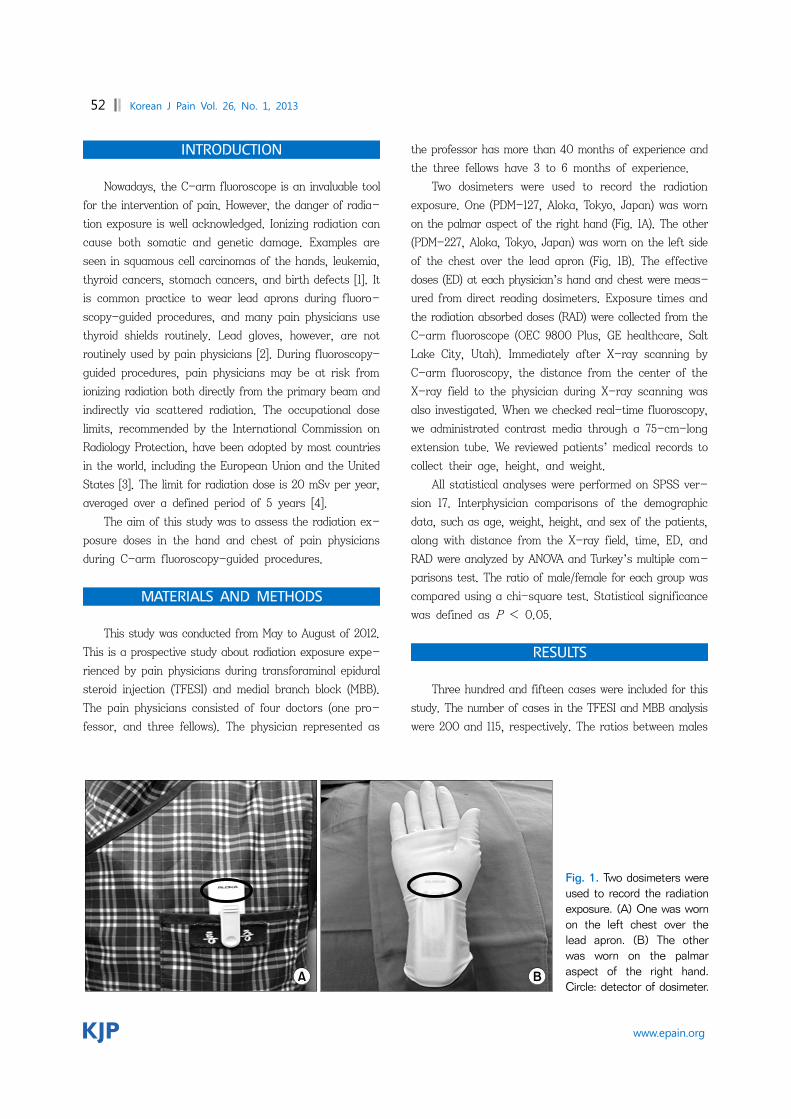
Fig. 1. Two dosimeters were used to record the radiation exposure. (A) One was wornon the left chest over the lead apron. (B) The other was worn on the palmar aspect of the right hand. Circle: detector of dosimeter.
INTRODUCTION
Nowadays, the C-arm fluoroscope is an invaluable tool
for the intervention of pain. However, the danger of radia-
tion exposure is well acknowledged. Ionizing radiation can
cause both somatic and genetic damage. Examples are
seen in squamous cell carcinomas of the hands, leukemia,
thyroid cancers, stomach cancers, and birth defects [1]. It
is common practice to wear lead aprons during fluoro-
scopy-guided procedures, and many pain physicians use
thyroid shields routinely. Lead gloves, however, are not
routinely used by pain physicians [2]. During fluoroscopy-
guided procedures, pain physicians may be at risk from
ionizing radiation both directly from the primary beam and
indirectly via scattered radiation. The occupational dose
limits, recommended by the International Commission on
Radiology Protection, have been adopted by most countries
in the world, including the European Union and the United
States [3]. The limit for radiation dose is 20 mSv per year,
averaged over a defined period of 5 years [4].
The aim of this study was to assess the radiation ex-
posure doses in the hand and chest of pain physicians
during C-arm fluoroscopy-guided procedures.
MATERIALS AND METHODS
This study was conducted from May to August of 2012.
This is a prospective study about radiation exposure expe-
rienced by pain physicians during transforaminal epidural
steroid injection (TFESI) and medial branch block (MBB).
The pain physicians consisted of four doctors (one pro-
fessor, and three fellows). The physician represented as
the professor has more than 40 months of experience and
the three fellows have 3 to 6 months of experience.
Two dosimeters were used to record the radiation
exposure. One (PDM-127, Aloka, Tokyo, Japan) was worn
on the palmar aspect of the right hand (Fig. 1A). The other
(PDM-227, Aloka, Tokyo, Japan) was worn on the left side
of the chest over the lead apron (Fig. 1B). The effective
doses (ED) at each physician’s hand and chest were meas-
ured from direct reading dosimeters. Exposure times and
the radiation absorbed doses (RAD) were collected from the
C-arm fluoroscope (OEC 9800 Plus, GE healthcare, Salt
Lake City, Utah). Immediately after X-ray scanning by
C-arm fluoroscopy, the distance from the center of the
X-ray field to the physician during X-ray scanning was
also investigated. When we checked real-time fluoroscopy,
we administrated contrast media through a 75-cm-long
extension tube. We reviewed patients’ medical records to
collect their age, height, and weight.
All statistical analyses were performed on SPSS ver-
sion 17. Interphysician comparisons of the demographic
data, such as age, weight, height, and sex of the patients,
along with distance from the X-ray field, time, ED, and
RAD were analyzed by ANOVA and Turkey’s multiple com-
parisons test. The ratio of male/female for each group was
compared using a chi-square test. Statistical significance
was defined as P < 0.05.
RESULTS
Three hundred and fifteen cases were included for this
study. The number of cases in the TFESI and MBB analysis
were 200 and 115, respectively. The ratios between males

Jung, et al / Radiation Exposure of the Hand and Chest 53
www.epain.org
Table 1. Demographic and Radiation Related Data in Transforaminal Epidural Steroid Injection
Physician A (n = 38) B (n =55) C (n = 65) D (n = 42) P value
Age (yr)Height (cm)Weight (kg)Distance (cm)Time/level (sec)Hand/level (μSv)Chest/level (μSv)RAD/level (radcm2)
55.8 ± 14.8165.2 ± 6.8
65.8 ± 10.074.1 ± 13.6a
17.5 ± 7.4a
3.4 ± 3.8a
6.4 ± 8.8a
103.5 ± 87.5a
60.6 ± 15.0162.1 ± 9.2
64.4 ± 12.540.7 ± 9.5b
27.0 ± 15.9b
13.1 ± 9.3b
20.1 ± 16.0b
229.0 ± 145.8b
62.7 ± 16.2162.6 ± 7.9
63.5 ± 10.243.9 ± 3.7b
28.2 ± 17.3b
12.7 ± 9.1ab
26.9 ± 19.6b
236.2 ± 148.5b
59.0 ± 13.8165.1 ± 8.3
68.1 ± 8.846.9 ± 1.4b
27.2 ± 15.3b
37.8 ± 35.3c
26.5 ± 36.5b
304.6 ± 250.0b
0.1530.1390.146
0.000* 0.003* 0.000* 0.000* 0.000*
Distance: the distance from center of X-ray field to physician, Time/level: exposure time per one level, RAD/level: radiation absorbeddose per one level, Hand/level, Chest/level: radiation dose at hand and chest per one level. Values are expressed as mean ± SD. N: number of the patients. *P < 0.05. Small letter: the same letters indicate non-significant difference between groups based on Turkey’smultiple comparison test.
Table 2. Demographic and Radiation Related Data in Medial Branch Block
Physician A (n = 15) B (n = 4) C (n = 33) D (n = 49) P value
Age (yr)Height (cm)Weight (kg)Distance (cm)Time/level (sec)Hand/level (μSv)Chest/level (μSv)RAD/level (radcm2)
56.7 ± 19.7162.5 ± 6.8
66.7 ± 11.369.0 ± 3.13a
2.1 ± 0.9a
0.6 ± 0.70.4 ± 0.5
12.3 ± 8.3
49.5 ± 2.9168.4 ± 11.2
62.8 ± 11.943.3 ± 2.8bc
8.5 ± 3.43b
2.6 ± 2.01.6 ± 1.9
22.8 ± 9.7
60.1 ± 16.8160.6 ± 6.1
60.1 ± 8.544.0 ± 4.2b
5.5 ± 3.8b
1.7 ± 5.81.2 ± 2.3
20.9 ± 16.7
56.4 ± 15.0163.0 ± 6.0
60.2 ± 6.739.40 ± 3.6c
4.9 ± 2.2b
0.9 ± 0.80.5 ± 0.4
20.0 ± 12.1
0.5530.0890.061
0.000* 0.000*0.5190.0620.174
Distance: the distance from center of X-ray field to physician, Time/level exposure time per one level, RAD/level: radiation absorbed doseper one level, Hand/level, Chest/level: radiation dose at hand and chest per one level. Values are expressed as mean ± SD. N: numberof the patients. *P < 0.05. Small letter: the same letters indicate non-significant difference between groups based on Turkey’s multiple comparison test.
Table 3. Radiation Dose at the Hand and Chest in TFESI
Physician Hand/level (μSv) Chest/level (μSv) P value
A (n = 38)B (n = 55)C (n = 65)D (n = 42)
3.4 ± 3.813.1 ± 9.313.2 ± 9.037.8 ± 35.3
6.4 ± 8.820.1 ± 16.026.5 ± 19.226.5 ± 36.5
0.003*0.000*0.000*0.010*
TFESI: Transforaminal Epidural Steroid Injection, Hand/level, Chest/level: radiation dose at hand and chest per one level. Values are expressed as mean ± SD. N: number of the patients. *P < 0.05.
and females for the two groups were 88:112 and 64:37.
There were no significant differences in the patient’s sex, age, height, and weight among each of the physicians
(Table 1, 2). In the TFESI group, there were statistically
significant differences in the distance from the X-ray, ex-
posure time, and ED at the hand and chest, as well as
RAD between the physicians (Table 1). The distance from
the center of the X-ray field to physician A was more than
that of the other physicians, and the exposure time, ED,
and RAD in physician A were less than that of the other
physicians. ED at the hand of physician D was more than
that of the other physicians. There were significant differ-
ences between ED of the hand and ED of chest in all the
physicians (Table 3). In physician A, B and C, the ED at
the chest was more than the ED at the hand. In physician
A, the lowest dose was received at the hand and chest,
while the distance from the X-ray field was the furthest
among the physicians (Table 1).
In the MBB group, there were no differences between the
ED at the hand and chest, except for physician D (Table 4).
There were statistically significant differences in the dis-
54 Korean J Pain Vol. 26, No. 1, 2013
www.epain.org
Table 4. Radiation Dose at the Hand and Chest in MBB
Physician Hand/level (μSv) Chest/level (μSv) P value
A (n = 15)B (n = 4)C (n = 33)D (n = 49)
0.6 ± 0.72.6 ± 2.01.7 ± 5.80.9 ± 0.8
0.4 ± 0.51.6 ± 1.91.2 ± 2.30.5 ± 0.4
0.0570.1550.654
0.000*
MBB: medial branch block, Hand/level, Chest/level: radiation doseat hand and chestper one level. Values are expressed as mean ±SD. N: number of the patients. *P < 0.05.
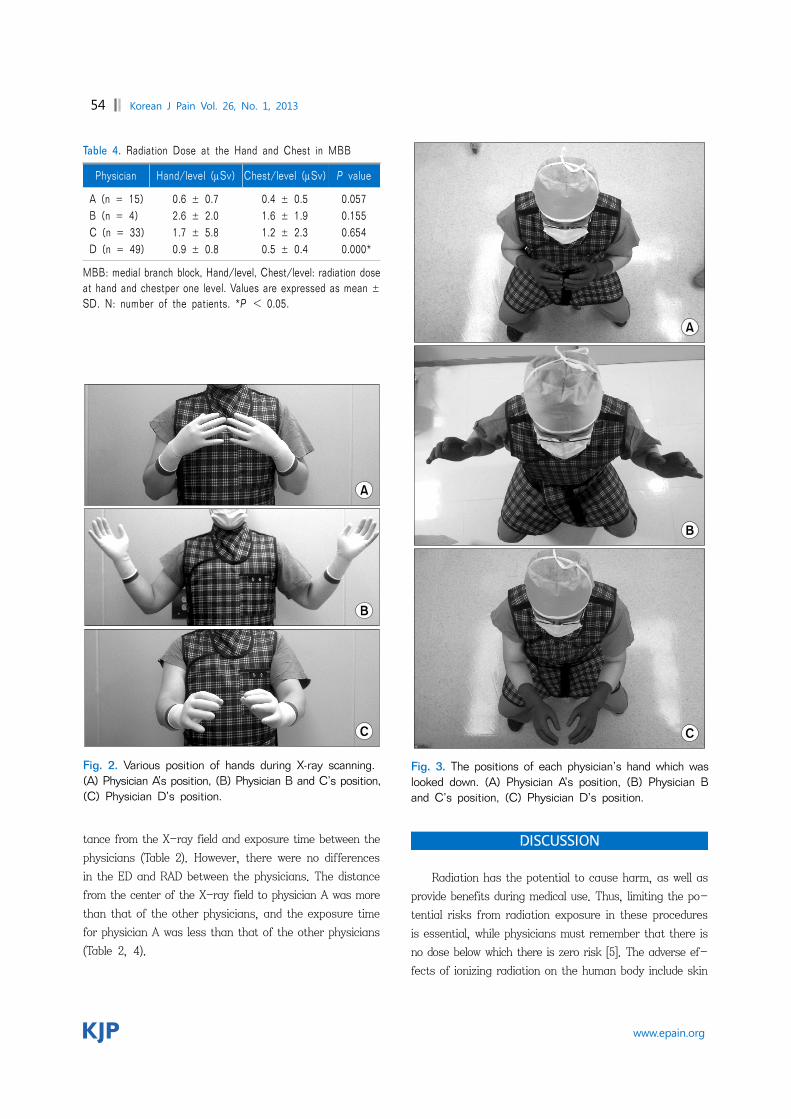
Fig. 2. Various position of hands during X-ray scanning. (A) Physician A’s position, (B) Physician B and C’s position,(C) Physician D’s position.
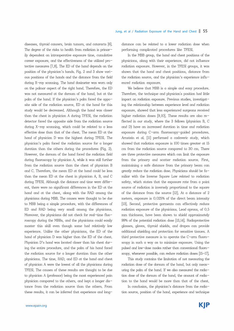
Fig. 3. The positions of each physician’s hand which waslooked down. (A) Physician A’s position, (B) Physician B and C’s position, (C) Physician D’s position.
tance from the X-ray field and exposure time between the
physicians (Table 2). However, there were no differences
in the ED and RAD between the physicians. The distance
from the center of the X-ray field to physician A was more
than that of the other physicians, and the exposure time
for physician A was less than that of the other physicians
(Table 2, 4).
DISCUSSION
Radiation has the potential to cause harm, as well as
provide benefits during medical use. Thus, limiting the po-
tential risks from radiation exposure in these procedures
is essential, while physicians must remember that there is
no dose below which there is zero risk [5]. The adverse ef-
fects of ionizing radiation on the human body include skin
Jung, et al / Radiation Exposure of the Hand and Chest 55
www.epain.org
diseases, thyroid cancers, brain tumors, and cataracts [6].
The degree of the risks to health from radiation is primar-
ily dependent on intraoperative exposure time, cumulative
career exposure, and the effectiveness of the utilized pro-
tective measures [7,8]. The ED of the hand depends on the
position of the physician’s hands. Fig. 2 and 3 show vari-
ous positions of the hands and the distance from the field
during X-ray scanning. The hand dosimeter was worn only
on the palmar aspect of the right hand. Therefore, the ED
was not measured at the dorsum of the hand, but at the
palm of the hand. If the physician’s palm faced the oppo-
site side of the radiation source, ED at the hand for this
study would be decreased. Although the hand was closer
than the chest in physician A during TFESI, the radiation
detector faced the opposite side from the radiation source
during X-ray scanning, which could be related to a less
effective dose than that of the chest. The mean ED at the
hand of physician D was the highest during TFESI. The
physician’s palm faced the radiation source for a longer
duration than the others during the procedures (Fig. 2).
However, the dorsum of the hand faced the radiation field
during fluoroscopy by physician A, while it was still further
from the radiation source than the chest of physician B
and C. Therefore, the mean ED at the hand could be less
than the mean ED at the chest in physician A, B, and C
during TFESI. Although the distance and time were differ-
ent, there were no significant differences in the ED at the
hand and at the chest, along with the RAD among the
physicians during MBB. The causes were thought to be due
to MBB being a simple procedure, with the differences of
ED and RAD being very small among the physicians.
Moreover, the physicians did not check for real-time fluo-
roscopy during the MBBs, and the physicians could easily
master this skill even though some had relatively low
experience. Unlike the other physicians, the ED of the
hand of physician D was higher than the ED of the chest.
Physician D’s hand was located closer than his chest dur-
ing the entire procedure, and the palm of his hand faced
the radiation source for a longer duration than the other
physicians. The time, RAD, and ED at the hand and chest
of physician A were the lowest of all the physicians during
TFESI. The causes of these results are thought to be due
to physician A (professor) being the most experienced pain
physician compared to the others, and kept a longer dis-
tance from the radiation source than the others. From
these results, it can be inferred that experience and long-
distance can be related to a lower radiation dose when
performing complicated procedures like TFESI.
In the MBB group, the hand and chest positions of the
physicians, along with their experience, did not influence
radiation exposure. However, in the TFESI groups, it was
shown that the hand and chest positions, distance from
the radiation source, and the physician’s experience influ-
enced radiation exposure.
We believe that MBB is a simple and easy procedure.
Therefore, the technique and physician's position had little
impact on radiation exposure. Previous studies, investigat-
ing the relationship between experience level and radiation
exposure, showed that less experienced surgeons received
higher radiation doses [9,10]. These results are also re-
flected in our study, where the 3 fellows (physician B, C
and D) have an increased duration in time and radiation
exposure during C-arm fluoroscopy-guided procedures.
Arnstein et al. [11] performed a cadaveric study, which
showed that radiation exposure is 100 times greater at 15
cm from the radiation source compared to 30 cm. There
are three protective measures which can limit the exposure
from the primary and scatter radiation source. First,
maintaining a safe distance from the primary beam can
greatly reduce the radiation dose. Physicians should be fa-
miliar with the Inverse Square Law related to radiation
safety, which states that the exposure rate from a point
source of radiation is inversely proportional to the square
of the distance from the source [12]. At a distance of 2
meters, exposure is 0.025% of the direct beam intensity
[13]. Second, protective garments can effectively reduce
radiation exposure of the physicians. Lead aprons, of 0.5
mm thickness, have been shown to shield approximately
99% of the potential radiation dose [13,14]. Radioprotective
glasses, gloves, thyroid shields, and drapes can provide
additional shielding and protection for sensitive tissues. A
third protective measure is to operate the C-arm fluoro-
scopy in such a way as to minimize exposure. Using the
pulsed and low-dose modes rather than conventional fluoro-
scopy, whenever possible, can reduce radiation doses [15-17].
This study contains the limitation of not measuring the
radiation dose of the dorsum of the hand, but only meas-
uring the palm of the hand. If we also measured the radia-
tion dose of the dorsum of the hand, the amount of radia-
tion to the hand would be more than that of the chest.
In conclusion, the physician’s distance from the radia-
tion source, position of the hand, experience and technique
56 Korean J Pain Vol. 26, No. 1, 2013
www.epain.org
can correlate with the radiation dose in complicated proce-
dures such as TFESI. Therefore, for radiation safety, we
have to make efforts to keep our hands and body at a dis-
tance from the radiation field, while attempting to master
the skills for interventional pain procedures.
REFERENCES
1. Andreassi MG. The biological effects of diagnostic cardiac imaging on chronically exposed physicians: the importance of being non-ionizing. Cardiovasc Ultrasound 2004; 2: 25.
2. Park PE, Park JM, Kang JE, Cho JH, Cho SJ, Kim JH, et al. Radiation safety and education in the applicants of the final test for the expert of pain medicine. Korean J Pain 2012; 25: 16-21.
3. Pradhan AS. Evolution of dosimetric quantities of International Commission on Radiological Protection (ICRP): Impact of the forthcoming recommendations. J Med Phys 2007; 32: 89- 91.
4. Miller DL, Vañó E, Bartal G, Balter S, Dixon R, Padovani R, et al. Occupational radiation protection in interventional radiology: a joint guideline of the Cardiovascular and Interventional Radiology Society of Europe and the Society of Interventional Radiology. J Vasc Interv Radiol 2010; 21: 607-15.
5. Harding LK, Thomson WH. International commission on radiation protection. Nucl Med Commun 1990; 11: 585-7.
6. Cousins C, Sharp C. Medical interventional procedures- reducing the radiation risks. Clin Radiol 2004; 59: 468-73.
7. Mroz TE, Yamashita T, Davros WJ, Lieberman IH. Radiation exposure to the surgeon and the patient during kyphoplasty. J Spinal Disord Tech 2008; 21: 96-100.
8. Theocharopoulos N, Perisinakis K, Damilakis J, Papadokostakis G, Hadjipavlou A, Gourtsoyiannis N. Occupational exposure
from common fluoroscopic projections used in orthopaedic surgery. J Bone Joint Surg Am 2003; 85: 1698-703.
9. Blattert TR, Fill UA, Kunz E, Panzer W, Weckbach A, Regulla DF. Skill dependence of radiation exposure for the orthopa-edic surgeon during interlocking nailing of long-bone shaft fractures: a clinical study. Arch Orthop Trauma Surg 2004; 124: 659-64.
10. Bahari S, Morris S, Broe D, Taylor C, Lenehan B, McElwain J. Radiation exposure of the hands and thyroid gland during percutaneous wiring of wrist and hand procedures. Acta Orthop Belg 2006; 72: 194-8.
11. Arnstein PM, Richards AM, Putney R. The risk from radiation exposure during operative X-ray screening in hand surgery. J Hand Surg Br 1994; 19: 393-6.
12. Siegel JA, Marcus CS, Sparks RB. Calculating the absorbed dose from radioactive patients: the line-source versus point-source model. J Nucl Med 2002; 43: 1241-4.
13. Bushberg JT, Seibert JA, Leidholdt EM, Boone JM. The essential physics of medical imaging. 3rd ed. Philadelphia, Lippincott Williams & Wilkins. 2011, pp 837-902.
14. Singer G. Occupational radiation exposure to the surgeon. J Am Acad Orthop Surg 2005; 13: 69-76.
15. Cho JH, Kim JY, Kang JE, Park PE, Kim JH, Lim JA, et al. A study to compare the radiation absorbed dose of the C-arm fluoroscopic modes. Korean J Pain 2011; 24: 199- 204.
16. Kallmes DF, O E, Roy SS, Piccolo RG, Marx WF, Lee JK, et al. Radiation dose to the operator during vertebroplasty: prospective comparison of the use of 1-cc syringes versus an injection device. AJNR Am J Neuroradiol 2003; 24: 1257-60.
17. Kruger R, Faciszewski T. Radiation dose reduction to medical staff during vertebroplasty: a review of techniques and methods to mitigate occupational dose. Spine 2003; 28: 1608-13.
![AM1808 ARM Microprocessor (Rev. C) · Title: AM1808 ARM Microprocessor (Rev. C) Author: Texas Instruments, Incorporated [SPRS653,C ] Subject: Data Manual on Single Products Keywords:](https://img.pdfslide.us/doc/110x75/60c343af5c1d0e57bf0df998/am1808-arm-microprocessor-rev-c-title-am1808-arm-microprocessor-rev-c-author.jpg)