Embed Size (px)
Citation preview
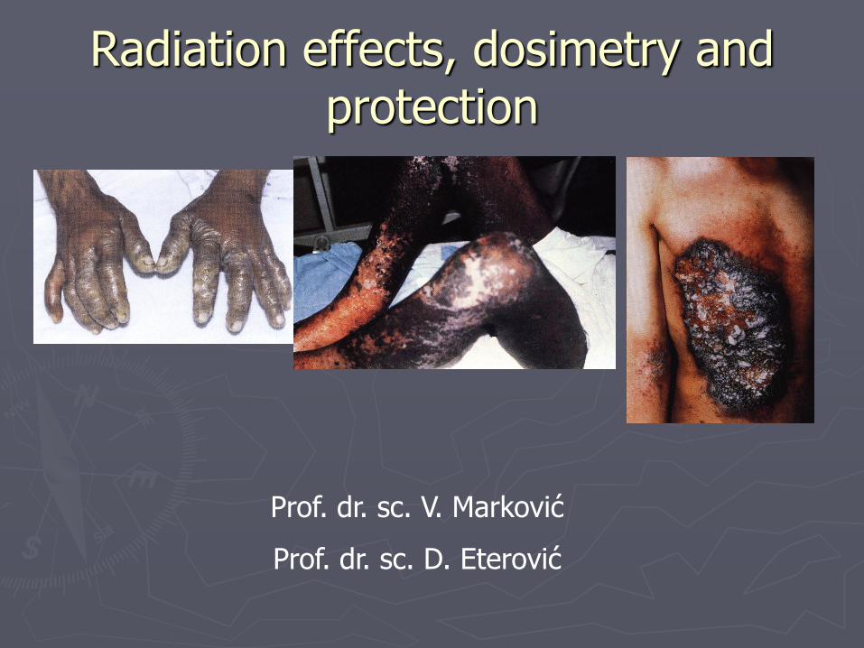
Radiation effects, dosimetry and protection
Prof. dr. sc. V. Marković
Prof. dr. sc. D. Eterović

Quantitation of radiation effects: dosimetry
Absorbed dose: energy delivered to unit mass of medium, unit:
Grey (Gy); 1 Gy=1 J/kg

Ionization is biologically harmful
Cells can be seriously impaired, depending on:
►(i) tissue exposed (proliferative cells are most sensitive)
►(ii) absorbed dose
►(iii) type of radiation
►(iv) dose rate (longer time enables reccuperation)

►Radiation impairs cells by ionizing vital mollecules, as DNA (direct effect), and by chemically altering intracellular water (indiret effect), giving rise to free radicals: H i OH.
►Indirect effects are much more common and thus more important.

Radiation effects can be hereditary or
somatic
►Hereditary effects are due to impairment of reproductive cells, do not affect the person exposed, but her (his) offspring
►Somatic effects affect only the person exposed to radiation

►Biological radiation effects are also classified as stochastic (randomly occuring) and non-stochastic (regularly occuring)
►Stochastic effects are due to mutation of cells. They have no threshold, i.e. may appear after minor exposure, however, with less probability than after large exposure (large absorbed dose). When they do apper, their seriousness does not depend on exposure.

Non-stochastic effects follow large exposure, inducing death of cells or permanent dammage, disabling cell division. They do have treshold (minor exposure cannot kill cells!) and their seriosness increase with absorbed dose. All non-stochastic effects are somatic (dead cells cannot play role in reproduction!)

Biological impairments of ionizing radiation, aside from absorbed dose
(energy delivered), depend on type of
radiation (mode of energy delivering)
►Neutrons and alpha particles ionize very densely, producing more commonly the irreparable, multiple impairments of macromolecules than gamma rays or beta particles

Equivalent dose = Q.Absorbed dose
Q is quality factor. For alpha particles, it
equals 20, for neutrons from 5 to 20 (depending on energy), and for beta particles, X and gamma photons- 1
Equivalent dose

►The unit for equivalent dose is
Sievert (Sv)
►Old unit is rem
1 rem = 0.01 Sv

We are most irradiated by natural radioactivity sources
►We are all constantly in the low intensity field of ionizing radiation from various sources (including our own tissues!). This is background radiation
►Mostly, the radiation sources are natural

Minor degree of irradiation is due to artificial sources
►95% of irradiation from artificial sources is due to medical diagnostics (radiotherapeutic exposure is not taken into account)
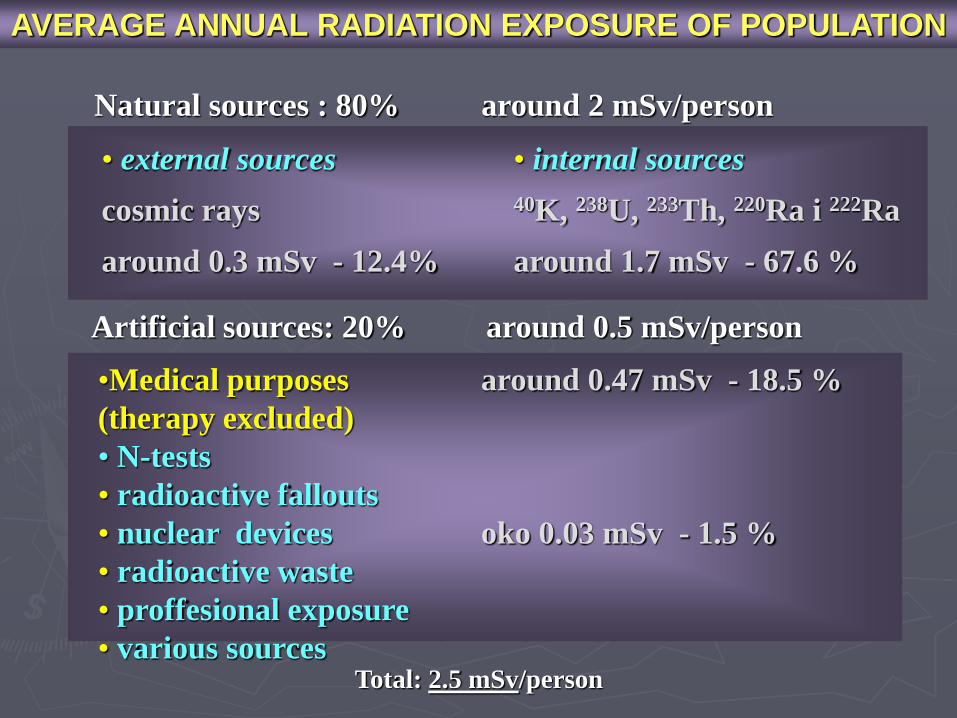
AVERAGE ANNUAL RADIATION EXPOSURE OF POPULATION
Artificial sources: 20% around 0.5 mSv/person
• external sources
cosmic rays
around 0.3 mSv - 12.4%
• internal sources
40K, 238U, 233Th, 220Ra i 222Ra
around 1.7 mSv - 67.6 %
•Medical purposes around 0.47 mSv - 18.5 %
(therapy excluded)
• N-tests
• radioactive fallouts
• nuclear devices oko 0.03 mSv - 1.5 %
• radioactive waste
• proffesional exposure
• various sources
Natural sources : 80% around 2 mSv/person
Total: 2.5 mSv/person

Effective equivalent dose
► Sensitivity to radiation exposure (radiosensitivity) is different in different tissues; to account for the part of body irradiated, to each body part one assigns the weight factor (wT) according to its specific radiosensitivity (the sum of all those factors equals 1):
►effective equivalent dose (Sv) =
= Σ specific factor x equivalent dose;
summing is over body parts irradiated

each WT
group WT
skin, bone surface 0,01 0,02
bile, liver, breast, esophagus, thyroid, other organs (except those below)
0,05 0,30
bone marrow, colon, lungs, stomach
0,12 0,48
gonads 0,20 0,20
total 1,0
Specific organ radiosensitivities

Internal dosimetry
►Assessment of absorbed doses due to radionuclides introduced into the body intentionally (diagnostics, therapy) or accidentally.
► Personal dosimetry vs. application of results for ‘average’ human.
►Methods: actual measurements or model-based calculations
►The results may not be accurate due to many unknowns
►Given the absorbed doses (and the dose rate!) there is large uncertainty in individual (patho)physilogical (clinical) response (wanted or unwnated)

►Absorbed doses are estimated separately for particle (alpha and beta) and non-particle ( X and gamma) radiation;
►In most cases both are present and the results are added.
►Distinguish: organ equivalent dose(s) and total body effective equivalent dose.
►It is important to report/note:
1. effective equivalent (body) dose
2. dose to target organ (organ aimed at diagnostically or therapeutically)
3. dose to critical organ (highest dose)
4. dose to gonads, bone marrow and eye lenses
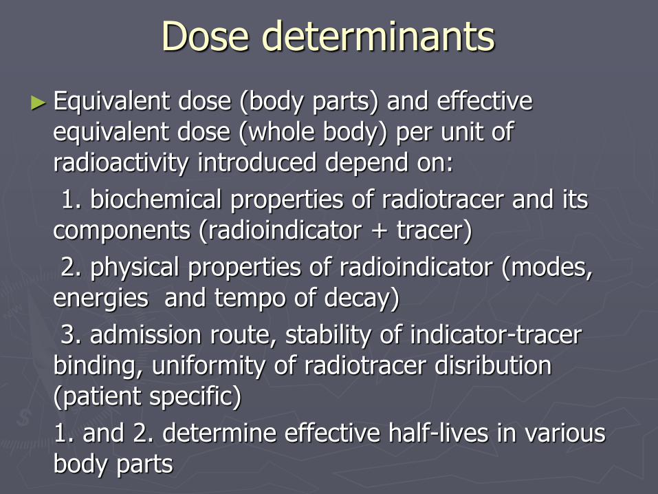
Dose determinants
► Equivalent dose (body parts) and effective equivalent dose (whole body) per unit of radioactivity introduced depend on:
1. biochemical properties of radiotracer and its components (radioindicator + tracer)
2. physical properties of radioindicator (modes, energies and tempo of decay)
3. admission route, stability of indicator-tracer binding, uniformity of radiotracer disribution (patient specific)
1. and 2. determine effective half-lives in various body parts

Particles Basic assumption: only target organs are affected since all energy is locally absorbed.
energy absorbed = average energy of a particle x total number of dissintegrations (‘cummulative activity’)
Cummulative activity equals the product of organ specific mean life of a compaund and activity that entered the organ (organ uptake x total activity introduced)

In convenient units, the later translates to:
Dose (Gy) = 14 x mean particle energy (MeV) x
cummulated activity (MBq/day)/ organ mass (g)
Equivalent organ dose (Sv) = Dose (Gy) x Q
Organ mass is usually assessed with aid of diagnostic imaging
Cummulated activity can be assessed by follow-up of
radiation intensity emanating from an organ (sequential external measurements of count-rates)

X and γ radiation Other organs, aside from target organ are affected, depending on proximity to target organ and photon energy (energies).
Complex calculations are required; the shapes of targets and sources (the same organ is irradiation source for other organs as well as their target) and their relative positions are taken into account.
Except in case of pure gamma emitters, the dose due to non-particle radiation is only a small part of total dose (the rest is due to particles).

Absorbed doses in common procedures
Procedure Radiotracer Activity Dose in critical
organ (Gy)a
Dose in gonads
(mGy)
Brain
scintigraphy pertechnetate 500 MBq
(~ 15 mCi) Intestines, 0.02 4
Liver
scintigraphy sulphor colloid 150 MBq
(~ 4 mCi) Liver, 0.02 0.85
Lung
scintigraphy macroaggregate 100 MBq
(~ 3 mCi) Lungs, 0.009 0.3
Bone
scintigraphy pyrophosphate 500 MBq
(~ 15 mCi) Bladder, 0.06 4
Renography hyppuric acid Bq
(~ 200 μCi) Bladder, 0.02 0.2
Thyroid
function sodium iodide
300 kBq
(~ 8 μCi) Thyroid, 0.08 0.6
Thyroid
scintigraphy pertechnetate 150 MBq
(~ 4 mCi) Intestines, 0.01 0.8
a critical organ has the largest absorbed dose
Tc99mTc99mTc99mTc99mI131I131 Tc99m

Whole body counters
In case of accidental intake of radioactive material the unwanted radiation exposure can be assessed by whole body counter: gamma counter large enough to accomodate a whole person inside of it.
►The devices aid in determing the type and quantity of radionculides introduced. These dana enable assessment of absorbed doses and expected clinical course.
► Pure alpha and β emitters can not be detected.
►The deviices are very sensitive, but have low spatial resolution.

Risks of radiation exposure in nuclear medicine
There are no serious non-stohastic effects of radiation due to nuclear medicine diagnostic procedures (effective doses mostly in range 0.5-5 mSv; exception is test of accumulation of I-131)

Probablitiies of stohasic effects of low-dose exposures to radiation
Lethal malignoma 5 x 10⁻² Sv-1 (1 in 20)
Curable malignoma 1 x 10⁻² Sv-1 (1 in 100)
Serious inherited disease in offspring
1,3 x 10⁻² Sv-1 (1 in 77)
All numbers are per 1 Sv of effective low dose, on a single occassion, uniformly accross the body, in case of an avearge world inhabitant.
For example a single, uniformly distributed effective dose of 3 mSv is expected to induce lethal malignoma in 1/6667 persons
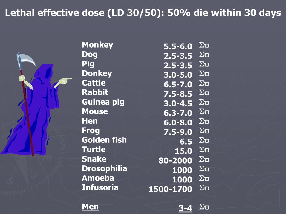
Lethal effective dose (LD 30/50): 50% die within 30 days
Monkey Dog Pig Donkey Cattle Rabbit Guinea pig Mouse Hen Frog Golden fish Turtle Snake Drosophilia Amoeba Infusoria Men
Sv
Sv
Sv
Sv
Sv
Sv
Sv
Sv
Sv
Sv
Sv
Sv
Sv
Sv
Sv
Sv
Sv
5.5-6.0 2.5-3.5 2.5-3.5 3.0-5.0 6.5-7.0 7.5-8.5 3.0-4.5 6.3-7.0 6.0-8.0 7.5-9.0
6.5 15.0
80-2000 1000 1000
1500-1700
3-4

“Small” doses
Up to 200 mGy for χ and γ radiation or 50 mGy for α
particles and neutrons, receieved acutely, on a single occasion. In practice we mostly encounter the yearly irradiation up to 20 mSv.
There are no evidences that very small doses
(couple of mGy) increase the risk of malignant diseases, but, to be on the safe side, we take it for granted that stohastic effects have no treshold.

Acute effects of radiation
1. Acute local radiation lesions
Lesions confied to smaller, superficial parts of body, due to accidental exposure to external radiation, mostly during professional activities

Erythema

Edema Develops due to vasculitis after the dose over 10 Sv

Epidermatitis Dry: dose treshold 5 Sv (lesion of basal skin layer) Moist: dose treshold 15 Sv, occurs after 20 days latency (plasma transudation, vesicules…)
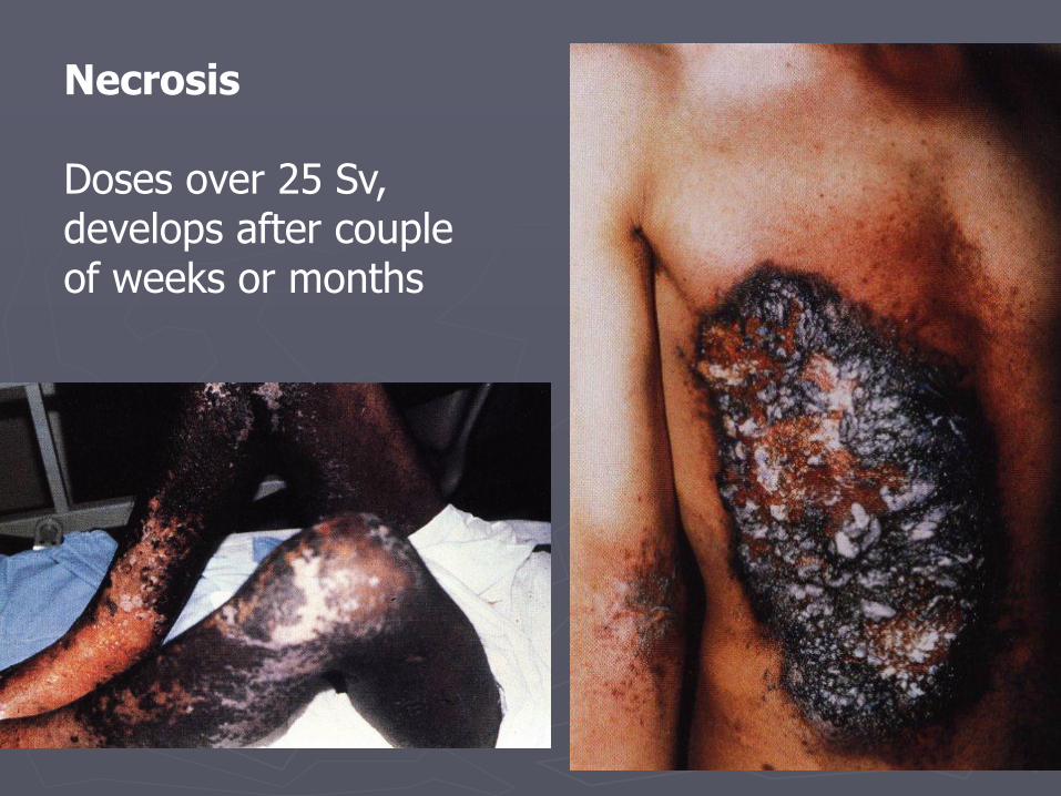
Necrosis Doses over 25 Sv, develops after couple of weeks or months

Alopecia Treshold is 3-4 Sv, above 7 Sv hair loss is permanent.

Skin effects

Develops after dose over 1 Sv accross whole body or its major part. Results in halt of reproductive processes, abnormal cytokine secretion and cell death
2. Acute radiation syndrome

Phases of Acute Radiation Syndrome
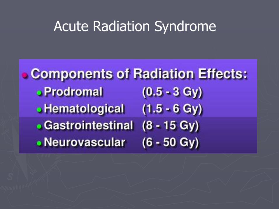
Acute Radiation Syndrome


Chronic radiodermatitis After long-term exposure to 10 Sv (but acutely under erythema treshold). Cataract External exposure to 2 Gy (acute) or 15 Gy (long term)
Late effects of radiation

Malignant tumors Firm evidences only after 1 Sv; effects of doses less than 0.1 Sv uncertain (although for protection purposes we assume no treshold, since a single mutated cell may induce carcinoma). Latency- around 8 years (leukemia) and 15-20 years (solid tumors). Genetic effects The effects are usually not expressed in first generation offspring

Sterilility
Acute or chronic- depending on dose
Deformity/retardation in newborn infants
During 8-1week of gestation the treshold is low (o.1 Sv)
Chromosome abberations
Circulating lymphocytes are most sensitive

50 mSv 500 mSv Skin equivalent dose
15 mSv 150 mSv Eye lenses equivalnet dose
1 mSv 20 mSv Effective dose
populaation professionals
Legal maximal tolerated yearly doses

Radiation protection principles
►Main principles:
There are no apriori acceptable levels: ALARA principle: As Low As Reasonably Achievable
Exposure of many persons to small radiation doses can induce the same number of genetic lesions as exposure of few persons to large doses

Reduction of external irradiation
►As short as possible
►As far away as possible: most efficient (inverse squared distance rule)
►Use of absorbers: not always efficient

1 cm of lead reduces Tc-99m radiation to 1/1012
but only to 1/12 in case of I-131
Isotope
Half- value thickness
lead (mm)
Half-values thickness
water(cm)
I-131
(364 keV) 2,3 6,3
Tc-99m
(140 keV) 0,28 4,5

Reduction of internal exposure
►Radionuclides may enter the body by ingestion, inhalation or through skin/skin lesions.
►Measures to reduce internal contamination should be strictly followed:
► -use of protective clothing, protective gloves, lead apron, lead glasses
► -the area with risk of contamination should be marked
► -radionuclide containers should be specified in terms of type, activity and date, etc.
The end!


![Fluoride toothpastes for preventing dental caries in ...neuron.mefst.hr/docs/katedre/znanstvena_metodologija/Fluoride... · [Intervention Review] Fluoride toothpastes for preventing](https://img.pdfslide.us/doc/110x75/5ac7a33f7f8b9aa3298b67ff/fluoride-toothpastes-for-preventing-dental-caries-in-intervention-review-fluoride.jpg)