Embed Size (px)
Citation preview

Basics of nuclear medicine
Prof. dr. Davor Eterović Prof. dr. Vinko Marković

Radioisotopes are used both in diagnostics and in therapy
• Diagnostics – gamma emitters are used since gamma rays can penetrate the body and be detected outside
• Therapy – beta emitters; short range => local delivery of high doses

Diagnostic activities correspond to sub-physiological masses
• Activities used: 370 KBq-740 MBq (10µCi-20 mCi)
• Corresponding masses <1g
• => No physiological effects, but enough for registration and diagnostic information

Tc-99m- the most used radioisotope in medicine
• Offspring of a nuclear reactor product Mo-99, which by beta-minus decay becomes Tc-99m, the meta-stable (long living) gamma emitter
• ADVANTAGES:
1. pure gamma emitter- no beta radiation
2. optimal photon energy- sufficient to penetrate body, not too large for efficient detection/protection
3. half life long enough to perform the investigation, with acceptable patient radiation dose
4. great chemical affinity
5. relatively cheap.

Technetium generator

Scintillation detectors: gamma rays are detected after conversion to visible light
• When some materials absorb ionizing radiation the part of absorbed energy is used to excite the atoms to higher energy states, the subsequent de-excitations produce flashes of visible light
• These flashes are called scintillations, and the materials scinillators
• => SCINTIGRAMS

Sodium iodide chrystal-the commonest gamma ray detector
1. High sensitivity of detection (owing to high density and atomic number of iodine)
2. Relatively high efficiency of conversion of gamma rays to visible light (around 10 %)
3. Short scintillation time (short dead-time) enables high registration rate (over 104 counts/sec)

Scintillation detectors can measure the energy of gamma rays
• The output electrical pulse is proportional to energy of the absorbed radiation, enabling:
1. using the detector as absorbed dose-meter (besides counting)
2. selective counting according to energy of photon (pulse height)- spectral analysis

Scintillator and photomultiplier tube comprise the scintillation counter
SCINTIGRAM – image of organ function
• Accumulation of radiotracer in part of a body depends only on chemical affinity and physical properties of the tracer to which the radioisotope is bound.
• Nuclear medicine image reflects the physiological function and is created by gamma rays that leave the body.

Scintigraphy
Image of distribution of radioisotope that is attached to a molecule that accumulates in the desired region, tissue or organ, to extent that depends on cellular function

•99m-Tc-pertechnetate- scintigram of normal thyroid distribution (left)
•201-Tl-chloride- scintigram of
•normal heart muscle (down)


• I-131 scintigraphy of toxic nodular goitter

Imaging equipment
• Scanner (middle)
• Gamma camera
• PET scanner (down)

Registration of gamma radiation requires massive detector
• Two purposes of radiactive measurements:
•
1. measurements of concentration in a sample
2. imaging distribution within the body
• Due to large energy and consequent penetrability in both cases one needs
• massive detector , which limits resolution and sensitivity of registration

Well counter

Pinhole collimator: imaging of small volumes


Gamma camera creates live scintigrams
• Parallel collimator – lead plate with many narrow parallel holes (channels) at right angle to plate and crystal bases
• => gamma ray hitting the crystal originates from the area directly bellow the place of absorption
• gamma camera is always sensitive to the toal area beneath the crystal
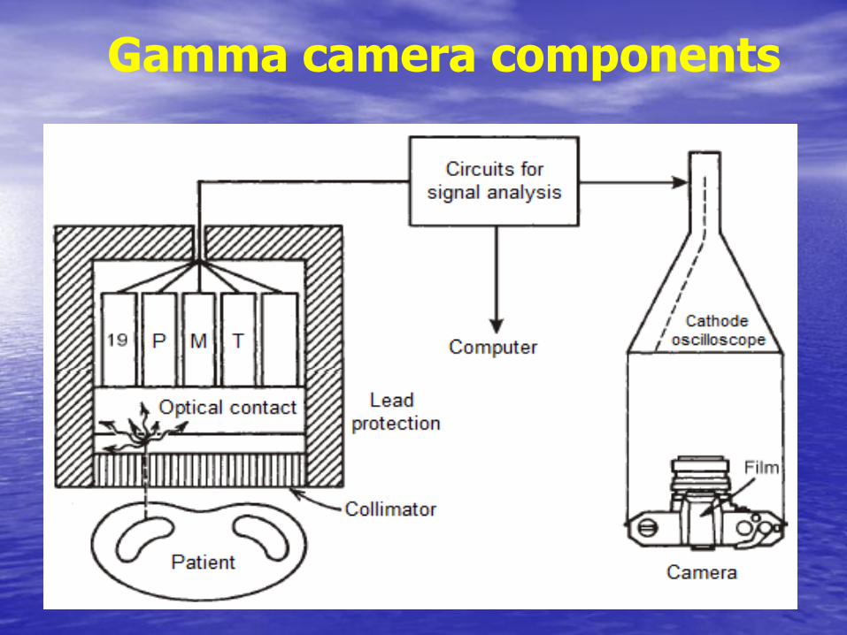
Gamma camera components


Two ways to present the gamma camera image:
1. analog image: the position of scintillation is projected on the screen of an oscilloscope, which illuminates the photograpic film
2. digital image: position and rate of scintillations are stored in computer memory as a digital matrix (64x64; 128x128;..)

Liver scintigraphy with Tc-99m-colloid in case of trauma causing tissue rupture
analog image digital image

The effect of matrix size

Distinctive scintigram features: functionality and quantitative aspect
• functionality: radiotracer accumulation mirrors the organ(s) function
• quantitative aspect: scintigram is basically a table of numbers

Factors of scintigram (dis)advantages
1. Large energy of a gamma ray: requires massive detector, but enables registration of small, deep sources of minute activities
2. spreadness/wide distribution of gamma ray sources: requires collimation of radiation, but enables functional organ images
=> Minute amounts of radioindicator, in short time, create diagnostically useful images

• Lesion detectability increases with its size, but primarily depends on its contrast against the neighboring tissue
• Resolution-important for small lesions
• Digital image can manipulated on a computer to enhance visual effects (caution: possibility of artifacts!)

Hot lesions are better seen than cold ones

Radiohistogram are analyzed with computer aid to diagnose and quantify various parameters

Radihistogram: time-activity curve
computer programs: algorithm codes to analyze (series of) scintigrams or time-activity curves

Typical mechanisms of ratiotracer localization
Mechanism Organ Radiotracer
active transport thyroid J-131
active transport kidney ortho-I-131-hypurric acid
active transport myocardium
Tl-201
capillary blockade lungs Tc-99m-macroaggregate
filtration kidney Tc-99m-diethilene-triamine-pentaacetic acid
dilution blood Tc-99m-human serum albumin
phagocytosis liver Tc-99m-sumphor colloid
sequestration spleen Tc-99m- erythrocytes (damaged by heating)

Effective half-life: determinant of absorbed dose due to radioacitivity introduced into the body
• constant of biological elimination (B)
• biological half-life (T1/2)B= ln(2)/B
• constant of effective elimination:
• EF = B + F (constant of radiactive decay)
• effective half-life (T1/2)EF= ln(2)/EF
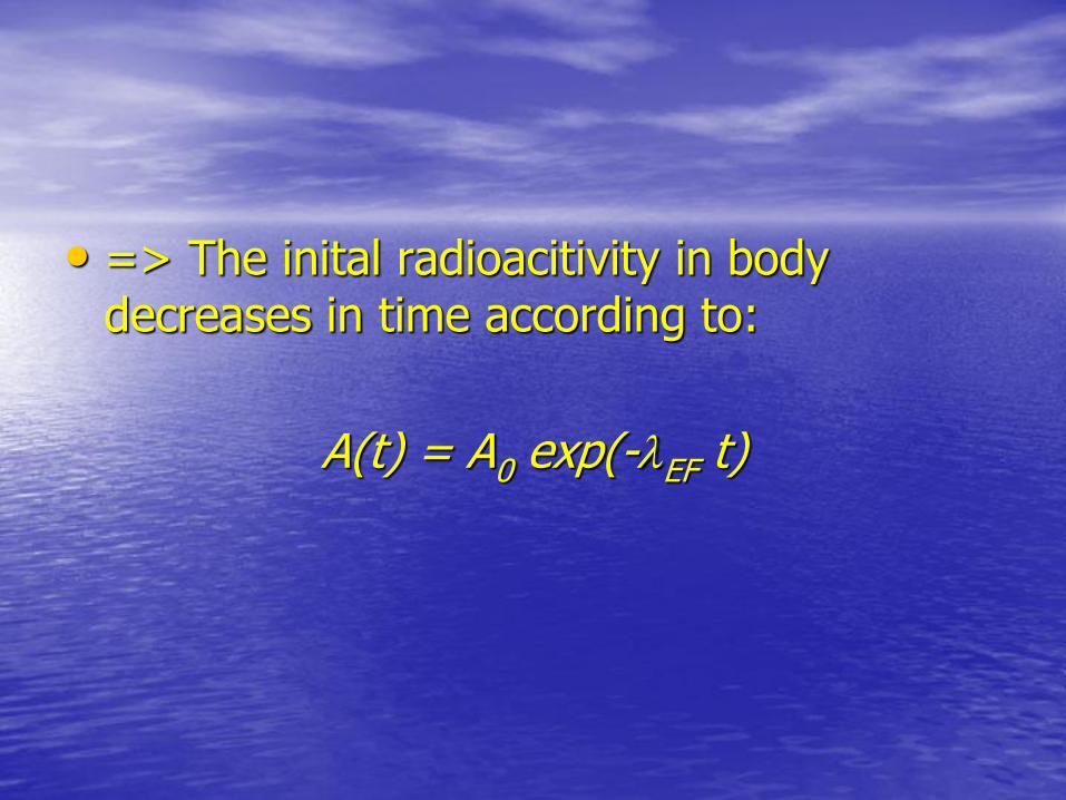
• => The inital radioacitivity in body decreases in time according to:
A(t) = A0 exp(-EF t)

Absorbed doses in common procedures Procedure Radiotracer Activity Dose in critical
organ (Gy)a Dose in gonads (mGy)
Brain scintigraphy pertechnetate
500 MBq
(~ 15 mCi) Intestines, 0.02
4
Liver scintigraphy sulphor colloid
150 MBq
(~ 4 mCi) Liver, 0.02 0.85
Lung scintigraphy macroaggregate
100 MBq
(~ 3 mCi) Lungs, 0.009 0.3
Bone scintigraphy pyrophosphate
500 MBq
(~ 15 mCi) Bladder, 0.06 4
Renography hyppuric acid Bq
(~ 200 μCi) Bladder, 0.02 0.2
Thyroid function
sodium iodide 300 kBq
(~ 8 μCi) Thyroid, 0.08 0.6
Thyroid scintigraphy pertechnetate
150 MBq
(~ 4 mCi) Intestines, 0.01
0.8
a critical organ has the largest absorbed dose
Tc99mTc99mI131

Imaging of body slices= tomography
• SPECT = single-photon emission computed tomography
• PET = positron emission tomography

Imaging from many directions enables slice isolation
• SPECT: gamma camera rotates around body, the series of scintigrams (projections) thus obtained are raw dana from which computer reconstructs slices

SPECT

Tomograms have inferior resolution compared to planar images
• Tomogram, being derived from series of planar images (projections) by non-perfect algorithm (absorption of radation in the body can be accounted for only approximately) has inferior resolution than the original (raw) images
• Nevertheless, due to increased contrasts, the overall diagnostic information is greater

Positron emissiion tomography-PET
PET is used to detect the (pato)physiological and biochemical processes by virtue of kinetics of positron emitters (F-18, C-11, O-15, N-13), or mollecules attached to them. The images reflect: tissue perfusion, the metabolism of oxygen, carbohydrates, fatty acids, amino acids, as well as neuroreceptors.

Positron emitters display metabolic processes:
Radionuclide Half-life (min)
Tracer Use
Carbon-11 20.5 Nitric acid Clinical research
Nitrogen-13 10 Cardiology Cardiology
Oxygen-15 2.1 H2O, CO, CO2
Clinical research
Fluorine-18 110 FDG and F-dopamine
Oncology, cardiology, neurology
Rubidium-82 1.3 Potassium analogs
Cardiology

Creation of PET image
The positions of sources (A,B) are reconstructed as sections of straight lines passing through detector pairs

Principle of coincident detection
•No collimation needed: resolution , sensitivity

PET image of a patient with liver metastasis

PET: pathologic focus in shoulder region

CT: liver metastasis
PET: liver metastasis

Image fusion: PET-CT

CT
PET
Lung metastasis

PET-CT fusion (lung metastasis)


Therapy
Radiotracers containg particle emitters
(mostly beta-emitters) can be used for
selective tissue destruction, by virtue of their
small range and consequent local energy
absorption

Nuclear medicine perspective
The future of nuclear medicine depends on development of new radiotracers: mollecular nuclear medicine
THE END