Embed Size (px)
Citation preview
RADIATION DOSIMETRY IN NUCLEAR MEDICINE
PART V - 752
Radiation Dosimetry in Nuclear MedicineEP Visser, Radboud University Nijmegen, Medical CentreJ de Jong, University of Groningen, University Medical Centre Groningen
Introduction
The main purpose of this chapter is to provide an overview of effective radiation doses to patients for all clinical protocols that are presented in part I and II. In almost all cases, the values were taken from the most recent publications of the International Commission on Radiological Protection (ICRP). In cases where ICRP data are not (yet) available, the authors have done their best to present data from the most recent scientifi c literature. In all cases, dose values are given for adults. In addition, dose values for 1, 5, 10 and 15 y old children are presented when available.
Furthermore, this chapter provides recommendations with regard to several issues that are related to the administration of these radiopharmaceuticals. These include doses to foetuses, infants (from breast milk and/or contact with the mother who has received a radiopharmaceutical), household members (after the patient has returned home), third parties (people in contact with the patient within a short time following the investigation), and pregnant or breast-feeding hospital personnel. This chapter does not cover radiation dosimetry for hospital personnel besides the pregnant or breast-feeding hospital personnel.
In the Netherlands, bone densitometry using Dual Energy X-ray Absorption (DEXA) is often carried out in Nuclear Medicine departments. For this reason, this chapter also contains dosimetric data related to DEXA investigations although these are not associated with the use of radiopharmaceuticals. The same holds for low dose CT scans made in PET/CT or SPECT/CT scanners for the purpose of anatomical localization and attenuation correction.
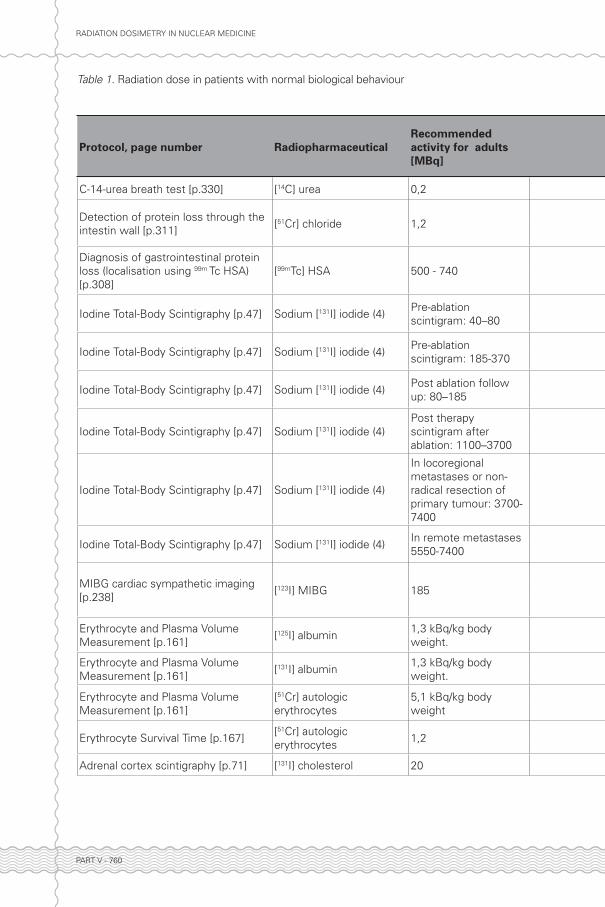
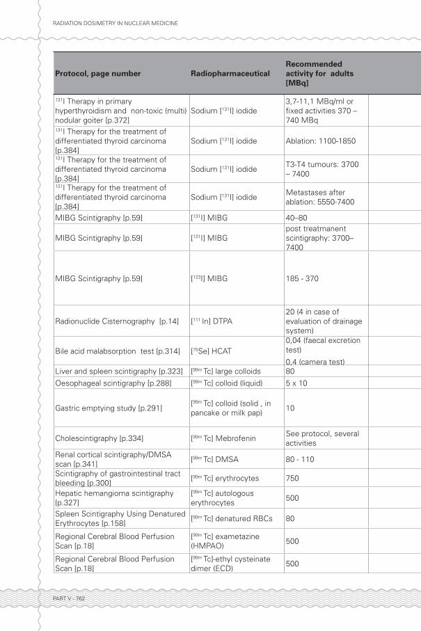
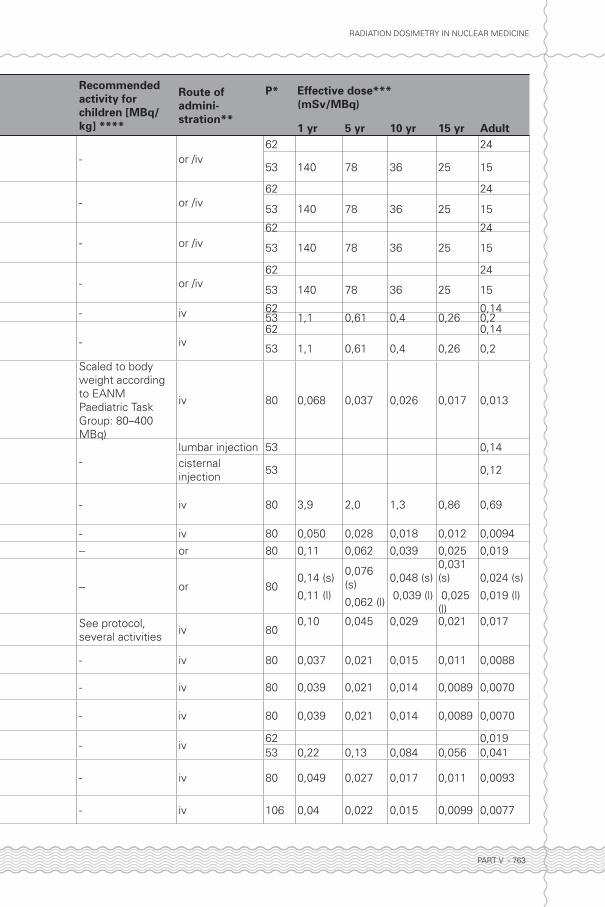
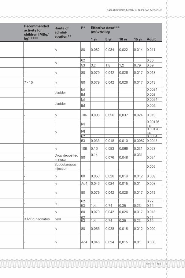
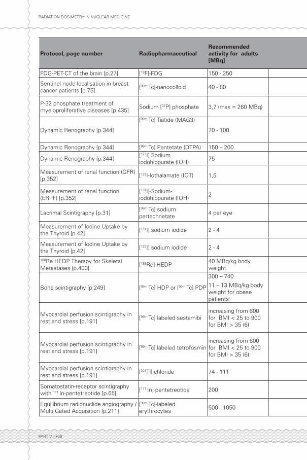
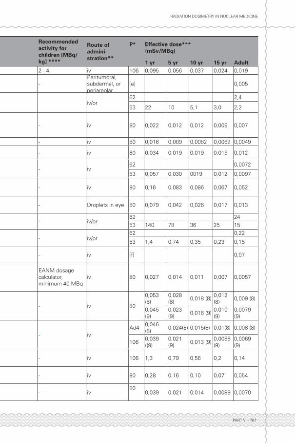
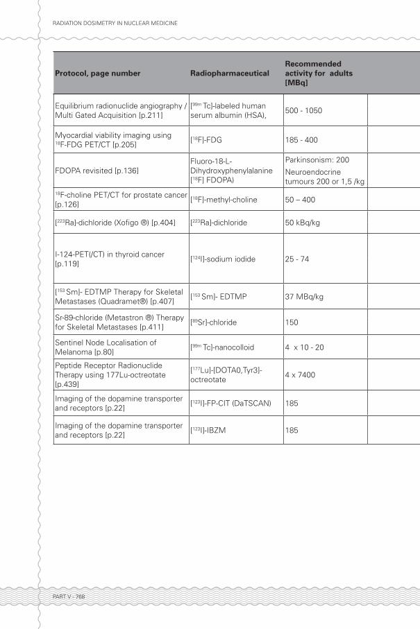
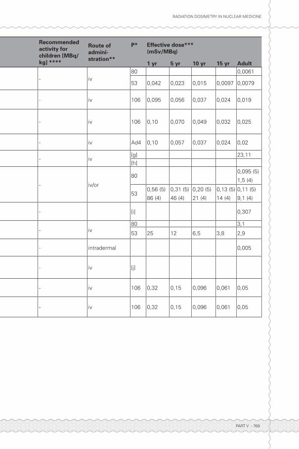
1. Patients1.1 Effective dose in patients with normal biokinetics and biodistributionTable 1 presents dosimetric data for patients with normal biokinetics and biodistribution for all protocols and radiopharmaceuticals that have been described in in part I and II of this book. The patient’s effective dose is calculated using models developed by the MIRD (Medical Internal Radiation Dose commission), which were subsequently adopted by the ICRP. In some cases the effective dose is calculated by a more recent model than that provided by the MIRD. Effective doses are calculated using organ or tissue weighting factors which provide the opportunity to represent the radiation-induced risk to patients undergoing different radio-diagnostic procedures by means of a single value. These weighting factors are proportional to the degree of risk to a particular organ, and sum to 1 for all organs together.
Column 1 presents the relevant protocol and page number. Column 2 gives the radiopharmaceutical with the radionuclide indicated in square brackets. Column 3 gives
Part V.indd 752 27-12-16 14:40
RADIATION DOSIMETRY IN NUCLEAR MEDICINE
PART V - 753
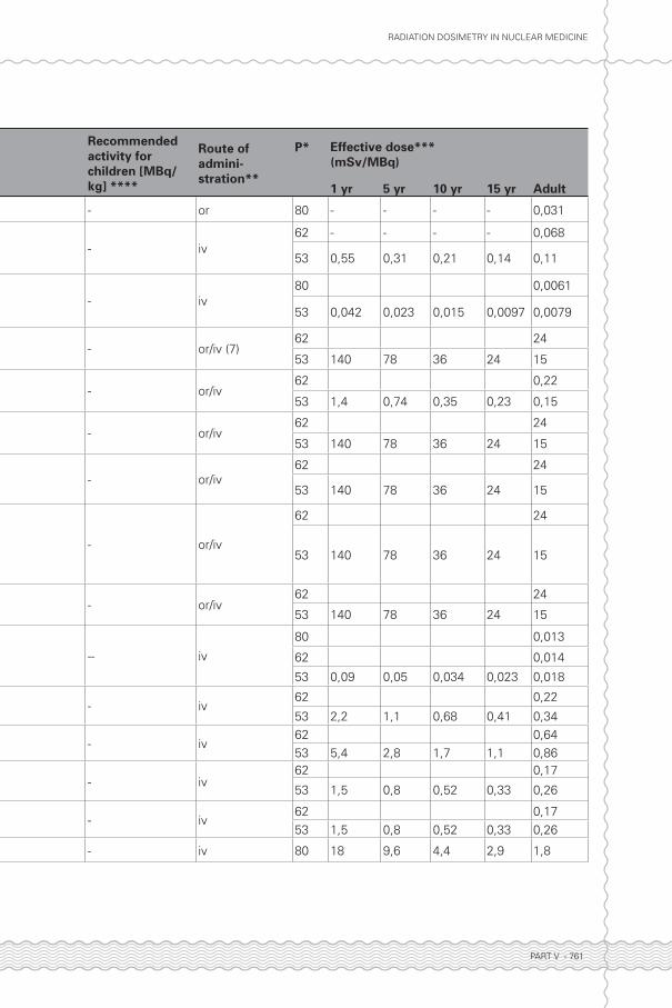
the activity for adults as recommended in the protocol. Column 4 gives the recommended activity for children, if available in the protocol. In general, it is recommended to use the latest version of the EANM paediatric dosage card (to be downloaded from www.eanm.org). Column 5 gives the route of administration. Column 6 gives the reference in which the data were published (e.g. ICRP publication 53, 62, 80 or 106). Note that effective doses from different publications can be based on different tissue weighting factors. Due to better measurements and/or models, tissue-weighting factors have changed between the IRCP publications 26 (1977), 60 (1990) and 103 (2007), most importantly for the gonads where a decrease from 0,25 to 0,20 to 0,08 was effected over the course of time. Also the weighting factor for the breast has changed: from 0,15 to 0,05 to 0,12. Effective doses in ICRP62 and ICRP80 are based on the tissue weighting factors of ICRP60, whereas those in ICRP53 are based on tissue weighting factors from ICRP26. The updated tissue weighting factors as published in ICRP103 have not yet led to a complete re-evaluation of the effective doses for the radiopharmaceuticals of ICRP80. ICRP106 (2008) lists effective dose values for a limited number of radiopharmaceuticals not yet described in earlier publications using the tissue weighting factors of ICRP103, but does not give an update for all other radiopharmaceuticals published earlier (ICRP80, ICRP62, and ICRP53).
Columns 7-11 give the effective dose per MBq of administered radiopharmaceutical for patients of different ages. It should be mentioned that for several radiopharmaceuticals, the age-dependent doses were taken from ICRP53, whereas effective doses for adults are from ICRP62, ICRP80, or ICRP106. The reason is that for these radiopharmaceuticals, age dependence was not included in the more recent ICRP publications. As mentioned earlier, please note that different tissue weighting factors were used in the different publications. Finally, please note the difference in wording: “effective dose equivalent” (ICRP53) versus “effective dose” (ICRP60), which refl ects the different tissue weighting factors (and also the use of different phantoms). The authors of this chapter have striven to present the most recently published dose values for all cases. Finally, column 12 gives the effective doses for adults when administering the recommended amount of activity.
It should be noted that table 1 is restricted to effective doses. Individual organ doses are available for many radiopharmaceuticals (see, e.g. ICRP53, ICRP62, ICRP80, ICRP106). but are not presented here. Furthermore, calculations are based on the assumption that the radiation energy absorbed by the organs is homogenous; micro-dosimetric effects are not taken into account. More information about the calculation methods is given in the references.
A factor which can highly infl uence doses is the micturition interval used in the models, which is mostly taken as 3,5 h for adults, but is shorter for children (3 h for a 5 y old child, 2 h for a 1 y old child and newborns (ICRP106). In general, good hydration and frequent micturition can reduce radiation dose considerably. The effective doses in individual patients may vary signifi cantly from the values shown in table 1, due to uncertainties of the quantitative description of uptake, distribution and retention of radiopharmaceuticals, and in radiation transport and absorption calculations and phantoms used. ICRP53 states that values can vary by up to a factor of 2.
Part V.indd 753 27-12-16 14:40
RADIATION DOSIMETRY IN NUCLEAR MEDICINE
PART V - 754
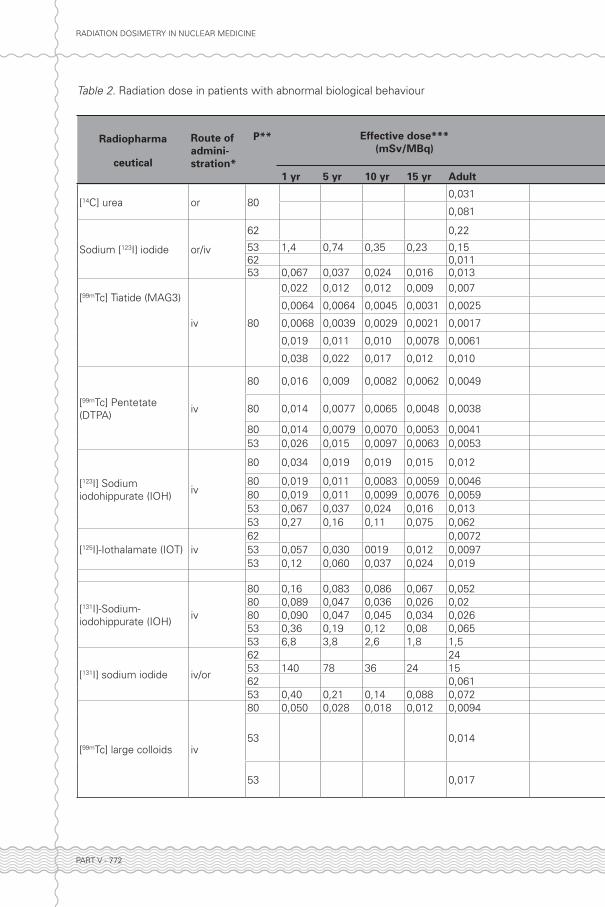
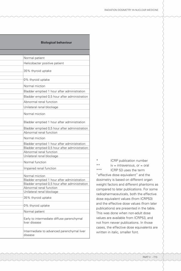
1.2 Effective dose in patients with deviating biokinetics and biodistributionTable 2 presents dosimetric data for patients with deviating biokinetics and biodistribution for a selected number of protocols and radiopharmaceuticals.
1.3 Radiopharmaceuticals administered via local routesThe absorbed dose for radiopharmaceuticals administered via other, local routes (that is, other than intravenously or orally) depends largely on their biodistribution and their clearance rate from the region of accumulation. It is not always possible to provide fi gures for the effective dose given the limited yet widely varying aspects of local accumulation. In general, such examinations involve small amounts of radioactivity, see table 1, resulting in low radiation doses.
1.4 Injection site extravasationExtravasation of radioactive material at the site of injection results in a high radiation dose to that area. The radiation dose depends on the amount of radioactivity and the volume of fl uid containing the radioactivity, but also the rate at which the material is cleared. Massage can help to reduce the radiation dose at the injection site. The effective half-life of extravasated material around the injection site is generally short (approximately 2 h), but may be longer (8-10 h) if the material is extravasated at a site that is poorly vascularised. In order to gain some idea of the clearance time, the half-life can be roughly calculated by measuring the activity at the injection site several times during the fi rst 24 h under standardised circumstances using a gamma camera, a scintillation counter or a radiation monitor. Extravasation of injection fl uid may result in the radiopharmaceutical having to be re-injected (at a later stage) for the same investigation.
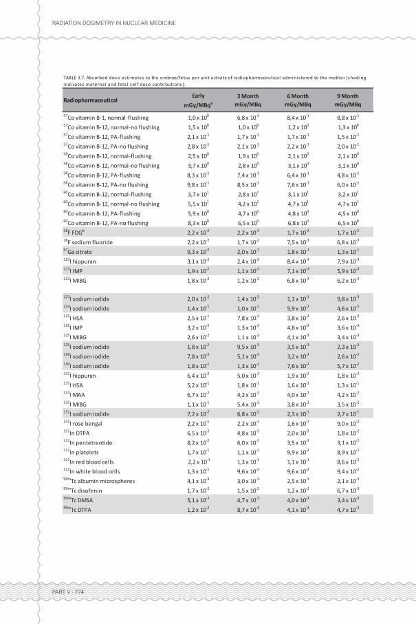
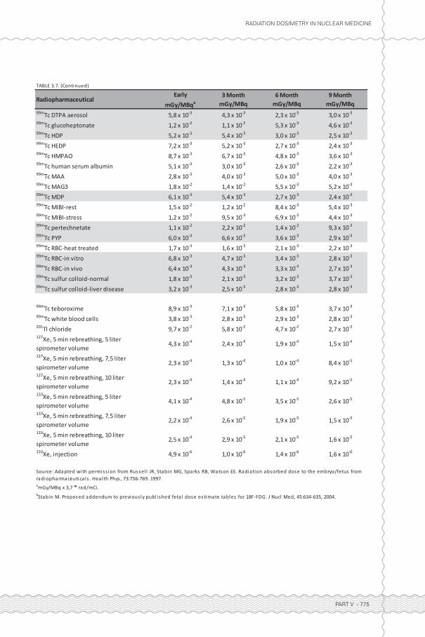
2. Foetuses 2.1 Dose to foetus in a pregnant woman undergoing administration of a radiopharmaceuticalThe absorbed dose to the embryo or foetus has long been an area of concern. The use of pregnant female phantom series has enabled the estimation of absorbed doses for the foetus in early pregnancy and at 3, 6, and 9 months gestation possible. Table 3 shows these doses as calculated by Russell et al for radiopharmaceuticals which might be given, whether intentionally or not, to women of childbearing age. Biokinetic data for these radiopharmaceuticals were gathered from various documents and other resources. The absorbed doses to the embryo and foetus at these different stages of gestation from radiation originating within the mother’s organs were then estimated. In addition, information about activity distributed within the placenta and foetus was included where quantitative data were available.
In many cases, bladder activity will comprise a relatively large proportion of the total radiation dose to the foetus. Extra micturition can reduce this dose signifi cantly, and should therefore be advised.
According to the Dutch Health Council, there is no reason to terminate a pregnancy when the foetal dose is lower than 100 mGy. However, doses above 500 mGy can induce considerable damage to the health of the unborn child. Based on the data of table 3,
Part V.indd 754 27-12-16 14:40
RADIATION DOSIMETRY IN NUCLEAR MEDICINE
PART V - 755
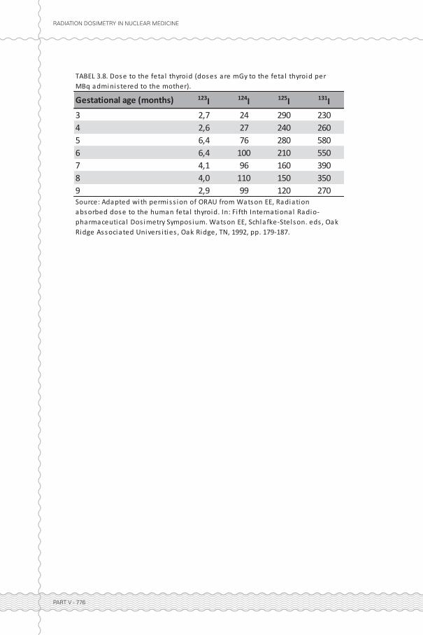
it is concluded that for most diagnostic administrations, the value of 100 mGy will not be reached. Further details with regard to the effects of radiation on the foetus with a subdivision into stochastic and deterministic effects for several stages of pregnancy, can be found in “Dutch Health Council”. When administering radiation to a woman who is over 10-13 weeks gestation. Special attention should be given to the foetal thyroid.
After this period, the foetal thyroid has already been formed, and concentrates iodine that crosses the placenta. Foetal thyroid dose for 123I, 124I, 125I and I 131I administered to the mother at 3-9 months of gestation are presented in Table 4. It should be mentioned that a period of 10-13 weeks of pregnancy will generally not be unnoticed. This implies that a careful trade-off between interruption of pregnancy and necessity of treatment or diagnosis of the pregnant patient can be made. Doses to the foetus from hyperthyroid and athyroid subjects for 131I can be found in, tables 3.9 and 3.10]
When administering therapeutic doses of radiopharmaceuticals to pregnant patient, much care should be taken. Recommendations for pregnant women are given in. The most important advice for 131I therapy being the prevention of pregnancy during the fi rst 4 months after therapy. More details can be found in.
2.2 Dose to foetus in pregnant hospital personnel, dose limit for pregnant hospital personnelAccording to section 80, paragraph 1 of the 2012 Radiation Protection Act (RPA), a woman may continue to work with ionizing radiation during her pregnancy as long as the effective dose to the unborn child is as low as reasonably achievable and this dose, calculated as of the date the pregnancy is fi rst reported, does not exceed 1mSv during the rest of the pregnancy. The risk assessment outlined in section 10 of the RPA describes the type of work for which this applies. In addition to the obligatory dosimetry, supplementary dosimetry may be carried out in order to determine whether these requirements can be met. In practice, this must be assessed for each individual situation. For instance, it could be essential to carry an electronic personal dosimeter with direct dose reading instead of a thermoluminescent detector that provides cumulative doses only.
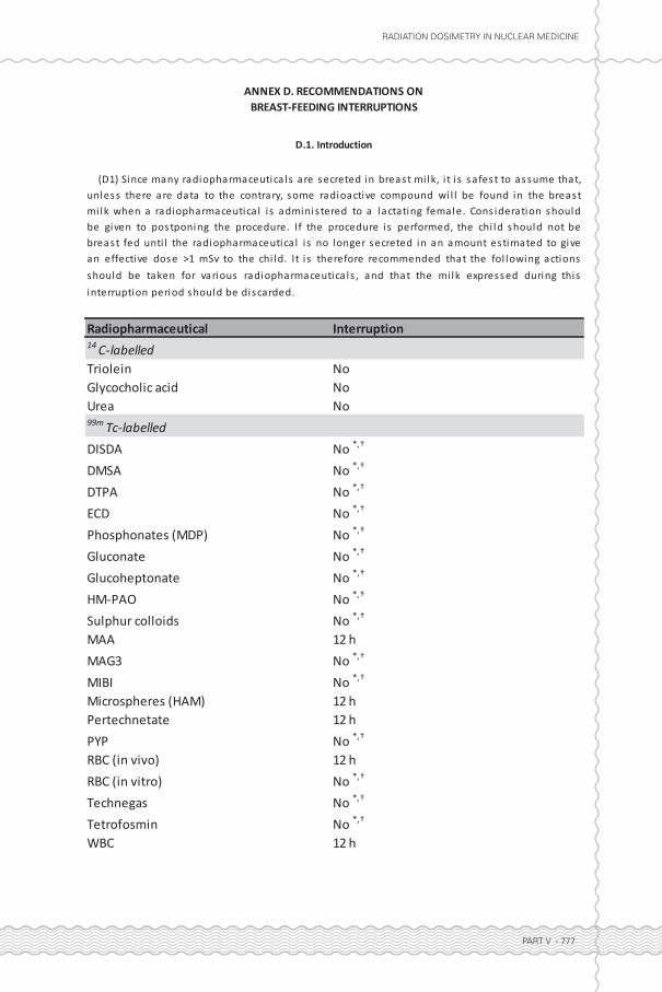
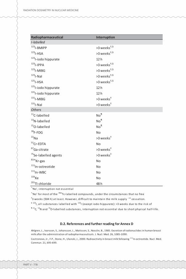
3. InfantsIf a radiopharmaceutical is administered to a breastfeeding mother, the child may be exposed to radiation through internal contamination by breast milk or by external irradiation during direct contact. For radionuclide therapy, any breastfeeding is prohibited: “breast feeding must be stopped before therapy and not be resumed after discharge from the hospital”. The following considerations hold for diagnostic examinations: 3.1 Internal contaminationIf a patient is breast-feeding, it is of interest how long breast-feeding should be interrupted (if at all) to protect the nursing infant. The most recent recommendations are given in table 5 (ICRP106). The radiation dose from internal contamination can be limited by expressing milk with a breast pump and storing it in a refrigerator until the radioactivity has decayed suffi ciently. Otherwise, breast-feeding should be temporarily discontinued and replaced by formula feed.
Part V.indd 755 27-12-16 14:40
RADIATION DOSIMETRY IN NUCLEAR MEDICINE
PART V - 756
3.2 External radiationThe radiation dose to which infants are exposed due to external radiation depends on the radiopharmaceutical used and its bio-distribution. Highest doses will of course be received from body parts of the mother that are close to the breasts. Not much literature data is available pertaining to this issue. However, references exist for 99mTc macroaggregates (lung scintigraphy), and for FDG. Both papers conclude that the external radiation dose from breasts and other body part close to the infant is higher than the dose from the ingested milk. This implies that in case of doubt, one could use expressed milk. Depending on the internal dose associated with the ingestion of expressed milk, one could use it to feed the child immediately (e.g. by the father or other person), or store the breast milk in a refrigerator as mentioned in the previous section.
3.3 Internal contamination of breastfeeding hospital personnelSection 80, paragraph 2, of the RPA states that employers must ensure any staff member who is breastfeeding is exempted from activities which pose a greater than negligible risk of radioactive contamination of the body. In the explanatory notes to the RPA, work with open sources is limited to two sources for which the total activity, and activity concentration, is equal to and no greater than the source strength and concentration, as indicated in the exclusion and exemption levels in table 1 of the RPA. These limits are extremely rigorous and are currently under discussion. The new version of the Dutch Decree on Radiation Protection from January 2014 states a less severe requirement: The employer must ensure that an employee who has mentioned that she is breastfeeding, does not perform work for which, based on risk analysis, a relevant risk is present for radioactive contamination of the body
4. Household members, third partiesFollowing administration of the radiopharmaceutical, patients become a temporary source of radiation to their environment due to the radiation emitted by the patients themselves and possibly by contamination through radioactive excreta. Contamination through excreta is, however, negligible if good hygiene practices are adhered to. The dose received through direct exposure to the radiation depends on the type and energy of the radiation, the amount of activity administered, and the effective (that is, biological and physical) half-life of the radiopharmaceutical. Precautions for the public are rarely required after diagnostic nuclear medicine procedures, as indicated in ICRP103. Examples of dose rates are given in IRCP68 for bone and liver scintigraphy, and blood pool determination using 99mTc coupled MDP, colloid and RBC respectively, and myocardial scintigraphy using 201Tl. Absorbed dose rates are around 10 nGy/MBq at a distance of 30 cm from to the patient, and are well below 10 nGy/MBq for distances of 1 m and farther. These numbers hold immediately after administration and for 2 h after administration, and will decrease in time due to physical decay and biological clearance. Given typically administered activities of several hundreds of MBq, dose rates will be several µGy/h at 30 cm and <1 µGy/h at 1 m from the patient. In view of the generally short half-lives of diagnostic radionuclides, the doses to household members and third parties can be neglected compared to the background radiation, which is around 2 mSv/y in the Netherlands.
When using ionizing radiation, the radiation dose to everyone should be kept as low as
Part V.indd 756 27-12-16 14:40
RADIATION DOSIMETRY IN NUCLEAR MEDICINE
PART V - 757
reasonably achievable. The Nuclear Energy Act (Decree on Radiation Protection) stipulates a maximum permitted dose such that the risks as a result of exposure to this radiation be negligible. This maximum dose is 1 mSv/y for members of the public. The same limit applies to the unborn child. Given the fact that the radiation dose to which household members and third parties are exposed is negligible compared to background radiation, measures to limit the contact between patient and household members (including pregnant women) are not required.
Please note that this holds for diagnostic examinations. On the contrary, rules are recommended for household members of patients who undergo radionuclide therapy. ICRP94] provides recommendations for the release of patients after radionuclide therapy. These recommendations include that young children and infants, as well as visitors not engaged in direct care or comforting, should be treated as members of the public for radiological protection purposes (i.e., be subject to the public dose limit of 1 mSv/y). For individuals directly involved in comforting and caring, other than young children and infants, a dose constraint of 5 mSv per episode (i.e., for the duration of a given release after therapy) is reasonable. The constraint needs to be used fl exibly. For example, higher doses may well be appropriate for parents of very sick children.
Recent developments in diagnostic investigations show the use of longer-lived PET nuclides such as 89 Zr and 124I. This led to a discussion on the need to provide rules for household members in these cases. At the time of writing no consensus has been reached.
The cornerstones of release criteria are dose limits for the public and dose constraints for relatives and caregivers. In spite of this, there is wide variation in criteria used to decide whether to release or hospitalise patients. At present, the two general forms of release criteria are those based on individual situations and projected doses to other people, and those based on retained activity (usually following conservative assumptions). It is interesting to note that the ICRP has not provided recommendations on the criteria to follow regarding the release of patients after radionuclide therapy. Instead, the recommendations have been directed at dose limits for occupationally exposed workers in hospitals, dose limits for the public, and dose constraints for caregivers. Thus, the ICRP has not set any retained activity level to require hospitalisation. A patient may be discharged regardless of the magnitude of retained activity provided that the dose limit and dose constraint issues are met.
Specifi cations of release criteria in the Netherlands for patients after radionuclide therapy can be found in (Het werken met therapeutische doses radionucliden (VROM, SZW, NVNG)). ICRP94 provides an overview of recommendations given in several countries and by several national and international organizations.
5. Bone densitometryDual energy x-ray absorption (DEXA) using x-ray sources of different energy (e.g. 100 and 140 keVp) is used for bone density measurements. As mentioned before, for historical reasons this kind of equipment is often operated by nuclear medicine departments
Part V.indd 757 27-12-16 14:40

RADIATION DOSIMETRY IN NUCLEAR MEDICINE
PART V - 758
instead of radiology departments. The dose to the patient depends on factors including source energy, tube current, scan speed, bundle size, size of scan area and the radiation fi lters used. Manufacturers of DEXA scanners usually specify skin entrance doses (mGy) for the protocols provided with their scanners. However, in dosimetric considerations, organ doses and effective doses with tissue weighting factors taken into account, should be available. Blake et al have provided organ doses and effective doses for hip, spine and total body DEXA scans. A physical phantom (Rando) was used to obtain depth dose data, which were mapped to the set of mathematical phantoms developed by Christy for adults and children of 5, 10 and 15 years old. Tissue weighting factors were taken from ICRP60. Although these doses were obtained for a specifi c scanner (Hologic Discovery), it may be assumed that doses obtained using other scanners will be in the same order of magnitude. Without going into all details of, it can be concluded that patient doses associated with DEXA scans are very low, the highest reported effective doses being in the order of 50 µSv, which is 34 times smaller than the yearly, background radiation in the Netherlands (around 2 mSv/y). 6. Low dose CT in PET/CT or SPECT/CT scansFor the purpose of PET or SPECT attenuation correction and for anatomical localization, low dose CT scans are made in conjunction with these nuclear scans. For these scans, CT settings resulting in lower doses than for high quality, diagnostic CT scans can be used.
Most CT scanners have the option to adapt the tube current setting (mA) to the total attenuation encountered in different body parts during the scan. That is, e.g. lungs, head, legs are scanned using lower mA values than e.g. the abdomen, where attenuation is less, thus lowering the total patient dose. Recently, modulation of the kV settings has also become available for several types of CT scanners.
Furthermore, iterative CT reconstruction techniques are available for most modern CT scanners. This allows to further reduce mA and/or kV settings whilst maintaining image quality. At present it is not yet clear as to how far the CT dose in PET/CT and SPECT/CT can be lowered using a combination of all available techniques.
Low dose CT scans contribute to the total effective dose of the multi-modality scan depending on the acquisition protocol being used by the institute which will in general be a locally determined optimum between low-dose and image quality. The effective dose resulting from the low dose CT scan may be calculated using the excel sheets provided by ImPACT. Since the number of different acquisition protocols is usually limited (e.g. low-dose CT for a whole-body PET/CT and low-dose CT for brain PET/CT), the parameters of these low dose CT scanning protocols need to be entered into the excel sheets only once. The dosimetry of stand-alone CT usage of a nuclear/CT camera, such as diagnostic CT, is beyond the scope of these recommendations and are at the time of writing usually performed under responsibility of radiology.
Part V.indd 758 27-12-16 14:40
RADIATION DOSIMETRY IN NUCLEAR MEDICINE
PART V - 759
7. References• ICRP, 1988. Radiation Dose to Patients from Radiopharmaceuticals. ICRP Publication 53. Ann. ICRP 18
(1-4).
• ICRP, 1992. Radiological Protection in Biomedical Research. ICRP Publication 62. Ann. ICRP 22 (3).
• ICRP, 1998. Radiation Dose to Patients from Radiopharmaceuticals (Addendum to ICRP Publication 53).
ICRP Publication 80. Ann. ICRP 28 (3).
• ICRP, 2008. Radiation Dose to Patients from Radiopharmaceuticals - Addendum 3 to ICRP Publication
53. ICRP Publication 106. Ann. ICRP 38 (1-2).
• ICRP, 1977. Recommendations of the ICRP. ICRP Publication 26. Ann. ICRP 1 (3).
• ICRP, 1991. 1990 Recommendations of the International Commission on Radiological Protection. ICRP
Publication 60. Ann. ICRP 21 (1-3).
• ICRP, 2007. The 2007 Recommendations of the International Commission on Radiological Protection.
ICRP Publication 103. Ann. ICRP 37 (2-4).
• Russell et al, Radiation absorbed dose to the embryo/fetus from radiopharmaceuticals. Health Phys.
1997 Nov;73[5]:756-69
• Health Council of the Netherlands. Risks of exposure to ionising radiation. The Hague: Health Council of
the Netherlands, 2007; publication no. 2007/03. ISBN 978-90-5549-633-4
• Stabin, Fundamentals of Nuclear Medicine Dosimetry, Springer 2008.
• VROM 2004, Aanbevelingen: Het werken met therapeutische doses radionucliden
• Besluit Stralingsbescherming 2002
• Berke et al, Radiation dose to breast-feeding child. J Nucl Med. 1973 Jan;14(1):51-2
• Hicks et al, Pattern of uptake and excretion of (18)F-FDG in the lactating breast. J Nucl Med. 2001
Aug;42(8):1238-42
• Besluit Stralingsbescherming 2014
• ICRP, 1994. Dose Coeffi cients for Intakes of Radionuclides by Workers. ICRP Publication 68. Ann. ICRP
24 (4).
• ICRP, 2004. Release of Patients after Therapy with Unsealed Radionuclides. ICRP Publication 94. Ann.
ICRP 34 (2)
• Blake et al, Comparison of effective dose to children and adults from dual X-ray absorptiometry
examinations. Bone 2006 Jun;38(6):935-42
• Christy M, Eckerman KF (1987) Specifi c absorbed fractions of energy at various ages from internal
photon sources. ORNL/TM 8381/V1-V7, Oak Ridge National Laboratory
• ImPACT CT dosimetry tool: http://www.impactscan.org/ctdosimetry.htm
Part V.indd 759 27-12-16 14:40
RADIATION DOSIMETRY IN NUCLEAR MEDICINE
PART V - 760
Table 1. Radiation dose in patients with normal biological behaviour
Protocol, page number RadiopharmaceuticalRecommended activity for adults [MBq]
Recommended activity for children [MBq/kg] ****
Route of admini-stration**
P* Effective dose***(mSv/MBq)
1 yr 5 yr 10 yr 15 yr Adult
C-14-urea breath test [p.330] [14C] urea 0,2 - or 80 - - - - 0,031
Detection of protein loss through the intestin wall [p.311]
[51Cr] chloride 1,2 - iv62 - - - - 0,068
53 0,55 0,31 0,21 0,14 0,11
Diagnosis of gastrointestinal protein loss (localisation using 99m Tc HSA) [p.308]
[99mTc] HSA 500 - 740 - iv80 0,0061
53 0,042 0,023 0,015 0,0097 0,0079
Iodine Total-Body Scintigraphy [p.47] Sodium [131I] iodide (4)Pre-ablation scintigram: 40–80
- or/iv (7)62 24
53 140 78 36 24 15
Iodine Total-Body Scintigraphy [p.47] Sodium [131I] iodide (4)Pre-ablation scintigram: 185-370
- or/iv62 0,22
53 1,4 0,74 0,35 0,23 0,15
Iodine Total-Body Scintigraphy [p.47] Sodium [131I] iodide (4)Post ablation follow up: 80–185
- or/iv62 24
53 140 78 36 24 15
Iodine Total-Body Scintigraphy [p.47] Sodium [131I] iodide (4)Post therapy scintigram after ablation: 1100–3700
- or/iv62 24
53 140 78 36 24 15
Iodine Total-Body Scintigraphy [p.47] Sodium [131I] iodide (4)
In locoregional metastases or non-radical resection of primary tumour: 3700-7400
- or/iv
62 24
53 140 78 36 24 15
Iodine Total-Body Scintigraphy [p.47] Sodium [131I] iodide (4)In remote metastases 5550-7400
- or/iv62 24
53 140 78 36 24 15
MIBG cardiac sympathetic imaging [p.238]
[123I] MIBG 185 -- iv
80 0,013
62 0,014
53 0,09 0,05 0,034 0,023 0,018
Erythrocyte and Plasma Volume Measurement [p.161]
[125I] albumin1,3 kBq/kg body weight.
- iv62 0,22
53 2,2 1,1 0,68 0,41 0,34
Erythrocyte and Plasma Volume Measurement [p.161]
[131I] albumin1,3 kBq/kg body weight.
- iv62 0,64
53 5,4 2,8 1,7 1,1 0,86
Erythrocyte and Plasma Volume Measurement [p.161]
[51Cr] autologic erythrocytes
5,1 kBq/kg body weight
- iv62 0,17
53 1,5 0,8 0,52 0,33 0,26
Erythrocyte Survival Time [p.167][51Cr] autologic erythrocytes
1,2 - iv62 0,17
53 1,5 0,8 0,52 0,33 0,26
Adrenal cortex scintigraphy [p.71] [131I] cholesterol 20 - iv 80 18 9,6 4,4 2,9 1,8
Part V.indd 760 27-12-16 14:40
RADIATION DOSIMETRY IN NUCLEAR MEDICINE
PART V - 761
Table 1. Radiation dose in patients with normal biological behaviour
Protocol, page number RadiopharmaceuticalRecommended activity for adults [MBq]
Recommended activity for children [MBq/kg] ****
Route of admini-stration**
P* Effective dose***(mSv/MBq)
1 yr 5 yr 10 yr 15 yr Adult
C-14-urea breath test [p.330] [14C] urea 0,2 - or 80 - - - - 0,031
Detection of protein loss through the intestin wall [p.311]
[51Cr] chloride 1,2 - iv62 - - - - 0,068
53 0,55 0,31 0,21 0,14 0,11
Diagnosis of gastrointestinal protein loss (localisation using 99m Tc HSA) [p.308]
[99mTc] HSA 500 - 740 - iv80 0,0061
53 0,042 0,023 0,015 0,0097 0,0079
Iodine Total-Body Scintigraphy [p.47] Sodium [131I] iodide (4)Pre-ablation scintigram: 40–80
- or/iv (7)62 24
53 140 78 36 24 15
Iodine Total-Body Scintigraphy [p.47] Sodium [131I] iodide (4)Pre-ablation scintigram: 185-370
- or/iv62 0,22
53 1,4 0,74 0,35 0,23 0,15
Iodine Total-Body Scintigraphy [p.47] Sodium [131I] iodide (4)Post ablation follow up: 80–185
- or/iv62 24
53 140 78 36 24 15
Iodine Total-Body Scintigraphy [p.47] Sodium [131I] iodide (4)Post therapy scintigram after ablation: 1100–3700
- or/iv62 24
53 140 78 36 24 15
Iodine Total-Body Scintigraphy [p.47] Sodium [131I] iodide (4)
In locoregional metastases or non-radical resection of primary tumour: 3700-7400
- or/iv
62 24
53 140 78 36 24 15
Iodine Total-Body Scintigraphy [p.47] Sodium [131I] iodide (4)In remote metastases 5550-7400
- or/iv62 24
53 140 78 36 24 15
MIBG cardiac sympathetic imaging [p.238]
[123I] MIBG 185 -- iv
80 0,013
62 0,014
53 0,09 0,05 0,034 0,023 0,018
Erythrocyte and Plasma Volume Measurement [p.161]
[125I] albumin1,3 kBq/kg body weight.
- iv62 0,22
53 2,2 1,1 0,68 0,41 0,34
Erythrocyte and Plasma Volume Measurement [p.161]
[131I] albumin1,3 kBq/kg body weight.
- iv62 0,64
53 5,4 2,8 1,7 1,1 0,86
Erythrocyte and Plasma Volume Measurement [p.161]
[51Cr] autologic erythrocytes
5,1 kBq/kg body weight
- iv62 0,17
53 1,5 0,8 0,52 0,33 0,26
Erythrocyte Survival Time [p.167][51Cr] autologic erythrocytes
1,2 - iv62 0,17
53 1,5 0,8 0,52 0,33 0,26
Adrenal cortex scintigraphy [p.71] [131I] cholesterol 20 - iv 80 18 9,6 4,4 2,9 1,8
Part V.indd 761 27-12-16 14:40
RADIATION DOSIMETRY IN NUCLEAR MEDICINE
PART V - 762
Protocol, page number RadiopharmaceuticalRecommended activity for adults [MBq]
Recommended activity for children [MBq/kg] ****
Route of admini-stration**
P* Effective dose***(mSv/MBq)
1 yr 5 yr 10 yr 15 yr Adult131I Therapy in primary hyperthyroidism and non-toxic (multi)nodular goiter [p.372]
Sodium [131I] iodide3,7-11,1 MBq/ml or fi xed activities 370 – 740 MBq
- or /iv62 24
53 140 78 36 25 15
131I Therapy for the treatment of differentiated thyroid carcinoma [p.384]
Sodium [131I] iodide Ablation: 1100-1850 - or /iv62 24
53 140 78 36 25 15
131I Therapy for the treatment of differentiated thyroid carcinoma [p.384]
Sodium [131I] iodideT3-T4 tumours: 3700 – 7400
- or /iv62 24
53 140 78 36 25 15
131I Therapy for the treatment of differentiated thyroid carcinoma [p.384]
Sodium [131I] iodideMetastases after ablation: 5550-7400
- or /iv62 24
53 140 78 36 25 15
MIBG Scintigraphy [p.59] [131I] MIBG 40–80 - iv 62 0,1453 1,1 0,61 0,4 0,26 0,2
MIBG Scintigraphy [p.59] [131I] MIBGpost treatmanent scintigraphy: 3700–7400
- iv62 0,14
53 1,1 0,61 0,4 0,26 0,2
MIBG Scintigraphy [p.59] [123I] MIBG 185 - 370
Scaled to body weight according to EANM Paediatric Task Group: 80–400 MBq)
iv 80 0,068 0,037 0,026 0,017 0,013
Radionuclide Cisternography [p.14] [111 In] DTPA20 (4 in case of evaluation of drainage system)
-lumbar injection 53 0,14cisternal injection
53 0,12
Bile acid malabsorption test [p.314] [75Se] HCAT0,04 (faecal excretion test)
0,4 (camera test)- iv 80 3,9 2,0 1,3 0,86 0,69
Liver and spleen scintigraphy [p.323] [99m Tc] large colloids 80 - iv 80 0,050 0,028 0,018 0,012 0,0094
Oesophageal scintigraphy [p.288] [99m Tc] colloid (liquid) 5 x 10 -- or 80 0,11 0,062 0,039 0,025 0,019
Gastric emptying study [p.291][99m Tc] colloid (solid , in pancake or milk pap)
10 -- or 800,14 (s)
0,11 (l)
0,076 (s)
0,062 (l)
0,048 (s)
0,039 (l)
0,031 (s)
0,025 (l)
0,024 (s)
0,019 (l)
Cholescintigraphy [p.334] [99m Tc] MebrofeninSee protocol, several activities
See protocol, several activities
iv 800,10 0,045 0,029 0,021 0,017
Renal cortical scintigraphy/DMSA scan [p.341]
[99m Tc] DMSA 80 - 110 - iv 80 0,037 0,021 0,015 0,011 0,0088
Scintigraphy of gastrointestinal tract bleeding [p.300]
[99m Tc] erythrocytes 750 - iv 80 0,039 0,021 0,014 0,0089 0,0070
Hepatic hemangioma scintigraphy [p.327]
[99m Tc] autologous erythrocytes
500 - iv 80 0,039 0,021 0,014 0,0089 0,0070
Spleen Scintigraphy Using Denatured Erythrocytes [p.158]
[99m Tc] denatured RBCs 80 - iv62 0,01953 0,22 0,13 0,084 0,056 0,041
Regional Cerebral Blood Perfusion Scan [p.18]
[99m Tc] exametazine (HMPAO)
500 - iv 80 0,049 0,027 0,017 0,011 0,0093
Regional Cerebral Blood Perfusion Scan [p.18]
[99m Tc]-ethyl cysteinate dimer (ECD)
500 - iv 106 0,04 0,022 0,015 0,0099 0,0077
Part V.indd 762 27-12-16 14:40
RADIATION DOSIMETRY IN NUCLEAR MEDICINE
PART V - 763
Protocol, page number RadiopharmaceuticalRecommended activity for adults [MBq]
Recommended activity for children [MBq/kg] ****
Route of admini-stration**
P* Effective dose***(mSv/MBq)
1 yr 5 yr 10 yr 15 yr Adult131I Therapy in primary hyperthyroidism and non-toxic (multi)nodular goiter [p.372]
Sodium [131I] iodide3,7-11,1 MBq/ml or fi xed activities 370 – 740 MBq
- or /iv62 24
53 140 78 36 25 15
131I Therapy for the treatment of differentiated thyroid carcinoma [p.384]
Sodium [131I] iodide Ablation: 1100-1850 - or /iv62 24
53 140 78 36 25 15
131I Therapy for the treatment of differentiated thyroid carcinoma [p.384]
Sodium [131I] iodideT3-T4 tumours: 3700 – 7400
- or /iv62 24
53 140 78 36 25 15
131I Therapy for the treatment of differentiated thyroid carcinoma [p.384]
Sodium [131I] iodideMetastases after ablation: 5550-7400
- or /iv62 24
53 140 78 36 25 15
MIBG Scintigraphy [p.59] [131I] MIBG 40–80 - iv 62 0,1453 1,1 0,61 0,4 0,26 0,2
MIBG Scintigraphy [p.59] [131I] MIBGpost treatmanent scintigraphy: 3700–7400
- iv62 0,14
53 1,1 0,61 0,4 0,26 0,2
MIBG Scintigraphy [p.59] [123I] MIBG 185 - 370
Scaled to body weight according to EANM Paediatric Task Group: 80–400 MBq)
iv 80 0,068 0,037 0,026 0,017 0,013
Radionuclide Cisternography [p.14] [111 In] DTPA20 (4 in case of evaluation of drainage system)
-lumbar injection 53 0,14cisternal injection
53 0,12
Bile acid malabsorption test [p.314] [75Se] HCAT0,04 (faecal excretion test)
0,4 (camera test)- iv 80 3,9 2,0 1,3 0,86 0,69
Liver and spleen scintigraphy [p.323] [99m Tc] large colloids 80 - iv 80 0,050 0,028 0,018 0,012 0,0094
Oesophageal scintigraphy [p.288] [99m Tc] colloid (liquid) 5 x 10 -- or 80 0,11 0,062 0,039 0,025 0,019
Gastric emptying study [p.291][99m Tc] colloid (solid , in pancake or milk pap)
10 -- or 800,14 (s)
0,11 (l)
0,076 (s)
0,062 (l)
0,048 (s)
0,039 (l)
0,031 (s)
0,025 (l)
0,024 (s)
0,019 (l)
Cholescintigraphy [p.334] [99m Tc] MebrofeninSee protocol, several activities
See protocol, several activities
iv 800,10 0,045 0,029 0,021 0,017
Renal cortical scintigraphy/DMSA scan [p.341]
[99m Tc] DMSA 80 - 110 - iv 80 0,037 0,021 0,015 0,011 0,0088
Scintigraphy of gastrointestinal tract bleeding [p.300]
[99m Tc] erythrocytes 750 - iv 80 0,039 0,021 0,014 0,0089 0,0070
Hepatic hemangioma scintigraphy [p.327]
[99m Tc] autologous erythrocytes
500 - iv 80 0,039 0,021 0,014 0,0089 0,0070
Spleen Scintigraphy Using Denatured Erythrocytes [p.158]
[99m Tc] denatured RBCs 80 - iv62 0,01953 0,22 0,13 0,084 0,056 0,041
Regional Cerebral Blood Perfusion Scan [p.18]
[99m Tc] exametazine (HMPAO)
500 - iv 80 0,049 0,027 0,017 0,011 0,0093
Regional Cerebral Blood Perfusion Scan [p.18]
[99m Tc]-ethyl cysteinate dimer (ECD)
500 - iv 106 0,04 0,022 0,015 0,0099 0,0077
Part V.indd 763 27-12-16 14:40

RADIATION DOSIMETRY IN NUCLEAR MEDICINE
PART V - 764
Protocol, page number RadiopharmaceuticalRecommended activity for adults [MBq]
Recommended activity for children [MBq/kg] ****
Route of admini-stration**
P* Effective dose***(mSv/MBq)
1 yr 5 yr 10 yr 15 yr Adult
Leukocyte scintigraphy [p.171][99m Tc] exametazine (HMPAO) labeled leukocytes
370 - 740 - iv 80 0,062 0,034 0,022 0,014 0,011
Leukocyte scintigraphy [p.171][111 In] oxine labeled
leucocytes 10 – 18,5 - iv
62 0,36
53 3,2 1,8 1,2 0,79 0,59
Salivary gland scintigraphy [p.284][99m Tc] sodium pertechnetate
100 - iv 80 0,079 0,042 0,026 0,017 0,013
Scintigraphy of ectopic gastric mucosa [p.300]
[99m Tc] sodium pertechnetate
200 7 - 10 iv 80 0,079 0,042 0,026 0,017 0,013
Micturition Cystourethrography using Scintigraphy [p.356]
[99m Tc] DTPA 30 - bladder[a] 0,0024
[b] 0,002
Micturition Cystourethrography using Scintigraphy [p.356]
[99m Tc] colloid 30 - bladder[a] 0,0024
[b] 0,002
18F-FDG for infl ammation and infection detection [p.187]
[18F]-FDG - iv 106 0,095 0,056 0,037 0,024 0,019
Myocardial perfusion PET/CT with Rubidium-82 [p.222]
[82Rb] 1110 - 1480 - iv
[c]0,00126 (8)
[d]0,00128 (9)
62 0,003453 0,033 0,018 0,010 0,0067 0,0048
16α-[18F]fl uoro-17β-oestradiol ([18F]FES) in oncology [p.90]
[18F]-FES 200 - iv 106 0,16 0,093 0,066 0,031 0,023
Nasal Mucociliary Clearance [p.280] [99m Tc] nanocolloid 2 -Drop deposited in nose
800,14
0,076 0,048 0,031
0,024
Lymphoscintigraphy of the Upper Extremities [p.145]
[99m Tc] nanocolloid 50 MBq per hand -Subcutaneous injection
0,005
Parathyroid gland scintigraphy [p.52] [99m Tc] labeled sestamibi 500 - 700 - iv 80 0,053 0,028 0,018 0,012 0,009
Parathyroid gland scintigraphy [p.52] [99m Tc] labeled tetrofosmin 500 - 700 - iv Ad4 0,046 0,024 0,015 0,01 0,008
Parathyroid gland scintigraphy [p.52]Dual tracer imaging using sodium [99m Tc] pertechnetate
35-75 - iv 80 0,079 0,042 0,026 0,017 0,013
Parathyroid gland scintigraphy [p.52]Dual tracer imaging using sodium [123I]
7,5 – 22 - iv62 0,2253 1,4 0,74 0,35 0,23 0,15
Thyroid Gland Scintigraphy [p.35][99m Tc] sodium pertechnetate
80-180 - iv 80 0,079 0,042 0,026 0,017 0,013
Thyroid Gland Scintigraphy [p.35] [123I] sodium iodide 10 - 20 3 MBq neonates iv/or 62 0,2253 1,4 0,74 0,35 0,23 0,15
Molecular Breast Imaging (MBI) using breast specifi c gamma cameras (BSGI) [p.85]
[99m Tc] labeled sestamibi 200 - 600 - iv 80 0,053 0,028 0,018 0,012 0,009
Molecular Breast Imaging (MBI) using breast specifi c gamma cameras (BSGI) [p.85]
[99m Tc] labeled tetrofosmin 200 - 600 - iv Ad4 0,046 0,024 0,015 0,01 0,008
Part V.indd 764 27-12-16 14:40
RADIATION DOSIMETRY IN NUCLEAR MEDICINE
PART V - 765
Protocol, page number RadiopharmaceuticalRecommended activity for adults [MBq]
Recommended activity for children [MBq/kg] ****
Route of admini-stration**
P* Effective dose***(mSv/MBq)
1 yr 5 yr 10 yr 15 yr Adult
Leukocyte scintigraphy [p.171][99m Tc] exametazine (HMPAO) labeled leukocytes
370 - 740 - iv 80 0,062 0,034 0,022 0,014 0,011
Leukocyte scintigraphy [p.171][111 In] oxine labeled
leucocytes 10 – 18,5 - iv
62 0,36
53 3,2 1,8 1,2 0,79 0,59
Salivary gland scintigraphy [p.284][99m Tc] sodium pertechnetate
100 - iv 80 0,079 0,042 0,026 0,017 0,013
Scintigraphy of ectopic gastric mucosa [p.300]
[99m Tc] sodium pertechnetate
200 7 - 10 iv 80 0,079 0,042 0,026 0,017 0,013
Micturition Cystourethrography using Scintigraphy [p.356]
[99m Tc] DTPA 30 - bladder[a] 0,0024
[b] 0,002
Micturition Cystourethrography using Scintigraphy [p.356]
[99m Tc] colloid 30 - bladder[a] 0,0024
[b] 0,002
18F-FDG for infl ammation and infection detection [p.187]
[18F]-FDG - iv 106 0,095 0,056 0,037 0,024 0,019
Myocardial perfusion PET/CT with Rubidium-82 [p.222]
[82Rb] 1110 - 1480 - iv
[c]0,00126 (8)
[d]0,00128 (9)
62 0,003453 0,033 0,018 0,010 0,0067 0,0048
16α-[18F]fl uoro-17β-oestradiol ([18F]FES) in oncology [p.90]
[18F]-FES 200 - iv 106 0,16 0,093 0,066 0,031 0,023
Nasal Mucociliary Clearance [p.280] [99m Tc] nanocolloid 2 -Drop deposited in nose
800,14
0,076 0,048 0,031
0,024
Lymphoscintigraphy of the Upper Extremities [p.145]
[99m Tc] nanocolloid 50 MBq per hand -Subcutaneous injection
0,005
Parathyroid gland scintigraphy [p.52] [99m Tc] labeled sestamibi 500 - 700 - iv 80 0,053 0,028 0,018 0,012 0,009
Parathyroid gland scintigraphy [p.52] [99m Tc] labeled tetrofosmin 500 - 700 - iv Ad4 0,046 0,024 0,015 0,01 0,008
Parathyroid gland scintigraphy [p.52]Dual tracer imaging using sodium [99m Tc] pertechnetate
35-75 - iv 80 0,079 0,042 0,026 0,017 0,013
Parathyroid gland scintigraphy [p.52]Dual tracer imaging using sodium [123I]
7,5 – 22 - iv62 0,2253 1,4 0,74 0,35 0,23 0,15
Thyroid Gland Scintigraphy [p.35][99m Tc] sodium pertechnetate
80-180 - iv 80 0,079 0,042 0,026 0,017 0,013
Thyroid Gland Scintigraphy [p.35] [123I] sodium iodide 10 - 20 3 MBq neonates iv/or 62 0,2253 1,4 0,74 0,35 0,23 0,15
Molecular Breast Imaging (MBI) using breast specifi c gamma cameras (BSGI) [p.85]
[99m Tc] labeled sestamibi 200 - 600 - iv 80 0,053 0,028 0,018 0,012 0,009
Molecular Breast Imaging (MBI) using breast specifi c gamma cameras (BSGI) [p.85]
[99m Tc] labeled tetrofosmin 200 - 600 - iv Ad4 0,046 0,024 0,015 0,01 0,008
Part V.indd 765 27-12-16 14:40
RADIATION DOSIMETRY IN NUCLEAR MEDICINE
PART V - 766
Protocol, page number RadiopharmaceuticalRecommended activity for adults [MBq]
Recommended activity for children [MBq/kg] ****
Route of admini-stration**
P* Effective dose***(mSv/MBq)
1 yr 5 yr 10 yr 15 yr AdultFDG-PET-CT of the brain [p.27] [18F]-FDG 150 - 250 2 - 4 iv 106 0,095 0,056 0,037 0,024 0,019
Sentinel node localisation in breast cancer patients [p.75]
[99m Tc]-nanocolloid 40 - 80 -Peritumoral, subdermal, or periareolar
[e] 0,005
P-32 phosphate treatment of myeloproliferative diseases [p.435]
Sodium [32P] phosphate 3,7 (max = 260 MBq) iv/or62 2,4
53 22 10 5,1 3,0 2,2
Dynamic Renography [p.344]
[99m Tc] Tiatide (MAG3)
70 - 100 - iv 80 0,022 0,012 0,012 0,009 0,007
Dynamic Renography [p.344] [99m Tc] Pentetate (DTPA) 150 – 200 - iv 80 0,016 0,009 0,0082 0,0062 0,0049
Dynamic Renography [p.344][123I] Sodium iodohippurate (IOH)
75 - iv 80 0,034 0,019 0,019 0,015 0,012
Measurement of renal function (GFR) [p.352]
[125I-Iothalamate (IOT) 1,5 - iv62 0,0072
53 0,057 0,030 0019 0,012 0,0097
Measurement of renal function (ERPF) [p.352]
[131I]-Sodium-iodohippurate (IOH)
2 - iv 80 0,16 0,083 0,086 0,067 0,052
Lacrimal Scintigraphy [p.31][99m Tc] sodium pertechnetate
4 per eye - Droplets in eye 80 0,079 0,042 0,026 0,017 0,013
Measurement of Iodine Uptake by the Thyroid [p.42]
[131I] sodium iodide 2 - 4 - iv/or62 24
53 140 78 36 25 15
Measurement of Iodine Uptake by the Thyroid [p.42]
[123I] sodium iodide 2 - 4 - iv/or62 0,22
53 1,4 0,74 0,35 0,23 0,15188Re HEDP Therapy for Skeletal Metastases [p.400]
[188Re]-HEDP40 MBq/kg body weight
- iv [f] 0,07
Bone scintigraphy [p.249] [99m Tc] HDP or [99m Tc] PDP
300 – 740
11 – 13 MBq/kg body weight for obese patients
EANM dosage calculator, minimum 40 MBq
iv 80 0,027 0,014 0,011 0,007 0,0057
Myocardial perfusion scintigraphy in rest and stress [p.191]
[99m Tc] labeled sestamibiincreasing from 600 for BMI < 25 to 900 for BMI > 35 (6)
- iv 80
0,053 (8)
0,028 (8)
0,018 (8)0,012 (8)
0,009 (8)
0,045 (9)
0,023 (9)
0,016 (9)0,010 (9)
0,0079 (9)
Myocardial perfusion scintigraphy in rest and stress [p.191]
[99m Tc] labeled tetrofosminincreasing from 600 for BMI < 25 to 900 for BMI > 35 (6)
- ivAd4
0,046 (8)
0,024(8) 0,015(8) 0,01(8) 0,008 (8)
1060,039 ((9)
0,021 (9)
0,013 (9)0,0088 (9)
0,0069 (9)
Myocardial perfusion scintigraphy in rest and stress [p.191]
[201Tl] chloride 74 - 111 - iv 106 1,3 0,79 0,56 0,2 0,14
Somatostatin-receptor scintigraphy with 111 In-pentetreotide [p.65]
[111 In] pentetreotide 200 - iv 80 0,28 0,16 0,10 0,071 0,054
Equilibrium radionuclide angiography / Multi Gated Acquisition [p.211]
[99m Tc]-labeled erythrocytes
500 - 1050 - iv80
0,039 0,021 0,014 0,0089 0,0070
Part V.indd 766 27-12-16 14:40
RADIATION DOSIMETRY IN NUCLEAR MEDICINE
PART V - 767
Protocol, page number RadiopharmaceuticalRecommended activity for adults [MBq]
Recommended activity for children [MBq/kg] ****
Route of admini-stration**
P* Effective dose***(mSv/MBq)
1 yr 5 yr 10 yr 15 yr AdultFDG-PET-CT of the brain [p.27] [18F]-FDG 150 - 250 2 - 4 iv 106 0,095 0,056 0,037 0,024 0,019
Sentinel node localisation in breast cancer patients [p.75]
[99m Tc]-nanocolloid 40 - 80 -Peritumoral, subdermal, or periareolar
[e] 0,005
P-32 phosphate treatment of myeloproliferative diseases [p.435]
Sodium [32P] phosphate 3,7 (max = 260 MBq) iv/or62 2,4
53 22 10 5,1 3,0 2,2
Dynamic Renography [p.344]
[99m Tc] Tiatide (MAG3)
70 - 100 - iv 80 0,022 0,012 0,012 0,009 0,007
Dynamic Renography [p.344] [99m Tc] Pentetate (DTPA) 150 – 200 - iv 80 0,016 0,009 0,0082 0,0062 0,0049
Dynamic Renography [p.344][123I] Sodium iodohippurate (IOH)
75 - iv 80 0,034 0,019 0,019 0,015 0,012
Measurement of renal function (GFR) [p.352]
[125I-Iothalamate (IOT) 1,5 - iv62 0,0072
53 0,057 0,030 0019 0,012 0,0097
Measurement of renal function (ERPF) [p.352]
[131I]-Sodium-iodohippurate (IOH)
2 - iv 80 0,16 0,083 0,086 0,067 0,052
Lacrimal Scintigraphy [p.31][99m Tc] sodium pertechnetate
4 per eye - Droplets in eye 80 0,079 0,042 0,026 0,017 0,013
Measurement of Iodine Uptake by the Thyroid [p.42]
[131I] sodium iodide 2 - 4 - iv/or62 24
53 140 78 36 25 15
Measurement of Iodine Uptake by the Thyroid [p.42]
[123I] sodium iodide 2 - 4 - iv/or62 0,22
53 1,4 0,74 0,35 0,23 0,15188Re HEDP Therapy for Skeletal Metastases [p.400]
[188Re]-HEDP40 MBq/kg body weight
- iv [f] 0,07
Bone scintigraphy [p.249] [99m Tc] HDP or [99m Tc] PDP
300 – 740
11 – 13 MBq/kg body weight for obese patients
EANM dosage calculator, minimum 40 MBq
iv 80 0,027 0,014 0,011 0,007 0,0057
Myocardial perfusion scintigraphy in rest and stress [p.191]
[99m Tc] labeled sestamibiincreasing from 600 for BMI < 25 to 900 for BMI > 35 (6)
- iv 80
0,053 (8)
0,028 (8)
0,018 (8)0,012 (8)
0,009 (8)
0,045 (9)
0,023 (9)
0,016 (9)0,010 (9)
0,0079 (9)
Myocardial perfusion scintigraphy in rest and stress [p.191]
[99m Tc] labeled tetrofosminincreasing from 600 for BMI < 25 to 900 for BMI > 35 (6)
- ivAd4
0,046 (8)
0,024(8) 0,015(8) 0,01(8) 0,008 (8)
1060,039 ((9)
0,021 (9)
0,013 (9)0,0088 (9)
0,0069 (9)
Myocardial perfusion scintigraphy in rest and stress [p.191]
[201Tl] chloride 74 - 111 - iv 106 1,3 0,79 0,56 0,2 0,14
Somatostatin-receptor scintigraphy with 111 In-pentetreotide [p.65]
[111 In] pentetreotide 200 - iv 80 0,28 0,16 0,10 0,071 0,054
Equilibrium radionuclide angiography / Multi Gated Acquisition [p.211]
[99m Tc]-labeled erythrocytes
500 - 1050 - iv80
0,039 0,021 0,014 0,0089 0,0070
Part V.indd 767 27-12-16 14:40
RADIATION DOSIMETRY IN NUCLEAR MEDICINE
PART V - 768
Protocol, page number RadiopharmaceuticalRecommended activity for adults [MBq]
Recommended activity for children [MBq/kg] ****
Route of admini-stration**
P* Effective dose***(mSv/MBq)
1 yr 5 yr 10 yr 15 yr Adult
Equilibrium radionuclide angiography / Multi Gated Acquisition [p.211]
[99m Tc]-labeled human serum albumin (HSA),
500 - 1050 - iv80 0,0061
53 0,042 0,023 0,015 0,0097 0,0079
Myocardial viability imaging using 18F-FDG PET/CT [p.205]
[18F]-FDG 185 - 400 - iv 106 0,095 0,056 0,037 0,024 0,019
FDOPA revisited [p.136]Fluoro-18-L-Dihydroxyphenylalanine [18F] FDOPA)
Parkinsonism: 200
Neuroendocrine tumours 200 or 1,5 /kg
- iv 106 0,10 0,070 0,049 0,032 0,025
18F-choline PET/CT for prostate cancer [p.126]
[18F]-methyl-choline 50 – 400 - iv Ad4 0,10 0,057 0,037 0,024 0,02
[223Ra]-dichloride (Xofi go ®) [p.404] [223Ra]-dichloride 50 kBq/kg - iv[g] 23,11
[h]
I-124-PET(/CT) in thyroid cancer [p.119]
[124I]-sodium iodide 25 - 74 - iv/or
800,095 (5)
1,5 (4)
530,56 (5)
86 (4)
0,31 (5)
46 (4)
0,20 (5)
21 (4)
0,13 (5)
14 (4)
0,11 (5)
9,1 (4)
[153 Sm]- EDTMP Therapy for Skeletal Metastases (Quadramet®) [p.407]
[153 Sm]- EDTMP 37 MBq/kg - [i] 0,307
Sr-89-chloride (Metastron ®) Therapy for Skeletal Metastases [p.411]
[89Sr]-chloride 150 - iv80 3,1
53 25 12 6,5 3,8 2,9
Sentinel Node Localisation of Melanoma [p.80]
[99m Tc]-nanocolloid 4 x 10 - 20 - intradermal 0,005
Peptide Receptor Radionuclide Therapy using 177Lu-octreotate [p.439]
[177Lu]-[DOTA0,Tyr3]-octreotate
4 x 7400 - iv [j]
Imaging of the dopamine transporter and receptors [p.22]
[123I]-FP-CIT (DaTSCAN) 185 - iv 106 0,32 0,15 0,096 0,061 0,05
Imaging of the dopamine transporter and receptors [p.22]
[123I]-IBZM 185 - iv 106 0,32 0,15 0,096 0,061 0,05
Part V.indd 768 27-12-16 14:40
RADIATION DOSIMETRY IN NUCLEAR MEDICINE
PART V - 769
Protocol, page number RadiopharmaceuticalRecommended activity for adults [MBq]
Recommended activity for children [MBq/kg] ****
Route of admini-stration**
P* Effective dose***(mSv/MBq)
1 yr 5 yr 10 yr 15 yr Adult
Equilibrium radionuclide angiography / Multi Gated Acquisition [p.211]
[99m Tc]-labeled human serum albumin (HSA),
500 - 1050 - iv80 0,0061
53 0,042 0,023 0,015 0,0097 0,0079
Myocardial viability imaging using 18F-FDG PET/CT [p.205]
[18F]-FDG 185 - 400 - iv 106 0,095 0,056 0,037 0,024 0,019
FDOPA revisited [p.136]Fluoro-18-L-Dihydroxyphenylalanine [18F] FDOPA)
Parkinsonism: 200
Neuroendocrine tumours 200 or 1,5 /kg
- iv 106 0,10 0,070 0,049 0,032 0,025
18F-choline PET/CT for prostate cancer [p.126]
[18F]-methyl-choline 50 – 400 - iv Ad4 0,10 0,057 0,037 0,024 0,02
[223Ra]-dichloride (Xofi go ®) [p.404] [223Ra]-dichloride 50 kBq/kg - iv[g] 23,11
[h]
I-124-PET(/CT) in thyroid cancer [p.119]
[124I]-sodium iodide 25 - 74 - iv/or
800,095 (5)
1,5 (4)
530,56 (5)
86 (4)
0,31 (5)
46 (4)
0,20 (5)
21 (4)
0,13 (5)
14 (4)
0,11 (5)
9,1 (4)
[153 Sm]- EDTMP Therapy for Skeletal Metastases (Quadramet®) [p.407]
[153 Sm]- EDTMP 37 MBq/kg - [i] 0,307
Sr-89-chloride (Metastron ®) Therapy for Skeletal Metastases [p.411]
[89Sr]-chloride 150 - iv80 3,1
53 25 12 6,5 3,8 2,9
Sentinel Node Localisation of Melanoma [p.80]
[99m Tc]-nanocolloid 4 x 10 - 20 - intradermal 0,005
Peptide Receptor Radionuclide Therapy using 177Lu-octreotate [p.439]
[177Lu]-[DOTA0,Tyr3]-octreotate
4 x 7400 - iv [j]
Imaging of the dopamine transporter and receptors [p.22]
[123I]-FP-CIT (DaTSCAN) 185 - iv 106 0,32 0,15 0,096 0,061 0,05
Imaging of the dopamine transporter and receptors [p.22]
[123I]-IBZM 185 - iv 106 0,32 0,15 0,096 0,061 0,05
Part V.indd 769 27-12-16 14:40
RADIATION DOSIMETRY IN NUCLEAR MEDICINE
PART V - 770
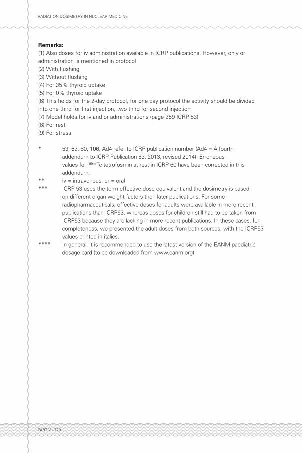
Remarks:(1) Also doses for iv administration available in ICRP publications. However, only or administration is mentioned in protocol(2) With fl ushing (3) Without fl ushing(4) For 35% thyroid uptake(5) For 0% thyroid uptake(6) This holds for the 2-day protocol, for one day protocol the activity should be divided into one third for fi rst injection, two third for second injection(7) Model holds for iv and or administrations (page 259 ICRP 53)(8) For rest(9) For stress
* 53, 62, 80, 106, Ad4 refer to ICRP publication number (Ad4 = A fourth addendum to ICRP Publication 53, 2013, revised 2014). Erroneous values for 99m Tc tetrofosmin at rest in ICRP 60 have been corrected in this addendum. ** iv = intravenous, or = oral *** ICRP 53 uses the term effective dose equivalent and the dosimetry is based on different organ weight factors then later publications. For some
radiopharmaceuticals, effective doses for adults were available in more recent publications than ICRP53, whereas doses for children still had to be taken from ICRP53 because they are lacking in more recent publications. In these cases, for completeness, we presented the adult doses from both sources, with the ICRP53 values printed in italics.
**** In general, it is recommended to use the latest version of the EANM paediatric dosage card (to be downloaded from www.eanm.org).
Part V.indd 770 27-12-16 14:40
RADIATION DOSIMETRY IN NUCLEAR MEDICINE
PART V - 771
References other than ICRP publications:• EANM guidelines for direct radionuclide cystography in children, Under the Auspices of the Paediatric
Committee of the European Association of Nuclear Medicine, Guidelines issued date: December 29, 2002,
• NVNG Aanbevelingen Nucleaire Geneeskunde 2007
• Senthamizhchelvan, S., P.E. Bravo, C. Esaias, M.A. Lodge, J. Merrill, R.F. Hobbs, G. Sgouros, and F.M.
Bengel, Human biodistribution and radiation dosimetry of 82Rb. J Nucl Med, 2010. 51(10): p. 1592-9.
• Senthamizhchelvan, S., P.E. Bravo, M.A. Lodge, J. Merrill, F.M. Bengel, and G. Sgouros, Radiation dosimetry
of 82Rb in humans under pharmacologic stress. J Nucl Med, 2011. 52(3): p. 485-91
• Law, M., W.H. Ma, R. Leung, S. Li, K.K. Wong, W.Y. Ho, and A. Kwong, Evaluation of patient effective dose
from sentinel lymph node lymphoscintigraphy in breast cancer: a phantom study with SPECT/CT and ICRP-
103 recommendations. Eur J Radiol, 2012. 81(5): p. e717-20.
• Liepe, K., J. Kropp, R. Runge, and J. Kotzerke, Therapeutic effi ciency of rhenium-188-HEDP in human
prostate cancer skeletal metastases. Br J Cancer, 2003. 89(4): p. 625-9.
• Patient brochure Bayer, 2013
• R. Bagheri, H. Afarideh, M. Ghannadi-Maragheh, A. Bahrami-Samani, S. P. Shirmardi, Dosimetric study of
radium-223 chloride and 153Sm-EDTMP for treatment of bone metastases using MCNPX code and available
experimental data, J Radioanal Nucl Chem (2015) 303:1991-1998
• CIS bio international, Summary of product characteristics, 2007
• Kam et al: Eur J Nucl Med Mol Imaging. Feb 2012; 39 (Suppl 1): 103-12.
Part V.indd 771 27-12-16 14:40
RADIATION DOSIMETRY IN NUCLEAR MEDICINE
PART V - 772
Table 2. Radiation dose in patients with abnormal biological behaviour
Radiopharma
ceutical
Route of admini-stration*
P** Effective dose***(mSv/MBq) Biological behaviour
1 yr 5 yr 10 yr 15 yr Adult
[14C] urea or 800,031 Normal patient
0,081 Helicobacter positive patient
Sodium [123I] iodide or/iv
62 0,2235% thyroid uptake
53 1,4 0,74 0,35 0,23 0,1562 0,011
0% thyroid uptake53 0,067 0,037 0,024 0,016 0,013
[99mTc] Tiatide (MAG3)
iv 80
0,022 0,012 0,012 0,009 0,007 Normal miction
0,0064 0,0064 0,0045 0,0031 0,0025 Bladder emptied 1 hour after administration
0,0068 0,0039 0,0029 0,0021 0,0017 Bladder emptied 0,5 hour after administration
0,019 0,011 0,010 0,0078 0,0061 Abnormal renal function
0,038 0,022 0,017 0,012 0,010 Unilateral renal blockage
[99mTc] Pentetate (DTPA)
iv
80 0,016 0,009 0,0082 0,0062 0,0049 Normal miction
80 0,014 0,0077 0,0065 0,0048 0,0038 Bladder emptied 1 hour after administration
80 0,014 0,0079 0,0070 0,0053 0,0041 Bladder emptied 0,5 hour after administration53 0,026 0,015 0,0097 0,0063 0,0053 Abnormal renal function
[123I] Sodium iodohippurate (IOH)
iv
80 0,034 0,019 0,019 0,015 0,012 Normal miction
80 0,019 0,011 0,0083 0,0059 0,0046 Bladder emptied 1 hour after administration:80 0,019 0,011 0,0099 0,0076 0,0059 Bladder emptied 0,5 hour after administration:53 0,067 0,037 0,024 0,016 0,013 Abnormal renal function53 0,27 0,16 0,11 0,075 0,062 Unilateral renal blockage
[125I]-Iothalamate (IOT) iv62 0,0072
Normal function53 0,057 0,030 0019 0,012 0,009753 0,12 0,060 0,037 0,024 0,019 Impaired renal function
[131I]-Sodium-iodohippurate (IOH)
iv
80 0,16 0,083 0,086 0,067 0,052 Normal miction80 0,089 0,047 0,036 0,026 0,02 Bladder emptied 1 hour after administration80 0,090 0,047 0,045 0,034 0,026 Bladder emptied 0,5 hour after administration53 0,36 0,19 0,12 0,08 0,065 Abnormal renal function53 6,8 3,8 2,6 1,8 1,5 Unilateral renal blockage
[131I] sodium iodide iv/or
62 2435% thyroid uptake
53 140 78 36 24 1562 0,061
0% thyroid uptake53 0,40 0,21 0,14 0,088 0,072
[99mTc] large colloids iv
80 0,050 0,028 0,018 0,012 0,0094 Normal patient
53 0,014Early to intermediate diffuse parenchymal liver disease
53 0,017Intermediate to advanced parenchymal liver disease
Part V.indd 772 27-12-16 14:40
RADIATION DOSIMETRY IN NUCLEAR MEDICINE
PART V - 773
Table 2. Radiation dose in patients with abnormal biological behaviour
Radiopharma
ceutical
Route of admini-stration*
P** Effective dose***(mSv/MBq) Biological behaviour
1 yr 5 yr 10 yr 15 yr Adult
[14C] urea or 800,031 Normal patient
0,081 Helicobacter positive patient
Sodium [123I] iodide or/iv
62 0,2235% thyroid uptake
53 1,4 0,74 0,35 0,23 0,1562 0,011
0% thyroid uptake53 0,067 0,037 0,024 0,016 0,013
[99mTc] Tiatide (MAG3)
iv 80
0,022 0,012 0,012 0,009 0,007 Normal miction
0,0064 0,0064 0,0045 0,0031 0,0025 Bladder emptied 1 hour after administration
0,0068 0,0039 0,0029 0,0021 0,0017 Bladder emptied 0,5 hour after administration
0,019 0,011 0,010 0,0078 0,0061 Abnormal renal function
0,038 0,022 0,017 0,012 0,010 Unilateral renal blockage
[99mTc] Pentetate (DTPA)
iv
80 0,016 0,009 0,0082 0,0062 0,0049 Normal miction
80 0,014 0,0077 0,0065 0,0048 0,0038 Bladder emptied 1 hour after administration
80 0,014 0,0079 0,0070 0,0053 0,0041 Bladder emptied 0,5 hour after administration53 0,026 0,015 0,0097 0,0063 0,0053 Abnormal renal function
[123I] Sodium iodohippurate (IOH)
iv
80 0,034 0,019 0,019 0,015 0,012 Normal miction
80 0,019 0,011 0,0083 0,0059 0,0046 Bladder emptied 1 hour after administration:80 0,019 0,011 0,0099 0,0076 0,0059 Bladder emptied 0,5 hour after administration:53 0,067 0,037 0,024 0,016 0,013 Abnormal renal function53 0,27 0,16 0,11 0,075 0,062 Unilateral renal blockage
[125I]-Iothalamate (IOT) iv62 0,0072
Normal function53 0,057 0,030 0019 0,012 0,009753 0,12 0,060 0,037 0,024 0,019 Impaired renal function
[131I]-Sodium-iodohippurate (IOH)
iv
80 0,16 0,083 0,086 0,067 0,052 Normal miction80 0,089 0,047 0,036 0,026 0,02 Bladder emptied 1 hour after administration80 0,090 0,047 0,045 0,034 0,026 Bladder emptied 0,5 hour after administration53 0,36 0,19 0,12 0,08 0,065 Abnormal renal function53 6,8 3,8 2,6 1,8 1,5 Unilateral renal blockage
[131I] sodium iodide iv/or
62 2435% thyroid uptake
53 140 78 36 24 1562 0,061
0% thyroid uptake53 0,40 0,21 0,14 0,088 0,072
[99mTc] large colloids iv
80 0,050 0,028 0,018 0,012 0,0094 Normal patient
53 0,014Early to intermediate diffuse parenchymal liver disease
53 0,017Intermediate to advanced parenchymal liver disease
* ICRP publication number** iv = intravenous, or = oral *** ICRP 53 uses the term “effective dose equivalent” and the dosimetry is based on different organ weight factors and different phantoms as compared to later publications. For some radiopharmaceuticals, both the effective dose equivalent values (from ICRP53) and the effective dose values (from later publications) are presented in the table. This was done when non-adult dose values are available from ICRP53, and not from newer publications. In those cases, the effective dose equivalents are written in italic, smaller font.
Part V.indd 773 27-12-16 14:40
RADIATION DOSIMETRY IN NUCLEAR MEDICINE
PART V - 774
RadiopharmaceuticalEarly
mGy/MBqa3MonthmGy/MBq
6MonthmGy/MBq
9MonthmGy/MBq
57CovitaminB-1,normal-flushing 1,0x100 6,8x10⁻¹ 8,4x10⁻¹ 8,8x10⁻¹
57CovitaminB-12,normal-noflushing 1,5x100 1,0x100 1,2x100 1,3x100
57CovitaminB-12,PA-flushing 2,1x10⁻¹ 1,7x10⁻¹ 1,7x10⁻¹ 1,5x10⁻¹
57CovitaminB-12,PA-noflushing 2,8x10⁻¹ 2,1x10⁻¹ 2,2x10⁻¹ 2,0x10⁻¹
58CovitaminB-12,normal-flushing 2,5x100 1,9x100 2,1x100 2,1x100
58CovitaminB-12,normal-noflushing 3,7x100 2,8x100 3,1x100 3,1x100
58CovitaminB-12,PA-flushing 8,3x10⁻¹ 7,4x10⁻¹ 6,4x10⁻¹ 4,8x10⁻¹
58CovitaminB-12,PA-noflushing 9,8x10⁻¹ 8,5x10⁻¹ 7,6x10⁻¹ 6,0x10⁻¹
60CovitaminB-12,normal-flushing 3,7x101 2,8x101 3,1x101 3,2x101
60CovitaminB-12,normal-noflushing 5,5x101 4,2x101 4,7x101 4,7x101
60CovitaminB-12,PA-flushing 5,9x100 4,7x100 4,8x100 4,5x100
60CovitaminB-12,PA-noflushing 8,3x100 6,5x100 6,8x100 6,5x100
18FFDGb 2,2x10⁻² 2,2x10⁻² 1,7x10⁻² 1,7x10⁻²
18Fsodiumfluoride 2,2x10⁻² 1,7x10⁻² 7,5x10⁻³ 6,8x10⁻³
67Gacitrate 9,3x10⁻² 2,0x10⁻¹ 1,8x10⁻¹ 1,3x10⁻¹
123Ihippuran 3,1x10⁻² 2,4x10⁻² 8,4x10⁻³ 7,9x10⁻³
123IIMP 1,9x10⁻² 1,1x10⁻² 7,1x10⁻³ 5,9x10⁻³
123IMIBG 1,8x10⁻² 1,2x10⁻² 6,8x10⁻³ 6,2x10⁻³
123Isodiumiodide 2,0x10⁻² 1,4x10⁻² 1,1x10⁻¹ 9,8x10⁻³
124Isodiumiodide 1,4x10⁻¹ 1,0x10⁻¹ 5,9x10⁻² 4,6x10⁻²
125IHSA 2,5x10⁻¹ 7,8x10⁻² 3,8x10⁻² 2,6x10⁻²
125IIMP 3,2x10⁻² 1,3x10⁻² 4,8x10⁻³ 3,6x10⁻³
125IMIBG 2,6x10⁻² 1,1x10⁻² 4,1x10⁻³ 3,4x10⁻³
125Isodiumiodide 1,8x10⁻² 9,5x10⁻³ 3,5x10⁻³ 2,3x10⁻³
126Isodiumiodide 7,8x10⁻² 5,1x10⁻² 3,2x10⁻² 2,6x10⁻²
130Isodiumiodide 1,8x10⁻¹ 1,3x10⁻¹ 7,6x10⁻² 5,7x10⁻²
131Ihippuran 6,4x10⁻² 5,0x10⁻² 1,9x10⁻² 1,8x10⁻²
131IHSA 5,2x10⁻¹ 1,8x10⁻¹ 1,6x10⁻¹ 1,3x10⁻¹
131IMAA 6,7x10⁻² 4,2x10⁻² 4,0x10⁻² 4,2x10⁻²
131IMIBG 1,1x10⁻¹ 5,4x10⁻² 3,8x10⁻² 3,5x10⁻²
131Isodiumiodide 7,2x10⁻² 6,8x10⁻² 2,3x10⁻¹ 2,7x10⁻¹
131Irosebengal 2,2x10⁻¹ 2,2x10⁻¹ 1,6x10⁻¹ 9,0x10⁻²
111InDTPA 6,5x10⁻² 4,8x10⁻² 2,0x10⁻² 1,8x10⁻²
111Inpentetreotide 8,2x10⁻² 6,0x10⁻² 3,5x10⁻² 3,1x10⁻²
111Inplatelets 1,7x10⁻¹ 1,1x10⁻¹ 9,9x10⁻² 8,9x10⁻²
111Inredbloodcells 2,2x10⁻¹ 1,3x10⁻¹ 1,1x10⁻¹ 8,6x10⁻²
111Inwhitebloodcells 1,3x10⁻¹ 9,6x10⁻² 9,6x10⁻² 9,4x10⁻²
99mTcalbuminmicrospheres 4,1x10⁻³ 3,0x10⁻³ 2,5x10⁻³ 2,1x10⁻³
99mTcdisofenin 1,7x10⁻² 1,5x10⁻² 1,2x10⁻² 6,7x10⁻³
99mTcDMSA 5,1x10⁻³ 4,7x10⁻³ 4,0x10⁻³ 3,4x10⁻³
99mTcDTPA 1,2x10⁻² 8,7x10⁻³ 4,1x10⁻³ 4,7x10⁻³
TABLE3.7.Absorbeddoseestimates totheembryo/fetus peruni tactivi tyofradiopharmaceutica l adminis teredtothemother(shadingindicates maternal andfeta l sel fdosecontributions).
Part V.indd 774 27-12-16 14:40
RADIATION DOSIMETRY IN NUCLEAR MEDICINE
PART V - 775
TABLE 3.7. (Continued)
RadiopharmaceuticalEarly
mGy/MBqa3 Month
mGy/MBq6 Month
mGy/MBq9 Month
mGy/MBq99mTc DTPA aerosol 5,8 x 10⁻³ 4,3 x 10⁻³ 2,3 x 10⁻³ 3,0 x 10⁻³
99mTc glucoheptonate 1,2 x 10⁻² 1,1 x 10⁻² 5,3 x 10⁻³ 4,6 x 10⁻³
99mTc HDP 5,2 x 10⁻³ 5,4 x 10⁻³ 3,0 x 10⁻³ 2,5 x 10⁻³
99mTc HEDP 7,2 x 10⁻³ 5,2 x 10⁻³ 2,7 x 10⁻³ 2,4 x 10⁻³
99mTc HMPAO 8,7 x 10⁻³ 6,7 x 10⁻³ 4,8 x 10⁻³ 3,6 x 10⁻³
99mTc human serum albumin 5,1 x 10⁻³ 3,0 x 10⁻³ 2,6 x 10⁻³ 2,2 x 10⁻³
99mTc MAA 2,8 x 10⁻³ 4,0 x 10⁻³ 5,0 x 10⁻³ 4,0 x 10⁻³
99mTc MAG3 1,8 x 10⁻² 1,4 x 10⁻² 5,5 x 10⁻³ 5,2 x 10⁻³
99mTc MDP 6,1 x 10⁻³ 5,4 x 10⁻³ 2,7 x 10⁻³ 2,4 x 10⁻³
99mTc MIBI-rest 1,5 x 10⁻² 1,2 x 10⁻² 8,4 x 10⁻³ 5,4 x 10⁻³
99mTc MIBI-stress 1,2 x 10⁻² 9,5 x 10⁻³ 6,9 x 10⁻³ 4,4 x 10⁻³
99mTc pertechnetate 1,1 x 10⁻² 2,2 x 10⁻² 1,4 x 10⁻² 9,3 x 10⁻³
99mTc PYP 6,0 x 10⁻³ 6,6 x 10⁻³ 3,6 x 10⁻³ 2,9 x 10⁻³
99mTc RBC-heat treated 1,7 x 10⁻³ 1,6 x 10⁻³ 2,1 x 10⁻³ 2,2 x 10⁻³
99mTc RBC-in vitro 6,8 x 10⁻³ 4,7 x 10⁻³ 3,4 x 10⁻³ 2,8 x 10⁻³
99mTc RBC-in vivo 6,4 x 10⁻³ 4,3 x 10⁻³ 3,3 x 10⁻³ 2,7 x 10⁻³
99mTc sulfur colloid-normal 1,8 x 10⁻³ 2,1 x 10⁻³ 3,2 x 10⁻³ 3,7 x 10⁻³
99mTc sulfur colloid-liver disease 3,2 x 10⁻³ 2,5 x 10⁻³ 2,8 x 10⁻³ 2,8 x 10⁻³
99mTc teboroxime 8,9 x 10⁻³ 7,1 x 10⁻³ 5,8 x 10⁻³ 3,7 x 10⁻³
99mTc white blood cells 3,8 x 10⁻³ 2,8 x 10⁻³ 2,9 x 10⁻³ 2,8 x 10⁻³
201Tl chloride 9,7 x 10⁻² 5,8 x 10⁻² 4,7 x 10⁻² 2,7 x 10⁻²
127Xe, 5 min rebreathing, 5 liter spirometer volume
4,3 x 10⁻⁴ 2,4 x 10⁻⁴ 1,9 x 10⁻⁴ 1,5 x 10⁻⁴
127Xe, 5 min rebreathing, 7,5 liter spirometer volume
2,3 x 10⁻⁴ 1,3 x 10⁻⁴ 1,0 x 10⁻⁴ 8,4 x 10⁻⁵
127Xe, 5 min rebreathing, 10 liter spirometer volume
2,3 x 10⁻⁴ 1,4 x 10⁻⁴ 1,1 x 10⁻⁴ 9,2 x 10⁻⁵
133Xe, 5 min rebreathing, 5 liter spirometer volume
4,1 x 10⁻⁴ 4,8 x 10⁻⁵ 3,5 x 10⁻⁵ 2,6 x 10⁻⁵
133Xe, 5 min rebreathing, 7,5 liter spirometer volume
2,2 x 10⁻⁴ 2,6 x 10⁻⁵ 1,9 x 10⁻⁵ 1,5 x 10⁻⁵
133Xe, 5 min rebreathing, 10 liter spirometer volume
2,5 x 10⁻⁴ 2,9 x 10⁻⁵ 2,1 x 10⁻⁵ 1,6 x 10⁻⁵
133Xe, injection 4,9 x 10⁻⁶ 1,0 x 10⁻⁶ 1,4 x 10⁻⁶ 1,6 x 10⁻⁶
amGy/MBq x 3,7 rad/mCi .
Source: Adapted with permiss ion from Russel l JR, Stabin MG, Sparks RB, Watson EE. Radiation absorbed dose to the embryo/fetus from radiopharmaceutica ls . Health Phys , 73:756-769. 1997.
bStabin M. Proposed addendum to previous ly publ ished feta l dose estimate tables for 18F-FDG. J Nucl Med, 45:634-635, 2004.
Part V.indd 775 27-12-16 14:40
RADIATION DOSIMETRY IN NUCLEAR MEDICINE
PART V - 776
Gestational age (months) 123I 124I 125I 131I3 2,7 24 290 2304 2,6 27 240 2605 6,4 76 280 5806 6,4 100 210 5507 4,1 96 160 3908 4,0 110 150 3509 2,9 99 120 270
TABEL 3.8. Dose to the feta l thyroid (doses are mGy to the feta l thyroid per MBq adminis tered to the mother).
Source: Adapted with permiss ion of ORAU from Watson EE, Radiation absorbed dose to the human feta l thyroid. In: Fi fth International Radio-pharmaceutica l Dos imetry Sympos ium. Watson EE, Schlafke-Stelson. eds , Oak Ridge Associated Univers i ties , Oak Ridge, TN, 1992, pp. 179-187.
Part V.indd 776 27-12-16 14:40
RADIATION DOSIMETRY IN NUCLEAR MEDICINE
PART V - 777
Radiopharmaceutical Interruption14 C-labelledTriolein NoGlycocholic acid NoUrea No99m Tc-labelledDISDA No *,†
DMSA No *,†
DTPA No *,†
ECD No *,†
Phosphonates (MDP) No *,†
Gluconate No *,†
Glucoheptonate No *,†
HM-PAO No *,†
Sulphur colloids No *,†
MAA 12 hMAG3 No *,†
MIBI No *,†
Microspheres (HAM) 12 hPertechnetate 12 hPYP No *,†
RBC (in vivo) 12 hRBC (in vitro) No *,†
Technegas No *,†
Tetrofosmin No *,†
WBC 12 h
(D1) Since many radiopharmaceutica ls are secreted in breast mi lk, i t i s safest to assume that,unless there are data to the contrary, some radioactive compound wi l l be found in the breastmi lk when a radiopharmaceutica l i s adminis tered to a lactating female. Cons ideration shouldbe given to postponing the procedure. If the procedure is performed, the chi ld should not bebreast fed unti l the radiopharmaceutica l i s no longer secreted in an amount estimated to givean effective dose >1 mSv to the chi ld. It i s therefore recommended that the fol lowing actionsshould be taken for various radiopharmaceutica ls , and that the mi lk expressed during thisinterruption period should be discarded.
D.1. Introduction
ANNEX D. RECOMMENDATIONS ON BREAST-FEEDING INTERRUPTIONS
Part V.indd 777 27-12-16 14:40
RADIATION DOSIMETRY IN NUCLEAR MEDICINE
Radiopharmaceutical InterruptionI-labelled123I-BMIPP >3 weeks‡,•
123I-HSA >3 weeks‡,•
123I-iodo hippurate 12 h123I-IPPA >3 weeks‡,•
123I-MIBG >3 weeks‡,•
123I-NaI >3 weeks‡,•
125I-HSA >3 weeks‡,•
125I-iodo hippurate 12 h131I-iodo hippurate 12 h131I-MIBG >3 weeks‡
131I-NaI >3 weeks‡
Others11C-labelled No‣
13N-labelled No‣
15O-labelled No‣
18F-FDG No22Na >3 weeks‡
51Cr-EDTA No67Ga-citrate >3 weeks‡
75Se-labelled agents >3 weeks‡
81mKr-gas No111In-octreotide No111In-WBC No133Xe No201Tl-chloride 48 h
‡3 weeks (504 h) at least. However, di fficul t to mainta in the mi lk supply cessation.
*'No', interruption not essentia l†'No' for most of the 99mTc-label led compounds , under the ci rcumstances that no free
• 123I, a l l substances label led with 123I (except iodo-hippurate): >3 weeks due to the ri sk of contamination of other iodine i sotopes‣ 11C, 13N and 15O-label led substances , interruption not essentia l due to short phys ica l ha l f-l i fe.
D.2. References and further reading for Annex D
Castronovo, Jr., F.P., Stone, H., Ulanski, J., 2000. Radioactivity in breast milk following 111In-octreotide. Nucl. Med. Commun. 21, 695-699.
Ahlgren, L., Ivarsson, S., Johansson, L., Mattsson, S., Nosslin, B., 1985. Excretion of radionuclides in human breast milk after the administration of radiopharmaceuticals. J. Nucl. Med. 26, 1085-1090.
PART V - 778
Part V.indd 778 27-12-16 14:40