Embed Size (px)
Citation preview
3 Quorum sensing
Chapter 1
Introduction
Atomiclevel
Chapter 2 Chapter 3 Chapter 4 Chapter 5 Chapter 6
Pathwaylevel
Proteomelevel
Cellularlevel
Spial Quorumsensing
Chemo-genomics
Descriptorrelationships
Conclusionsand
perspectives

Chapter 3: Quorum sensing
3-1
Contents of chapter 3
Contents of chapter 3.................................................................................................. 1 Summary of chapter 3................................................................................................. 3 Introduction to quorum sensing................................................................................... 3
What quorum sensing is .......................................................................................... 4 Cell-to-cell communication in bacteria..................................................................... 6
Acyl homoserine lactones.................................................................................... 7 Autoinducer-2....................................................................................................... 9 Processed oligopeptides.................................................................................... 10 Other systems.................................................................................................... 11 Quorum quenching ............................................................................................ 12 Cross-genome interactions ................................................................................ 12
Cell-to-cell communication in yeast ....................................................................... 14 Farnesol ............................................................................................................. 14 Aromatic alcohol derivatives .............................................................................. 15
Tools and Techniques ........................................................................................... 15 Chemical ............................................................................................................ 15 Genetic............................................................................................................... 15 Computational.................................................................................................... 16
A survey of quorum sensing protein domains ........................................................... 16 Results and discussion.......................................................................................... 16
A γ-butyrolactone quorum sensing system in Rhodococcus? ........................... 18 Materials and methods .......................................................................................... 19
The agr quorum sensing system in firmicutes........................................................... 20 Results and discussion.......................................................................................... 21
The agr system is widespread among firmicutes............................................... 23 New agr-like systems......................................................................................... 25 Multiple paralogous copies of AgrB in Clostridia genomes................................ 26 A novel gene associated with the agr system in Clostridia ................................ 27 RNAIII and the agr system................................................................................. 27 agr-regulated genes........................................................................................... 28 Concluding remarks ........................................................................................... 29
Materials and methods .......................................................................................... 29 Homologue identification.................................................................................... 30 Tree building ...................................................................................................... 30 RNAIII identification ........................................................................................... 31
Chapter 3: Quorum sensing
3-2
Gene expression data analysis.......................................................................... 31 Detection of orthologues.................................................................................... 32
The Saccharomyces cerevisiae quorum sensing system ......................................... 32 Results and discussion.......................................................................................... 34
Attributing differential expression to transcription factors .................................. 35 Quorum sensing and entry into stationary phase .............................................. 37 Genomic organization and nucleosome modification of the differentially
expressed genes................................................................................................ 38 Conservation of quorum sensing components in fungal genomes .................... 40
Materials and methods .......................................................................................... 42 Gene expression data........................................................................................ 42 Identification of key transcription factors............................................................ 43 Genomic distribution of differentially expressed genes...................................... 44 Orthologue detection.......................................................................................... 44 Epigenetic modifications .................................................................................... 45
References................................................................................................................ 46
Chapter 3: Quorum sensing
3-3
Summary of chapter 3
Cell-to-cell signalling is a prerequisite for the development of multicellular organisms
such as animals and plants, but has also evolved in groups which would not usually
be described as multicellular, such as bacteria and unicellular fungi. Quorum sensing
is a process of cell-to-cell communication by which individual cells regulate their
phenotype in response to the extracellular concentration of small molecules. This is
achieved by the secretion of small molecules into the environment that bind sensory
proteins and directly or indirectly affect transcription and translation. In this chapter, I
use a combination of computational methods to identify quorum sensing systems in
sequenced bacterial genomes. Furthermore, I establish a framework for the
identification of transcription factors involved in the quorum sensing response in the
yeast Saccharomyces cerevisiae.
Parts of this chapter appear in the following peer-reviewed articles:
Wuster, A. and M. Madan Babu (2008). “Conservation and evolutionary dynamics of
the agr cell-to-cell communication system across firmicutes.” J Bacteriol 190(2): 743-
6.
Wuster, A. and M. Madan Babu (2008). “Chemical Molecules that Regulate
Transcription and Facilitate Cell-to-cell Communication.” book chapter in Wiley
Encyclopedia of Chemical Biology, Hoboken, New Jersey: John Wiley and Sons, Inc.
Wuster, A. and M. Madan Babu (2009). “Transcriptional control of the quorum
sensing response in yeast.” Mol BioSyst, DOI: 10.1039/b913579k.
Introduction to quorum sensing Quorum sensing (QS) is a way for individual cells to exchange information using
small molecules (SMs) that bind sensory proteins and thus directly or indirectly affect
transcription and translation. The binding threshold is assumed to be reached once
the growing population, and hence the concentration of the secreted SM, attains a
certain level. In the following, I use the term 'QS system' to mean a cell-to-cell
communication system in unicellular organisms that functions via the secretion of
communication molecules into the environment and their subsequent binding to
sensor proteins. The term ‘communication molecule’ as I use it here refers to any SM
that is secreted by one organism and changes the behaviour of another one. As I will
Chapter 3: Quorum sensing
3-4
discuss later, it is not necessary that the communication molecule synthesis or
detection machinery has evolved for this purpose, although this is often the case.
Different systems can be distinguished by the different types of communication
molecule they use, which are normally also associated with different types of signal
synthesis, import and export, reception and response machinery (Pai and You 2009). The study of cell-to-cell communication and its effects on transcription in unicellular
organisms promises a variety of practical applications. One of them is the possibility
of interfering with intercellular communication systems in pathogenic microbes, a
process also referred to as quorum quenching. For this, it is first necessary to know
which quorum systems are present in which genomes. To answer this is the main
aim of this chapter. I approach the problem in three stages.
The first stage is a knowledge-based approach to discovering QS systems in a library
of 384 fully sequenced microbial genomes. The second stage focuses on two specific
QS systems, which are the γ-butyrolactone and the agr QS systems. I present
evidence that the agr system is present across the bacterial phylum of firmicutes,
including the human pathogen Clostridium perfringens. The third stage deals with the
QS system of the yeast Saccharomyces cerevisiae. By employing an integrated in
silico approach that makes use of large-scale genomics datasets, I identify key
transcriptional regulators that control the differential expression of the genes affected
by QS in S. cerevisiae.
For the remainder of this introduction, I will define QS more rigorously and introduce
the QS systems that I will analyse in more detail later in this chapter.
What quorum sensing is
Whilst the current consensus seems to be that QS is a way of signalling between
bacterial cells, there are a number of reasons why this may not always be the case.
Firstly, signalling is the exchange of information between two willing partners (Mullard
2009). In other words, both the sender and the receiver of a communication molecule
have to benefit from the process for it to qualify as signalling (table 3-1). This is not
necessarily always the case in QS. It is often not clear whether the exchange of SMs
has evolved for the purpose of signalling, or whether the SMs are, for example,
metabolic side-products in one cell that just happen to have an influence on another
cell.
Chapter 3: Quorum sensing
3-5
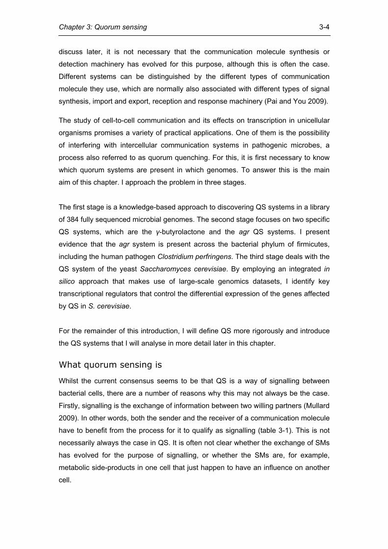
... sender Communication fitness effect to the... Beneficial Neutral/deleterious
Beneficial A. Signal B. Cue/Spying ... receiver Neutral/deleterious C. Manipulation D. ? Table 3-1. Only the exchange of information that is beneficial to both the receiver and the sender should be called signal. Considering this, some SMs that have been called QS signals may not actually be signals. Secondly, bacteria may secrete SMs that change their and other’s behaviour without
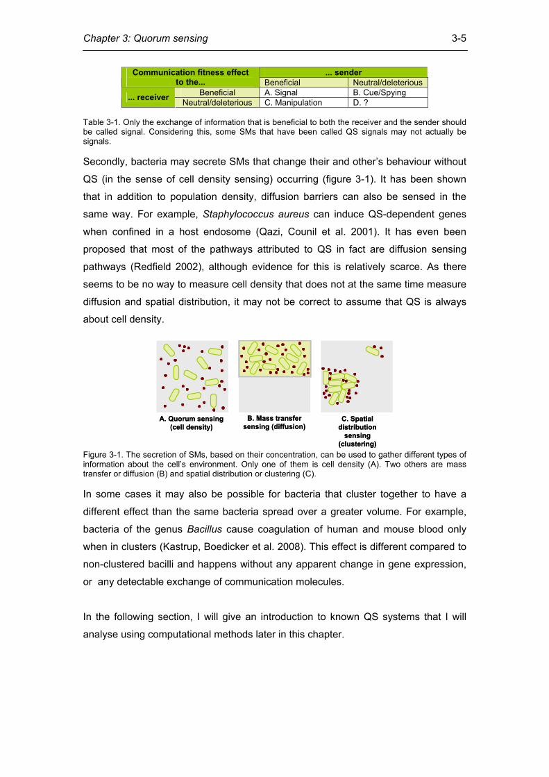
QS (in the sense of cell density sensing) occurring (figure 3-1). It has been shown
that in addition to population density, diffusion barriers can also be sensed in the
same way. For example, Staphylococcus aureus can induce QS-dependent genes
when confined in a host endosome (Qazi, Counil et al. 2001). It has even been
proposed that most of the pathways attributed to QS in fact are diffusion sensing
pathways (Redfield 2002), although evidence for this is relatively scarce. As there
seems to be no way to measure cell density that does not at the same time measure
diffusion and spatial distribution, it may not be correct to assume that QS is always
about cell density.
A. Quorum sensing(cell density)
A. Quorum sensing(cell density)
B. Mass transfer sensing (diffusion)
B. Mass transfer sensing (diffusion)
C. Spatial distribution
sensing (clustering)
C. Spatial distribution
sensing (clustering)
Figure 3-1. The secretion of SMs, based on their concentration, can be used to gather different types of information about the cell’s environment. Only one of them is cell density (A). Two others are mass transfer or diffusion (B) and spatial distribution or clustering (C). In some cases it may also be possible for bacteria that cluster together to have a
different effect than the same bacteria spread over a greater volume. For example,
bacteria of the genus Bacillus cause coagulation of human and mouse blood only
when in clusters (Kastrup, Boedicker et al. 2008). This effect is different compared to
non-clustered bacilli and happens without any apparent change in gene expression,
or any detectable exchange of communication molecules.
In the following section, I will give an introduction to known QS systems that I will
analyse using computational methods later in this chapter.
Chapter 3: Quorum sensing
3-6
Cell-to-cell communication in bacteria
QS involves dedicated cellular systems for the production and detection of
communication molecules, sometimes called quormones (quorum sensing
pheromones). In bacterial species that employ QS, each cell secretes a basal
amount of communication molecules at low cell density. As cell density increases,
communication molecule concentration also increases, provided that the cells are not
too far apart. Communication molecules bind to special receptors once their
concentration exceeds a certain threshold. This in turn produces the physiological
response.
In bacteria, QS regulated phenotypes include bioluminescence, exopolysaccharide
production, virulence, conjugal plasmid transfer, antibiotic and exoenzyme production,
biofilm formation, and growth inhibition (Lazdunski, Ventre et al. 2004). Types of
communication molecules discussed in this section are acyl homoserine lactones
(AHLs), AI-2 molecules, and modified oligopeptides (table 3-2). AHLs mostly affect
transcription via a one-component signal transduction system, where the protein
domain that binds the SMs is fused to a DNA binding domain. Peptide
communication molecules and AI-2, on the other hand, often affect transcription via
two-component signal transduction systems composed of a histidine kinase and a
response regulator protein (Ulrich, Koonin et al. 2005). As we shall see in the
example of Vibrio harveyi, in some instances the AI-2 signal transduction cascade
can also be composed of more than two components.
Chapter 3: Quorum sensing
3-7
Acyl homoserine lactone (AHL)Autoinducing peptide (AIP, encoded by agrD)AI-2 (a furanosyl borate diester)γ-butyrolactone
ComELuxOAgrATranscription regulator
ComD transmembrane sensor kinase binds extracellular CSP and phosphorylates cytoplasmic ComE
ArpALuxQ transmembrane sensor kinase binds periplasmic complex LuxP-AI-2, and phosphorylates cytoplasmic LuxU
AgrC transmembrane sensor kinase binds extracellular AIP and phosphorylates cytoplasmic AgrA
LuxRSensor
ComABNo transporter knownNo exporter for AI-2.no importer in Vibrio harveyi, but importer is present in Escherichia coli and Salmonella(Lsr ABC-type transporter)
ABC exporterNo transporter – AHL can diffuse through membranes
Transporters (exporters and importer)
Not applicableAfsALuxSSynthesis from S-adenosyl methionine (SAM)
Prepeptide modified by AgrB, which adds thiolactone ring
LuxI, synthesis from S-adenosyl methionine (SAM) and fatty acid carrier protein
Synthase
Competence stimulating peptide (CSP, encoded by comC)Glu-Met-Arg-Leu-Ser-Lys-Phe-Phe-Arg-Asp-Phe-Ile-Leu-Gln-Arg-Lys-Lys
γ-butyrolactone. R is an acyl side chain
AI-2 (a furanosyl borate diester)
Autoinducing peptide (AIP, encoded by agrD)
Acyl homoserine lactone (AHL). R is acyl chain with 4 to 18 carbons, for example 3OC12
Signalling molecule
Streptococcus pneumoniae
Streptomyces griseusVibrio harveyiStaphylococcus aureusVibrio fischeriExample of bacterial species
E. Competence stimulating peptide (CSP) system
D. γ-butyrolactone system
C. AI-2 systemB. Autoinducing peptide (AIP) system
A. Acyl homoserine lactone (AHL) system
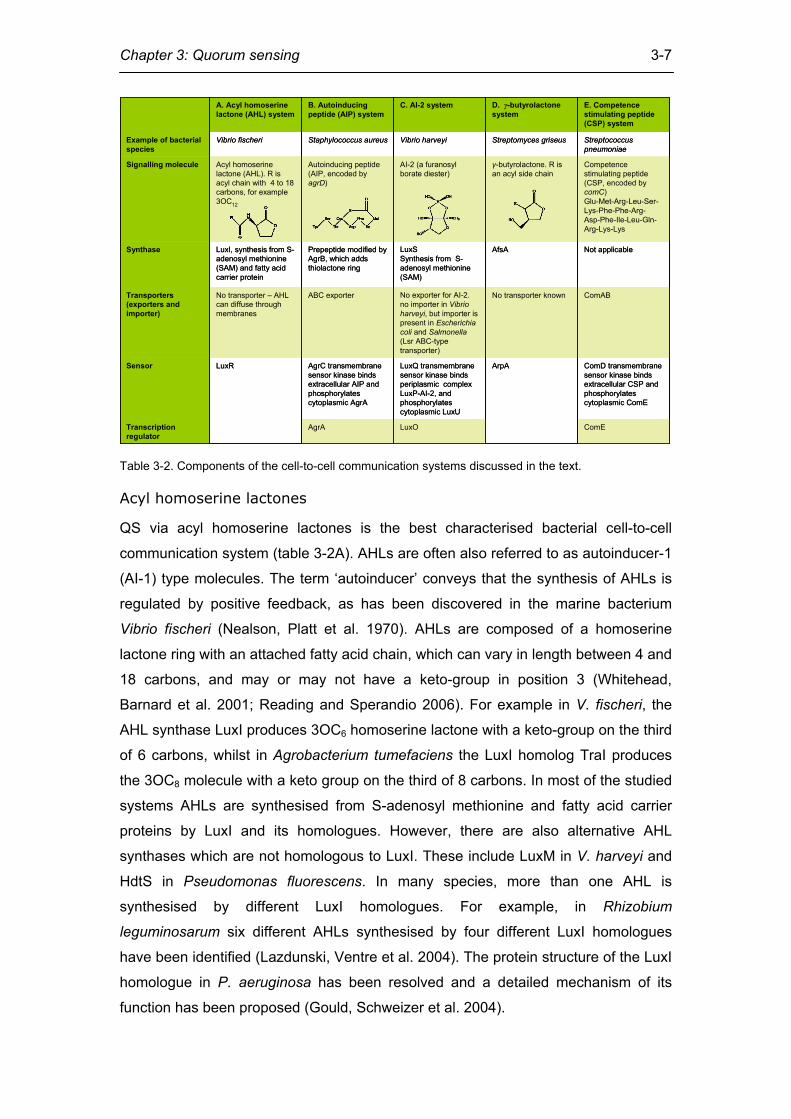
Table 3-2. Components of the cell-to-cell communication systems discussed in the text.
Acyl homoserine lactones
QS via acyl homoserine lactones is the best characterised bacterial cell-to-cell
communication system (table 3-2A). AHLs are often also referred to as autoinducer-1
(AI-1) type molecules. The term ‘autoinducer’ conveys that the synthesis of AHLs is
regulated by positive feedback, as has been discovered in the marine bacterium
Vibrio fischeri (Nealson, Platt et al. 1970). AHLs are composed of a homoserine
lactone ring with an attached fatty acid chain, which can vary in length between 4 and
18 carbons, and may or may not have a keto-group in position 3 (Whitehead,
Barnard et al. 2001; Reading and Sperandio 2006). For example in V. fischeri, the
AHL synthase LuxI produces 3OC6 homoserine lactone with a keto-group on the third
of 6 carbons, whilst in Agrobacterium tumefaciens the LuxI homolog TraI produces
the 3OC8 molecule with a keto group on the third of 8 carbons. In most of the studied
systems AHLs are synthesised from S-adenosyl methionine and fatty acid carrier
proteins by LuxI and its homologues. However, there are also alternative AHL
synthases which are not homologous to LuxI. These include LuxM in V. harveyi and
HdtS in Pseudomonas fluorescens. In many species, more than one AHL is
synthesised by different LuxI homologues. For example, in Rhizobium
leguminosarum six different AHLs synthesised by four different LuxI homologues
have been identified (Lazdunski, Ventre et al. 2004). The protein structure of the LuxI
homologue in P. aeruginosa has been resolved and a detailed mechanism of its
function has been proposed (Gould, Schweizer et al. 2004).
Chapter 3: Quorum sensing
3-8
Most AHLs cross membranes by diffusion and bind LuxR-like response regulators.
LuxR-like response regulators simultaneously act as sensors and transcription
factors (Choi and Greenberg 1991). This group of signal transduction systems, where
the signal binding domain and the transcription-regulating DNA-binding domain are
fused, are referred to as one-component signal transduction systems. They have
been shown to be the most common sort of bacterial signal transduction systems
(Madan Babu and Teichmann 2003; Aravind, Anantharaman et al. 2005; Ulrich,
Koonin et al. 2005).
The N-terminal signal-binding domain of LuxR-like proteins has an αβα-sandwich
GAF domain-like fold. This is particularly interesting as GAF domains are usually
found in signalling and sensor proteins (Aravind and Ponting 1997). The structure of
the A. tumefaciens LuxR homolog TraR (Zhang, Pappas et al. 2002) shows that,
upon binding, AHL is deeply embedded in the protein. Contact with conserved
hydrophobic and aromatic residues is established via a number of hydrogen bonds,
which are either direct or via water. AHL binding is high-affinity, which means that
bacteria are able to sense relatively small communication molecule concentrations.
The specificity of the LuxR-like protein for AHL is determined by the acyl binding
pocket of the LuxR homologue (Camilli and Bassler 2006), as AHLs differ only in
their acyl chains. The C-terminal domain belongs to the DNA and RNA binding helix-
turn-helix fold. LuxR-like proteins use this domain to bind their cognate DNA motifs
such as the palindromic lux box in the case of V. fischeri, where it activates
transcription of the luxICDABE operon. Because this operon encodes the AHL
synthase LuxI, AHL synthesis is subject to positive feedback. However, in other
species, LuxR orthologues can act as transcriptional repressors, such as is the case
for P. aeruginosa RhlR and LasR, which have been shown by microarray analysis
(Schuster, Lostroh et al. 2003; Clark, Acharya et al. 2009) to negatively regulate
multiple genes.
Due to the positive feedback effect AHLs have on regulating the transcription of their
own synthases, concentrations of AHLs can vary enormously between lower-density
and higher-density cultures such as biofilms. P. aeruginosa 3OC12 homoserine
lactone (with a keto-group on the third of the 12 carbons of the acyl chain) has been
measured to have a concentration of 2-10 µM in a standard lab culture and of up to
600 µM in the vicinity of an in vitro biofilm. However, experimental sensitivity and
Chapter 3: Quorum sensing
3-9
issues with AHL stability might have influenced the above results (Shiner, Rumbaugh
et al. 2005).
Although the basic AHL QS system only consists of two proteins – LuxI and LuxR –
there is a whole ensemble of proteins modulating it. These include the AiiA AHL
lactonase of Bacillus thuringiensis, which inactivates AHL by hydrolysing the
homoserine lactone ring. This is an instance of quorum quenching, more examples of
which I will give later. Other LuxR homologues, such as Escherichia coli SdiA,
recognise more than one AHL (Whitehead, Barnard et al. 2001; Reading and
Sperandio 2006). This might be because SdiA is probably used to detect AHL
produced by other species, as no LuxI homologue has been detected in E. coli. This
may be a case of E. coli ‘spying’ on the communication molecules of other species
(table 3-1).
Autoinducer-2
There is an overlap of AHL signalling with other QS systems, such as seen in V.
harveyi. Apart from an AHL system, V. harveyi has a parallel system whose
signalling molecules are referred to as autoinducer-2 (AI-2; table 3-2C). In V. harveyi,
AI-2 is a furanosyl borate diester, whose precursor is 4,5-dihydroxy-2,3-pentanedione
(DPD). DPD is synthesised by LuxS from S-adenosyl methionine (SAM). Thus, SAM
is a precursor both in AHL synthesis and AI-2 synthesis. It is assumed that DPD
spontaneously rearranges into AI-2 when borate is available (Chen, Schauder et al.
2002). In other species this rearrangement does not occur, and AI-2 has a different
structure.
In V. harveyi, in the absence of AI-2, the membrane-bound kinase LuxQ undergoes
autophosphorylation on a conserved histidine residue (Neiditch, Federle et al. 2005;
Neiditch, Federle et al. 2006). The phosphoryl group is then transferred from the
histidine of LuxQ to an aspartate of the response regulator LuxU. Phospho-LuxU in
turn phosphorylates LuxO. Phospho-LuxO together with the sigma factor σ54 then
activates transcription of a set of small RNAs. These small RNAs, together with the
RNA chaperone Hfq, contribute to the degradation of the mRNA of the LuxR
transcription factor. LuxR is therefore the ultimate effector of the system in the
presence of AI-2 in V. harveyi. It is important to distinguish this LuxR from the non-
homologous LuxR protein that binds AHL in V. fischeri as discussed in the previous
section. When AI-2 is present, it is bound by LuxP, which is a periplasmic binding
protein. LuxP-AI-2 binding to the LuxQ kinase triggers a dephosphorylation cascade
Chapter 3: Quorum sensing
3-10
by turning the kinases into phosphatases. LuxQ dephosphorylates LuxU, which in
turn dephosphorylates LuxO. The result is that the mRNA of the transcription factor
LuxR is no longer degraded by sRNAs, and it can regulate its target genes (Waters
and Bassler 2006). Interestingly, LuxU can also be phosphorylated by two other
mechanisms. The first mechanism involves 3OC4 homoserine lactone that binds to
the LuxN sensor kinase, which in turn dephosphorylates LuxU. The second
mechanism acts via the unidentified autoinducer CAI-1. Thus, AI-2, 3OC4
homoserine lactone and CAI-1 funnel their signals into one common system.
Furthermore, sensing of the AHL 3OC4 homoserine lactone by a membrane-bound
kinase in V. harveyi shows that AHLs can also be sensed by pathways which are
dissimilar to the one-component pathway outlined in the previous section.
Processed oligopeptides
Many Gram-positive bacteria use processed oligopeptides as communication
molecules. The precursor peptides are typically between 40 and 65 amino acid
residues in length. These pre-peptides are processed (cleaved) in all known cases
and the resulting peptide communication molecules are typically 5 to 34 residues in
length. In many cases the peptides are also modified. The minimum components for
peptide communication, apart from the peptide signal itself, are a membrane-bound
histidine kinase, and a response regulator with an aspartate phosphorylation residue.
These components constitute a two-component signal transduction system, as
opposed to the one-component system observed in AHL-based communication.
Peptide communication systems include the competence stimulating peptide (CSP)
of Streptococcus pneumoniae (Havarstein, Coomaraswamy et al. 1995) (table 3-2E),
and the peptide antibiotic nisin in Lactococcus lactis, which positively regulates its
own expression. Another peptide communication molecule discovered in
Staphylococcus aureus is the autoinducing peptide (AIP; table 3-2C), which is
derived from the precursor AgrD to which a thiolactone ring is added between the
carboxyl residue and a Cysteine residue at position 5 by AgrB. The AIP is sensed by
the AgrC sensor kinase, which may dimerise upon signal binding. The signal is then
passed on to the AgrA response regulator, which activates transcription of the agr
operon (Ji, Beavis et al. 1997; Reading and Sperandio 2006), therefore again leading
to autoinduction (figure 3-3B). Both AgrD and ComX, another peptide communication
molecule precursor found in B. subtilis, have an amphipathic motif in their N-terminal
region, which might serve the purpose of recruiting the peptide to the membrane
where it can be processed (Zhang, Lin et al. 2004). This is often done by a dedicated
Chapter 3: Quorum sensing
3-11
ATP binding cassette (ABC) transporter, which in some cases recognises a so-called
GG leader sequence (consensus sequence LSxxELxxIxGG) at the N-terminal side of
the region encoding the actual signal (Dirix, Monsieurs et al. 2004). Peptide signals may be more flexible than the small communication molecules
discussed above, as they do not require a specialised synthase and can change to
adapt to ecological niches by simple codon mutation (Morrison 2002). The
observation that some genes encoding peptide signals are more variable than others
might be an indication that this actually happens.
Other systems
Other bacterial QS systems include the P. aeruginosa quinolone signal (PQS; 2-
heptyl-3-hydroxy-4-quinolone) synthesised by PqsH, which is quite hydrophobic and
is exported out of the cell by a vesicular transport system analogous to the ones used
by eukaryotes. It seems that PQS is directly facilitating vesicle formation, as pqsH
mutants do not produce vesicles. However, the phenotype can be rescued by the
addition of exogenous PQS (Mashburn and Whiteley 2005). The pqs genes have
also been found in Burkholderia, although the production of PQS in these species
could not be confirmed.
Fruiting body formation in the bacterium Myxococcus xanthus is facilitated by the
diffusible A-signal, which seems to consist of six different amino acids generated by
extracellular proteolysis, and the contact-dependent C-signal. The C-signal is a cell-
surface protein (Kaiser 2004). Contact-dependent communication has also been
observed in E. coli, where it is facilitated by CdiA and CdiB and leads to growth
inhibition (Aoki, Pamma et al. 2005).
Another QS signal that I will discuss in more detail later is the γ-butyrolactone system
found in Actinobacteria (table 3-2D).
The list discussed here is by no means complete. Other QS molecules include
Bacillus subtilis ComX, Streptomyces coelicolor SapB, and the Rhizobium meliloti
Nod factor (Shapiro 1998; Bassler and Losick 2006). It can be expected that even
more bacterial QS systems await discovery, especially when considering the vast
amounts of microbial diversity made available by metagenomic sequencing projects.
Chapter 3: Quorum sensing
3-12
Quorum quenching
In the human pathogen P. aeruginosa it has been shown that disrupting its QS
system diminishes virulence (Kastrup, Boedicker et al. 2008). This ‘quorum
quenching’ takes the form of enzymes degrading the communication molecules. Two
different sorts of enzymes doing this for AHLs have been described: AHLases (e.g.
AiiA) hydrolyse the lactone ring, resulting in acyl homoserine, whilst AHL-acylases
(e.g. AiiD) break the amide bond, cleaving homoserine lactone from the acyl side
chain (Leadbetter and Greenberg 2000; Park, Hwang et al. 2006). Communication
molecules can also be quenched by organisms which do not produce the molecules,
presumably in order to gain an advantage over communicating bacterial species in
the same ecological niche. For example, Rhodococcus is able to degrade AHL
signals without having any known ability to produce them (Jafra, Przysowa et al.
2006).
Cross-genome interactions
Several instances of the exchange of SMs between bacteria and eukaryotes are
known. This can either take the form of bacterial communication molecules being
received by eukaryotic cells, or eukaryotic communication molecules being received
by bacterial cells (Mullard 2009).
For example, QseC is a sensor protein that in enterohaemorrhagic Escherichia coli is
activated by both AI-3 and the mammalian hormones adrenaline and noradrenaline.
In both cases, the expression of virulence genes is up-regulated (Clarke, Hughes et
al. 2006). It is however not clear if QseC in E. coli has evolved to respond to
adrenaline and noradrenaline in bacteria, or whether it does so because there has
been horizontal gene transfer between eukaryotes and bacteria (Iyer, Aravind et al.
2004). Another example of a eukaryote interfering with bacterial cell-to-cell
communication is Apolipoprotein B (APOB), which smothers the AIP in S. aureus and
prevents it from becoming virulent. Mice that lack APOB are more susceptible to S.
aureus infection (Peterson, Mack et al. 2008). This may explain why one quarter of
humans can permanently live with S. aureus in their noses.
Chapter 3: Quorum sensing
3-13
Beneficial to bacterium Beneficial to eukaryote Bacterium produces SM, eukaryote receives it
A. The pathogenic bacterium Pseudomonas aeruginosa produces C12, which inhibits the NF-κB pathway in the mammalian host, therefore interfering with the immune response (Kravchenko, Kaufmann et al. 2008).
B. The mobile zoospores of the green alga Ulva intestinalis preferentially settle on Vibrio biofilms. They may detect these biofilms by the AHL they produce (Tait, Joint et al. 2005; Joint, Tait et al. 2007).
Eukaryote produces SM, bacterium receives it
C. Agrobacterium recognises several SMs produced by plants. This includes phenolic compounds, which stimulate the transcription of genes needed for infection (Winans 1992).
D. The red alga Delisea pulchra produces a halogenated furanone, which inhibits QS in the bacterium Pseudomonas aeruginosa. This in turn promotes the disintegration of biofilms (Hentzer, Riedel et al. 2002; Hentzer, Wu et al. 2003).
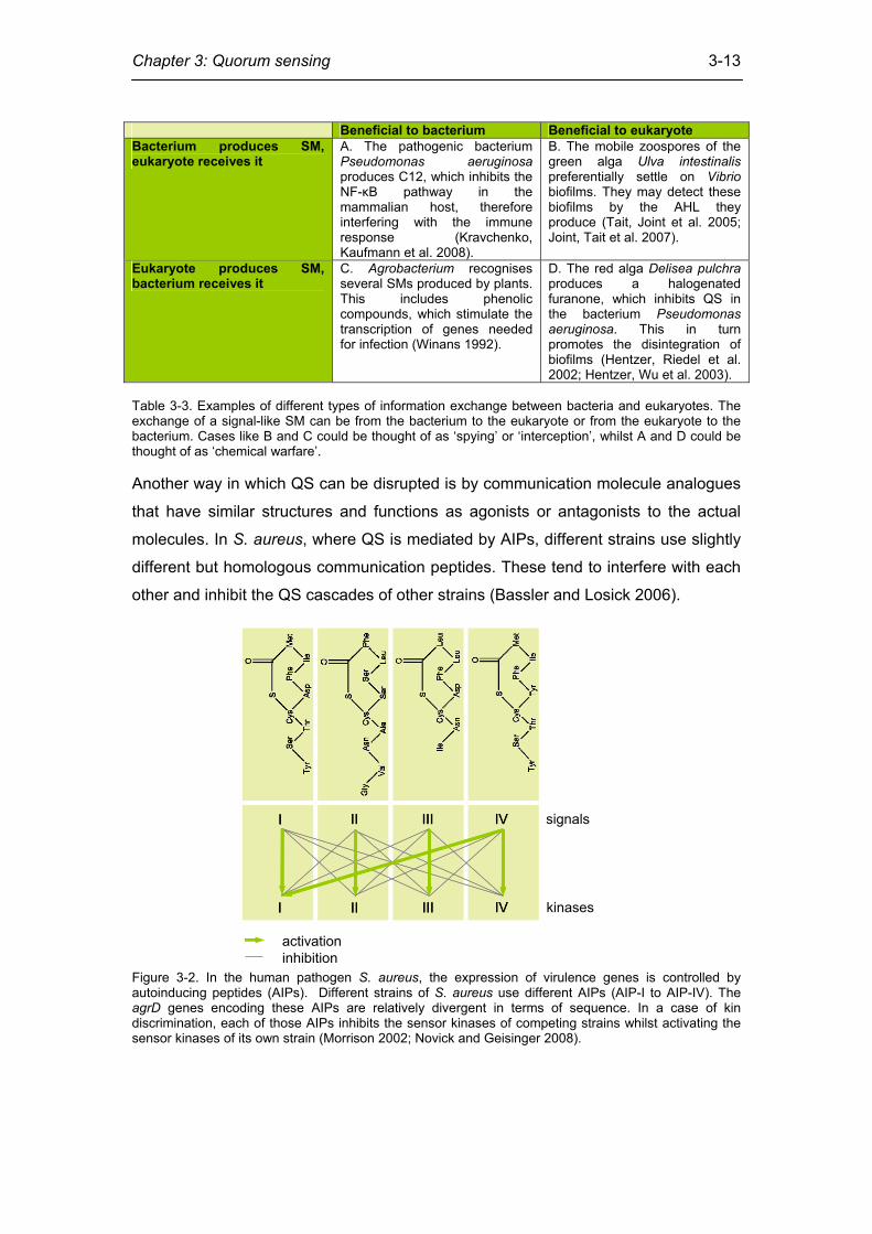
Table 3-3. Examples of different types of information exchange between bacteria and eukaryotes. The exchange of a signal-like SM can be from the bacterium to the eukaryote or from the eukaryote to the bacterium. Cases like B and C could be thought of as ‘spying’ or ‘interception’, whilst A and D could be thought of as ‘chemical warfare’. Another way in which QS can be disrupted is by communication molecule analogues
that have similar structures and functions as agonists or antagonists to the actual
molecules. In S. aureus, where QS is mediated by AIPs, different strains use slightly
different but homologous communication peptides. These tend to interfere with each
other and inhibit the QS cascades of other strains (Bassler and Losick 2006).
signals
kinases
II IIII IV
I II III IV
II IIII IV
I II III IV
activationinhibition
Figure 3-2. In the human pathogen S. aureus, the expression of virulence genes is controlled by autoinducing peptides (AIPs). Different strains of S. aureus use different AIPs (AIP-I to AIP-IV). The agrD genes encoding these AIPs are relatively divergent in terms of sequence. In a case of kin discrimination, each of those AIPs inhibits the sensor kinases of competing strains whilst activating the sensor kinases of its own strain (Morrison 2002; Novick and Geisinger 2008).
Chapter 3: Quorum sensing
3-14
Cell-to-cell communication in yeast
In yeasts, several QS-like mechanisms have been reported. In all cases, the major
phenotype affected by QS is the transition between the filamentous form and the
solitary yeast form. For example, Histoplasma capsulatum is a parasitic yeast that
can either exist in its filamentous form in soil or in its yeast form as a parasite of
humans. Once it enters the host, its morphology switches to the yeast form which
synthesises cell-wall polysaccharides such as α-(1,3)-glucan. It has been shown that
the glucan concentration increases in a cell-density dependent fashion. A culture
grown in fresh medium to which filtrate from a dense culture is added produces
glucan, which suggests the existence of a factor that promotes glucan incorporation
into the cell wall (see section Tools and Techniques for more details on this
approach).
Farnesol
The existence of a number of different QS molecules has been reported for the
human pathogen Candida albicans. At low densities, the cells develop germ tubes
(filamentous protrusions) which are not normally observed at high cell densities,
which suggests that the switch between unicellular yeast and filamentous form
depends on cell density. A molecule which blocks the formation of these germ tubes
at high cell densities has been identified as farnesol, which has only been observed
to have this function in C. albicans [53]. Farsenol, as used for communication in C.
albicans, consists of an OH group and a branched C15 side chain. Farnesol acts by
affecting transcription. The product of TUP1 is a transcriptional repressor regulating
the yeast-to-filamentous transition by negatively regulating the transcription of
hyphal-specific genes. TUP1 expression has been shown to be increased by farnesol.
In tup1∆/tup1∆, as well as other obligatorily filamentous mutants, farnesol production
is increased (Nickerson, Atkin et al. 2006), indicating some sort of negative feedback.
Another molecule involved in the high-to-low cell density transition identified in C.
albicans is tyrosol (Chen, Fujita et al. 2004). As opposed to farnesol it promotes cell
growth and the development of germ tubes at low cell densities. Expression profiling
of cultures under conditions of reduced tyrosol concentration showed reduced
expression of proteins involved in DNA synthesis and cell cycle regulation (Sprague
and Winans 2006). Other putative C. albicans QS molecules include the substance
MARS of unknown identity and, with diminutive effect, farsenoic acid (Nickerson,
Atkin et al. 2006).
Chapter 3: Quorum sensing
3-15
Aromatic alcohol derivatives
An altogether more detailed picture of density-dependent cell-to-cell communication
has been uncovered for Saccharomyces cerevisiae, where phenylethanol (a
phenylalanine aromatic alcohol derivative) and tryptophol (a tryptophan aromatic
alcohol derivative) have been implicated in QS (Chen and Fink 2006). As in the other
examples of yeast QS discussed above, these molecules regulate the transition to
the filamentous form. They synergistically affect the upregulation of FLO11 via the
cAMP-dependent kinase Tpk2p (a PKA subunit) and the transcription factor Flo8p.
Flo11p, the product of FLO11, is the GPI anchored cell surface flocculin protein and
is essential for filamentous growth. S. cerevisiae strains with deletions of either TPK2
or FLO8 do not form filaments in response to aromatic alcohols (Chen and Fink
2006). I present my research on the S. cerevisiae aromatic alcohol QS system in
more detail later in this chapter.
Tools and Techniques
Chemical
The identification of cell-to-cell communication molecules is a non-trivial task. At an
abstract level, a simple assay that allows us to ascertain if a certain phenotype is
influenced by QS is to grow cells in culture in stationary phase for some time. During
that period, potential communication molecules can accumulate. The cells are then
filtered and/or centrifuged out of the growth medium and the remainder is purified. If
the addition of this conditioned medium to fresh, exponentially growing cells induces
the phenotype in question, one possible explanation is that QS molecules affecting
the phenotype were present in the filtrate. High-resolution liquid
chromatography/mass spectrometry (LC/MS) and nuclear magnetic resonance
(NMR) spectroscopy can aid in the identification of the actual QS molecules. This is
achieved by enabling the researcher to compare the spectra of synthetic molecules
to molecules purified from the conditioned medium.
Genetic
Genetic techniques to investigate QS include the disruption of the genes in a
pathway that produces QS molecules, knock-out of sensor and response regulator
genes, addition of purified or synthetic QS molecules, or addition of quorum
quenchers. The identity of synthetic and actual (purified) communication molecules
can be verified by adding synthetic molecules to a mutant culture which cannot
produce its own signals. If the phenotypic effects are the same, this indicates that the
Chapter 3: Quorum sensing
3-16
synthetic and purified molecule share the same structure. It can however also
happen that chemically similar but not identical SMs elicit similar effects. For example,
the QS homoserine lactone 3OC12, as produced by the bacterium P. aeruginosa, has
been shown to mimic the effects of farsenol in C. albicans, probably due to it being
somewhat similar in its side chain structure.
Computational
QS can also be studied by computational methods. The structure of many QS
molecules as well as that of their cognate synthases and receptor molecules has
been solved by X-ray diffraction analysis or NMR. Gene expression microarray
datasets under QS conditions are also publicly available. Integrating structural and
gene expression data with the abundantly available genome sequence data will
therefore be of great value for the discovery of new QS systems as well as for the
integration of QS with intracellular signalling pathways, and later in this chapter I give
examples of this in bacteria and in yeast.
A survey of quorum sensing protein domains
Until now, I have introduced different known QS systems and their components,
including communication molecule synthase and receptor proteins. A knowledge of
the diverse components of different QS systems allows the identification of new
systems in fully sequenced genomes using a computational approach.
I used a hand-curated library of hidden Markov models (HMMs) of protein domains
that are unique to QS (table 3-4) to identify new QS systems in 384 fully sequenced
microbial genomes. The result was the discovery of a number of putative QS
systems in genomes that were not previously known to possess such systems.
Results and discussion
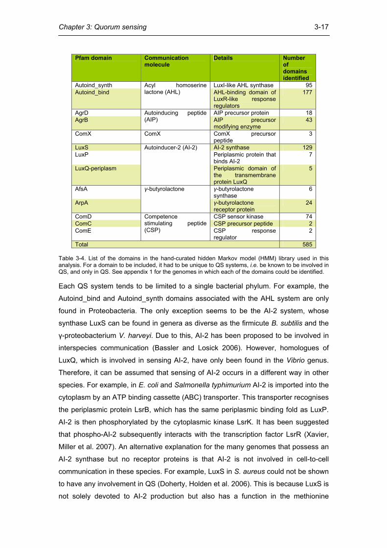
I identified 585 QS protein domains, which are listed by genome in appendix 1. The
most common domains were the AHL-binding Autoind_bind domain (177 instances)
and the AI-2 synthase LuxS (129 instances).
Chapter 3: Quorum sensing
3-17
Table 3-4. List of the domains in the hand-curated hidden Markov model (HMM) library used in this analysis. For a domain to be included, it had to be unique to QS systems, i.e. be known to be involved in QS, and only in QS. See appendix 1 for the genomes in which each of the domains could be identified. Each QS system tends to be limited to a single bacterial phylum. For example, the
Autoind_bind and Autoind_synth domains associated with the AHL system are only
found in Proteobacteria. The only exception seems to be the AI-2 system, whose
synthase LuxS can be found in genera as diverse as the firmicute B. subtilis and the
γ-proteobacterium V. harveyi. Due to this, AI-2 has been proposed to be involved in
interspecies communication (Bassler and Losick 2006). However, homologues of
LuxQ, which is involved in sensing AI-2, have only been found in the Vibrio genus.
Therefore, it can be assumed that sensing of AI-2 occurs in a different way in other
species. For example, in E. coli and Salmonella typhimurium AI-2 is imported into the
cytoplasm by an ATP binding cassette (ABC) transporter. This transporter recognises
the periplasmic protein LsrB, which has the same periplasmic binding fold as LuxP.
AI-2 is then phosphorylated by the cytoplasmic kinase LsrK. It has been suggested
that phospho-AI-2 subsequently interacts with the transcription factor LsrR (Xavier,
Miller et al. 2007). An alternative explanation for the many genomes that possess an
AI-2 synthase but no receptor proteins is that AI-2 is not involved in cell-to-cell
communication in these species. For example, LuxS in S. aureus could not be shown
to have any involvement in QS (Doherty, Holden et al. 2006). This is because LuxS is
not solely devoted to AI-2 production but also has a function in the methionine
Pfam domain Communication molecule
Details Number of domains identified
Autoind_synth LuxI-like AHL synthase 95 Autoind_bind
Acyl homoserine lactone (AHL) AHL-binding domain of
LuxR-like response regulators
177
AgrD AIP precursor protein 18 AgrB
Autoinducing peptide (AIP) AIP precursor
modifying enzyme 43
ComX ComX ComX precursor peptide
3
LuxS AI-2 synthase 129 LuxP Periplasmic protein that
binds AI-2 7
LuxQ-periplasm
Autoinducer-2 (AI-2)
Periplasmic domain of the transmembrane protein LuxQ
5
AfsA γ-butyrolactone synthase
6
ArpA
γ-butyrolactone
γ-butyrolactone receptor protein
24
ComD CSP sensor kinase 74 ComC CSP precursor peptide 2 ComE
Competence stimulating peptide (CSP) CSP response
regulator 2
Total 585
Chapter 3: Quorum sensing
3-18
metabolic pathway (Rezzonico and Duffy 2008). Therefore, many of the LuxS
homologues identified here may not produce communication molecules but simply
metabolic side-products that are not meant to convey information between cells.
A γ-butyrolactone quorum sensing system in Rhodococcus?
One QS system that seems to be limited to Actinobacteria is the γ-butyrolactone
system. It is similar to the AHL-based system in the sense that there is some
structural similarity between γ-butyrolactone and AHL, but also in the sense that it is
a one-component system where the communication molecule sensing protein is also
the response regulator.
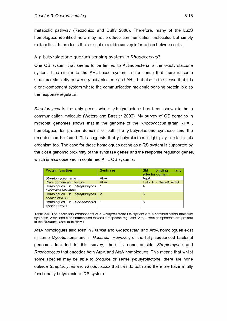
Streptomyces is the only genus where γ-butyrolactone has been shown to be a
communication molecule (Waters and Bassler 2006). My survey of QS domains in
microbial genomes shows that in the genome of the Rhodococcus strain RHA1,
homologues for protein domains of both the γ-butyrolactone synthase and the
receptor can be found. This suggests that γ-butyrolactone might play a role in this
organism too. The case for these homologues acting as a QS system is supported by
the close genomic proximity of the synthase genes and the response regulator genes,
which is also observed in confirmed AHL QS systems.
Protein function Synthase SM binding and effector domain
Streptomyces name AfsA ArpA Pfam domain architecture AfsA TetR_N - Pfam-B_4709 Homologues in Streptomyces avermitilis MA-4680
1 4
Homologues in Streptomyces coelicolor A3(2)
2 6
Homologues in Rhodococcus species RHA1
1 8
Table 3-5. The necessary components of a γ-butyrolactone QS system are a communication molecule synthase, AfsA, and a communication molecule response regulator, ArpA. Both components are present in the Rhodococcus strain RHA1. AfsA homologues also exist in Frankia and Gloeobacter, and ArpA homologues exist
in some Mycobacteria and in Nocardia. However, of the fully sequenced bacterial
genomes included in this survey, there is none outside Streptomyces and
Rhodococcus that encodes both ArpA and AfsA homologues. This means that whilst
some species may be able to produce or sense γ-butyrolactone, there are none
outside Streptomyces and Rhodococcus that can do both and therefore have a fully
functional γ-butyrolactone QS system.
Chapter 3: Quorum sensing
3-19
Although so far there has been no confirmation of the γ-butyrolactone QS system in
Rhodococcus strain RHA1, there is further evidence that supports the case that such
a system is present. Firstly, Rhodococcus produces γ-butyrolactone in high cell
density cultures (Curragh, Flynn et al. 1994). Secondly, Rhodococcus has an
unusually high number of AHL-inactivating lactonases. The number genes encoding
AHL-inactivating Lactamase_B Pfam domains in the genomes of Actinobacteria
included in this survey varies between one and 21. Only the Rhodococcus strain
RHA1 is an outlier and has 28 genes encoding proteins with such domains (data not
shown). These lactonases may play a role in the intracellular metabolism of lactone
compounds such as γ-butyrolactone (Uroz, Chhabra et al. 2005). In this context, it is
also interesting that Rhodococcus strain RHA1 has the only Autoind_synth domain
coding sequence found outside the Proteobacteria.
In summary, the identification of all necessary components of a QS system in
Rhodococcus strain RHA1 demonstrates that computational methods can give
information about previously uncharacterised QS systems. Later in this chapter I use
similar approaches to discover further QS systems in the bacterial phylum of
firmicutes.
Materials and methods
All sequence data were obtained from the NCBI genome database
(ftp://ftp.ncbi.nih.gov/genbank/genomes/Bacteria/). For most of this analysis I used a
set of 384 fully sequenced microbial genomes. Through an in-depth survey of the
literature available on QS, I identified protein domains that are unique to QS
processes (table 3-4). For each of these domains, Hidden Markov Models (HMMs)
were available on the protein families database Pfam (Finn, Mistry et al. 2006). I
used these HMM models to search the 384 genomes for further instances of the
same domains. These were then used to refine the HMM model and to re-search the
genomes in an iterative process until saturation. Both the hmmbuild and hmmsearch
programs that I used are part of the HMMER suite of programmes
(http://hmmer.janelia.org/) (Durbin, Eddy et al. 1998).
Some of the methods I use in the next section are similar to the ones used in this one,
but its focus is narrower. Instead of dealing with a wide range of bacterial genomes, it
concentrates on firmicutes only, and instead of dealing with a wide range of QS
systems, it deals with the agr system only.
Chapter 3: Quorum sensing
3-20
The agr quorum sensing system in firmicutes
As already discussed above, the pathogenic firmicute S. aureus has a well-
characterised cell-to-cell communication system whose signals are modified peptides
(Ji, Beavis et al. 1997). This system is encoded in the accessory gene regulator (agr)
locus (Recsei, Kreiswirth et al. 1986). The agr system is an important regulator of
biofilm formation and virulence factor expression (Kleerebezem, Quadri et al. 1997),
and consists of four genes encodeding a polycistronic transcript (figure 3-3A). The
agrD and agrB genes encode the precursor signalling peptide and the enzyme that
cleaves and modifies the precursor peptide, respectively. The resultant signal is
called autoinducing peptide (AIP). The agrC gene encodes the transmembrane
kinase that senses the AIP, and the agrA gene encodes the response regulator that
is phosphorylated by AgrC and effects changes in gene expression, respectively
(figure 3-3B).
The AIP signal has seven to nine residues, depending on the strain. The only
consistently conserved residue of the AIP is a cysteine at the 5th position from the C-
terminus (figure 3-4). This cysteine forms a thiolactone ring with the residue at the C-
terminus (Mayville, Ji et al. 1999). The AIP is produced from the precursor peptide
AgrD. The N- and C-terminal portions of AgrD are cleaved off by the membrane-
associated enzyme AgrB, which also forms the thiolactone ring, hence producing the
AIP. AgrB might also be involved in the export of the AIP across the cell membrane
(Qiu, Pei et al. 2005).
However, overexpression of AgrB and AgrD does not result in increased AIP
production. AgrB therefore does not seem to be the only processing enzyme required
for the synthesis of AIP (Saenz, Augsburger et al. 2000). Once it is in the intercellular
space, AIP diffuses until it binds the AgrC sensor kinase. This kinase can either be
located in the membrane of the cell which produces AIP, or in the membrane of
another S. aureus cell. The AgrC kinase is part of a two-component signal
transduction system. The second component is the cytoplasmic response regulator
AgrA. AgrA has two domains: The N-terminal domain, which is a receiver domain
that becomes phosphorylated by AgrC, and the C-terminal domain, which is the DNA
binding LytTR domain. Phosphorylation of the receiver domain leads to AgrA
activation and DNA binding. AgrA positively regulates transcription of the agr operon.
This means that a positive feedback effect takes place, with a high level of AIP
leading to even higher agr expression. Apart from the agrBDCA promoter, another
Chapter 3: Quorum sensing
3-21
promoter positively regulated by AgrA is the one controlling expression of RNAIII.
The RNAIII promoter is located next to the agrBDCA promoter, but is transcribed in
the opposite direction (figure 3-3A). Until recently, it was believed that RNAIII rather
than AgrA is the immediate regulator of most of the genes regulated by the agr
operon (Benito, Kolb et al. 2000). However, it has recently been shown that the
genes regulated by the agr operon can be divided into RNAIII-dependent and RNAIII-
independent, AgrA-dependent genes (Queck, Jameson-Lee et al. 2008). Although
RNAIII is a regulatory RNA, its transcript also contains the δ-haemolysin gene hld. In
some strains of S. aureus, the staphylococcal accessory regulator (SarA) protein is
also necessary to activate transcription of agrBDCA and RNAIII (Cheung and Projan
1994). SarA is a relatively abundant protein that seems to be a global regulator
modulating the expression of many genes.
Systems that are similar to the agr system either by homology or analogy have been
identified in firmicutes outside Staphylococcus. In some cases they have different
names. For example, the homologous agr-like system in Enterococcus faecalis is
known as fsr system (Qin, Singh et al. 2000), and the one in Lactobacillus plantarum
is called lam system (Diep, Havarstein et al. 1994). Here, I investigate the distribution
of agr-like systems outside the Staphylococcaceae in order to understand the
conservation and plasticity of their interaction with other cellular components and
signal transduction and communication systems.
Results and discussion
In order to define agr homologues in taxa other than Staphylococcus, I built hidden
Markov models (HMMs) of protein domains which are considered to be unique to the
agr system. In a complementary approach, I identified agr homologues with PSI-
BLAST searches. Protein domains which I considered to be unique to the agr system
are the AgrB domain and the AgrD domain. To my knowledge, no function of AgrB
apart from processing of AgrD has been reported so far. The remaining protein
domains of the agr system are the HATPase_c Pfam domain of the histidine kinase
and the Response_reg and LytTR domains of the response regulator. However,
these domains are not unique to the agr system and were therefore not considered to
be prognostic of it. I built HMMs of the AgrB and the AgrD domains, starting with the
published Pfam domains and adding homologous sequences identified in the
literature.
Chapter 3: Quorum sensing
3-22
Streptococcus pyogenes M1 GAS Streptococcus pyogenes SSI-1 Streptococcus pyogenes MGAS8232 Streptococcus pyogenes MGAS5005 Streptococcus pyogenes MGAS10394 Streptococcus pyogenes MGAS315 Streptococcus pyogenes MGAS10270 Streptococcus pyogenes MGAS9429 Streptococcus pyogenes MGAS6180 Streptococcus pyogenes MGAS2096
Streptococcus agalactiae 2603 Streptococcus agalactiae A909 Streptococcus agalactiae NEM316
Streptococcus pneumoniae R6 Streptococcus pneumoniae TIGR4
Streptococcus mutans Streptococcus thermophilus CNRZ1066 Streptococcus thermophilus LMG 18311 Lactococcus lactis
Enterococcus faecalis V583 Lactobacillus acidophilus NCFM Lactobacillus johnsonii NCC 533
Lactobacillus sakei 23K Lactobacillus plantarum Lactobacillus salivarius UCC118
Lactobacillales
Listeria monocytogenes 4b F2365 Listeria innocua Listeria monocytogenes
Listeriaceae
Bacillus cereus ZK Bacillus anthracis Ames Bacillus anthracis str Sterne Bacillus cereus ATCC 10987 Bacillus thuringiensis konkukian Bacillus anthracis Ames 0581 Bacillus cereus ATCC14579 Bacillus subtilis Geobacillus kaustophilus HTA426
Oceanobacillus iheyensis Bacillus halodurans Bacillus clausii KSM-K16
Bacillaceae
Staphylococcus saprophyticus Staphylococcus epidermidis ATCC 12228 Staphylococcus epidermidis RP62A Staphylococcus haemolyticus Staphylococcus aureus USA300 Staphylococcus aureus N315 Staphylococcus aureus RF122 Staphylococcus aureus COL Staphylococcus aureus NCTC 8325 Staphylococcus aureus aureus MSSA476 Staphylococcus aureus aureus MRSA252 Staphylococcus aureus MW2 Staphylococcus aureus Mu50
Staphylococcaceae
Carboxydothermus hydrogenoformans Z-2901 Moorella thermoacetica ATCC 39073
Syntrophomonas wolfei Goettingen Desulfitobacterium hafniense Y51
Thermoanaerobacter tengcongensis Clostridium acetobutylicum Clostridium perfringens SM101 Clostridium perfringens ATCC 13124 Clostridium perfringens
Clostridia
Acholeplasmatales Aster yellows witches-broom phytoplasma Mycoplasma capricolum ATCC 27343 Mesoplasma florum L1
Mycoplasma genitalium Mycoplasma pneumoniae
Mycoplasma gallisepticum Mycoplasma penetrans
Ureaplasma urealyticum Mycoplasma synoviae 53
Mycoplasma mobile 163K Mycoplasma pulmonis
Mycoplasma hyopneumoniae 7448 Mycoplasma hyopneumoniae 232 Mycoplasma hyopneumoniae J
Mycoplasmatales
Escherichia coli K1271
100
10099
5370
100
70
100
100
92100
100
100
99
100
100
51
85
100
99
47100
99
100
4753
61
51
100
79
99
64
49
42
92
90
60
99
62
5247
99
99
91
100100
100
100
100
56
42
99
100
63
pathogenicnot pathogenic
agrBagrD
1 – agrB/agrD fusion2 – agrB overlaps with other gene3 – mobile genetic element insertion4 – potential agrD homologueother gene
agr-associatedagrC
agrApathogenicnot pathogenic
agrBagrD
1 – agrB/agrD fusion2 – agrB overlaps with other gene3 – mobile genetic element insertion4 – potential agrD homologueother gene
agr-associatedagrC
agrA
157 15 218
2 0 0 50
2000
1 0 5 46
157 15 218
2 0 0 50
2000
1 0 5 46
Sta
phyl
ococ
cus
aure
usS
taph
yloc
occu
sep
ider
mid
is
Entero-coccusfaecalis
Lacto-bacillusplantarum
A
B
C
D
RNAIII agrA-C
AIP
AgrB
AgrD
AgrA
AgrC
target genes
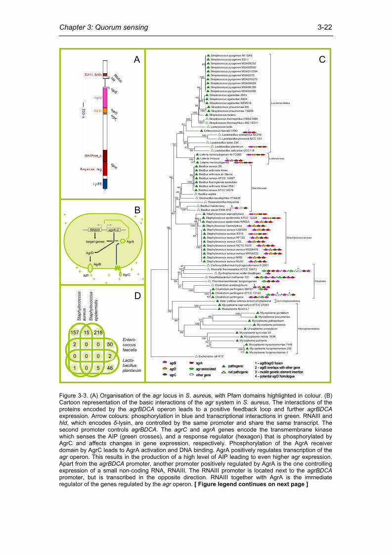
Figure 3-3. (A) Organisation of the agr locus in S. aureus, with Pfam domains highlighted in colour. (B) Cartoon representation of the basic interactions of the agr system in S. aureus. The interactions of the proteins encoded by the agrBDCA operon leads to a positive feedback loop and further agrBDCA expression. Arrow colours: phosphorylation in blue and transcriptional interactions in green. RNAIII and hld, which encodes δ-lysin, are controlled by the same promoter and share the same transcript. The second promoter controls agrBDCA. The agrC and agrA genes encode the transmembrane kinase which senses the AIP (green crosses), and a response regulator (hexagon) that is phosphorylated by AgrC and affects changes in gene expression, respectively. Phosphorylation of the AgrA receiver domain by AgrC leads to AgrA activation and DNA binding. AgrA positively regulates transcription of the agr operon. This results in the production of a high level of AIP leading to even higher agr expression. Apart from the agrBDCA promoter, another promoter positively regulated by AgrA is the one controlling expression of a small non-coding RNA, RNAIII. The RNAIII promoter is located next to the agrBDCA promoter, but is transcribed in the opposite direction. RNAIII together with AgrA is the immediate regulator of the genes regulated by the agr operon. [ Figure legend continues on next page ]
Chapter 3: Quorum sensing
3-23
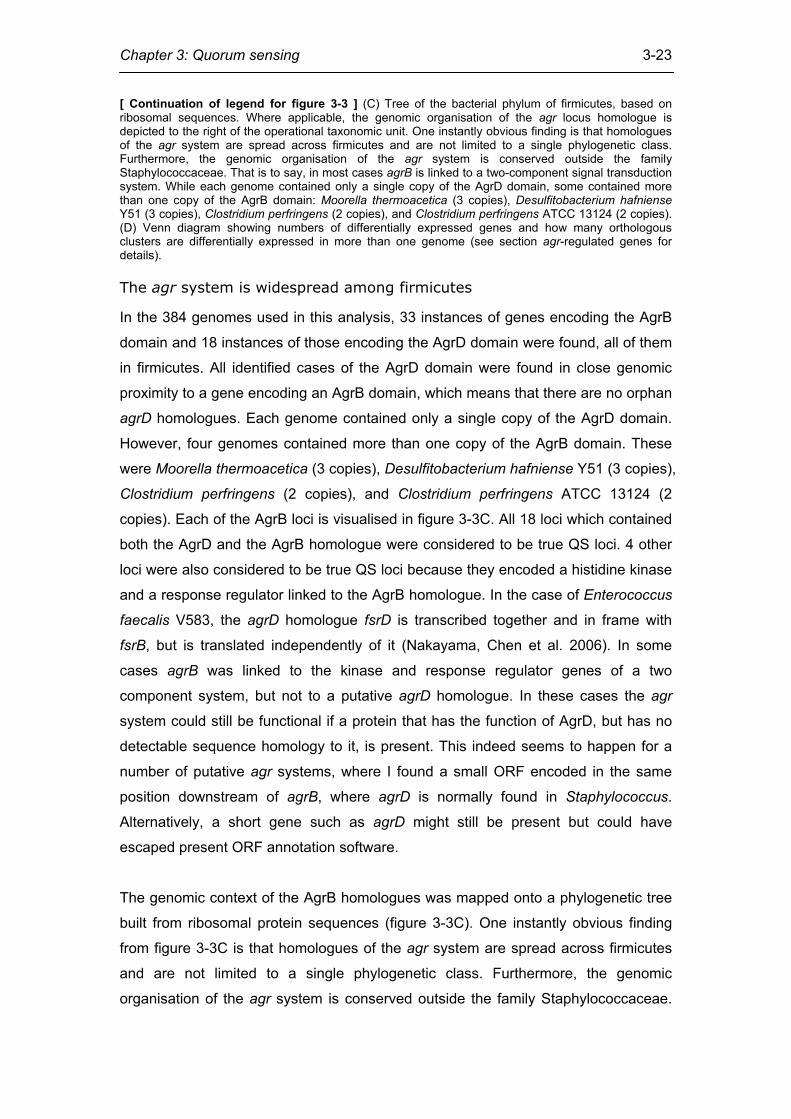
[ Continuation of legend for figure 3-3 ] (C) Tree of the bacterial phylum of firmicutes, based on ribosomal sequences. Where applicable, the genomic organisation of the agr locus homologue is depicted to the right of the operational taxonomic unit. One instantly obvious finding is that homologues of the agr system are spread across firmicutes and are not limited to a single phylogenetic class. Furthermore, the genomic organisation of the agr system is conserved outside the family Staphylococcaceae. That is to say, in most cases agrB is linked to a two-component signal transduction system. While each genome contained only a single copy of the AgrD domain, some contained more than one copy of the AgrB domain: Moorella thermoacetica (3 copies), Desulfitobacterium hafniense Y51 (3 copies), Clostridium perfringens (2 copies), and Clostridium perfringens ATCC 13124 (2 copies). (D) Venn diagram showing numbers of differentially expressed genes and how many orthologous clusters are differentially expressed in more than one genome (see section agr-regulated genes for details).
The agr system is widespread among firmicutes
In the 384 genomes used in this analysis, 33 instances of genes encoding the AgrB
domain and 18 instances of those encoding the AgrD domain were found, all of them
in firmicutes. All identified cases of the AgrD domain were found in close genomic
proximity to a gene encoding an AgrB domain, which means that there are no orphan
agrD homologues. Each genome contained only a single copy of the AgrD domain.
However, four genomes contained more than one copy of the AgrB domain. These
were Moorella thermoacetica (3 copies), Desulfitobacterium hafniense Y51 (3 copies),
Clostridium perfringens (2 copies), and Clostridium perfringens ATCC 13124 (2
copies). Each of the AgrB loci is visualised in figure 3-3C. All 18 loci which contained
both the AgrD and the AgrB homologue were considered to be true QS loci. 4 other
loci were also considered to be true QS loci because they encoded a histidine kinase
and a response regulator linked to the AgrB homologue. In the case of Enterococcus
faecalis V583, the agrD homologue fsrD is transcribed together and in frame with
fsrB, but is translated independently of it (Nakayama, Chen et al. 2006). In some
cases agrB was linked to the kinase and response regulator genes of a two
component system, but not to a putative agrD homologue. In these cases the agr
system could still be functional if a protein that has the function of AgrD, but has no
detectable sequence homology to it, is present. This indeed seems to happen for a
number of putative agr systems, where I found a small ORF encoded in the same
position downstream of agrB, where agrD is normally found in Staphylococcus.
Alternatively, a short gene such as agrD might still be present but could have
escaped present ORF annotation software.
The genomic context of the AgrB homologues was mapped onto a phylogenetic tree
built from ribosomal protein sequences (figure 3-3C). One instantly obvious finding
from figure 3-3C is that homologues of the agr system are spread across firmicutes
and are not limited to a single phylogenetic class. Furthermore, the genomic
organisation of the agr system is conserved outside the family Staphylococcaceae.
Chapter 3: Quorum sensing
3-24
That is to say, in most cases agrB is linked to a two-component signal transduction
system.
These findings pose the question of whether the agr QS system is an evolutionarily
ancient system which was present in the common ancestor of firmicutes, or whether
it is a more recent innovation and has been transferred between groups by horizontal
gene transfer. To investigate this, I built two more phylogenetic trees (not shown).
The first tree was built from an alignment of AgrB homologues. The second tree was
built from an alignment of AgrB, AgrD, AgrC, and AgrA homologues, whose
sequences were concatenated. The topology of these trees should agree with the
topology of the tree built from ribosomal sequences if agr is inherited only vertically.
Although these trees generally agree with each other and the ribosomal tree, the agr
locus of Clostridium acetobutylicum clusters with the Listeriaceae. C. acetobutylicum
however is a member of the Clostridia, therefore making its agr system a possible
candidate for horizontal gene transfer. To my knowledge, this is the first reported
instance of a possible case of horizontal gene transfer between the Clostridia and the
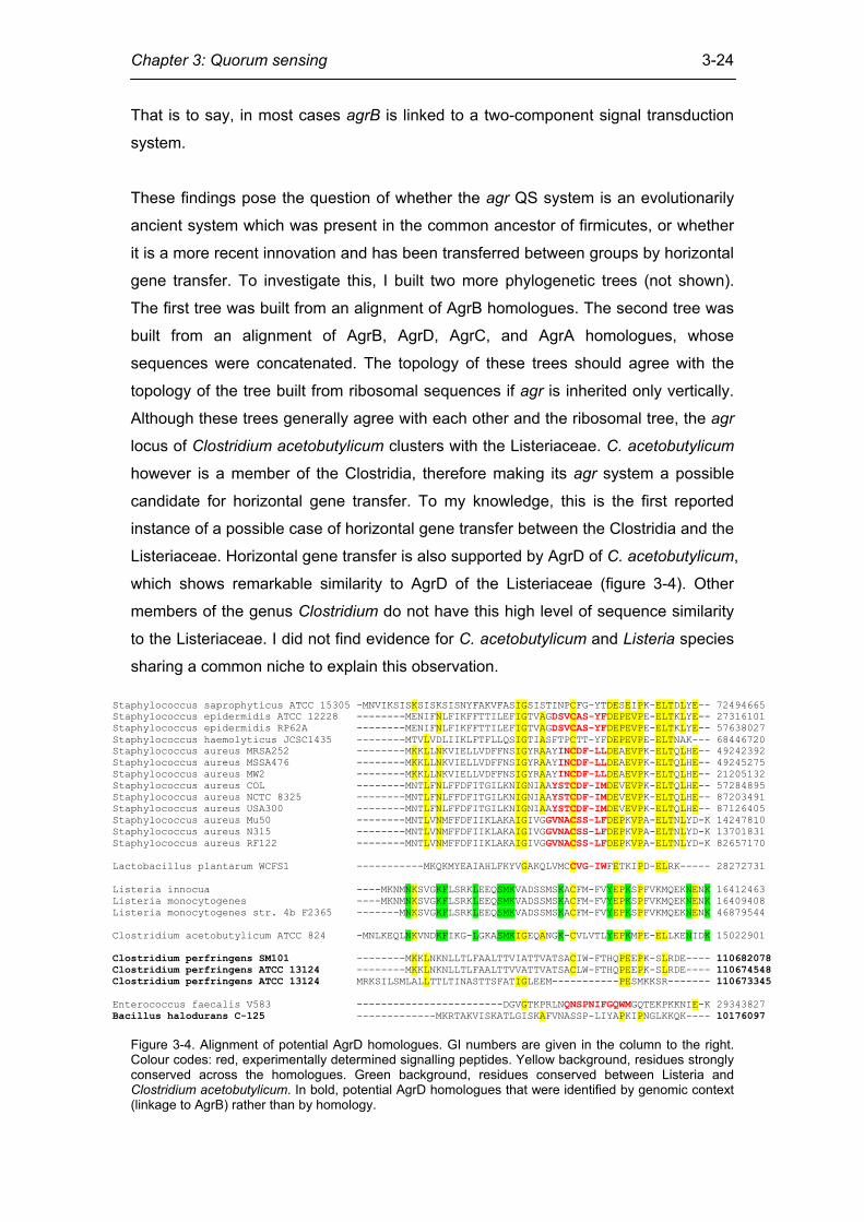
Listeriaceae. Horizontal gene transfer is also supported by AgrD of C. acetobutylicum,
which shows remarkable similarity to AgrD of the Listeriaceae (figure 3-4). Other
members of the genus Clostridium do not have this high level of sequence similarity
to the Listeriaceae. I did not find evidence for C. acetobutylicum and Listeria species
sharing a common niche to explain this observation.
Staphylococcus saprophyticus ATCC 15305 -MNVIKSISKSISKSISNYFAKVFASIGSISTINPCFG-YTDESEIPK-ELTDLYE-- 72494665 Staphylococcus epidermidis ATCC 12228 --------MENIFNLFIKFFTTILEFIGTVAGDSVCAS-YFDEPEVPE-ELTKLYE-- 27316101 Staphylococcus epidermidis RP62A --------MENIFNLFIKFFTTILEFIGTVAGDSVCAS-YFDEPEVPE-ELTKLYE-- 57638027 Staphylococcus haemolyticus JCSC1435 --------MTVLVDLIIKLFTFLLQSIGTIASFTPCTT-YFDEPEVPE-ELTNAK--- 68446720 Staphylococcus aureus MRSA252 --------MKKLLNKVIELLVDFFNSIGYRAAYINCDF-LLDEAEVPK-ELTQLHE-- 49242392 Staphylococcus aureus MSSA476 --------MKKLLNKVIELLVDFFNSIGYRAAYINCDF-LLDEAEVPK-ELTQLHE-- 49245275 Staphylococcus aureus MW2 --------MKKLLNKVIELLVDFFNSIGYRAAYINCDF-LLDEAEVPK-ELTQLHE-- 21205132 Staphylococcus aureus COL --------MNTLFNLFFDFITGILKNIGNIAAYSTCDF-IMDEVEVPK-ELTQLHE-- 57284895 Staphylococcus aureus NCTC 8325 --------MNTLFNLFFDFITGILKNIGNIAAYSTCDF-IMDEVEVPK-ELTQLHE-- 87203491 Staphylococcus aureus USA300 --------MNTLFNLFFDFITGILKNIGNIAAYSTCDF-IMDEVEVPK-ELTQLHE-- 87126405 Staphylococcus aureus Mu50 --------MNTLVNMFFDFIIKLAKAIGIVGGVNACSS-LFDEPKVPA-ELTNLYD-K 14247810 Staphylococcus aureus N315 --------MNTLVNMFFDFIIKLAKAIGIVGGVNACSS-LFDEPKVPA-ELTNLYD-K 13701831 Staphylococcus aureus RF122 --------MNTLVNMFFDFIIKLAKAIGIVGGVNACSS-LFDEPKVPA-ELTNLYD-K 82657170 Lactobacillus plantarum WCFS1 -----------MKQKMYEAIAHLFKYVGAKQLVMCCVG-IWFETKIPD-ELRK----- 28272731 Listeria innocua ----MKNMNKSVGKFLSRKLEEQSMKVADSSMSKACFM-FVYEPKSPFVKMQEKNENK 16412463 Listeria monocytogenes ----MKNMNKSVGKFLSRKLEEQSMKVADSSMSKACFM-FVYEPKSPFVKMQEKNENK 16409408 Listeria monocytogenes str. 4b F2365 -------MNKSVGKFLSRKLEEQSMKVADSSMSKACFM-FVYEPKSPFVKMQEKNENK 46879544 Clostridium acetobutylicum ATCC 824 -MNLKEQLNKVNDKFIKG-LGKASMKIGEQANGK-CVLVTLYEPKMPE-ELLKENIDK 15022901 Clostridium perfringens SM101 --------MKKLNKNLLTLFAALTTVIATTVATSACIW-FTHQPEEPK-SLRDE---- 110682078 Clostridium perfringens ATCC 13124 --------MKKLNKNLLTLFAALTTVVATTVATSACLW-FTHQPEEPK-SLRDE---- 110674548 Clostridium perfringens ATCC 13124 MRKSILSMLALLTTLTINASTTSFATIGLEEM-----------PESMKKSR------- 110673345 Enterococcus faecalis V583 ------------------------DGVGTKPRLNQNSPNIFGQWMGQTEKPKKNIE-K 29343827 Bacillus halodurans C-125 -------------MKRTAKVISKATLGISKAFVNASSP-LIYAPKIPNGLKKQK---- 10176097
Figure 3-4. Alignment of potential AgrD homologues. GI numbers are given in the column to the right. Colour codes: red, experimentally determined signalling peptides. Yellow background, residues strongly conserved across the homologues. Green background, residues conserved between Listeria and Clostridium acetobutylicum. In bold, potential AgrD homologues that were identified by genomic context (linkage to AgrB) rather than by homology.
Chapter 3: Quorum sensing
3-25
New agr-like systems
In three genomes I was able to identify a putative agr system where, to my
knowledge, none had been reported before. These genomes are Moorella
thermoacetica ATCC 39070, Desulfitobacterium hafniense Y51, and
Thermoanaerobacter tengcongensis. All three species are members of the taxonomic
class Clostridia. In all three genomes, agrB occurs as a genomic neighbour of a gene
encoding a response regulator and a gene encoding a histidine kinase, which makes
a function of the locus in cell-to-cell communication plausible (Bassler and Losick
2006). I propose that these genomes might contain a functional peptide QS circuit. In
the following, I discuss the implications of QS in these organisms. Like all Clostridia,
these species are anaerobic, and Moorella and Thermoanaerobacter are
thermophilic (Xue, Xu et al. 2001; Drake and Daniel 2004). All three species are non-
pathogenic. The genus Desulfitobacterium consists of bacteria that survive in a wide
range of environments. Desulfitobacteria are metabolically diverse and use a variety
of compounds as electron acceptors, including metals, nitrate, sulphite, and
halogenated compounds. This makes the Desulfitobacteria a candidate for anaerobic
bioremediation processes. In anaerobic fixed-film reactors D. hafniense has been
shown to occur in a biofilm together with sulphate reducing bacteria (Villemur,
Lanthier et al. 2006). If QS via the agr locus indeed occurs in D. hafniense, it can be
assumed that it would modulate biofilm formation as it does in other species
(Dunman, Murphy et al. 2001; Bourgogne, Hilsenbeck et al. 2006; Cassat, Dunman
et al. 2006).
In the genomes that contain an AgrB homologue that is not linked to a complete two-
component system, agr mediated QS might also be functional. This would require the
two-component sensing system to be encoded somewhere else in the genome rather
than being linked to agrB. Genomes which have an agrB homologue that is not linked
to a two-component system are those of Bacillus halodurans, Syntrophomonas wolfei,
and three C. perfringens strains. However, in the Clostridium perfringens strain
ATCC 13124 and strain 13 one of the two paralogues of AgrB co-occurs with a
histidine sensor kinase. Peptide-mediated QS in the Clostridium perfringens species
would be of particular interest, as this species is the causative agent of the human
disease gas gangrene (Present, Meislin et al. 1990). The possibility of agr-like
systems in the genus Clostridium has recently been explored (Burrell 2006). Burrell
examined uncultured Clostridium species isolated from landfill leachate reactor
biomass by cloning PCR products. Some homologues of agrC were identified using
Chapter 3: Quorum sensing
3-26
primers from the agrC homologue identified in C. thermocellum. However it was not
established if these agrC homologues are functional components of a QS circuit.
Multiple paralogous copies of AgrB in Clostridia genomes
Four of the genomes I examined in the Clostridia have more than one copy of AgrB.
These were M. thermoacetica ATCC 39073 (3 copies), D. hafniense Y51 (3 copies),
C. perfringens (2 copies), and C. perfringens ATCC 13124 (2 copies). The
phylogenetic evidence suggests that the two C. perfringens strains diverged after a
duplication event which created two agrB paralogues. This duplication event in the
common ancestor of the two strains must have been ancient, as the two copies are
phylogenetically distant. The same applies to the duplication events that have given
rise to multiple copies of AgrB in D. hafniense and M. thermoacetica. In all genomes
only one paralogue of AgrB is linked with a histidine kinase as well as a response
regulator, whilst the other paralogues seem to be orphans. This raises the question
what the function, if any, of the paralogous copies might be. I did not find any genes
which were consistently linked to the orphans or were predicted to be in the same
operon. Something which might explain the presence of multiple paralogous AgrB
homologues in the same genomes is that peptide 'signals' do not only serve the
purpose of cell-to-cell signalling, but are also involved in environmental sensing. It
can be speculated that the structure of peptide signals can be altered by the
extracellular conditions, subsequently passing this information on to two-component
systems (Morrison 2002). If this is the case, selection pressures other than signalling
efficiency and reliability act on peptide signalling systems. This provides a possible
explanation why for example M. thermoacetica or D. hafniense possess multiple
AgrB homologues: In an instance of subfunctionalisation they might use one or more
of the paralogues for environment sensing rather than cell-to-cell signalling. That all
genomes with multiple copies of agrB are found in the Clostridia could be because
many species in this class are versatile and can persist in a wide range of
environments, therefore having an increased need for environmental sensing. It
would not be necessary that the two-component system sensing the peptide is
encoded in close genomic proximity to the processing enzyme, AgrB. An alternative
hypothesis is that AgrB produces a peptide which interferes with QS in related but
competing strains. This would explain why one of the agrB genes appears to be an
orphan without a linked two-component system. Another explanation would be that
additional AgrB paralogues are used to increase signal production.
Chapter 3: Quorum sensing
3-27
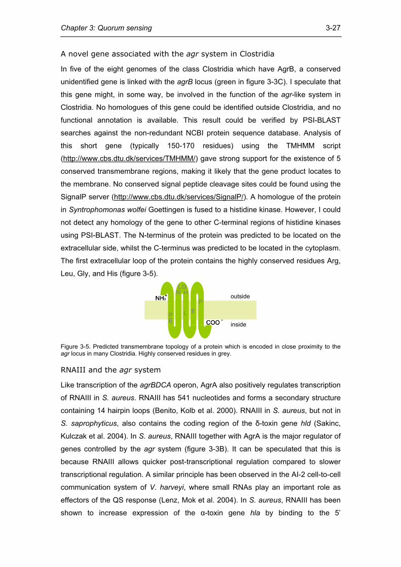
A novel gene associated with the agr system in Clostridia
In five of the eight genomes of the class Clostridia which have AgrB, a conserved
unidentified gene is linked with the agrB locus (green in figure 3-3C). I speculate that
this gene might, in some way, be involved in the function of the agr-like system in
Clostridia. No homologues of this gene could be identified outside Clostridia, and no
functional annotation is available. This result could be verified by PSI-BLAST
searches against the non-redundant NCBI protein sequence database. Analysis of
this short gene (typically 150-170 residues) using the TMHMM script
(http://www.cbs.dtu.dk/services/TMHMM/) gave strong support for the existence of 5
conserved transmembrane regions, making it likely that the gene product locates to
the membrane. No conserved signal peptide cleavage sites could be found using the
SignalP server (http://www.cbs.dtu.dk/services/SignalP/). A homologue of the protein
in Syntrophomonas wolfei Goettingen is fused to a histidine kinase. However, I could
not detect any homology of the gene to other C-terminal regions of histidine kinases
using PSI-BLAST. The N-terminus of the protein was predicted to be located on the
extracellular side, whilst the C-terminus was predicted to be located in the cytoplasm.
The first extracellular loop of the protein contains the highly conserved residues Arg,
Leu, Gly, and His (figure 3-5).
RLG
H
L EP
PE
outside
inside
NH3+NH3+
COO -COO -
Figure 3-5. Predicted transmembrane topology of a protein which is encoded in close proximity to the agr locus in many Clostridia. Highly conserved residues in grey.
RNAIII and the agr system
Like transcription of the agrBDCA operon, AgrA also positively regulates transcription
of RNAIII in S. aureus. RNAIII has 541 nucleotides and forms a secondary structure
containing 14 hairpin loops (Benito, Kolb et al. 2000). RNAIII in S. aureus, but not in
S. saprophyticus, also contains the coding region of the δ-toxin gene hld (Sakinc,
Kulczak et al. 2004). In S. aureus, RNAIII together with AgrA is the major regulator of
genes controlled by the agr system (figure 3-3B). It can be speculated that this is
because RNAIII allows quicker post-transcriptional regulation compared to slower
transcriptional regulation. A similar principle has been observed in the AI-2 cell-to-cell
communication system of V. harveyi, where small RNAs play an important role as
effectors of the QS response (Lenz, Mok et al. 2004). In S. aureus, RNAIII has been
shown to increase expression of the α-toxin gene hla by binding to the 5’
Chapter 3: Quorum sensing
3-28
untranslated region (UTR) of its mRNA. This is achieved by a high level of sequence
complementarity between the UTR and the 5’ end of RNAIII (Morfeldt, Taylor et al.
1995). I investigated how widespread this sort of regulation might be amongst
firmicutes by scanning whole genomic nucleotide sequences for RNAIII. I searched
the nucleotide sequences of complete genomes which encode a copy of AgrB for
RNAIII homologues. This resulted in highly significant hits for all S. aureus and S.
epidermidis strains. Putative hits were also found for S. haemolyticus and S.
saprophyticus, albeit at a lower significance level. In all cases, RNAIII was encoded
in close genomic proximity to the agr locus, with its promoter pointing in the opposite
direction, as depicted in Figure 3-3A. Putative RNAIII in Staphylococcus was also
found when no hld gene was predicted. The expression of RNAIII has been
confirmed experimentally in S. epidermidis (Tegmark, Morfeldt et al. 1998) and S.
saprophyticus (Sakinc, Kulczak et al. 2004). To my knowledge, RNAIII function has
not yet been examined in S. haemolyticus. It should be noted that the fact that I did
not identify RNAIII outside Staphylococcaceae does not necessarily mean that agr
does not exert its effect via noncoding RNAs in these species. However, I conclude
that RNAIII is an innovation which was made in the common ancestor of the
Staphylococcaceae.
agr-regulated genes
Differences in gene expression levels between wildtype and agr knockout cells have
previously been compared in Lactobacillus plantarum WCFS1 (Sturme, Nakayama et
al. 2005), Enterococcus faecalis OG1RF (Bourgogne, Hilsenbeck et al. 2006), S.
aureus UAMS-1 (Cassat, Dunman et al. 2006), and S. epidermidis (Yao, Vuong et al.
2006). According to my criteria (see Materials and Methods), 175 genes are
differentially expressed in S. aureus UAMS-1, 54 in L. plantarum, as well as 54 in E.
faecalis, and 238 in S. epidermidis. After defining clusters of orthologous genes, I
asked if there are any orthologues which are differentially expressed in all four
genomes. I did not find any cluster that is differentially regulated in all four genomes.
There are two clusters that are upregulated by agr in both E. faecalis and L.
plantarum. These two clusters correspond to AgrB and AgrC orthologues. There are
also two clusters which are upregulated in S. aureus N315 and E. faecalis. These
clusters correspond to a sugar ABC transporter and a prephenate dehydratase,
which is an enzyme involved in phenylalanine synthesis (MetCyc database (Caspi,
Foerster et al. 2006)). There is one cluster that is upregulated in both S. aureus N315
and L. plantarum, corresponding to a short chain dehydrogenase/reductase.
Chapter 3: Quorum sensing
3-29
Therefore, there is no significant propensity for orthologous genes to be regulated by
the agr system in the four different species. Genes that are involved in biofilm formation, such as those encoding
exopolysaccharide biosynthesis proteins, are downregulated in S. aureus and E.
faecalis. Some exopolysaccharide biosynthesis genes, such as those of the cps2
cluster, are upregulated in L. plantarum. This seems to contradict the observation
that deletion of lamA, the agrA homologue, has the phenotypic effect of reduced
adherence to a glass substratum compared to the wildtype. However the
polysaccharides might be membrane associated and capsular rather than secreted.
This in turn could prevent adhesion factors from increasing the cell's propensity to
form biofilms, as has been observed in S. aureus (Sturme, Nakayama et al. 2005).
However, it is not clear why agr would promote biofilm formation in the pathogen E.
faecalis but not in S. aureus N315, which is also a pathogen. The production of
virulence proteins is up-regulated by the agr-like system in both virulent species. A
clear conclusion is that the agr system is not a regulator of virulence factors only and
is also active in non-pathogenic firmicutes.
It should be noted that the platforms for L. plantarum and E. faecalis were dual-
channel cohybridisation arrays, while the platform for Staphylococcus aureus UAMS-
1 was an Affymetrix GeneChip. The absolute ratios by which genes were up-or
downregulated with respect to the wildtype are therefore not directly comparable.
However, the identity of differentially expressed genes should be platform
independent, and therefore comparable across experiments.
Concluding remarks
The number of completely sequenced microbial genomes is increasing steadily. In
addition, an enormous amount of sequence data is generated from metagenomics
projects. As it is very unlikely that all bacterial cell-to-cell communication systems
have already been described, new methods for predicting these communication
systems are required. Here I have applied an integrated approach which is capable
of identifying previously unknown systems.
Materials and methods
All sequence data used was obtained from the NCBI genome database. For most of
this analysis I used a set of 384 fully sequenced genomes.
Chapter 3: Quorum sensing
3-30
Homologue identification
In order to define agr homologues in taxa other than Staphylococcus, I built HMMs of
protein domains that I considered to be unique to the agr system. Protein domains
which I considered to be unique to the agr system are the AgrB domain and the AgrD
domain, each found in proteins with the respective name in Staphylococcus. The
remaining protein domains of the agr system are the HATPase_c domain of the
histidine kinase and the Response_reg and LytTR domains of the response regulator.
However, these domains are not unique to the agr system and were therefore not
taken to be indicative of it. As before, I built HMMs of the AgrB and the AgrD domains,
starting with published Pfam models (Finn, Mistry et al. 2006) and adding
homologous sequences identified in the literature. These sequences were aligned
using the MUSCLE multiple alignment programme (Edgar 2004) and manually
corrected. Following this, HMMs were created from the multiple sequence alignments
using the hmmbuild programme. These HMMs were then used to search the
microbial genomes for further homologues using the hmmsearch programme. These
homologues were then aligned again and used to build another model. This process
was repeated until further iterations did not identify new homologues. Both hmmbuild
and hmmsearch are part of the HMMER suite of programmes
(http://hmmer.janelia.org/; (Durbin, Eddy et al. 1998).
To validate my findings, I also did PSI-BLAST searches (Altschul, Madden et al.
1997) against the nonredundant NCBI protein sequence database, which is more
exhaustive than the database of fully sequenced genomes used here. All searches
were iterated until saturation. The results of the PSI-BLAST searches are available
on the supplementary material website.
Tree building
A guide tree of firmicutes was constructed using ribosomal proteins. Escherichia coli
K12 was used as an outgroup to root the tree. The ribosomal proteins used were L1,
L3, S2, and S5. The corresponding genes were determined by Ciccarlli et al. to be
very likely to occur only once in each genome and to be unlikely to be affected by
horizontal gene transfer (Ciccarelli, Doerks et al. 2006). HMMs for these proteins
were downloaded from Pfam and used to search the firmicute genomes for
homologues. Each genome was found to contain no more than one copy of each
protein. The four proteins were concatenated in each genome and aligned using
MUSCLE. The alignment was inspected by eye and gaps were removed.
Chapter 3: Quorum sensing
3-31
The tree in Figure 3-3C was constructed using the Neighbour Joining algorithm. A
Poisson correction model was used as a model of amino acid substitution. The tree
building procedure was repeated using a PAM matrix and a Jones-Taylor-Thornton
matrix as substitution models, but the topology of the resulting tree was identical in all
cases. Furthermore, I calculated bootstrap values by replicating the tree-building
procedure 1000 times. To evaluate the robustness of the method I constructed the
same tree using Maximum Parsimony and Minimum Evolution and compared the
results with the NJ tree. The topology within the seven groups Lactobacillales,
Listeriaceae, Bacillaceae, Staphylococcaceae, Clostridia, Acholeplasmatales, and
Mycoplasmatales was largely the same with these methods, although the relationship
between the groups was not.
For all tree building procedures MEGA 3.1 (Kumar, Tamura et al. 2004) was used.
RNAIII identification
Noncoding RNAs can tentatively be identified by using an approach that combines
sequence homology with secondary structure information. For this, I used the
programme INFERNAL, which predicts noncoding RNAs by using covariance models
that take into account both sequence homology and RNA secondary structure. The
model of RNAIII used by INFERNAL was obtained from the Rfam database (Griffiths-
Jones, Bateman et al. 2003). Whole genomic DNA sequences of those genomes that
have a predicted agr system were searched.
Gene expression data analysis
Gene expression data was collected from four previously published experiments
(Dunman, Murphy et al. 2001; Sturme, Nakayama et al. 2005; Bourgogne,
Hilsenbeck et al. 2006; Cassat, Dunman et al. 2006; Yao, Vuong et al. 2006). Genes
that were expressed at least three-fold differently in the agr mutants compared to the
wildtype were identified. For the two cohybridisation arrays this was done by using
the data as published in the respective papers. For the GeneChip experiment
(Cassat, Dunman et al. 2006), data was downloaded from the Gene Expression
Omnibus database (http://www.ncbi.nlm.nih.gov/projects/geo/), where its identifier is
GSE5466. The GeneChip used for these hybridisations was the Saur2a array, which
uses genomic sequences and ORF predictions from the Staphylococcus aureus
strains N315, MRSA252, MSSA476, NCTC8325, COL, and Mu50, as well as N315
intergenic regions which are longer than 50 nucleotides. For simplicity, strain N315
Chapter 3: Quorum sensing
3-32
was used as a reference strain in this analysis. Because this was not done by
Dunman et al., the list of genes which are differentially expressed differ from the one
reported here. Furthermore, only genes which showed a three-fold difference in
expression level in the agr mutant were included. For all three experiments,
expression data from the transition between late exponential growth phase and early
stationary phase was used.
Detection of orthologues
Orthologues between the genomes S. aureus N315, L. plantarum, E. faecalis, and S.
epidermidis were detected using the Inparanoid programme (Remm, Storm et al.
2001).
The Saccharomyces cerevisiae quorum sensing system
Outside of bacteria, QS has also been reported in fungi. Although in fungi the
chemical identity of the communication molecules varies, in all reported cases the
major phenotype reported is the transition between the filamentous growth form and
the solitary yeast form (Chen, Fujita et al. 2004; Chen and Fink 2006; Nickerson,
Atkin et al. 2006; Sprague and Winans 2006). In the model organism S. cerevisiae,
the aromatic alcohols phenylethanol and tryptophol, derived from the amino acids
phenylalanine and tryptophan, serve as communication molecules in low nitrogen
conditions (Chen and Fink 2006; Hogan 2006). Although the molecular machinery for
synthesising phenylethanol and tryptophol is known, it is not yet clear how S.
cerevisiae senses and transduces the signal. What we do know is that phenylethanol
and tryptophol synergistically affect the up-regulation of FLO11 via the cAMP-
dependent PKA subunit Tpk2p and the transcription factor Flo8p (Chen and Fink
2006) (figure 3-6). Flo11p, the product of FLO11, is a glycosylphosphatidylinositol
(GPI) anchored cell surface flocculin protein and is essential for filamentous growth
(Barrales, Jimenez et al. 2008; Fischer, Valerius et al. 2008; Vinod, Sengupta et al.
2008). S. cerevisiae strains with deletions of either TPK2 or FLO8 do not form
filaments in response to the aromatic alcohols (Chen and Fink 2006).
The production of phenylethanol and tryptophol is cell-density dependent. ARO9 and
ARO10 are two genes required for their synthesis. The expression of these aromatic
aminotransferases is induced by tryptophol via the Aro80p transcription factor. Since
tryptophol induces the enzymes required for its own synthesis, this results in a
positive feedback loop.
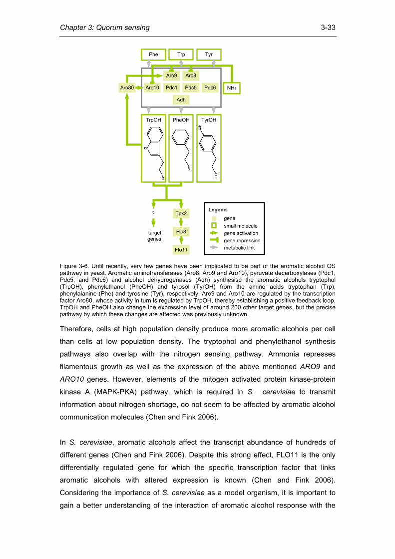
Chapter 3: Quorum sensing
3-33
NH4Aro80
Tpk2
Flo11
Flo8
TrpPhe Tyr
TrpOH PheOH TyrOH
Aro9 Aro8
Aro10
Adh
Pdc1 Pdc5 Pdc6
targetgenes
gene?
Legend
small moleculegene activationgene repressionmetabolic link
Figure 3-6. Until recently, very few genes have been implicated to be part of the aromatic alcohol QS pathway in yeast. Aromatic aminotransferases (Aro8, Aro9 and Aro10), pyruvate decarboxylases (Pdc1, Pdc5, and Pdc6) and alcohol dehydrogenases (Adh) synthesise the aromatic alcohols tryptophol (TrpOH), phenylethanol (PheOH) and tyrosol (TyrOH) from the amino acids tryptophan (Trp), phenylalanine (Phe) and tyrosine (Tyr), respectively. Aro9 and Aro10 are regulated by the transcription factor Aro80, whose activity in turn is regulated by TrpOH, thereby establishing a positive feedback loop. TrpOH and PheOH also change the expression level of around 200 other target genes, but the precise pathway by which these changes are affected was previously unknown. Therefore, cells at high population density produce more aromatic alcohols per cell
than cells at low population density. The tryptophol and phenylethanol synthesis
pathways also overlap with the nitrogen sensing pathway. Ammonia represses
filamentous growth as well as the expression of the above mentioned ARO9 and
ARO10 genes. However, elements of the mitogen activated protein kinase-protein
kinase A (MAPK-PKA) pathway, which is required in S. cerevisiae to transmit
information about nitrogen shortage, do not seem to be affected by aromatic alcohol
communication molecules (Chen and Fink 2006).
In S. cerevisiae, aromatic alcohols affect the transcript abundance of hundreds of
different genes (Chen and Fink 2006). Despite this strong effect, FLO11 is the only
differentially regulated gene for which the specific transcription factor that links
aromatic alcohols with altered expression is known (Chen and Fink 2006).
Considering the importance of S. cerevisiae as a model organism, it is important to
gain a better understanding of the interaction of aromatic alcohol response with the
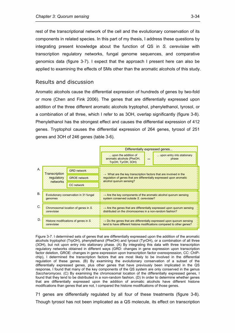
Chapter 3: Quorum sensing
3-34
rest of the transcriptional network of the cell and the evolutionary conservation of its
components in related species. In this part of my thesis, I address these questions by
integrating present knowledge about the function of QS in S. cerevisiae with
transcription regulatory networks, fungal genome sequences, and comparative
genomics data (figure 3-7). I expect that the approach I present here can also be
applied to examining the effects of SMs other than the aromatic alcohols of this study.
Results and discussion
Aromatic alcohols cause the differential expression of hundreds of genes by two-fold
or more (Chen and Fink 2006). The genes that are differentially expressed upon
addition of the three different aromatic alcohols tryptophol, phenylethanol, tyrosol, or
a combination of all three, which I refer to as 3OH, overlap significantly (figure 3-8).
Phenylethanol has the strongest effect and causes the differential expression of 412
genes. Tryptophol causes the differential expression of 264 genes, tyrosol of 251
genes and 3OH of 246 genes (table 3-6).
Differentially expressed genes...
... upon the addition of aromatic alcohols (PheOH,
TrpOH, TyrOH, 3OH)
... upon entry into stationary phase
GRD network
GROE network
CC network
Evolutionary conservation in 31 fungalgenomes
Chromosomal location of genes in S.cerevisiae
Transcriptionregulatorynetworks
→ What are the key transcription factors that are involved in the regulation of genes that are differentially expressed upon aromatic alcohol quorum sensing?
→ Are the key components of the aromatic alcohol quorum sensing system conserved outside S. cerevisiae?
→ Are the genes that are differentially expressed upon quorum sensing distributed on the chromosomes in a non-random fashion?
A.
B.
C.
D. Histone modifications of genes in S.cerevisiae
→ Do the genes that are differentially expressed upon quorum sensing tend to have different histone modifications compared to other genes?
Figure 3-7. I determined sets of genes that are differentially expressed upon the addition of the aromatic alcohols tryptophol (TrpOH), phenylethanol (PheOH) and tyrosol (TyrOH), or a combination of all three (3OH), but not upon entry into stationary phase. (A) By integrating this data with three transcription regulatory networks obtained in different ways (GRD: changes in gene expression upon transcription factor deletion, GROE: changes in gene expression upon transcription factor overexpression, CC: ChIP-chip), I determined the transcription factors that are most likely to be involved in the differential regulation of these genes. (B) By examining the evolutionary conservation of a subset of the differentially expressed genes, plus other genes that have previously been implicated in the QS response, I found that many of the key components of the QS system are only conserved in the genus Saccharomyces. (C) By examining the chromosomal location of the differentially expressed genes, I found that they tend to be distributed in a non-random fashion. (D) In order to determine whether genes that are differentially expressed upon the addition of aromatic alcohols have different histone modifications than genes that are not, I compared the histone modifications of those genes. 71 genes are differentially regulated by all four of these treatments (figure 3-8).
Though tyrosol has not been implicated as a QS molecule, its effect on transcription
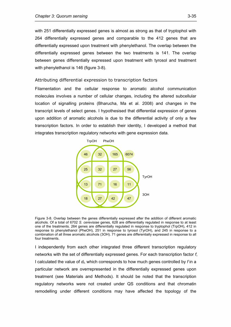
Chapter 3: Quorum sensing
3-35
with 251 differentially expressed genes is almost as strong as that of tryptophol with
264 differentially expressed genes and comparable to the 412 genes that are
differentially expressed upon treatment with phenylethanol. The overlap between the
differentially expressed genes between the two treatments is 141. The overlap
between genes differentially expressed upon treatment with tyrosol and treatment
with phenylethanol is 146 (figure 3-8).
Attributing differential expression to transcription factors
Filamentation and the cellular response to aromatic alcohol communication
molecules involves a number of cellular changes, including the altered subcellular
location of signalling proteins (Bharucha, Ma et al. 2008) and changes in the
transcript levels of select genes. I hypothesised that differential expression of genes
upon addition of aromatic alcohols is due to the differential activity of only a few
transcription factors. In order to establish their identity, I developed a method that
integrates transcription regulatory networks with gene expression data.
46 32 165 6074
56
11
47
27
16
42
32
71
27
25
13
18
TrpOH PheOH
TyrOH
3OH
Figure 3-8. Overlap between the genes differentially expressed after the addition of different aromatic alcohols. Of a total of 6702 S. cerevisiae genes, 628 are differentially regulated in response to at least one of the treatments. 264 genes are differentially regulated in response to tryptophol (TrpOH), 412 in response to phenylethanol (PheOH), 251 in response to tyrosol (TyrOH), and 245 in response to a combination of all three aromatic alcohols (3OH). 71 genes are differentially expressed in response to all four treatments. I independently from each other integrated three different transcription regulatory
networks with the set of differentially expressed genes. For each transcription factor f,
I calculated the value of df, which corresponds to how much genes controlled by f in a
particular network are overrepresented in the differentially expressed genes upon
treatment (see Materials and Methods). It should be noted that the transcription
regulatory networks were not created under QS conditions and that chromatin
remodelling under different conditions may have affected the topology of the
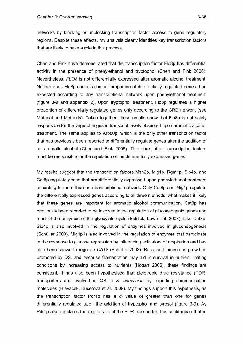
Chapter 3: Quorum sensing
3-36
networks by blocking or unblocking transcription factor access to gene regulatory
regions. Despite these effects, my analysis clearly identifies key transcription factors
that are likely to have a role in this process.
Chen and Fink have demonstrated that the transcription factor Flo8p has differential
activity in the presence of phenylethanol and tryptophol (Chen and Fink 2006).
Nevertheless, FLO8 is not differentially expressed after aromatic alcohol treatment.
Neither does Flo8p control a higher proportion of differentially regulated genes than
expected according to any transcriptional network upon phenylethanol treatment
(figure 3-9 and appendix 2). Upon tryptophol treatment, Flo8p regulates a higher
proportion of differentially regulated genes only according to the GRD network (see
Material and Methods). Taken together, these results show that Flo8p is not solely
responsible for the large changes in transcript levels observed upon aromatic alcohol
treatment. The same applies to Aro80p, which is the only other transcription factor
that has previously been reported to differentially regulate genes after the addition of
an aromatic alcohol (Chen and Fink 2006). Therefore, other transcription factors
must be responsible for the regulation of the differentially expressed genes.
My results suggest that the transcription factors Msn2p, Mig1p, Rgm1p, Sip4p, and
Cat8p regulate genes that are differentially expressed upon phenylethanol treatment
according to more than one transcriptional network. Only Cat8p and Mig1p regulate
the differentially expressed genes according to all three methods, what makes it likely
that these genes are important for aromatic alcohol communication. Cat8p has
previously been reported to be involved in the regulation of gluconeogenic genes and
most of the enzymes of the glyoxylate cycle (Biddick, Law et al. 2008). Like Cat8p,
Sip4p is also involved in the regulation of enzymes involved in gluconeogenesis
(Schüller 2003). Mig1p is also involved in the regulation of enzymes that participate
in the response to glucose repression by influencing activators of respiration and has
also been shown to regulate CAT8 (Schüller 2003). Because filamentous growth is
promoted by QS, and because filamentation may aid in survival in nutrient limiting
conditions by increasing access to nutrients (Hogan 2006), these findings are
consistent. It has also been hypothesised that pleiotropic drug resistance (PDR)
transporters are involved in QS in S. cerevisiae by exporting communication
molecules (Hlavacek, Kucerova et al. 2009). My findings support this hypothesis, as
the transcription factor Pdr1p has a df value of greater than one for genes
differentially regulated upon the addition of tryptophol and tyrosol (figure 3-9). As
Pdr1p also regulates the expression of the PDR transporter, this could mean that in
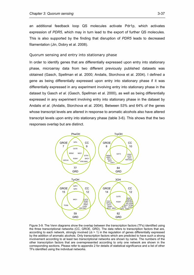
Chapter 3: Quorum sensing
3-37
an additional feedback loop QS molecules activate Pdr1p, which activates
expression of PDR5, which may in turn lead to the export of further QS molecules.
This is also supported by the finding that disruption of PDR5 leads to decreased
filamentation (Jin, Dobry et al. 2008).
Quorum sensing and entry into stationary phase
In order to identify genes that are differentially expressed upon entry into stationary
phase, microarray data from two different previously published datasets was
obtained (Gasch, Spellman et al. 2000; Andalis, Storchova et al. 2004). I defined a
gene as being differentially expressed upon entry into stationary phase if it was
differentially expressed in any experiment involving entry into stationary phase in the
dataset by Gasch et al. (Gasch, Spellman et al. 2000), as well as being differentially
expressed in any experiment involving entry into stationary phase in the dataset by
Andalis et al. (Andalis, Storchova et al. 2004). Between 53% and 64% of the genes
whose transcript levels are altered in response to aromatic alcohols also have altered
transcript levels upon entry into stationary phase (table 3-6). This shows that the two
responses overlap but are distinct.
GROE CC
GRD
PheOH
77
97GROE CC
GRD
TrpOH
84
108
GROE CC
GRD
TyrOH
59
1215GROE CC
GRD
3OH
82
1214
SPS18
STP4
SKO1RGM1
GAT3
MSN2
SIP4
YPR196W
MIG1
CAT8
PDR1
YER184C
ADR1
CAT8
TOS8
GZF3
MIG1RGM1
SIP4
CHA4
RFX1MIG1
CRZ1
PDR1GAT3MSN2 CAT8
YER184CSIP4
CAT8
RGM1
GAT3
YOX1SPS18
RGM1RFX1 MIG1
RDR1ARO80
YER184CYPL230W
RGT1STP2
STB4CAD1
SIP4 PDR1
Figure 3-9. The Venn diagrams show the overlap between the transcription factors (TFs) identified using the three transcriptional networks (CC, GROE, GRD). The data refers to transcription factors that are, according to each network, strongly involved (df > 1) in the regulation of genes differentially expressed by the addition of aromatic alcohols. Only transcription factors which are predicted to have such a strong involvement according to at least two transcriptional networks are shown by name. The numbers of the other transcription factors that are overrepresented according to only one network are shown in the corresponding sections. Please refer to appendix 2 for details of statistical significance and a list of other TFs identified using the individual networks.
Chapter 3: Quorum sensing
3-38
S. cerevisiae
Treatment All differentially expressed genes
Genes that are not differentially
expressed upon entry into
stationary phase
Proportional decrease
C. albicans orthologues of differentially expressed S.
cerevisiae genes phenylethanol 412 147 64% 125 tryptophol 264 114 57% 63 tyrosol 251 118 53% 62 3OH (combination) 246 99 60% 61 Table 3-6. In order to identify genes that are differentially expressed by aromatic alcohol treatment but that are not also differentially expressed during entry into stationary phase, I removed all stationary phase-specific genes from the dataset (column 2). The number of C. albicans genes whose orthologues are differentially expressed in S. cerevisiae is given in the last column.
Genomic organization and nucleosome modification of the differentially
expressed genes
Next, I investigated whether the genes that are differentially expressed upon the
addition of aromatic alcohol communication molecules are distributed across the
chromosomes randomly. Non-random distribution of these genes may allow quicker
access of transcription factors to accessible regions of chromatin whenever this is
required (Janga, Collado-Vides et al. 2008). To assess whether genes differentially
expressed after aromatic alcohol addition are distributed across chromosomes in a
specific way, I created random sets of genes and compared their chromosomal
locations to the actually observed locations. I found that genes that are differentially
expressed when aromatic alcohols are added are not distributed amongst
chromosomes randomly. Genes that are differentially expressed upon the addition of
phenylethanol tend to reside on chromosome 15 more often than expected by
chance (figure 3-10). This also holds when genes that are differentially expressed
upon entry into stationary phase are removed as described in the methods. No highly
significant effect was found for tyrosol, which is in agreement with the finding that
only tryptophol and phenylethanol are likely to be QS molecules (Chen and Fink
2006).
I also found that genes that are differentially expressed in response to phenylethanol
are clustered within chromosomes. 48.3% of genes that are differentially regulated in
response to phenylethanol are located within 10 genes of another gene (Pal and
Hurst 2003; Janga, Collado-Vides et al. 2008) regulated by this aromatic alcohol (PI
= 0.483, p = 0.03; figure 3-10). This drops to 26.1% when filtering removing genes
that are also differentially regulated upon entry into stationary phase (PI = 0.261, p =
0.02). This suggests that genes regulated by the communication molecule
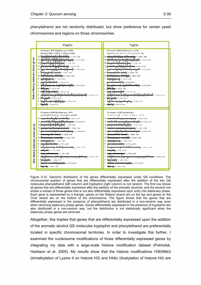
Chapter 3: Quorum sensing
3-39
phenylethanol are not randomly distributed, but show preference for certain yeast
chromosomes and regions on those chromosomes.
Figure 3-10. Genomic distribution of the genes differentially expressed under QS conditions. The chromosomal position of genes that are differentially expressed after the addition of the two QS molecules phenylethanol (left column) and tryptophol (right column) is not random. The first row shows all genes that are differentially expressed after the addition of the aromatic alcohols, and the second row shows a subset of those genes that is not also differentially expressed upon entry into stationary phase. Each gene is represented by a triangle; genes on the Watson strand are on the top and genes on the Crick strand are on the bottom of the chromosome. The figure shows that the genes that are differentially expressed in the presence of phenylethanol are distributed in a non-random way even when removing stationary phase genes. Genes differentially expressed in the presence of tryptophol are also distributed in a non-random way, but the distribution is not statistically significant when the stationary phase genes are removed. Altogether, this implies that genes that are differentially expressed upon the addition
of the aromatic alcohol QS molecules tryptophol and phenylethanol are preferentially
located in specific chromosomal territories. In order to investigate this further, I
examined the nucleosome modifications of those differentially expressed genes by
integrating my data with a large-scale histone modification dataset (Pokholok,
Harbison et al. 2005). My results show that the histone modifications H3K4Me3
(trimethylation of Lysine 4 on histone H3) and H4Ac (Acetylation of histone H4) are
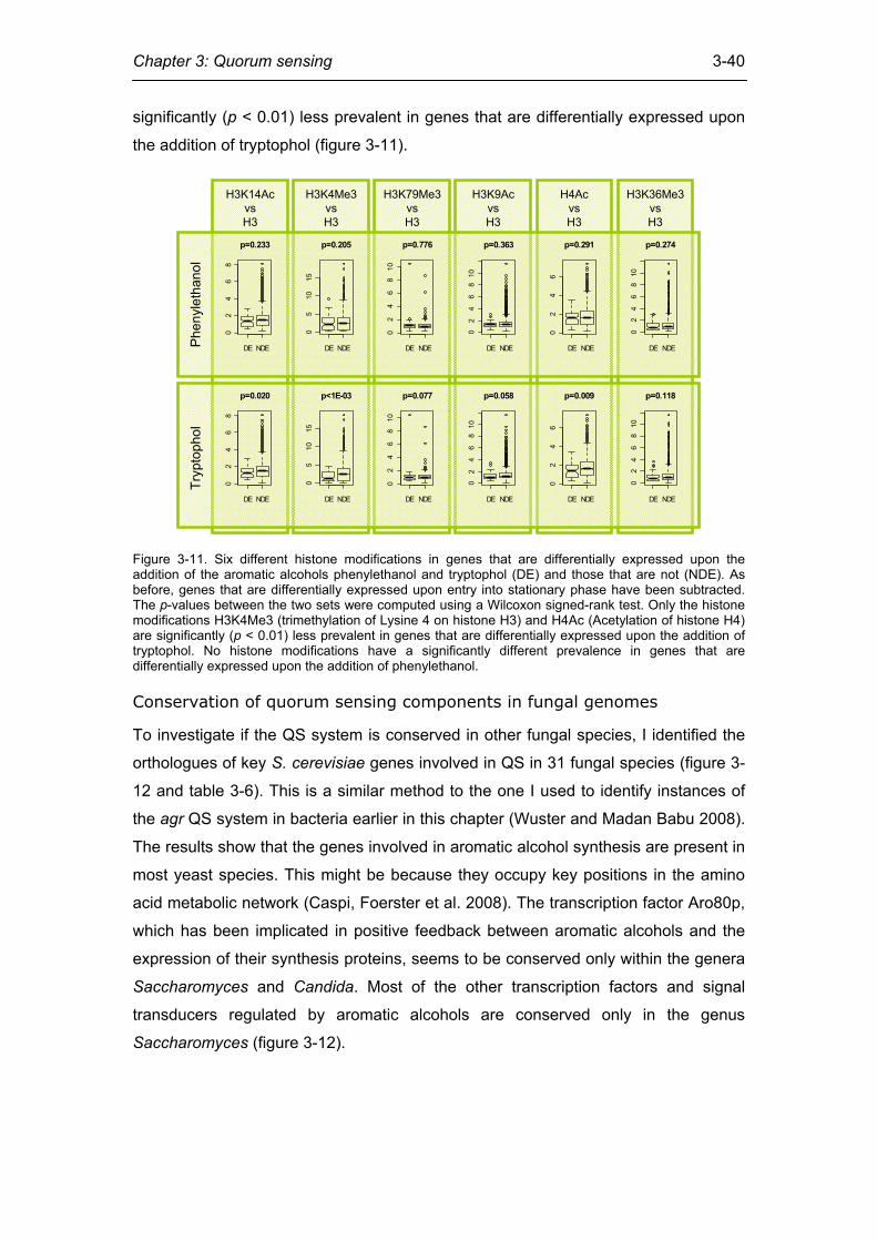
Chapter 3: Quorum sensing
3-40
significantly (p < 0.01) less prevalent in genes that are differentially expressed upon
the addition of tryptophol (figure 3-11).
Phe
nyle
than
olTr
ypto
phol
H3K14AcvsH3
H3K4Me3vsH3
H3K79Me3vsH3
H3K9AcvsH3
H4AcvsH3
H3K36Me3vsH3
DE NDE
02
46
8
p=0.233
DE NDE
05
1015
p=0.205
DE NDE
02
46
810
p=0.776
DE NDE
02
46
810
p=0.363
DE NDE
02
46
p=0.291
DE NDE
02
46
810
p=0.274
DE NDE
02
46
8
p=0.020
DE NDE
05
1015
p<1E-03
DE NDE
02
46
810
p=0.077
DE NDE
02
46
810
p=0.058
DE NDE
02
46
p=0.009
DE NDE
02
46
810
p=0.118
Figure 3-11. Six different histone modifications in genes that are differentially expressed upon the addition of the aromatic alcohols phenylethanol and tryptophol (DE) and those that are not (NDE). As before, genes that are differentially expressed upon entry into stationary phase have been subtracted. The p-values between the two sets were computed using a Wilcoxon signed-rank test. Only the histone modifications H3K4Me3 (trimethylation of Lysine 4 on histone H3) and H4Ac (Acetylation of histone H4) are significantly (p < 0.01) less prevalent in genes that are differentially expressed upon the addition of tryptophol. No histone modifications have a significantly different prevalence in genes that are differentially expressed upon the addition of phenylethanol.
Conservation of quorum sensing components in fungal genomes
To investigate if the QS system is conserved in other fungal species, I identified the
orthologues of key S. cerevisiae genes involved in QS in 31 fungal species (figure 3-
12 and table 3-6). This is a similar method to the one I used to identify instances of
the agr QS system in bacteria earlier in this chapter (Wuster and Madan Babu 2008).
The results show that the genes involved in aromatic alcohol synthesis are present in
most yeast species. This might be because they occupy key positions in the amino
acid metabolic network (Caspi, Foerster et al. 2008). The transcription factor Aro80p,
which has been implicated in positive feedback between aromatic alcohols and the
expression of their synthesis proteins, seems to be conserved only within the genera
Saccharomyces and Candida. Most of the other transcription factors and signal
transducers regulated by aromatic alcohols are conserved only in the genus
Saccharomyces (figure 3-12).
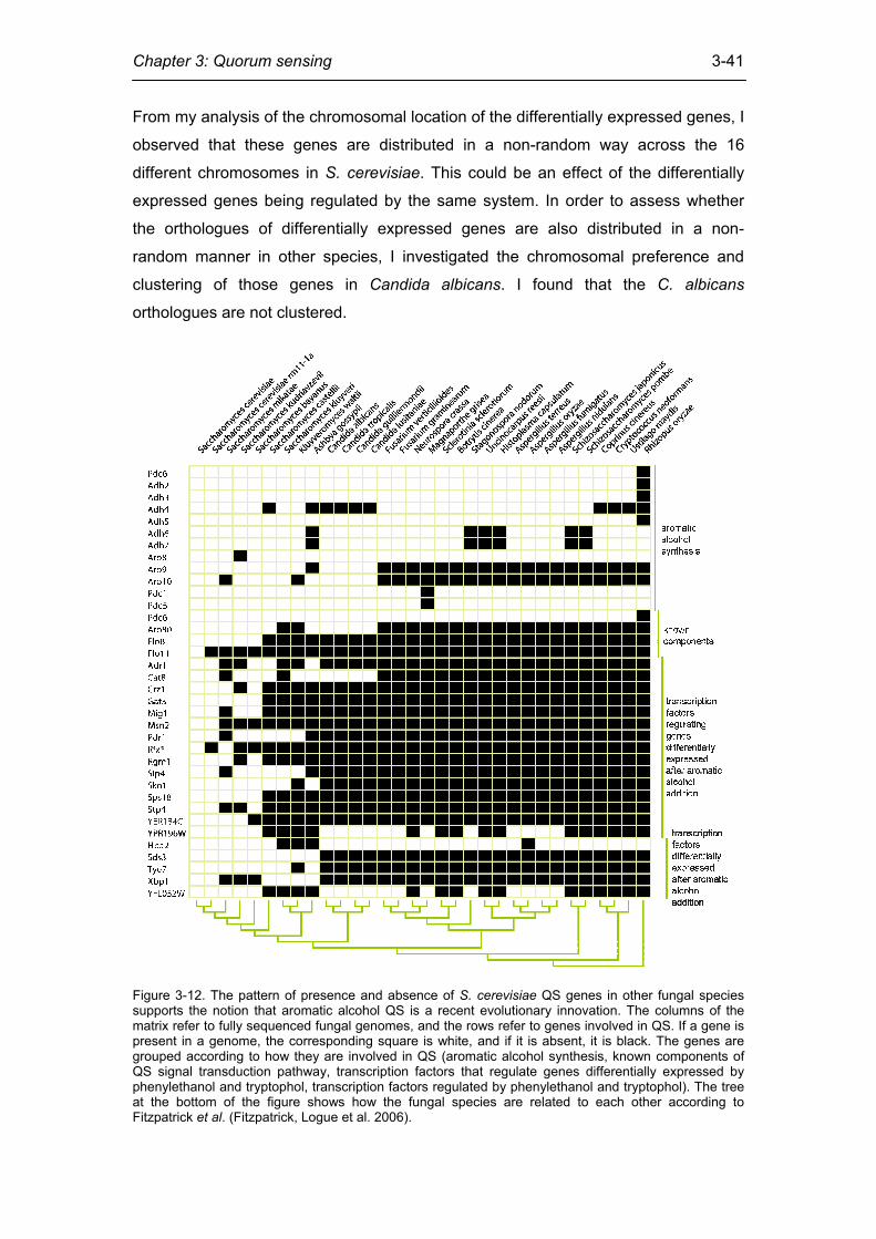
Chapter 3: Quorum sensing
3-41
From my analysis of the chromosomal location of the differentially expressed genes, I
observed that these genes are distributed in a non-random way across the 16
different chromosomes in S. cerevisiae. This could be an effect of the differentially
expressed genes being regulated by the same system. In order to assess whether
the orthologues of differentially expressed genes are also distributed in a non-
random manner in other species, I investigated the chromosomal preference and
clustering of those genes in Candida albicans. I found that the C. albicans
orthologues are not clustered.
Figure 3-12. The pattern of presence and absence of S. cerevisiae QS genes in other fungal species supports the notion that aromatic alcohol QS is a recent evolutionary innovation. The columns of the matrix refer to fully sequenced fungal genomes, and the rows refer to genes involved in QS. If a gene is present in a genome, the corresponding square is white, and if it is absent, it is black. The genes are grouped according to how they are involved in QS (aromatic alcohol synthesis, known components of QS signal transduction pathway, transcription factors that regulate genes differentially expressed by phenylethanol and tryptophol, transcription factors regulated by phenylethanol and tryptophol). The tree at the bottom of the figure shows how the fungal species are related to each other according to Fitzpatrick et al. (Fitzpatrick, Logue et al. 2006).
Chapter 3: Quorum sensing
3-42
When also taking into account experimental data, which shows that filamentation of
C. albicans is not stimulated by tryptophol or phenylethanol (Chen and Fink 2006),
this could suggest that these genes do not act in the same pathway (Janga, Collado-
Vides et al. 2008).
Together with my finding that many of the genes that are involved in QS pathways in
S. cerevisiae are not present in C. albicans and other more distantly related fungi, I
suggest that it is unlikely that QS by aromatic alcohols is conserved in the same form
outside the genus Saccharomyces.
Taken together, my results show that although QS using aromatic alcohols is a
relatively recent evolutionary innovation in S. cerevisiae, and that the genes that are
differentially expressed are not randomly distributed. Instead they show a clear
chromosomal preference and tend to be clustered on the chromosomal arms.
Through the integrated analysis of large-scale genomic datasets, I propose several
transcription factors previously not known to participate in QS, such as Cat8p and
Mig1p, to be involved in the process. My observation that the key elements in the
circuit are conserved only in the genus Saccharomyces, but not in other genomes,
suggests that the system is unlikely to operate in other species in the same form.
Materials and methods
Gene expression data
In order to identify genes that are differentially expressed in the presence of aromatic
alcohols, I used microarray data of yeast cultures treated with the aromatic alcohols
tryptophol, phenylethanol, tyrosol, or a combination of all three, as published recently
by Chen and Fink (Chen and Fink 2006). Genes were considered to be differentially
expressed if their intensity on the microarray differed 2-fold or more on treatment with
the aromatic alcohol compared to the control condition. To identify genes that are
differentially expressed only under aromatic alcohol communication conditions, it is
important to identify and remove genes that are differentially expressed upon entry
into stationary phase. To this end, genes that are differentially expressed upon entry
into stationary phase were identified using gene expression data from two previously
published experiments by Gasch et al. (Gasch, Spellman et al. 2000) and by Andalis
et al. (Andalis, Storchova et al. 2004). In both experiments, the expression level of
genes in different conditions, including entry into stationary phase, was determined
using DNA microarrays. Genes that are uniquely differentially expressed upon
Chapter 3: Quorum sensing
3-43
aromatic alcohol communication, but not upon entry into stationary phase, were thus
identified by integrating these datasets. I defined a gene as being differentially
expressed upon entry into stationary phase if it was differentially expressed in any
experiment involving entry into stationary phase in the dataset by Gasch et al., as
well as being differentially expressed in any experiment involving entry into stationary
phase in the dataset by Andalis et al.
Identification of key transcription factors
In order to identify the transcription factors (TFs) that regulate the genes that are
differentially expressed by the addition of aromatic alcohols, I integrated previously
published gene expression data with transcriptional regulatory interactions that were
reconstructed using three experimental methods that exploit different principles. One
of them is based on ChIP-chip data (Lee, Rinaldi et al. 2002; Harbison, Gordon et al.
2004; Balaji, Madan Babu et al. 2006), which identifies direct in vivo DNA binding
events for TFs. The other two are genetic methods that identify differentially
expressed genes upon over-expression(Chua, Morris et al. 2006) or deletion (Hu,
Killion et al. 2007) of TFs using microarrays. I refer to the networks obtained in this
way as the CC (for Chip-Chip; 144 TFs regulating 4347 targets via 12230 regulatory
interactions), GRD (for Genetic Reconstruction upon gene Deletion; 157 TFs
regulating 1992 targets via 5589 regulatory interactions) and GROE (for Genetic
Reconstruction upon Over-Expression; 55 TFs regulating 1951 targets via 5802
regulatory interactions) network (Balaji, Iyer et al. 2008). In the CC network, nodes
represent TFs or target genes (TGs) and edges represent direct binding of the TF in
the promoter region of the TG. Likewise, in the GRD and GROE networks, nodes
represent TFs or TGs and a TF is linked to a TG if it is differentially expressed upon
deletion or over-expression of the TF, respectively. For each of the three networks
independently, I calculated the score df, for each transcription factor f, where
df = log2((tdiffex / tall) / (adiffex / aall))
tdiffex is the number of differentially regulated genes controlled by f according to the
specific network, tall is the number of all genes regulated by f in the same network.
adiffex is the number of all differentially expressed genes upon treatment, and aall is the
number of all genes in the network. In other words, this value corresponds to how
much genes controlled by the TF f in a particular network are overrepresented in the
differentially expressed genes upon treatment. In this way, transcription factors that
are likely to be responsible for regulating the genes that are differentially expressed
Chapter 3: Quorum sensing
3-44
upon the addition of the aromatic alcohols can be identified. In my analysis, I chose
TFs that were more than two-fold overrepresented (df > 1) as those which are likely
to regulate the differentially expressed genes. I also calculated the statistical
significance by estimating a p-value that each TF controls a higher proportion of the
differentially expressed genes than what would be expected using a hypergeometric
distribution as a null model (appendix 2).
Genomic distribution of differentially expressed genes
The chromosomal location of genes was obtained from http://www.yeastgenome.org/
for S. cerevisiae and from http://www.candidagenome.org/ for C. albicans. In order to
assess whether an input set of genes is distributed randomly across the
chromosomes or whether there is a preference for certain chromosomes, I performed
two different types of statistical test. The first one is a χ2 test that has as a null
hypothesis that the differentially expressed genes are distributed across the 16 yeast
chromosomes in an unbiased manner. The second test computes the expected
distribution of genes on the 16 chromosomes by repeatedly (1000 times) and
randomly picking the same number of genes as the set of differentially expressed
genes. This is compared to the observed distribution of the differentially expressed
genes to obtain statistical significance for chromosomal preference (p-values and Z-
scores). In order to assess whether the distribution of genes within chromosomes is
random or clustered, I calculated the proximity index (PI) for the differentially
expressed set of genes. The PI is defined as the ratio of proximal genes to the set of
differentially expressed genes. Two genes are defined to be proximal if they are
separated by less than 10 genes on the chromosome (Pal and Hurst 2003; Janga,
Collado-Vides et al. 2008). To assess statistical significance, the PI value was
calculated from random sets of genes (1000 in this case) and compared to the
observed value.
Orthologue detection
In order to assess whether the genes involved in aromatic alcohol communication are
well-conserved within fungi, I identified their orthologues across 31 different fungal
genomes (Cornell, Alam et al. 2007). The complete genome sequences were
downloaded from http://fungal.genome.duke.edu/ and the orthologues were identified
using OrthoMCL with the default parameters (Gao, Teplitski et al. 2003). The
OrthoMCL output was filtered and visualised using an in-house Perl script (figure 3-
12).
Chapter 3: Quorum sensing
3-45
Epigenetic modifications
In order to determine whether genes that are differentially expressed upon the
addition of the QS aromatic alcohols tryptophol and phenylethanol have different
histone modifications compared to those that are not differentially expressed, I
integrated my data with a large-scale nucleosome modification dataset (Pokholok,
Harbison et al. 2005). As in many cases each S. cerevisiae gene is covered by more
than one tile, in those cases I chose only to take into account the modification of the
5’-most tile. The p-value that the nucleosome modification in genes that are
differentially expressed genes and those that are not are identical was computed
using the Wilcoxon signed-rank test as implemented in the statistical programming
package R.
Chapter 3: Quorum sensing
3-46
References
Altschul, S. F., T. L. Madden, et al. (1997). "Gapped BLAST and PSI-BLAST: a new generation of protein database search programs." Nucleic Acids Res 25(17): 3389-402.
Andalis, A. A., Z. Storchova, et al. (2004). "Defects arising from whole-genome duplications in Saccharomyces cerevisiae." Genetics 167(3): 1109-21.
Aoki, S. K., R. Pamma, et al. (2005). "Contact-dependent inhibition of growth in Escherichia coli." Science 309(5738): 1245-8.
Aravind, L., V. Anantharaman, et al. (2005). "The many faces of the helix-turn-helix domain: transcription regulation and beyond." FEMS Microbiol Rev 29(2): 231-62.
Aravind, L. and C. P. Ponting (1997). "The GAF domain: an evolutionary link between diverse phototransducing proteins." Trends Biochem Sci 22(12): 458-9.
Balaji, S., L. M. Iyer, et al. (2008). "Comparison of transcription regulatory interactions inferred from high-throughput methods: what do they reveal?" Trends Genet 24(7): 319-23.
Balaji, S., M. Madan Babu, et al. (2006). "Comprehensive analysis of combinatorial regulation using the transcriptional regulatory network of yeast." J Mol Biol 360(1): 213-27.
Barrales, R. R., J. Jimenez, et al. (2008). "Identification of novel activation mechanisms for FLO11 regulation in Saccharomyces cerevisiae." Genetics 178(1): 145-56.
Bassler, B. L. and R. Losick (2006). "Bacterially speaking." Cell 125(2): 237-46. Benito, Y., F. A. Kolb, et al. (2000). "Probing the structure of RNAIII, the
Staphylococcus aureus agr regulatory RNA, and identification of the RNA domain involved in repression of protein A expression." Rna 6(5): 668-79.
Bharucha, N., J. Ma, et al. (2008). "Analysis of the yeast kinome reveals a network of regulated protein localization during filamentous growth." Mol Biol Cell 19(7): 2708-17.
Biddick, R. K., G. L. Law, et al. (2008). "Adr1 and cat8 mediate coactivator recruitment and chromatin remodeling at glucose-regulated genes." PLoS ONE 3(1): e1436.
Bourgogne, A., S. G. Hilsenbeck, et al. (2006). "Comparison of OG1RF and an isogenic fsrB deletion mutant by transcriptional analysis: the Fsr system of Enterococcus faecalis is more than the activator of gelatinase and serine protease." J Bacteriol 188(8): 2875-84.
Burrell, P. C. (2006). "The detection of environmental autoinducing peptide quorum-sensing genes from an uncultured Clostridium sp. in landfill leachate reactor biomass." Lett Appl Microbiol 43(4): 455-60.
Camilli, A. and B. L. Bassler (2006). "Bacterial small-molecule signaling pathways." Science 311(5764): 1113-6.
Caspi, R., H. Foerster, et al. (2006). "MetaCyc: a multiorganism database of metabolic pathways and enzymes." Nucleic Acids Res 34(Database issue): D511-6.
Caspi, R., H. Foerster, et al. (2008). "The MetaCyc Database of metabolic pathways and enzymes and the BioCyc collection of Pathway/Genome Databases." Nucleic Acids Res 36(Database issue): D623-31.
Cassat, J., P. M. Dunman, et al. (2006). "Transcriptional profiling of a Staphylococcus aureus clinical isolate and its isogenic agr and sarA mutants reveals global differences in comparison to the laboratory strain RN6390." Microbiology 152(Pt 10): 3075-90.
Chapter 3: Quorum sensing
3-47
Chen, H. and G. R. Fink (2006). "Feedback control of morphogenesis in fungi by aromatic alcohols." Genes Dev 20(9): 1150-61.
Chen, H., M. Fujita, et al. (2004). "Tyrosol is a quorum-sensing molecule in Candida albicans." Proc Natl Acad Sci U S A 101(14): 5048-52.
Chen, X., S. Schauder, et al. (2002). "Structural identification of a bacterial quorum-sensing signal containing boron." Nature 415(6871): 545-9.
Cheung, A. L. and S. J. Projan (1994). "Cloning and sequencing of sarA of Staphylococcus aureus, a gene required for the expression of agr." J Bacteriol 176(13): 4168-72.
Choi, S. H. and E. P. Greenberg (1991). "The C-terminal region of the Vibrio fischeri LuxR protein contains an inducer-independent lux gene activating domain." Proc Natl Acad Sci U S A 88(24): 11115-9.
Chua, G., Q. D. Morris, et al. (2006). "Identifying transcription factor functions and targets by phenotypic activation." Proc Natl Acad Sci U S A 103(32): 12045-50.
Ciccarelli, F. D., T. Doerks, et al. (2006). "Toward automatic reconstruction of a highly resolved tree of life." Science 311(5765): 1283-7.
Clark, M. A., R. A. Acharya, et al. (2009). "Design, synthesis and selection of DNA-encoded small-molecule libraries." Nat Chem Biol.
Clarke, M. B., D. T. Hughes, et al. (2006). "The QseC sensor kinase: a bacterial adrenergic receptor." Proc Natl Acad Sci U S A 103(27): 10420-5.
Cornell, M. J., I. Alam, et al. (2007). "Comparative genome analysis across a kingdom of eukaryotic organisms: specialization and diversification in the fungi." Genome Res 17(12): 1809-22.
Curragh, H., O. Flynn, et al. (1994). "Haloalkane degradation and assimilation by Rhodococcus rhodochrous NCIMB 13064." Microbiology 140 ( Pt 6): 1433-42.
Diep, D. B., L. S. Havarstein, et al. (1994). "The gene encoding plantaricin A, a bacteriocin from Lactobacillus plantarum C11, is located on the same transcription unit as an agr-like regulatory system." Appl Environ Microbiol 60(1): 160-6.
Dirix, G., P. Monsieurs, et al. (2004). "Screening genomes of Gram-positive bacteria for double-glycine-motif-containing peptides." Microbiology 150(Pt 5): 1121-6.
Doherty, N., M. T. Holden, et al. (2006). "Functional analysis of luxS in Staphylococcus aureus reveals a role in metabolism but not quorum sensing." J Bacteriol 188(8): 2885-97.
Drake, H. L. and S. L. Daniel (2004). "Physiology of the thermophilic acetogen Moorella thermoacetica." Res Microbiol 155(10): 869-83.
Dunman, P. M., E. Murphy, et al. (2001). "Transcription profiling-based identification of Staphylococcus aureus genes regulated by the agr and/or sarA loci." J Bacteriol 183(24): 7341-53.
Durbin, R., S. Eddy, et al. (1998). Biological sequence analysis: probabilistic models of proteins and nucleic acids. Cambridge, Cambridge University Press.
Edgar, R. C. (2004). "MUSCLE: a multiple sequence alignment method with reduced time and space complexity." BMC Bioinformatics 5: 113.
Finn, R. D., J. Mistry, et al. (2006). "Pfam: clans, web tools and services." Nucleic Acids Res 34(Database issue): D247-51.
Fischer, C., O. Valerius, et al. (2008). "Posttranscriptional regulation of FLO11 upon amino acid starvation in Saccharomyces cerevisiae." FEMS Yeast Res 8(2): 225-36.
Fitzpatrick, D. A., M. E. Logue, et al. (2006). "A fungal phylogeny based on 42 complete genomes derived from supertree and combined gene analysis." BMC Evol Biol 6: 99.
Gao, M., M. Teplitski, et al. (2003). "Production of substances by Medicago truncatula that affect bacterial quorum sensing." Mol Plant Microbe Interact 16(9): 827-34.
Chapter 3: Quorum sensing
3-48
Gasch, A. P., P. T. Spellman, et al. (2000). "Genomic expression programs in the response of yeast cells to environmental changes." Mol Biol Cell 11(12): 4241-57.
Gould, T. A., H. P. Schweizer, et al. (2004). "Structure of the Pseudomonas aeruginosa acyl-homoserinelactone synthase LasI." Mol Microbiol 53(4): 1135-46.
Griffiths-Jones, S., A. Bateman, et al. (2003). "Rfam: an RNA family database." Nucleic Acids Res 31(1): 439-41.
Harbison, C. T., D. B. Gordon, et al. (2004). "Transcriptional regulatory code of a eukaryotic genome." Nature 431(7004): 99-104.
Havarstein, L. S., G. Coomaraswamy, et al. (1995). "An unmodified heptadecapeptide pheromone induces competence for genetic transformation in Streptococcus pneumoniae." Proc Natl Acad Sci U S A 92(24): 11140-4.
Hentzer, M., K. Riedel, et al. (2002). "Inhibition of quorum sensing in Pseudomonas aeruginosa biofilm bacteria by a halogenated furanone compound." Microbiology 148(Pt 1): 87-102.
Hentzer, M., H. Wu, et al. (2003). "Attenuation of Pseudomonas aeruginosa virulence by quorum sensing inhibitors." Embo J 22(15): 3803-15.
Hlavacek, O., H. Kucerova, et al. (2009). "Putative role for ABC multidrug exporters in yeast quorum sensing." FEBS Lett 583(7): 1107-13.
Hogan, D. A. (2006). "Quorum sensing: alcohols in a social situation." Curr Biol 16(12): R457-8.
Hu, Z., P. J. Killion, et al. (2007). "Genetic reconstruction of a functional transcriptional regulatory network." Nat Genet 39(5): 683-7.
Iyer, L. M., L. Aravind, et al. (2004). "Evolution of cell-cell signaling in animals: did late horizontal gene transfer from bacteria have a role?" Trends Genet 20(7): 292-9.
Jafra, S., J. Przysowa, et al. (2006). "Detection and characterization of bacteria from the potato rhizosphere degrading N-acyl-homoserine lactone." Can J Microbiol 52(10): 1006-1015.
Janga, S. C., J. Collado-Vides, et al. (2008). "Transcriptional regulation constrains the organization of genes on eukaryotic chromosomes." Proc Natl Acad Sci U S A 105(41): 15761-6.
Ji, G., R. Beavis, et al. (1997). "Bacterial interference caused by autoinducing peptide variants." Science 276(5321): 2027-30.
Jin, R., C. J. Dobry, et al. (2008). "Large-scale analysis of yeast filamentous growth by systematic gene disruption and overexpression." Mol Biol Cell 19(1): 284-96.
Joint, I., K. Tait, et al. (2007). "Cross-kingdom signalling: exploitation of bacterial quorum sensing molecules by the green seaweed Ulva." Philos Trans R Soc Lond B Biol Sci 362(1483): 1223--33.
Kaiser, D. (2004). "Signaling in myxobacteria." Annu Rev Microbiol 58: 75-98. Kastrup, C. J., J. Q. Boedicker, et al. (2008). "Spatial localization of bacteria controls
coagulation of human blood by 'quorum acting'." Nat Chem Biol 4(12): 742-50. Kleerebezem, M., L. E. Quadri, et al. (1997). "Quorum sensing by peptide
pheromones and two-component signal-transduction systems in Gram-positive bacteria." Mol Microbiol 24(5): 895-904.
Kravchenko, V. V., G. F. Kaufmann, et al. (2008). "Modulation of gene expression via disruption of NF-kappaB signaling by a bacterial small molecule." Science 321(5886): 259-63.
Kumar, S., K. Tamura, et al. (2004). "MEGA3: Integrated software for Molecular Evolutionary Genetics Analysis and sequence alignment." Brief Bioinform 5(2): 150-63.
Lazdunski, A. M., I. Ventre, et al. (2004). "Regulatory circuits and communication in Gram-negative bacteria." Nat Rev Microbiol 2(7): 581-92.
Chapter 3: Quorum sensing
3-49
Leadbetter, J. R. and E. P. Greenberg (2000). "Metabolism of acyl-homoserine lactone quorum-sensing signals by Variovorax paradoxus." J Bacteriol 182(24): 6921-6.
Lee, T. I., N. J. Rinaldi, et al. (2002). "Transcriptional regulatory networks in Saccharomyces cerevisiae." Science 298(5594): 799-804.
Lenz, D. H., K. C. Mok, et al. (2004). "The small RNA chaperone Hfq and multiple small RNAs control quorum sensing in Vibrio harveyi and Vibrio cholerae." Cell 118(1): 69-82.
Madan Babu, M. and S. A. Teichmann (2003). "Evolution of transcription factors and the gene regulatory network in Escherichia coli." Nucleic Acids Res 31(4): 1234-44.
Mashburn, L. M. and M. Whiteley (2005). "Membrane vesicles traffic signals and facilitate group activities in a prokaryote." Nature 437(7057): 422-5.
Mayville, P., G. Ji, et al. (1999). "Structure-activity analysis of synthetic autoinducing thiolactone peptides from Staphylococcus aureus responsible for virulence." Proc Natl Acad Sci U S A 96(4): 1218-23.
Morfeldt, E., D. Taylor, et al. (1995). "Activation of alpha-toxin translation in Staphylococcus aureus by the trans-encoded antisense RNA, RNAIII." Embo J 14(18): 4569-77.
Morrison, D. A. (2002). Is anybody here? Cooperative bacterial regulation via peptide signals between Gram-positive bacteria. Signals, Switches, Regulons, and Cascades. D. A. Hodgson and C. M. Thomas. Cambridge, Cambridge University Press: 231-249.
Mullard, A. (2009). "Microbiology: Tinker, bacteria, eukaryote, spy." Nature 459(7244): 159-61.
Nakayama, J., S. Chen, et al. (2006). "Revised model for Enterococcus faecalis fsr quorum-sensing system: the small open reading frame fsrD encodes the gelatinase biosynthesis-activating pheromone propeptide corresponding to staphylococcal agrd." J Bacteriol 188(23): 8321-6.
Nealson, K. H., T. Platt, et al. (1970). "Cellular control of the synthesis and activity of the bacterial luminescent system." J Bacteriol 104(1): 313-22.
Neiditch, M. B., M. J. Federle, et al. (2005). "Regulation of LuxPQ receptor activity by the quorum-sensing signal autoinducer-2." Mol Cell 18(5): 507-18.
Neiditch, M. B., M. J. Federle, et al. (2006). "Ligand-induced asymmetry in histidine sensor kinase complex regulates quorum sensing." Cell 126(6): 1095-108.
Nickerson, K. W., A. L. Atkin, et al. (2006). "Quorum sensing in dimorphic fungi: farnesol and beyond." Appl Environ Microbiol 72(6): 3805-13.
Novick, R. P. and E. Geisinger (2008). "Quorum sensing in staphylococci." Annu Rev Genet 42: 541-64.
Pai, A. and L. You (2009). "Optimal tuning of bacterial sensing potential." Mol Syst Biol 5: 286.
Pal, C. and L. D. Hurst (2003). "Evidence for co-evolution of gene order and recombination rate." Nat Genet 33(3): 392-5.
Park, S. Y., B. J. Hwang, et al. (2006). "N-acylhomoserine lactonase producing Rhodococcus spp. with different AHL-degrading activities." FEMS Microbiol Lett 261(1): 102-8.
Peterson, M. M., J. L. Mack, et al. (2008). "Apolipoprotein B Is an innate barrier against invasive Staphylococcus aureus infection." Cell Host Microbe 4(6): 555-66.
Pokholok, D. K., C. T. Harbison, et al. (2005). "Genome-wide map of nucleosome acetylation and methylation in yeast." Cell 122(4): 517-27.
Present, D. A., R. Meislin, et al. (1990). "Gas gangrene. A review." Orthop Rev 19(4): 333-41.
Chapter 3: Quorum sensing
3-50
Qazi, S. N., E. Counil, et al. (2001). "agr expression precedes escape of internalized Staphylococcus aureus from the host endosome." Infect Immun 69(11): 7074-82.
Qin, X., K. V. Singh, et al. (2000). "Effects of Enterococcus faecalis fsr genes on production of gelatinase and a serine protease and virulence." Infect Immun 68(5): 2579-86.
Qiu, R., W. Pei, et al. (2005). "Identification of the putative staphylococcal AgrB catalytic residues involving the proteolytic cleavage of AgrD to generate autoinducing peptide." J Biol Chem 280(17): 16695-704.
Queck, S. Y., M. Jameson-Lee, et al. (2008). "RNAIII-independent target gene control by the agr quorum-sensing system: insight into the evolution of virulence regulation in Staphylococcus aureus." Mol Cell 32(1): 150-8.
Reading, N. C. and V. Sperandio (2006). "Quorum sensing: the many languages of bacteria." FEMS Microbiol Lett 254(1): 1-11.
Recsei, P., B. Kreiswirth, et al. (1986). "Regulation of exoprotein gene expression in Staphylococcus aureus by agar." Mol Gen Genet 202(1): 58-61.
Redfield, R. J. (2002). "Is quorum sensing a side effect of diffusion sensing?" Trends Microbiol 10(8): 365-70.
Remm, M., C. E. Storm, et al. (2001). "Automatic clustering of orthologs and in-paralogs from pairwise species comparisons." J Mol Biol 314(5): 1041-52.
Rezzonico, F. and B. Duffy (2008). "Lack of genomic evidence of AI-2 receptors suggests a non-quorum sensing role for luxS in most bacteria." BMC Microbiol 8: 154.
Saenz, H. L., V. Augsburger, et al. (2000). "Inducible expression and cellular location of AgrB, a protein involved in the maturation of the staphylococcal quorum-sensing pheromone." Arch Microbiol 174(6): 452-5.
Sakinc, T., P. Kulczak, et al. (2004). "Cloning of an agr homologue of Staphylococcus saprophyticus." FEMS Microbiol Lett 237(1): 157-61.
Schüller, H. J. (2003). "Transcriptional control of nonfermentative metabolism in the yeast Saccharomyces cerevisiae." Curr Genet 43(3): 139-60.
Schuster, M., C. P. Lostroh, et al. (2003). "Identification, timing, and signal specificity of Pseudomonas aeruginosa quorum-controlled genes: a transcriptome analysis." J Bacteriol 185(7): 2066-79.
Shapiro, J. A. (1998). "Thinking about bacterial populations as multicellular organisms." Annu Rev Microbiol 52: 81-104.
Shiner, E. K., K. P. Rumbaugh, et al. (2005). "Inter-kingdom signaling: deciphering the language of acyl homoserine lactones." FEMS Microbiol Rev 29(5): 935-47.
Sprague, G. F., Jr. and S. C. Winans (2006). "Eukaryotes learn how to count: quorum sensing by yeast." Genes Dev 20(9): 1045-9.
Sturme, M. H., J. Nakayama, et al. (2005). "An agr-like two-component regulatory system in Lactobacillus plantarum is involved in production of a novel cyclic peptide and regulation of adherence." J Bacteriol 187(15): 5224-35.
Tait, K., I. Joint, et al. (2005). "Disruption of quorum sensing in seawater abolishes attraction of zoospores of the green alga Ulva to bacterial biofilms." Environ Microbiol 7(2): 229-40.
Tegmark, K., E. Morfeldt, et al. (1998). "Regulation of agr-dependent virulence genes in Staphylococcus aureus by RNAIII from coagulase-negative staphylococci." J Bacteriol 180(12): 3181-6.
Ulrich, L. E., E. V. Koonin, et al. (2005). "One-component systems dominate signal transduction in prokaryotes." Trends Microbiol 13(2): 52-6.
Uroz, S., S. R. Chhabra, et al. (2005). "N-Acylhomoserine lactone quorum-sensing molecules are modified and degraded by Rhodococcus erythropolis W2 by both amidolytic and novel oxidoreductase activities." Microbiology 151(Pt 10): 3313-22.
Chapter 3: Quorum sensing
3-51
Villemur, R., M. Lanthier, et al. (2006). "The Desulfitobacterium genus." FEMS Microbiol Rev 30(5): 706-33.
Vinod, P. K., N. Sengupta, et al. (2008). "Integration of global signaling pathways, cAMP-PKA, MAPK and TOR in the regulation of FLO11." PLoS ONE 3(2): e1663.
Waters, C. M. and B. L. Bassler (2006). "The Vibrio harveyi quorum-sensing system uses shared regulatory components to discriminate between multiple autoinducers." Genes Dev 20(19): 2754-67.
Whitehead, N. A., A. M. Barnard, et al. (2001). "Quorum-sensing in Gram-negative bacteria." FEMS Microbiol Rev 25(4): 365-404.
Winans, S. C. (1992). "Two-way chemical signaling in Agrobacterium-plant interactions." Microbiol Rev 56(1): 12-31.
Wuster, A. and M. Madan Babu (2008). "Conservation and evolutionary dynamics of the agr cell-to-cell communication system across firmicutes." J Bacteriol 190(2): 743-6.
Xavier, K. B., S. T. Miller, et al. (2007). "Phosphorylation and Processing of the Quorum-Sensing Molecule Autoinducer-2 in Enteric Bacteria." ACS Chem Biol.
Xue, Y., Y. Xu, et al. (2001). "Thermoanaerobacter tengcongensis sp. nov., a novel anaerobic, saccharolytic, thermophilic bacterium isolated from a hot spring in Tengcong, China." Int J Syst Evol Microbiol 51(Pt 4): 1335-41.
Yao, Y., C. Vuong, et al. (2006). "Characterization of the Staphylococcus epidermidis accessory-gene regulator response: quorum-sensing regulation of resistance to human innate host defense." J Infect Dis 193(6): 841-8.
Zhang, L., J. Lin, et al. (2004). "Membrane anchoring of the AgrD N-terminal amphipathic region is required for its processing to produce a quorum-sensing pheromone in Staphylococcus aureus." J Biol Chem 279(19): 19448-56.
Zhang, R. G., T. Pappas, et al. (2002). "Structure of a bacterial quorum-sensing transcription factor complexed with pheromone and DNA." Nature 417(6892): 971-4.


![Research Article Broad Spectrum Anti-Quorum Sensing ...downloads.hindawi.com/journals/scientifica/2016/5823013.pdf · isms is called quorum sensing (QS) []. Quorum sensing is a process](https://img.pdfslide.us/doc/110x75/5edbc5d7ad6a402d66662749/research-article-broad-spectrum-anti-quorum-sensing-isms-is-called-quorum-sensing.jpg)


![Natural Anti-Quorum Sensing agents against Pseudomonas ... · 2. Quorum Sensing: a Novel Target Vfr Quorum sensing (QS) is a population-dependent event [13]. The capability to sense](https://img.pdfslide.us/doc/110x75/5edbcc02ad6a402d66663060/natural-anti-quorum-sensing-agents-against-pseudomonas-2-quorum-sensing-a.jpg)