Embed Size (px)
Citation preview

Ph.D. Thesis
Quality of life of head and neck cancer patients
after tumor treatment
and subsequent maxillofacial rehabilitation
Judit Kádár-Nagy D.D.S.
Szeged, Hungary
2011

Quality of life of head and neck cancer patients after tumor treatment and
subsequent maxillofacial rehabilitation
Judit Nagy D.D.S.
Ph.D. Thesis
Supervisor: Prof. Katalin Nagy D.D.S., Ph.D.
Department of Oral Surgery and Maxillofacial Rehabilitation, Faculty of Dentistry,
University of Szeged
Graduate School of Clinical Science
Head of the program: Prof. Zoltán Rakonczay Ph.D., DSc.
Szeged, Hungary
2011

1
List of scientific publications related to the subject of the thesis
Articles:
I. Nagy, J., Feher, L., Sonkodi, I., Lesznyak, J., Ivanyi, B., Puskas, L.: A second field
metachronous Merkel-cell carcinoma of the lip and the palatine tonsil confirmed
by microarray-based CGH.
Virchows Arch 446: (3) 278-286, 2005.
IF: 2.224
II. Nagy J., Iványi B., Sonkodi I.: Merkel-sejtes carcinoma.
Fogorvosi Szle, 99; (4) 135-139, 2006.
III. Nagy J., Seres L., Novák P., Nagy K.: Implantáció a szájüregi rák miatt
sugárkezelésben részesült betegeken.
Fogorvosi Szle 102; (1): 7-11, 2009.
Published abstracts:
I. Nagy J, Piffkó J, Nagy K: Quality of life of H&N cancer patients after prosthetic
rehabilitation. ID: 0294 CED- IADR Budapest, August 31- September 4, 2011. J
Dent Res 90, Spec. Is B (IF abstrakt: 3, 773)

2
CONTENTS
I. Abbreviations ………………………………………………………………………. 4
II. Hypothesis …………………………………………………………………………... 5
III.Introduction ………………………………………………………………………… 6
III.1. Maxillofacial rehabilitation ……………………………………………………….. 6
III.1.1.Intraoral rehabilitation …………………………………………………………… 8
III.1.1.1. Prosthetic rehabilitation of patients with oral malignancy-acquired maxillary defects ………………………………………………………………... 8
III.1.1.2. Prosthetic rehabilitation of patients with oral malignancy-acquired mandibular defects ……………………………………………………………… 11
III.1.2. Extraoral rehabilitation …………………………………………………………. 13
III.2. Quality of life …………………………………………………………………….. 16
III.3. Measurement of quality of life …………………………………………………… 17
III.3.1. UW QOL questionnaire ………………………………………………………… 18
III.3.2.EORTC H&N 35 questionnaire …………………………………………………. 18
IV. Aims of the study and questions to be answered ……………………………….. 20
V. Materials and methods …………………………………………………………… 21
V.1. Clinical study …………………………………………………………………….. 21
V.1.1. Patient selection ………………………………………………………………… 21
V.1.2. Data collection ………………………………………………………………….. 21
V.1.3. Patient self-report questionnaires ……………………………………………….. 21
V.2. Statistical analysis ………………………………………………………………... 22
VI. Results ……………………………………………………………………………... 23
VI.1. Demographic results / Patient characteristics …………………………………….. 23
VI.2. Statistical results of QOL questionnaires ………………………………………… 30
VI.2.1. Results of UW QOL questionnaire ……………………………………………... 30
VI.2.2. Results of EORTC H&N 35 QOL questionnaire ………………………………. 32
VII. Discussion ………………………………………………………………………... 37
VII.1. Sociodemographic and epidemiological analysis ………………………………. 37

3
VII.2. Comparative analysis of measurements of QOL questionnaires ……………….. 40
VII.2.1. Results of the UW QOL questionnaire ………………………………………... 40
VII.2.2. Results of EORTC H&N 35 QOL questionnaires …………………………….. 41
VII.2.3. Comparison of results of UW QOL and EORTC H&N 35 QOLquestionnaires ………………………………………………………………….. 43
VIII. Summary and conclusions ……………..……………….………………………. 44
IX. Acknowledgments ………………………………………………………………… 46
X. References …………………………………………………………………………. 47
XI. Articles …………………………………………………………………………….. 53

4
I. ABBREVIATIONS
AR: after rehabilitation
BR: before rehabilitation
CARES: Cancer Rehabilitation Evaluation System
EORTC: European Organization for Research and Treatment of Cancer
EORTC C30: QLQ of EORTC for cancer patients as concerns the general staging
EORTC H&N 35: QLQ of EORTC for head and neck cancer patients
HRQOL: health- related quality of life
IARC International Agency for Research on Cancer
KPS: Karnofsky Performance Scale
PSS-H&N: Performance Status Scale - Head and Neck
UW QOL: University of Washington Quality of Life Questionnaire
QOL: quality of life
QLQ: quality of life questionnaire

5
II. HYPOTHESIS
Head and neck cancer and its associated treatment regimens can decrease the quality of life
(QOL) of patients in consequence of the loss of structural and functional integrity in this
region. Important functions such as eating, speech and aesthetics can be damaged by surgical
treatment, irradiation or chemotherapy, with resultant adverse effects on the patient’s
physical, psychological and social functioning.
We hypothesize that methods of maxillofacial rehabilitation can improve the QOL of head
and neck cancer patients through reconstruction of the damaged anatomical parts in this
region.

6
III. INTRODUCTION
Head and neck cancer is a very common tumor worldwide. The statistical analysis by the
International Agency for Research on Cancer (IARC) indicated that the lip and oral cavity is
the tenth most common tumor site in the human organism. Annually, more than 640 000
patients worldwide are diagnosed with primary cancer in this area, and approximately
350 000 die of this disease (Parkin et al., Rinkel et al., 2009). It can be treated surgically, with
irradiation, with chemotherapy, or with a combination of these. Progress achieved in the
treatment of oral cancer has made it possible to reduce the post-treatment mortality, and the
survival rate has increased (Hassanein et al., 2005). Hovewer, the length of survival alone is
an unsatisfactory measure of success (Kazi et al., 2010). Despite major advances in cancer
biology and therapeutics, cancer and its treatment continue to cause devastating suffering, not
only for patients who die from their illness, but also for those who are successfully treated
(Morton et al., 2003, Kazi et al., 2010). This is especially true as regards the treatment of
head and neck cancer: important anatomical parts of the face or oral cavity can be removed
surgically because of the tumor, and this may be accompanied by severe problems relating to
eating, swallowing and speech. Facial disfigurement can cause huge aesthetic problems for
the patient. Moreover radiotherapy has side-effects, such as destruction of the salivary glands,
causing xerostomia extending to the whole of the survival time. Xerostomia can be associated
with oral infections, dental caries, pain and discomfort (Murphy et al., 2009). Defects after
surgical treatment and the side-effects of irradiation decrease the QOL, and if this post-
treatment status is left without medical and prosthetic rehabilitation, the physical, psychic and
social state of head and neck cancer patients can suffer a major deterioration.
III.1. Maxillofacial rehabilitation
Maxillofacial rehabilitation is the final step in the treatment of head and neck cancer. It is a
complex process of restoration of a previous state following a major change. It is very
important after tumor treatment to strive to attain a return to the pre-illness function. As a
result of treatment such as surgery and/or radiation therapy, chemotherapy, cryosurgery or
laser surgery, many patients are left with various defects in this area. Oral tumor resection

7
often results in serious disabilities, and aesthetic and functional disorders, as concerns
mastication, phonation, swallowing, breathing, etc. The degree of disability varies with the
location and extent of the defect (Watson et al.,1984, Hurst, 1985, Kudo et al.1978).
Anatomical damage and functional integrity of the oral cavity or face can be restored either
with microvascular reconstruction flaps or with prosthethic methods when surgery is not
feasible. Maxillofacial prosthetics is used as an adjunct to or a replacement for reconstructive
surgery (Converse, 1977).
By definition, „Maxillofacial prosthetics is the art and science of anatomic, functional and
cosmetic reconstruction, by the use of non-living substitutes, of those regions in the maxilla
and mandible and face that are missing or defective.” (Bulbulian, 1965) The field of plastic
and maxillofacial reconstructive surgery has now developed to the stage where gross
deformities can be corrected or improved by surgical means. When this is possible, it offers
the best solution and is always preferable when a satisfactory result can be obtained. Use of
the patient’s own tissues is far more desirable than employing synthetic materials. However,
reconstructive surgery alone produces satisfactory results in only a very limited number of
cases.
Maxillofacial rehabilitation, and hence prosthodontics, occupies a special position in the
achievement of a complex somatic, psychic and social improvement. The deterioration in the
QOL can lead to socio-economic failure, depression and suicide (Shontz, 1975, Finesinger et
al., 1952, Baile et al,.1992). The maximal rehabilitative effort is essential in order to correct
the physiological deficit whenever possible and to provide the necessary emotional and
occupational support in returning these patients to society. A team approach is required to
attain successful rehabilitation. A key role should be played throughout this process by the
maxillofacial prosthodontist, who can establish early contact with the patient prior to surgery
and be actively involved in the planning of the surgical treatment. During the healing period,
the prosthodontist makes the first daily contact with the patient, when several temporary
prostheses are required. In the long- term management, the prosthodontist can aid in restoring
the physiological function and the facial aesthetics to enable the patient to return to normal
life as fully as possible.
There are two main aspects of maxillofacial rehabilitation: intraoral and extraoral
reconstruction, depending on the site of the defect.

8
III.1.1. Intraoral rehabilitation
Surgical treatment of malignancies in the oral cavity and subsequent radiotherapy can result
in a challenging environment for prosthodontic rehabilitation (Rogers et al., 1999,
Paze-Balzan et al., 2004, 2006). Maxillary and mandibular tumor patients after surgical
treatment may exhibit intraoral defect differences as regards the method of rehabilitation, the
postsurgical QOL and the psychosocial function (Sprangers et al.,1993). Patients who have
undergone some form of surgical treatment can have various problems involving important
functions such as eating, swallowing and speech. Which function suffers the greatest
deterioration, depends on the location of the defect.
III.1.1.1. Prosthetic rehabilitation of patients with oral malignancy-acquired maxillary defects
Postsurgical maxillary defects can cause food and fluid leakage into the nasal cavity, an
impaired masticatory function, inadequate swallowing, hypernasal speech and various degrees
of cosmetic deformity (Beumer et al., 1990, Huryn et al., 1989, Kornblith et al., 1996). The
size of the defect influences the method of reconstruction. Small defects can be closed
surgically with local flaps, but if it is necessary to check on the cavity directly during the
follow-up, an obturator must be made to keep the defect open. Larger defects are more
suitable for prosthetic rehabilitation with an obturator (Rahn et al., 1979). The intraoral
disabilities are minimized or eliminated almost immediately on obturation, and maxillary
resection prostheses also reduce the cosmetic deformity by supplying the missing teeth and
supporting the lip and cheek. Facial changes can arise from the treatment: the surgical
procedure may result in some loss of facial form due to the removal of the zygoma from the
cheeks, and in drooping of the eyelid and clasps becoming visible due to the incision causing
contracture of the upper lip (Kornblith et al., 1996). Prosthetic rehabilitation of maxillary
surgical defects is so effective that neither reconstructive surgery nor osseointegrated implant
use is usually indicated in most cases (Steadman, 1957). In other cases, such as those
involving edentulous patients, implants can be used as retaining elements of obturators to
9
improve their stability. The most suitable sites for implant placement are the remaining
premaxillary segment and the maxillary tuberosity (Davis et al., 1995). In cases after bilateral
maxillectomy, zygoma implants can be used, or more rarely pterygoid implants to bear the
obturator without any hard and soft tissue retention and support (Bidra et al., 2011).
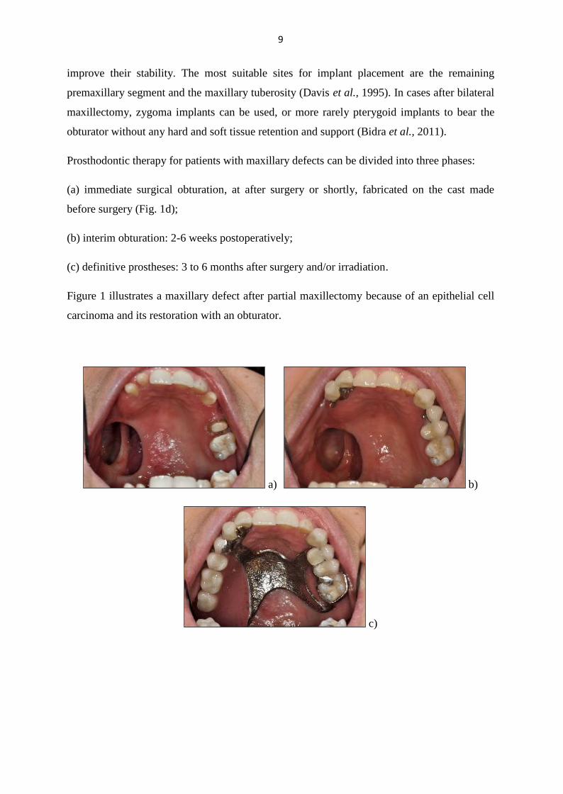
Prosthodontic therapy for patients with maxillary defects can be divided into three phases:
(a) immediate surgical obturation, at after surgery or shortly, fabricated on the cast made
before surgery (Fig. 1d);
(b) interim obturation: 2-6 weeks postoperatively;
(c) definitive prostheses: 3 to 6 months after surgery and/or irradiation.
Figure 1 illustrates a maxillary defect after partial maxillectomy because of an epithelial cell
carcinoma and its restoration with an obturator.
a) b)
c)

10
d)
e) f)
g)
h)
Figure 1. a) Maxillary defect. b) Intraoral status with bridges and preci-vertix. c) Intraoral
status with obturator. d) Immediate surgical obturation. e) f) Definitive prosthesis.
g) Occlusion with obturator. h) Full-face frontal view after rehabilitation.

11
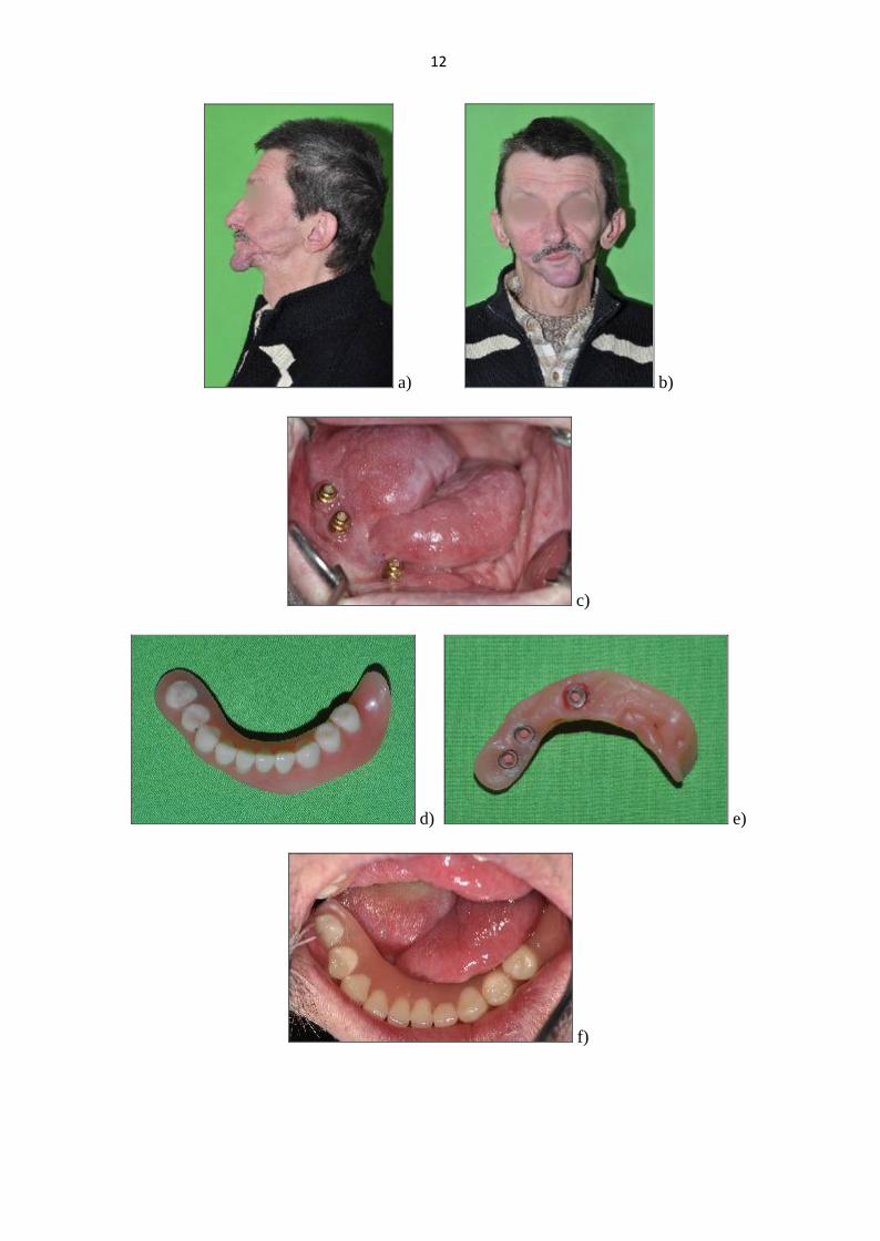
III.1.1.2. Prosthetic rehabilitation of patients with oral malignancy-acquired mandibular
defects
An other intraoral functional problem arises after the surgical treatment of a tumor of the
tongue or floor of the mouth. Both locations predispose the mandible to tumor invasion, often
necessitating its resection in conjunction with large portions of the tongue and surrounding
sublingual tissues and regional lymphatics (Harold, 1971). Because of the problem of tongue
movement the speech can be ununderstandable and the lack of the vestibulum causes
difficulties in the fixation of prostheses. In cases affecting the mandible, the involved segment
must be resected. The major causes of mandibular discontinuity are tumor resection, trauma
and, to a lesser degree, osteoradionecrosis and osteomyelitis. Loss of a mandibular segment
results in serious disabilities, including impairments of chewing, swallowing and speech,
drooling and a cosmetic disfigurement. The oral rehabilitation of these patients with
mandibular discontinuity defects is the most challenging problem facing both the surgeon and
the prosthodontist. The remaining mandibular segment is often displaced medially, causing an
inappropriate occlusal position (Figure 2).
The conventional denture fitted on the remaining mandibular segment is frequently unstable
and the unsatisfactory result can be frustrating to both patient and restorative dentist. It is
recommended to replace the missing bone and to reconstruct the functional and aesthetic
demands of the patients. The best method for this comprises free vascularized bone grafting,
such as an iliac or a fibula graft as vital bone graft (Keller et al., 1998, Urken et al.,1991).
Often, however, reconstruction of the bony defect alone does not guarantee an adequate
foundation for successful conventional prosthetic rehabilitation. Osseointegrated implants
placed into the microvascularized grafted bone offer an opportunity for an improved function
and patient satisfaction.
12
a) b)
c)
d) e)
f)

13
g)
h)
Figure 2. a) Full-face lateral view after operation and osteoradionecrosis. b) Full-face frontal
view after operation and osteoradionecrosis. c) Intraoral situation after operation and
osteoradionecrosis (with implants). d) e) Implant-retained defect prosthesis. f) Implant-
retained prosthesis in situ. g) Occlusion after rehabilitation. h) Full-face frontal view after
rehabilitation.
III.1.2. Extraoral rehabilitation
The restoration in cases of persons who have lost a portion of their faces through surgical
removal of a malignant tumor or through a congenital absence or trauma poses one of the
greatest challenges for the maxillofacial prosthodontist (McKinstry, 1995). A defect of the
face, as the most conspicuous body part, means a huge handicap for patients. It leads to a
decreased QOL, depression and barriers in resocialization. Restoration of these defects is very
important from functional and aesthetic aspects (Kadar, Nagy, 2009).

14
The success of the prosthetic restoration of any part of the body, including the head, depends
on the availability of a method of attaching the artificial substitute securely in the appropriate
place without causing discomfort or irritation to the tissues with which it comes in contact
(Bulbulian, 1973). Methods of retention used for facial prostheses fall into four categories: (a)
adhesive, when adhesive materials are used to retain the prosthesis, (b) mechanical, (c)
anatomical, when the retentive contours existing at the site of the deformity are used to retain
the prosthesis, and (d) extraoral implant, when implant fixtures anchored into the bone are
used to fix the facial prosthesis (McKinstry, 1995) Which method of retention is chosen
depends on the anatomical situation of the facial defect, the treatment method and the general
staging of the patient.
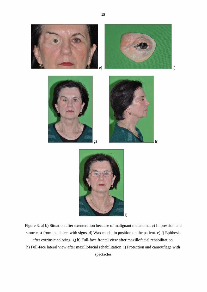
Figure 3 depicts the case of a facial defect after exenteration because of malignant melanoma
and the maxillofacial rehabilitation with an adhesive-retained epithesis.
a) b)
c) d)
15
e) f)
g) h)
i)
Figure 3. a) b) Situation after exenteration because of malignant melanoma. c) Impression and
stone cast from the defect with signs. d) Wax model in position on the patient. e) f) Epithesis
after extrinsic coloring. g) h) Full-face frontal view after maxillofacial rehabilitation.
h) Full-face lateral view after maxillofacial rehabilitation. i) Protection and camouflage with
spectacles

16
III.2. Quality of life
The QOL in patients treated for head and neck cancer is an important outcome parameter in
the post-treatment follow-up. QOL has been defined in many ways by numerous of groups.
The WHO originally defined QOL in 1947 as a „complete physical, mental and social welfare
state and not only the absence of the disease” (Torres Carranza et al., 2008). Nowdays, it is
defined by the WHO as „an individual’s perception of their own position in life, in the context
of the culture and value systems in their life and in relation to their goals, expectations,
standards and concerns” (Kazi et al., 2008, Sayed et al., 2009).
QOL can be defined as a concept that reflects several aspects of life, and an individual’s
perception of overall well-being with regard to disease and treatment-related symptoms is
specifically called the „health-related HRQOL”. (Boscolo-Rizzo et al., 2009, Kim et al.,
2010).
Revicki et al define QOL as a „broad range of human experiences related to one’s overall
well-being that minimally includes the broadly-defined assessments of the physical,
psychological and social domains of functioning”. (Revicki et al., 1997, Sayed et al., 2009).
QOL has also been defined as a multidimensional construct that includes, at a minimum,
physical, functional, psychological and social well-being. Other dimensions include
spirituality, sexuality, occupational functioning, treatment satisfaction and the overall rating
of the QOL. (Montazeri, 2009) Cella defined it as an individual’s perceived physical, mental
and social health status.
Cancer and its treatment regimens can result in the disruption of one or more dimensions of
the QOL. That is why the QOL is a parameter increasingly used in daily clinical practice to
assess the effectiveness of a treatment and has possibly become a parameter that helps
patients and physicians make therapeutic decisions (Lopez et al., 2009).

17
III.3. Measurement of quality of life
The European Organization for Research and Treatment of Cancer (EORTC) Quality of Life
Study Group has developed a measurement strategy for the assessment of QOL in clinical
trials (Bjordal et al., 1999).
The ideal measurement procedure for routine clinical practice should be short, easy for
patients to understand, address pertinent QOL issues, and be reliable and responsive to change
(Rogers et al., 1998) Patients are themselves unable to complete exhaustive questionnaires
and a short, simple measurement which takes less than 10 minutes to complete is ideal for
routine review (Sadura et al., 1992).
There are specific instruments with which to measure the QOL of head and neck cancer
patients, e.g. questionnaires- the University of Washington Quality of Life Questionnaire
(UW QLQ), the QLQ of EORTC for head and neck cancer patients (EORTC H&N 35),
the QLQ of the Cancer Rehabilitation Evaluation System (CARES) and the Performance
Status Scale- Head and Neck (PSS-H&N), indices such as the Karnofsky Performance Scale
(KPS), the Obturator Functioning Scale and the quantity of saliva measure. The QLQ
measures the individuals’ perceptions of their own physical, mental and social health status,
or some aspects of their health status resulting from cancer and its treatment. Sayed et al. have
given a list of 10 attributes necessary in the selection of QLQs as a final study tool: valid:
appropriateness, meaningfulness and usefulness of a measure for a specific purpose; reliable:
stability and reproducibility of a measure over time; interpretable: clinically relevant;
sensitive: responsive to change; short: minimal time-burden; easy to score; have an overall
global score and domain scores; multidimensional: covers a broad range of items in multiple
domains; self-administered; no floor or ceiling effect: ability to detect changes at two
extremes of QOL.
The questionnaires are self-administered but, depending on the patient input, with minimal
assistance from a health-worker if absolutely necessary.

18
III.3.1. UW- QOL questionnaire
The UW QOL questionnaire is a well-validated QOL instrument. It is potentially suitable as
an instrument for busy clinical practice as it is quick and simple for patients to complete and
is easy to process (Rogers et al., 1999).
The UW QOL questionnaire is a simple, brief, well-validated and widely-used head and neck
cancer-specific, self-administered scale (Hassan et al., 1993, Kazi, 2008). Version 1
comprises 9 domains that cover a range of disease-specific functional items including pain,
disfigurement, activity, recreation/entertainment, employment, speech, chewing, swallowing
and shoulder disability. It was revised in 2002-2003 to its current version 4 by Rogers SN
et al. in an attempt to eliminate inconsistencies and improve on important missing elements in
the spectrum of disease-specific responses to treatment (Rogers SN, 2002, Hassan et al.,
1993, Kazi et al. 2010). Version 4 contains 12 domains: pain, appearance, activity, recreation,
swallowing, chewing, speech, shoulder function, taste, saliva, mood and anxiety.
III.3.2. EORTC H&N 35 questionnaire
The EORTC H&N 35 (version 1.0) questionnaire, which includes 35 items, has been
translated into many languages, including Hungarian, following the EORTC QOL Study
Group guidelines (Bjordal et al., 1999, Cull et al., 1998). The original questionnaire was
validated by Bjordal and co-workers (Bjordal et al., 1992, Bjordal et al., 1994).
The EORTC H&N 35 QLQ is sometimes used together with the EORTC C30 QLQ, which
comprises physical, role, emotional, cognitive and social functioning scales and other items
such us fatigue, nausea, vomiting, pain, dyspnea, insomnia, loss of appetite, constipation,
diarrhea and financial difficulties.
The EORTC H&N 35 QLQ comprises 35 tumor- specific questions assessing symptoms and
side-effects of treatment. Most items are scored on a four-point response scale: 1 (not at all) to
4 (very much). 25 questions are organized into 7 multi-item subscales: pain (HNPA:

19
items 1-4 regarding pain in the mouth, pain in the jaw, soreness in the mouth and painful
throat), swallowing (HNSW: items 5-8 and 17 items that assess different degrees of
swallowing problems: problems in swallowing liquid, pureed food or solid food, and choking
when swallowing), senses (HNSE: items 13-14 regarding smell and taste), speech (HNSP:
items 16 and 23-24 assess hoarseness and problems with talking to other people or on the
phone), social eating (HNSO: items 19-22 regarding trouble in eating, individually or in front
of family or others), social contact (HNSC: items 18 and 25-28 regarding trouble with body
image and having physical and social contact with family and others) and sexuality (HNSX:
items 29-30 assess interest in sex and sexual enjoyment). The remaining 10 single items
address problems with teeth, dry mouth, sticky saliva, cough, mouth opening, weight loss,
weight gain, use of nutritional supplements, feeding tubes and pain medication
(Rinkel et al., 2009).

20
IV. AIMS OF THE STUDY AND QUESTIONS TO BE ANSWERED
The aims of my study were to examine the patients treated and rehabilitated at our
Maxillofacial Rehabilitation Department, to establish how the QOL of head and neck cancer
patients deteriorates after treatment (operation, radiotherapy and chemotherapy) and to
determine how it can be improved through maxillofacial rehabilitation.
Questions to be answered
I set out to collect epidemiological data on head and neck cancer patients in order to learn
their distribution and information concerning their smoking and alcohol drinking habits and
oncological characteristics. I wished to establish which treatment and rehabilitation methods
are most frequent.
I wished to know which of the important functions such as speech, eating, swallowing and
aesthetics are mainly impaired after treatment.
A further question to be answered related to whether the QOL of head and neck cancer
patients can be improved through maxillofacial rehabilitation and, if so, which of the impaired
functions is most improved by rehabilitation.
I additionally wished to learn whether the QLQ can be used as a routine examination in the
post- treatment follow- up of head and neck cancer patients, and which questionnaire is best
or gives more information about the QOL.

21
V. MATERIALS AND METHODS
The study protocol and the informed consent form were approved by the Ethics Committee of
the Faculty of Medicine, at the University of Szeged.
V. 1. Clinical study
V. 1. 1. Patient selection
In the period between 1994 and 2010, 92 head and neck cancer patients were rehabilitated
following tumor treatment at the Maxillofacial Rehabilitation Unit, Departement of Oral
Surgery, Faculty of Dentistry, University of Szeged. In the above period, 21 of the patients
subsequently died and 12 patients failed to respond to the invitation letter. The remaining 59
patients completed two QLQs. The eligibility criteria included tumor treatment administration
due to head and neck cancer, followed by maxillofacial rehabilitation, and the patient’s ability
to understand written and spoken Hungarian.
V.1.2. Data collection
The following data were obtained from patients who had undergone rehabilitation and from
them who later died: (a) socio-demographic characteristics such as age at treatment, and
gender; (b) behavior: smoking and drinking habits and (c) clinical status: site of primary
tumor, type of treatment and nature of rehabilitation. The information on these patients was
recorded retrospectively from the clinical documentation.
Additional investigations were performed to review the changes in QOL after maxillofacial
rehabilitation in comparison with the QOL status after tumor treatment without rehabilitation.
Two questionnaires were used for this study.
V.1.3. Patient self-report questionnaires
Two QLQs were completed: one of them was the UW QOL, version 1.0 questionnaire and the
other was the EORTC QOL H&N 35 questionnaire. Both of them were the official translated
Hungarian version. We did not wish to utilize the EORTC C30 together with EORTC QLQ
H&N 35 because we wished to use these other two special questionniares for head and neck
22
cancer patients and we concidered that three questionnaires would be too much for the
patients. The questionnaires were completed on two occasions: first, following treatment but
before rehabilitation, and then following maxillofacial rehabilitation. On both occasions, the
patients were recalled to complete the QLQs as part of an interview and follow-up. All the
patients completed the questionnaires themselves, but received helpful instructions if this was
necessary. A doctor who rehabilitates head and neck cancer patients and had been specifically
trained in connection with the questionnaires was therefore present at the interview.
The completed forms were carefully checked.
V.2. Statistical analysis
Statistical analysis based on the program Stata was carried out by the Statistics Team of the
Faculty of Medicine at the University of Szeged.
The collated data were entered into an Excel worksheet.
The sociodemographic data such as the age at treatment, the gender and mortality were
collected in Tables. The site of the primary tumor, the treatment mode and the rehabilitation
methods were recorded in other Tables. The program Stata was used. Descriptive statistics
were utilized to describe the mean, the standard deviation (SD), and the distributions of the
treatment and rehabilitation methods.
The Wilcoxon signed-rank test was used to compare the situations after tumor treatment with
and without maxillofacial rehabilitation. A p value less than/equal to 0.05 was considered
significant.
23
VI. RESULTS
VI.1. Demographic results / Patient characteristics
In the period in question, 92 patients underwent tumor treatment and maxillofacial
rehabilitation at the Faculty of Medicine and the Faculty of Dentistry, at the University of
Szeged. 21 of the 80 processed patients had died before the start of the present study. The
related mortality was therefore 26.25%. However since these patients had received treatment
for their tumor and also undergone maxillofacial rehabilitation, their epidemiological data
were nevertheless included in the study. The surviving patients were recalled several times
during the follow-up period for control purposes and to complete the UW QLQ and the
EORTC H&N 35 QLQ. 12 patients failed to reply to the invitation letter. The epidemiological
data on 80 patients were therefore processed. The recorded information included the age at
treatment, the gender, the tumor localization, the treatment method and the type of
rehabilitation. 53 (66.25%) of the patients were men and 27 (33.75%) women. The male:
female ratio was therefore 2:1. The average age was 53.86 years (ranging from 9 to 74 years),
with more than half of the patients (55 (56.25%)) aged between 50 and 69.9 years.
The epidemiological data are listed in Table 1. The age distribution is presented in Figure 4.
The incidence of smoking and alcohol consumption was rather high. 60 (75%) of the patients
were smokers, and 45 patients (56.25%) drank alcohol regularly.

24
Categories n(%)
Gender Male
Female
53 (66.25%)
27 (33.75%)
Age
at tumor treatment
<40
40-49.9
50-59.9
60-69.9
>70
9 (11.25%)
18 (22.5%)
26 (32.5%)
19 (23.75%)
8 (10%)
Tobacco use Yes
No
60 (75%)
20 (25%)
Alcohol
consumption
Yes
No
45 (56.25%)
35 (43.75%)
Mortality 21 (26.25%)
Table 1. Epidemiological characteristics of study population

25
0
5
1 0
1 5
2 0
2 5
3 0
n
< 4 0 4 0 -4 9 .9 5 0 -5 9 .9 6 0 -6 9 .9 > 7 0
A ge (years)
Figure 4. Age distribution among rehabilitated head and neck cancer patients.
The oncological data are to be found in Table 2. The distribution of the tumor localization and
the types of treatment methods are shown in Figures 5 and 6. As far as the locations were
concerned, the cancer developed most frequently in the floor of the mouth area, in 21 patients
(26.25%), followed by the mandibular or maxillar gingiva in 17 cases (21.25%), the maxilla
in 12 cases (15%) and the tongue in 9 cases (11.25%).
41 patients (51.25%) received combined surgery and radiotherapy. 26 patients (32.5%) were
treated surgically alone, and 2 patients (2.5%) with radiotherapy alone. 11 patients (13.75%)
participated in other forms of combined therapy.

26
Categories n (%)
Tumor localization maxilla
tongue
floor of the mouth
facial defect (eye-ear-nose)
others
12 (15%)
9 (11.25%)
21 (26.25%)
3-2-1 (3.75%- 2.5%-1.25%)
32 (40%)
Type of treatment surgery alone
radiotherapy alone
surgery and radiotherapy
surgery and chemotherapy
surgery, radiation and chemotherapy
26 (32.5%)
2 (2.5%)
41 (51.25%)
2 (2.5%)
9 (11.25%)
Table 2. Oncological characteristics of study population

27
2
2
1
5
6
2
8
9
9
21
3
12
0 5 10 15 20 25buccaph
arynx
laryn
xlower
lipfac
e
tonsil
loling
ual a
rea
maxilla
ry gin
giva
mandib
ular g
ingiva
tongu
e
floor
of the
mou
thpa
latemax
illa
Site
of p
rimar
y tu
mor
Number of cases
Figure 5. Distribution of sites of primary tumor

28
9
2
41
2
26
0 5 10 15 20 25 30 35 40 45
Surgery, radiation and chemotherapycombination
Surgery and chemotherapy
Surgery and radiation
Irradiation alone
Surgery alone
Trea
tmen
t met
hod
Number of cases
Figure 6. Distribution of treatment methods.
The types of prostheses are presented in Table 3, and their distribution in Figure 7. In the
course of rehabilitation, a special defect prosthesis was prepared for about half of the patients
(43.75%): an obturator was fitted in 14 patients (12.5%), an implant-retained removable
denture was applied in 23 patients (20.54%), and reconstruction with an epithesis was applied
in 12 patients (10.71%). Most of the epitheses were for an orbital defect, in 7 cases (6.25%).
Other parts of the face were also rehabilitated, with an ear epithesis in 3 cases (2.7%) and
a nasal epithesis in 2 cases (1.8%).

29
Type of prosthesis n (%)
Obturator prosthesis 14 (12.5%)
Removable denture (lower and/or upper) 32 (28.57%)
Implant-retained removable denture 23 (20.54%)
Combined prosthesis 28 (25%)
Bridge (fixed, cemented prosthesis) 3 (2.7%)
Orbital epithesis 7 (6.25%)
Nasal epithesis 2 (1.8%)
Aural epithesis 3 (2.7%)
Total number of prostheses 112
Table 3. Type of rehabilitation

30
12
3
28
32
23
14
0 10 20 30 40
Epithesis
Fixed bridge
Combined prosthesis
Removable denture
Implant-retained removabledenture
ObturatorTy
pe o
f reh
abili
tatio
n
Number of cases
Figure 7. Distribution of rehabilitation methods
VI.2. Statistical results of QOL questionnaires
The UW QOL and EORTC H&N 35 questionnaires were well accepted by the patients, who
appeared cooperative; none of the eligible participants refused to complete the questionnaire.
VI.2.1. Results of UW QOL questionnaire
The UW QOL questionnaire, which was well accepted by the patients, included 9 questions,
each answer is scaled from 0 (best) to 100 (worst). A composite score was calculated by
adding together the scores for 9 answers for the various domains and then dividing by 9 to
give a result on the scale from 0 to 100. The composite score, which before rehabilitation was

31
reasonably high, at 66.62, improved to 36.2 following rehabilitation. The change was
significant (p=0.000)
The scores before (BR) and after rehabilitation (AR) are reported in Table 4, and the
improvement in the QOL is shown in Figure 8.
The greatest problems after treatment but before rehabilitation were associated with chewing
(BR: 88.58), activity (BR: 68.8) and recreation (BR: 68.2). All of these improved
considerably after rehabilitation. Nevertheless, especially the subscale of pain was increased
after rehabilitation.
Employment displayed a high score both before and after rehabilitation (87.8 and 92), and
tended to deteriorate in the course of time after rehabilitation. As concerns the question of
family relations, the scores were good in both situations (BR: 25.4 and AR: 21.4), as was the
shoulder function (BR: 26 and AR: 23.6).
HRQOL domain BR score AR score Variation
Pain 67.4 25.6 p=0.000
Appearance 64.6 39.2 p=0.000
Activity 68.8 31.6 p=0.000
Recreation 68.2 32.8 p=0.000
Employment 87.8 92 p=0.732
Chewing 88.58 53.94 p=0.000
Swallowing 65.25 33.25 p=0.000
Speech 62.25 39.5 p=0.000
Shoulder function 26 23.6 p=0.452
Family relations 25.4 21.4 p=0.062
Resocialization with friends 36.2 24.8 p=0.000
Overall (Composite score) 66.62 36.2 p=0.000
Table 4. Results of UW QLQ before and after rehabilitation, with the level significance

32
0102030405060708090
100
Pain
AppearanceActivity
RecreationChewing
SwallowingSpeech
Shoulder function
FamilyFriends
EmploymentOverall
Scor
e
Before rehabilitation After rehabilitation
Figure 8. Changes in results of UW QLQ after rehabilitation
Significant improvements after rehabilitation were observed as regards pain, appearance,
activity, recreation, chewing, swallowing, speech, resocialization with friends and the overall
score. There was no significance from the aspects of employment (p=0.732), shoulder
function (p=0.452) and family relations (p=0.062).
VI.2.2. Results of EORTC H&N 35 QOL questionnaire
The mean EORTC H&N 35 scores, standard deviations and ranges after treatment but before
rehabilitation are presented in Table 5, the corresponding statistical results after rehabilitation
in Table 6, and the improvement in QOL in Figures 9 and 10.

33
EORTC H&N 35 QLQ Mean Standard deviation Range
Pain (HNPA) 69.95763 18.63425 26-104
Swallowing (HNSW) 77.22881 20.83935 26-104
Senses (HNSE) 56.99153 24.1528 25-100
Speech (HNSP) 61.52542 17.28649 24.75-99
Social eating (HNSO) 75.24576 17.91127 32.5-104
Social contacts (HNSC) 53.81356 17.64934 25-90
Sexuality (HNSX) 47.0339 26.09034 25-100
Teeth (HNTE) 2.271186 1.095925 1-4
Mouth opening (HNOM) 2.728814 .9970734 1-4
Dry mouth (HNDR) 3.033898 .889907 1-4
Sticky saliva (HNSS) 2.79661 .9961938 1-4
Coughing (HNCO) 1.79661 .9961938 1-4
Feeling ill (HNFI) 3.508475 .6531853 1-4
Pain killers (HNPK) 1.847458 .3626321 1-2
Nutritional supplements (HNNU) 1.576271 .4983902 1-2
Feeding tube (HNFE) 1.627119 .4877218 1-2
Weight loss (HNWL) 1.79661 .4059752 1-2
Weight gain (HNWG) 1.067797 .2535545 1-2
Table 5. Means, standard deviations and ranges after treatment, but before rehabilitation

34
Table 6. Statistical results relating to the changes in the QOL after rehabilitation
EORTC H&N 35 QLQ Mean Standard deviation Range
Pain (HNPA) 32.62264 7.908677 26-58.5
Swallowing (HNSW) 30.66038 6.421698 26-58.5
Senses (HNSE) 34.66981 15.43471 25-100
Speech (HNSP) 30.50943 8.650389 24.75-57.75
Social eating (HNSO) 32.37736 8.5978 26-65
Social contacts (HNSC) 28.67925 5.895806 25-55
Sexuality (HNSX) 32.54717 14.1548 25-75
Teeth (HNTE) 1.396226 .7162837 1-4
Mouth opening (HNOM) 1.509434 .7751586 1-3
Dry mouth (HNDR) 1.962264 .8311777 1-4
Sticky saliva (HNSS) 1.830189 .8023008 1-4
Coughing (HNCO) 1.301887 .6380531 1-4
Feeling ill (HNFI) 1.264151 .4863895 1-3
Pain killers (HNPK) 1.075472 .2666788 1-2
Nutritional supplements (HNNU) 1.150943 .3614196 1-2
Feeding tube (HNFE) 1 0 1-1
Weight loss (HNWL) 1.056604 .2332953 1-2
Weight gain (HNWG) 1.415094 .4974536 1-2

35
0
10
20
30
40
50
60
70
80
HNPA HNSW HNSE HNSP HNSO HNSC HNSX
Before rehabilitation After rehabilitation
Figure 9. Means of results of 7 scales in EORTC H&N 35 QLQ before and after rehabilitation
(HNPA: pain, HNSW: swallowing, HNSE: senses, HNSP: speech, HNSO: social eating,
HNSC: social contacts, HNSX: sexuality)
0,00
0,50
1,00
1,50
2,00
2,50
3,00
3,50
4,00
HNTE HNOM HNDR HNSS HNCO HNFI HNPK HNNU HNFE HNWL HNWG
Before rehabilitation After rehabilitation
Figure 10. Means of results of single items in EORTC H&N 35 QLQ before and after
rehabilitation
(HNTE: teeth, HNOM: mouth opening, HNDR: dry mouth, HNSS: sticky saliva, HNCO:
coughing, HNFI: feeling ill, HNPK: pain killers, HNNU: nutritional supplements, HNFE:
feeding tube, HNWL: weight loss, HNWG: weight gain)

36
After tumor treatment, the worst score in the subgroups was that for HNSW (swallowing),
followed by HNSO (social eating) and HNSP (pain). From the single items, the worst
problem was an illness feeling and dry mouth with sticky saliva as side-effects of irradiation.
The rehabilitation led to considerable effects on swallowing, social eating and speech. The
highest improvement for the single items after rehabilitation was in the illness feeling. Only
the weight gain gave an inverse result.
All of the items showed a significant increase (p<0.05) after rehabilitation in comparison with
the results before rehabilitation.

37
VII. DISCUSSION
In my study, I set out to collect data on rehabilitated head and neck cancer patients in the
descriptive part of the search. I was looking for the most affected age, the primary tumor site,
and the treatment and rehabilitation methods, and was seeking answers concerning which
primary tumor site and its treatment need special maxillofacial rehabilitation most frequently.
A further aim of my study was to examine the changes in the QOL of head and neck cancer
patients through comparisons before and after maxillofacial rehabilitation and to investigate
whether our prosthetic methods can improve the QOL of head and neck cancer patients
significantly.
Head and neck cancer and its treatment can have a profound effect on the patient’s physical,
functional and emotional well-being, especially decreasing the QOL. (Evans et al., 2003,
Jones et al., 1992, Rogers et al., 1999, Kazi et al., 2010). QOL evaluation has increasingly
become an important supplement in the interpretation of the outcome information in head and
neck cancer treatment (Hassan et al., 1993, Murphy, 2009, Vartanian et al., 2004, Nazar
et al., 2010) It can be measured by the administration of specific questionnaires to the
affected patients. In Hungary, there have been no such examinations of the QOL. A survey of
the international literature revealed numerous papers related to the comparison and validation
of different QLQs, the comparative analysis of the QOL before and after treatment, and the
comparison of the outcome following several treatment methods, but I have found no studies
involving a review of the QOL of head and neck cancer patients before and after maxillofacial
rehabilitation. This was the background in my selection of the goals in my study.
VII.1. Sociodemographic and epidemiologic analysis
I found that the male:female ratio in this patient group was 2:1. This correlates with the
results of Gritz et al., Hassan et al., Hassanein et al., Kornblith et al., Lam Tang et al. and
Yang et al., but differs from the findings of Head et al. (6.88:1), Silveira et al. (5:1), de Graeff
et al. (4:1), Alicikus et al. (4:1), Lopez et al. (4:1), Kim et al. (3.6:1), Arstaad et al. (3:1),

38
Nazar et al. (3:1), Scharloo et al. (3:1), Thomas et al. (3:1), Hammerlid et al. (3:1), Schoen et
al. (1.2:1) and Kruse et al. (1.15:1). Hammerlid et al. examined patients with oral, pharyngeal
and laryngeal cancer and found that the oral cavity was more common as tumor location
among females (52%) than among males.
The mean age of the patients was 53.8 years (SD: 12.8 years). This correlates with the results
of Hassan et al. (55 years), Hassanein et al. (58 years), Lam Tang et al. (55.5 years), Gritz
et al. (58.4 years), Kim et al. (60.6 years), Scharloo et al., (59.5 years), Silveira et al.
(59.4 years), Alicikus et al. (53 years), Lopez et al. (55.78 years), Stevens et al. (56.1 years),
Verdonck-de Leeuw et al. (59 years) and Kornblith et al. (59.5 years). In several studies, the
mean age was over 60 years: Bjordal et al. (61 years), Rinkel et al. (62 years), Head et al.
(60.2 years), Nalbadian et al. (62.57 years), Rogers et al. (62 years), Rinkel et al. (62 years),
Nazar et al. (64.4 years), Hammerlid et al. (63 years) and Schoen (63.5 years). Only one study
reported a mean age under 50 years: Kazi et al. found it to be 49.6 years.
Among the present head and neck cancer patients, 60 (75%) were smokers and 20 (25%) were
non-smokers, and 45 patients (56.25%) consumed alcohol regularly. These habits have been
considered in only a small proportion of the analogous investigations, although they are very
important factors in the development of tumors in the head and neck region, and later play a
considerable role in the changes in the QOL after treatment, as confirmed by the xerostomia.
In the study by Hammerlid et al., 29% of the patients had never smoked. Our result correlates
with this finding. Meyer et al. found a 64% incidence of tobacco use among their studied
patient group. Vartanian et al. (2006) started that 80% of their patients reported previous
tobacco use and 75.7% alcohol consumption. Gritz et al. observed a significant reduction in
smoking status after a 1-year follow-up, and a significant decline in alcohol use following
treatment, with a significant increase in alcohol use between 1 month and 1 year.
The most commonly affected primary site was the floor of the mouth, in 21 patients (26.25%),
followed by the gingiva in 17 patients (21.25%), the maxilla in 12 patients (15%) and the
tongue in 9 patients (11.25%). Our results are similar to those of Hassanein et al.: the floor of
the mouth (29%), the tongue (21%) and the mandibular alveolus (18%). Lopez et al. found
that the tongue was the most affected (38%), followed by the floor of the mouth, with 10%. In
the study by Lam Tang et al., the mandible was the most affected area (44%). Hassan et al.
examined patients with pharyngeal and laryngeal tumors, too and found the oral cavity to be
most affected (36%). Thomas et al. examined 77 patients, in 34 (44%) of whom the tumor
39
was in the tonsillar fossa, and in 20 patients (26%) in the tongue. Kruse et al. studied 99
elderly patients with head and neck cancer and found that the maxillary and mandibular
alveolar ridges (24% each) were the most affected, followed by the tongue (18.9%). In the 47
patients in the study by Biazevic et al., the tumor was in the oral cavity (the floor of the
mouth, the gingiva, the retromolar area or the palate) in 19 cases (40%) and, in the
oropharynx in 12 cases (25.5%), with 11 in the tongue (23.4%). Kim et al. conducted a study
on 133 patients, and found the tonsillar area to be affected in 89 cases (66.9%), the base of the
tongue in 23 (17.29%) and the soft palate in 15 patients (11.28%).
The most frequently applied treatment method was surgery together with radiotherapy
(51.25%). This confirms with the results of Rinkel et al. (50%), Nazar et al. (47.2%) and Kim
et al. (71.2%). Scharloo et al. found that the use of irradiation alone was the most frequent
treatment method (40.7%). In the investigation by Thomas et al., 88.3% of the patients
received primary or adjuvant radiotherapy. Vartanian et al. (2004) examined 301 patients,
158 of whom (52.5%) underwent only surgery, 34 (11.3%) were irradiated, and 98 (32.6%)
recieved a combination of surgery and radiotherapy. Nalbadian et al. found surgery alone to
be the most commonly applied treatment method (54.1%). In the study by Verdonck-de
Leeuw et al., radiotherapy was the most frequently applied treatment (32%), followed by a
combination of surgery and radiotherapy (27%). Hassanein et al. reported surgery as the most
common (70%) treatment method, with surgery combined with radiotherapy (18%) in second
place.
The tumor localization and the treatment method, together with the general disease stage, play
essential roles not only in the treatment of head and neck cancer, but also in the incidence and
intensity of the side-effects and the QOL (Alicikus et al., 2009, Zackrisson et al., 2003).
During the period examined, 106 prosthetic rehabilitations were performed on 92 patients.
Some of patients received both upper and lower prostheses, or several prostheses during this
period. Most of these prostheses were special prostheses for defect situations (43.75%):
23 implant-retained removable dentures were prepared for mandibular or maxillary defects,
14 obturators (12.5%) were made for maxillary defects after maxillectomy, and in 12
(10.71%) cases epitheses were made for facial defects: 7 orbital (6.25%), 3 aural (2.7%) and 2
nasal (1.8%) epitheses. The other rehabilitations involved 63 conventional prostheses
(56.25%): 32 total removable dentures (28.7%), 28 combined prosthesis (25%) and 3 fixed

40
cemented bridges (2.7%). I do not have exact information about whether some of the 28
combined prostheses were made for an intraoral defect situation.
Schoen et al. studied a group of 67 patients with an edentulous mandible after the treatment of
squamous cell carcinoma in the lower region of the oral cavity (the tongue, floor of the
mouth, mandibular gingiva, buccal mucosa or oropharynx). Half of the patients (n=33) never
wore their mandibular conventional prosthesis, or at most for only a few hours per day for
cosmetic reasons. Insufficient retention of the mandibular prosthesis was noted in 55% of the
patients, and diminished stability in 23% of the patients. Interforaminal located implants in
the mandible for improvement of the stability of a fully removable lower denture are
increasingly used by healthy patients. This is more to be expected in cases of mandibular
defects because of the decayed mucosal supplement and diminished vestibulum. Most of our
head and neck cancer patients with this intraoral situation are rehabilitated with an implant-
retained removable denture on 2 or 4 interforaminal implants.
VII.2. Comparative analysis of measurements of QOL questionnaires
In my study, I analyzed which function is especially damaged by tumor treatment and
measured the changes in the QOL through a comparison before and after rehabilitation.
Most of the available studies made comparisons between some special QLQs
(e.g. comparative studies with KPS, CARES or UW QOL questionnaires) or with only one or
two domains (e.g. the speech domain), or between healthy and tumor patient groups, or
between the pretreatment and the posttreatment situation, or on the longitudinal effects of
cancer treatment. Merely a few studies extended to the changes in the QOL after maxillofacial
rehabilitation. This study can give a new comparison profile and data for the Hungarian and
international literature.
VII.2.1. Results with the UW QOL questionnaire
The mean composite score in my study was reasonably high, at 66.62. This is lower than the
result determined in 2008 by Kazi et al. (73.6), who studied a subgroup of patients after
partial glossectomy.

41
The worst results in the UW QOL questionnaire before rehabilitation in my study related to
employment (BR: 87.8) and chewing domain (BR: 88.58). In the employment domain, a
common answer was ’’I am retired-due to the cancer treatment or not related to it”. It was
connected with the basic tumor disease, with habitual problems of smoking and drinking
alcohol, and the age and general health status of the patients. It means that most of the
patients were retired and the majority of the treatment did not seem to alter the employment
status. This scale was the only one for which the result after rehabilitation was decreased.
Rehabilitation had much less influence at this stage of life.
I observed a worse result in the scale of chewing, which correlated with the result of Kazi
et al (2010). Other wise, Biazevic et al. found chewing (48%) and speech (44%) to be the
most prevalent complaints at the time of treatment, and chewing (60%) and swallowing (24%)
at the 1-year follow-up. In their study, chewing was the QOL domain which exhibited the
largest reduction in rating, from 74.0 at baseline to 34.0 1 year after surgery. It is interesting
that Rogers et al. (1999) found no trouble with chewing in 45% of the patients in their study
group.
There was no significant change in shoulder function before and after rehabilitation and in
this scale the score was already low after treatment (BR: 26). This means that most of the
surgical procedures do not affect the accessorial nerve which is responsible for the abductor
movement of the shoulder.
The family relations did not show any significant change and the BR and AR answers were
equally positive. This is good from the aspect of the QOL because it means that the family
stands up for the patients in their enormous problems and help them in the healing period.
The best improvements following rehabilitation were in activity and recreation. This is related
with the overall feeling ill, mood and global health status. A great improvement in pain
emerges with the passage of time.
VII.2.2. Results of EORTC H&N 35 QOL questionnaire
The international literature relating to the QOL most frequently involves studies with the
EORTC H&N 35 QLQ. It is usually used together with EORTC C30, but I decided to apply

42
two questionnaires specific for head and neck tumors, and did not wish to overburden the
patients with too many questionnaires demanding a long completion time.
The questionnaire has 35 items concerning tumor and treatment-related physical symptoms.
The worst subscale scores after treatment were observed for swallowing (BR: 77.22), social
eating (BR: 75.24), pain (BR: 69.95) and speech (BR: 61.52), while the worst score for a
single item was that for dry mouth (BR: 3.03). The maxillofacial rehabilitation resulted in the
best effects on swallowing (AR: 30.66, change: 46.56), social eating (AR: 32.37, change:
42.87) and speech (AR: 30.50, change: 31.02). Other wise all of the examined subscales and
single items displayed significant changes in comparison with the situation before
rehabilitation.
Hammerlid et al. studied the QOL domains in connection with tumor localization, stage, sex
and age. They found that different primary tumor sites were associated with different scores:
Patients with nasopharyngeal carcinoma exhibited the worst social and role functioning and
highest pain score and they felt ill more often than patients with other tumor locations.
Patients with laryngeal carcinoma had the highest scores after treatment as regards speech and
coughing problems, while patients with tongue carcinoma scored highest on the pain scale
and for nutritional supplements. Their study revealed statistically significant differences in
connection with gender (all in favor of men), pain, social eating, social contacts and painkiller
use. Older patients tended to score more poorly than younger ones. De Graeff et al. conducted
a longitudinal study and found significantly increased problems involving pain, swallowing,
social eating, speech and taste/smell at 12 months after treatment. They observed a correlation
between the results on age and gender: women and older patients furnished worse scores.
Alicikus et al. carried out a study with EORTC H&N35 on factors influencing the QOL. They
found that the treatment modality had a major impact on speech ability and dry mouth:
postoperative irradiation led to a worse score for speech, and chemoradiotherapy did so for
sticky saliva and social eating. They further determined that the primary tumor site influenced
the results of EORTC H&N 35: Patients with nasopharyngeal tumor had the worst scores for
mouth opening, dry mouth, sticky saliva, swallowing and social eating, whereas patients with
laryngeal tumor indicated that speech was the worst problem. Murphy found through the use
of the EORTC H&N 35 QLQ that a number of symptoms remained problematic 12 and 24
months post-treatment: swallowing, taste/smell, speech, social eating, sexuality, trismus,
xerostomia and sticky saliva. They did not study patients with or without rehabilitation, and
examined only the treatment’s longitudinal effects. Nalbadian et al. studied Greek patients

43
with pharyngeal or laryngeal carcinoma after treatment and found the most common problems
with the EORTC H&N 35 QLQ in the areas of speech, sexuality, dry mouth, sticky saliva and
coughing. Speech and dry mouth were in the worst problem group after treatment in my
study. Kim et al. compared the outcome of surgery-based and radiation-based therapy. They
found no significant differences in the results of the EORTC H&N 35 QLQ between the two
groups, although members of the irradiated group had more problems with dry mouth, and
more difficulties in weight gain and were more dependent on pain killers.
VII.2.3. Comparison of results of UW QOL and EORTC H&N 35 questionnaires
For my study, I chose these two questionnaires because they complement each other well, and
both of them are very extensively applied in their own field. The UW QLQ contains more
questions about the psychological and social well-being of the patients. The EORTC H&N 35
questionnaire deals much more with the physical tumor- and treatment-related symptoms of
head and neck cancer patients. This causes difficulties in comparisons of the answers of the
two questionnaires: In the former, swallowing, activity, recreation and pain gave the worst
results before rehabilitation, while activity and recreation displayed the best increases after
maxillofacial rehabilitation. In the other questionnaire, swallowing, social eating, pain and dry
mouth were the worst problems for the patients, and the rehabilitation led to the greatest
changes in swallowing, social eating and speech. Swallowing and pain proved to be the most
serious problems before rehabilitation in both QLQs.
The study by Kanatas et al. demonstrated that the UW QOL was the most frequently used
questionnaire (72%) among members of the British Association of Head and Neck
Oncologists, followed by the EORTC C30 and the EORTC H&N 35 (52%).
44
VIII. SUMMARY AND CONCLUSIONS
Progress in the treatment of oral cancer has made it possible to reduce the post-treatment
mortality, and the survival rate has increased. However, the length of survival alone is an
unsatisfactory measure of the success. The tumor treatment of head and neck cancer patients
causes the QOL of the patients to deteriorate considerably after treatment, owing to the
impairment of such important functions as eating, swallowing and speech on the one hand,
and aesthetic aspects related to socialization on the other. This is why maxillofacial
rehabilitation has such an important place as the last step in the tumor treatment procedure.
In our study, the gender difference, with a male:female patient ratio of 2:1, appeared to be
significantly less marked than reported in earlier studies, which is explained by increasingly
higher rates of women smoking and drinking alcohol.
The majority of our patients consumed alcohol and smoked on a regular basis, which further
worsen the QOL through increase of the risk (and the related stress) of a local recurrence, and
affect the patients’ family and social relations.
A majority of the patients (51.25%) had received a combination of surgery and radiation as
therapy, which is in line with the oncotherapy protocol applied nowadays.
In the course of the rehabilitation, about half (43.75%) of the cases involved the preparation
of a special prosthesis as a solution: the application of obturators after maxillectomy
(14 cases/12.5%), implant-retained dentures (23 cases/20.54%) in cases of an acquired
mandibular defect or after surgery on a tumor of the tongue or the floor of the mouth, or
epitheses (12 cases/10.75%) in cases of facial defects.
As a means of assessing changes in the QOL with the aim of a subsequent improvement,
QOL questionnaires appear to provide an easily applicable, routine procedure in the care of
head and neck cancer patients. We conclude that the UW QLQ and the EORTC H&N 35
questionnaires are useful tools for the evaluation of the HRQOL in patients with cancer in this
region.
45
Statistical analysis of the results of the questionnaires suggests that post-treatment patients
awaiting rehabilitation experienced the greatest difficulties in the areas of eating and speech.
The results of the UW QLQ demonstrated that the worst problems after treatment related to
chewing, employment, activity and recreation, and the best increase after rehabilitation was
experienced as concerns pain, with additional significant improvements in activity and
recreation. There was no change in the level of family relations. This means that tumor as a
disease does not affect personal contacts in the family in a negative way and it does not need
improvement. There was no positive change in employment, because most of the head and
neck cancer patients had already retired before the tumor treatment, because of the general
staging or some other illness. There was no significant difference between the results before
and after rehabilitation as concerns the shoulder function.
The EORTC H&N 35 questionnaire was somewhat easier to complete. It indicated that the
worst subscale problems after tumor treatment were the swallowing and social eating,
followed by pain. Among the single items, the worst problems were dry mouth and sticky
saliva as side-effects of irradiation. The rehabilitation resulted in the greatest changes in
swallowing, social eating and speech and feeling ill.
Overall, maxillofacial rehabilitation leads to significant improvements in all impaired
functions and to positive changes affecting the QOL. The results of my investigations allow
me to state that prosthetic rehabilitation can play a key role in the life of head and neck cancer
patients through the resulting improvement in their QOL.

46
IX. ACKNOWLEDGMENTS
First and foremost, I would like to express my sincere thanks to Professor Katalin Nagy, Head
of the Faculty of Dentistry, for providing me with the opportunity to work on this special field
of dentistry and for her enormous help, encouraging support and constant enthusiasm and
professionalism in the supervision on my professional work in general and this study in
particular.
I am very grateful to Professor István Sonkodi, Head of the Department of Oral Medicine, for
his endless knowledge, support and tolerance throughout my whole clinical and scientific life.
I express my very special thanks to Professor József Piffkó, Head of the Department of
Maxillofacial Surgery, for his creation of an unique oasis in Hungarian medicine, and for his
efforts and support relating to my work. Many thanks too to the other members of the staff of
the Department of Maxillofacial Surgery for their co-operation.
I wish to express my appreciation to Professor Béla Iványi, Head of the Department of
Pathology, and to Dr. László Puskás, molecular researcher at the Biological Research Center
in Szeged, for adding their professional knowledge to my articles.
Many thanks are also due to Tibor Nyári at the Statistics Team of the Faculty of Medicine at
the University of Szeged for his work by statistical analysis.
I would like to express my thanks to my assistants and primarily Éva Körmendi and Györgyi
Vágó, without whom I could not have worked during the weekdays in this clinical
professional field.
I am grateful to Katalin Márton Pappné for her library help, and to Zsuzsa Kiss-Dózsai for the
photos and her technical help in my practical work and completion of this study.
I owe many thanks to my head and neck cancer patients, who have taught me love and respect
for health and human life.
Last, but not least, I am most grateful to my parents for their encouragement and especially
my daughter, Anna for her endless love and childlike adoration for my profession.

47
X. REFERENCES
1. Aarstad AK, Beisland E, Osthus AA, Aarstad HJ: Distress, quality of life, neuroticismand psychological coping are related in head and neck cancer patients duringfollow-up. Acta Oncologica 2010.
2. Alicikus ZA, Akman F, Ataman OU, Dag N, Orcin E, Bakis B, Kinay M: Importanceof patient, tumour and treatment related factors on quality of life in head and neckcancer patients after definitive treatment. Eur Arch Otorhinolaryngol 266: 1461-1468,2009.
3. Baile W, Gibertini M, Scott L: Depression and tumour stage in cancer of the head andneck. Psychooncol. 1: 15, 1992.
4. Beumer J, Zlotolow I, Curtis TA: Rehabilitation. In Silverman S Jr, ed. Oral cancer,3rd ed. Atlanta: American Cancer Society. 127-148, 1990.
5. Biazevic MG, Antunes JL, Togni J, de Andrade FP, de Carvalho MB, Wünsch-FilhoV: Survival and quality of life of patients with oral and oropharyngeal cancer at1-year follow-up of tumor resection. J Appl Oral Sci 18(3): 279-284, 2010.
6. Bidra AS, May GW, Tharp GE, Chambers MS: Pterygoid implants for maxillofacialrehabilitation of a patient with a bilateral maxillectomy defect. J Oral Implantol. 2011.
7. Bjordal K, Kaasa S: Psychometric validation of the EORTC Core Quality of LifeQuestionnaire, 30-item version and a diagnosis-specific module for head and neckcancer patients. Acta Oncologica 31: 311-321, 1992.
8. Bjordal K, Ahlner-Elmquist M, Toleson E, et al.: Development of a EuropeanOrganisation for Research and Treatment of Cancer (EORTC) questionnaire moduleto be used in quality of life assessments in head and neck cancer patients.Acta Oncologica 33: 879-885, 1994.
9. Bjordal K, Hammerlid E, Ahlner-Elmquist M: Quality of life in head and neck cancerpatients: Validation of the European organization for research and treatment of cancerquality of life questionnaire- H&N 35 J Clin Onc. 17(3),1008-1019, 1999.
10. Boscolo- Rizzo P, Stellin M, Fuson R, Marchiori C, Gava A, Da Mosto MC: Long-term quality of life after treatment for locally advanced oropharyngeal carcinoma:surgery and postoperative radiotherapy versus concurrent chemoradiation.Oral Oncol 45(11): 953-957, 2009.
11. Bulbulian AH.: Maxillofacial prosthetics: Evolution and practical appliction in patientrehabilitation. J Prosthet Dent 15: 554-569, 1965.
12. Bulbulian AH: Facial prosthetics, Springfield, II, Charles C Thomas. 1973.
13. Cella DF, Tulsky DS: Measuring quality of life today: Methodological aspects.Oncology 4: 29-38, 1990.

48
14. Coyne JC, Pajak TF, Harris J, Konski A, Movsas B, Ang K, Watkins Bruner D,Radiation Therapy Oncology Group: Emotional well-being does not predict survivalin head and neck cancer patients: a Radiation Therapy Oncology Group Study.Cancer 110: 2568-2575, 2007.
15. Converse JM: Reconstructive Plastic Surgery. W. B. Saunders Co., Philadelphia,1977.
16. Cull A, Sprangers MAG, Bjordal K et al: EORTC Quality of Life Study GroupTranslation Procedure. Brussels, Belgium, European Organization for Research andTreatment of Cancer. 1998.
17. Davis B, Roumanas E, Hong S, Nishimura R: Stress distribution of implants used forretention of maxillary obturators. In: Proceedings of 1st International Congress onMaxillofacial Prosthetics. 1995.
18. De Boer MF, Borne B Van Den, Pruyn JF, Ryckman RM, Volovics L, Knegt PP,Meeuwis CA, Mesters I, Verwoerd CD: Psychosocial and physical correlates ofsurvival and recurrence in patients with head and neck carcinoma: results of a 6-yearlongitudinal study. Cancer 83: 2567-2579, 1998.
19. De Graeff A, de Leeuw JR, Ros WJG, Hordijk GJ, Blijham GH, Winnubst JA:Pretreatment factors predicting quality of life after treatment for head and neck cancer.Head neck 22(4): 398-407, 2000.
20. De Graeff, de Leeuw JR, Row WJ, Hordijk GJ, Blijham GH, Winnubst JA:Sociodemographic factors and quality of life as prognostic indicators in head and neckcancer. Eur J Cancer 37: 332-339, 2001.
21. Evans PR, Montgomery PQ, Gullane PJ: Principles and practice of head and neckoncology. UK: Martin Dunitz; 2003.
22. Fang FM, Liu YT, Tang Y, Wang CJ, Ko SF: Quality of life as a survival predictor forpatients with advanced head and neck carcinoma treated with radiotherapy.Cancer 100: 425-432, 2004.
23. Finesinger J, Shands H, Abrams R: Managing the emotional problems of cancerpatient. In: Clinical problems in cancer research. Sloan-Kettering Cancer Centre,New York. 1952.
24. Gritz ER, Carmeck CL, de Moor C, Coscarelli A, Schacherer CW, Meyers EG,Abemayor E: First year after head and neck cancer: Quality of life. J Clin Onc 17(1):352-360, 1999.
25. Hammerlid E, Bjordal K, Ahlner-Elmqvist M, Boysen M et al.: A prospective study ofquality of life in head and neck cancer patients. Part I: At diagnosis. Laryngoscope111(4): 669-680, 2001.
26. Harold CC: Management of cancer of the floor of the mouth. Am J Surg 122: 487,1971.
27. Hassan SJ, Weymuller EA Jr: Assessment of quality of life in head and neck cancerpatients. Head Neck 15(6): 485-496, 1993.

49
28. Hassanein KAM, Musgrove BT, Bradbury E: Psychological outcome of patientsfollowing treatment of oral cancer and its relation with functional status and copingmechanisms. J Cran Max Surg 33: 404-409, 2005.
29. Head BA, Heitz L, Keeney C, Myers J, Appana SN, Studts JL, Bumpous J, Pfeifer M:The relationship between weight loss and health-related quality of life in personstreated for head and neck cancer. Support Care Cancer. 2010.
30. Hurst PS: Dental considerations in management of head and neck cancer. Otol ClinNorth Am 18(3): 573-603, 1985.
31. Huryn JM, Piro JD: The maxillary immediate surgical obturator prosthesis. J ProsthetDent 61: 343-347, 1989.
32. Jones E, Lund VJ, Howard DJ, Greenberg MP, McCarthy M: Quality of life ofpatients treated surgically for head and neck cancer. J Laryngol Otol 106: 238-242,1992.
33. Kadar L, Nagy K: Extraoral implants. In: Oral implantology Divinyi T. Medicina.2009.
34. Kanatas AN, Mehanna HM, Lowe D, Rogers SN: A second national survey of health-related quality of life questionnaires in head and neck oncology. Ann R Coll Surg Eng91: 420-425, 2009.
35. Karvonen-Gutierrez CA, Ronis DI, Fowler KE, Terrell JE, Gruber SB, Duffy SA:Quality of life scores predict survival among patients with head and neck cancer.J Clin Oncol 26: 2754-2760, 2008.
36. Kazi R, Johnson C, Prasad V, De Cordova J, Venkitaraman R, Nutting CM, Clarke P,Rhys Evans P, Harrington KJ: Quality of life outcome measures following partialglossectomy: using the UW- QOL scale. J Cancer Res Ther 4: 116-120, 2008.
37. Kazi R, Sayed S, Dwivedi RC: Clinical importance of quality of life measures in headand neck cancer. Indian J Cancer 47: 237-238, 2010.
38. Keller EE, Desjardins RP, Eckert SE: Composite bone grafts and titanium implants inmandibular discontinuity reconstruction. Int J Oral Maxillofac Implants 3(4): 261-267, 1988.
39. Kim TW, Youm HY, Byun H, Son YI, Baek CH: Treatment outcomes and quality oflife in oropharyngeal cancer after surgery-based versus radiation-based treatment. ClinExp Otorhinolaryng 3: 153-160, 2010.
40. Kornblith AB, Zlotolow IM, Goon J, Huryn JM et al: Quality of life of maxillectomypatients using an obturator prosthesis. Head Neck 18: 323-334, 1996.
41. Kruse AL, Bredell M, Luebbers HT, Gratz KW: Head and neck cancer in the elderly:A retrospective study over 10 years (1999-2008). Head Neck Oncol 2: 25, 2010.
42. Kudo K, Fujioka Y: Review of bone grafting for reconstruction of discontinuitydefects of the mandible. J Oral Surg 36(10): 791-793, 1978.

50
43. Lam-Thang JA, Rieger JM, Wolfaardt JF: A review of functional outcomes related toprosthetic treatment after maxillary and mandibular reconstruction in patients withhead and neck cancer. Int J Prosthodont 21(4): 337-354, 2008.
44. Lopez JH, Mayordomo AR, Rosado RL, Fernandez CIS, Gallana S: Quality of life inlong-term oral cancer survivors: A comparison with Spanish general populationnorms. J Oral Maxillofac Surg 67: 1607-1614, 2009.
45. McKinstry RE: Fundamentals of facial prosthetics. ABI Professional PublicationsUSA. 1995.
46. Mehanna HM, Morton RP: Does quality of life predict long-term survival in head andneck cancer patients. Head Otolaryngol Head Neck Surg 132: 27-31, 2006.
47. Meyer F, Fortin A, Gélinas M, Nabid A, Brochet F, Tetu B, Bairati I: Health-relatedquality of life as a survival predictor for patients with localized head and neck cancertreated with radiation therapy. J Clin Onc 27(18): 2970-2976, 2009.
48. Montazeri A: Quality of life data as prognostic indicators of survival in cancerpatients: an overview of the literature from 1982 to 2008. Health Quality LifeOutcomes. 7: 102, 2009.
49. Morton RP, Izzard ME: Quality of life outcomes in head and neck cancer patients.World J Surg 27: 884-889, 2003.
50. Murphy BA: Advances in quality of life and symptom management for head and neckcancer patients. Curr Opin Oncol 21: 242-247, 2009.
51. Nalbadian M, Nikolaidis V, Nikolaou A, Themelis C, Kouloulas A, Vital V:Psychometric properties of the EORTC head and neck-specific quality of lifequestionnaire in disease-free Greek patients with cancer of pharynx and larynx. Qual Life Res 19: 761-768, 2010.
52. Nazar G, Garmendia ML, Royer M, McDowell JA, Weymuller EA Jr, Yueh B:Spanish validation of the University of Washington Quality of life questionnaire forhead and neck cancer patients. Otolaryng Head Neck Surg 143: 801-807, 2010.
53. Nordgren M, Jannert M, Boysen M et al: Health-related quality of life in patients withpharyngeal carcinoma: a five year follow-up. Head Neck 28: 339-349, 2006.
54. Paze-Balzan A, Cawood JI, Howell R, Lowe D, Rogers SN: The Liverpool OralRehabilitation Questionnaire: a pilot study. J Oral Rehabil 31(6): 609-617, 2004.
55. Paze-Balzan A, Cawood JI, Howell R, Butterworth CW, Lowe D, Rogers SN: Thefurther development and validation of the Liverpool Oral RehabilitationQuestionnaire: a cross-sectional survey of patients attending for oral rehabilitation andgeneral dental practice. Int J Oral Maxillofacial Surg 35(1): 72-78, 2006.
56. Pace-Balzan A, Butterworth CJ, Dawson L, Lowe D, Rogers SN: A survey ofconsultants and specialists in restorative dentistry on their use of health-related qualityof life questionaires. Eur J Prosthodont Rest Dent 15(3): 122-126, 2007.
57. Rahn A, Goldman B, Parr G: Prosthetic principles in surgical planning for maxillaryand mandibular resection patients. J Prosthet Dent 42: 429, 1979.

51
58. Revicki DA, Cella DF: Health status assessment for the twenty-first century: itemresponse theory, item banking and computer adaptive testing. Qual Life Res 6:595-600, 1997.
59. Rinkel RN, Verdonck-de Leeuw IM, Langendijk JA, van REij EJ, Aaronson NK,Leemans CR: The psychometric and clinical validity of the SWAL-QOL questionnairein evaluating swallowing problems experienced by patients with oral andoropharyngeal cancer. Oral Onc 45: 67-71, 2009.
60. Rogers SN, Lowe D, Brown JS, Vaughan ED: A comparison between the Universityof Washington Head and Neck Disease-Specific Measure and the Medical Short Form36, EORTC QOQ-C33 and EORTC Head and Neck 35. Oral Oncology 34: 361-372,1998.
61. Rogers SN, Hannah L, Lowe D, Magennis P: Quality of life after 5-10 years afterprimary surgery for oral and oropharyngeal cancer. J Craniomaxillofac Surg27: 187-191, 1999.
62. Rogers SN, Gwanne S, Lowe D, Humphris G, Yueh B, Weymuller EA Jr:The addition of mood and anxiety domains to the University of Washington quality oflife scale. Head Neck 24(6): 521- 529, 2002.
63. Sadura A, Pater J, Osoba D, et al: Quality of life assessment: patient compliance withquestionnaire completion. J National Cancer Inst 84: 1023-1026, 1992.
64. Sayed SI, Elmiyeh B, Rhys-Evans P, Syrigos KN, Nutting CM, Harrington KJ,Kazi R: Quality of life and outcomes research in head and neck cancer: A review ofthe state of the discipline and likely future directions. Cancer Treat Rev. 35(5):397-402, 2009.
65. Scharloo M, Baatenburg de Jong RJ, Langeveld TP, van Velzen-Verkaik E, Doorn- opden Akker MM, Kaptein AA: Illness cognitions in head and neck squamous cellcarcinoma: predicting quality of life outcome. Support Care Cancer 18(9): 1137-1145,2010.
66. Schoen PJ, Reintsema H, Bouma J, Roodenburg JLN, Vissink A, Raghoebar GM:Quality of life related to oral function in edentulous head and neck cancer patientsposttreatment. Int J Prosthodont 20(5): 469-477, 2007.
67. Shontz F: The psychological aspect of physical illness and disability. MacmillanPublishing Co., Inc., New York. 1975.
68. Siddiqui F, Pajak TF, Watkins- Bruner D, Konski AA, Coyne JC, Gwede CK, GardenAS, Spencer SA, Jones C, Movsas B: Pretreatment quality of life predicts forlocoregional control in head and neck cancer patients: a radiation therapy oncologygroup analysis. Int J Radiat Oncol Biol Phys 70: 353-360, 2008.
69. Silveira AP, Goncalves J, Sequeira T, Ribeiro C, Lopes C, Monteiro E, Pimentel FL:Patient reported outcomes in head and neck cancer: selecting instruments for qualityof life integration in clinical protocols. Head Neck Oncol 2: 32, 2010.
70. Sprangers M, Cull A, Bjordal K, Groenwold M, Aaronson NK: The EuropeanOrganisation for Research and Treatment of Cancer. Approach to quality of life

52
assessment: guidelines for developing questionnaire modules. EORTC Study Groupon Quality of Life. Qual Life Res 2(4): 287-295, 1993.
71. Steadman B St.J: Construction of prostheses after resection of the maxilla.Int Dent J 7: 560, 1957.
72. Stevens CS, Lemon B, Lockwood GA, Waldron JN, Bezjak A, Ringash J: Thedevelopment and validation of a quality-of-life questionnaire for head and neck cancerpatients with enteral feeding tubes: the QOL-EF. Support Care Cancer 24 Jun 2010.
73. Thomas L, Jones TM, Tandon S, Carding P, Lowe D, Rogers S: Speech and voiceoutcomes in oropharyngeal cancer and evaluation of the University of WashingtonQuality of Life speech domain. Clin Otolaryngol 34: 34-42, 2009.
74. Torres-Carranza E, Infante-Cosso P, Hernández-Guisado JM, Hens Aumente E,Gutiérrez-Pérez JL: Assessment of quality of life in oral cancer. Med Oral Patol OralCir Bucal 13(11): E735-741, 2008.
75. Urken ML, Buchbinder D, Weinberg H: Functional evaluation followingmicrovascular oromandibular reconstruction of the oral cancer patients: a comparativestudy of reconstructed and nonreconstructed patients. Laryngoscope 101: 935-950,1991.
76. Yang Z, ChenW, Huang H, Pan C, Li J: Quality of life of patients with tongue cancer1 year after surgery. J Oral Maxillofac Surg 68: 2164-2168, 2010.
77. Vartanian JG, Carvalho AL, Yueh B et al.: Long-term quality of life evaluation afterhead and neck cancer treatment in a developing country. Arch Otolaryngol Head NeckSurg 130: 1209-1213, 2004.
78. Vartanian JG, Carvalho AL, Toyota J, Kowalski ISG, Kowalski LP: Socioeconomiceffects of and risk factors for disability in long-term survivors of head and neckcancer. Arch Otolaryngol Head Neck Surg 132: 32-35, 2006.
79. Verdonck-de Leeuw, van Bleek WJ, Leemans CR, Bree R: Employment and return towork in head and neck cancer survivors. Oral Onc 46: 56-60, 2010.
80. Watson RM, Welfare RD, Islami A: The difficulties of the prosthetic management ofedentoulous cases with hemi-mandibulectomy following cancer treatment. J OralRehabil 11(3): 201-214, 1984.
81. Zackrisson B, Mercke C, Strander H, Wennerberg J, Cavallin-Stahl E: A systemicoverview of radiation therapy effects in head and neck cancer. Acta Oncol 42(5-6):443-461, 2003.

53
I.

54

55

56
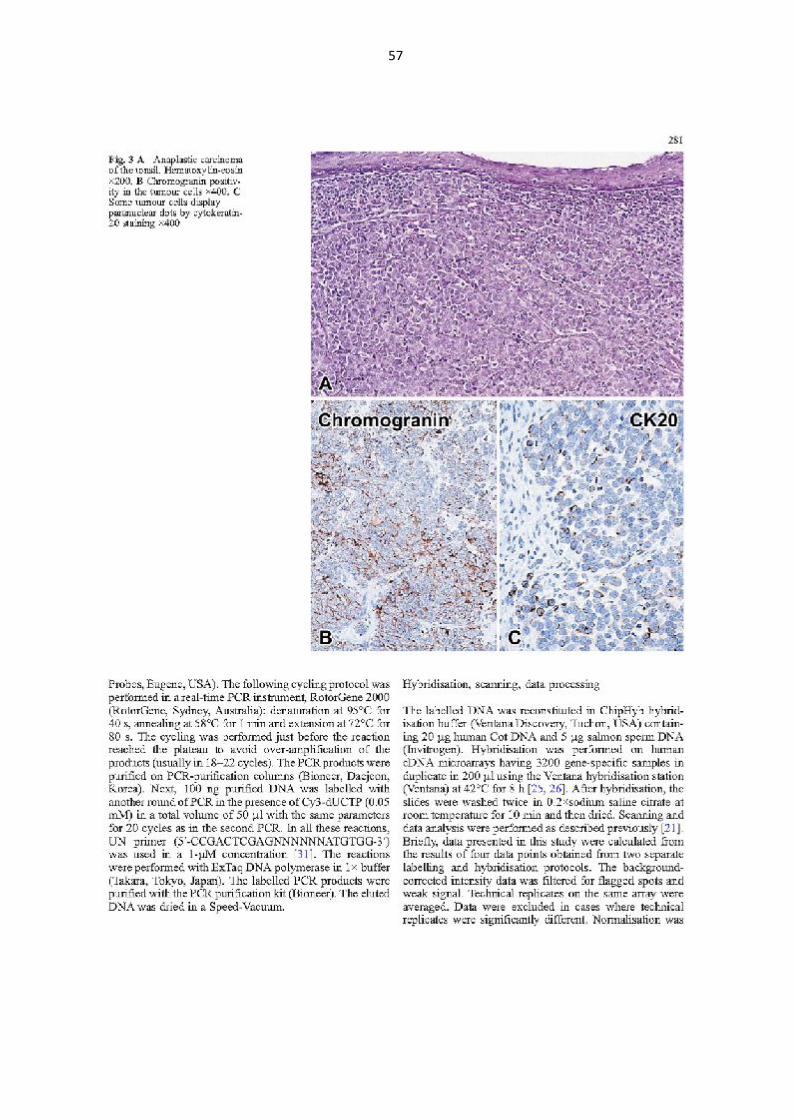
57

58

59
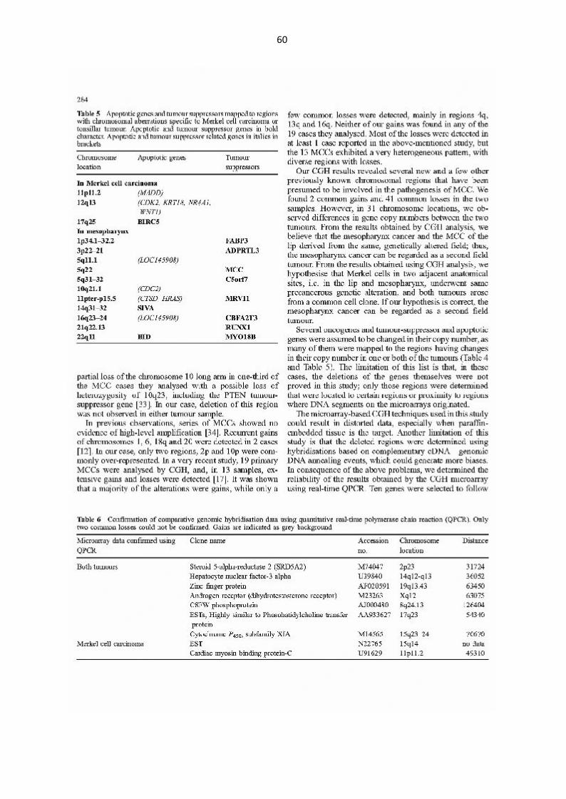
60

61

62

63
II.

64

65
66

67
68

69
III.

70
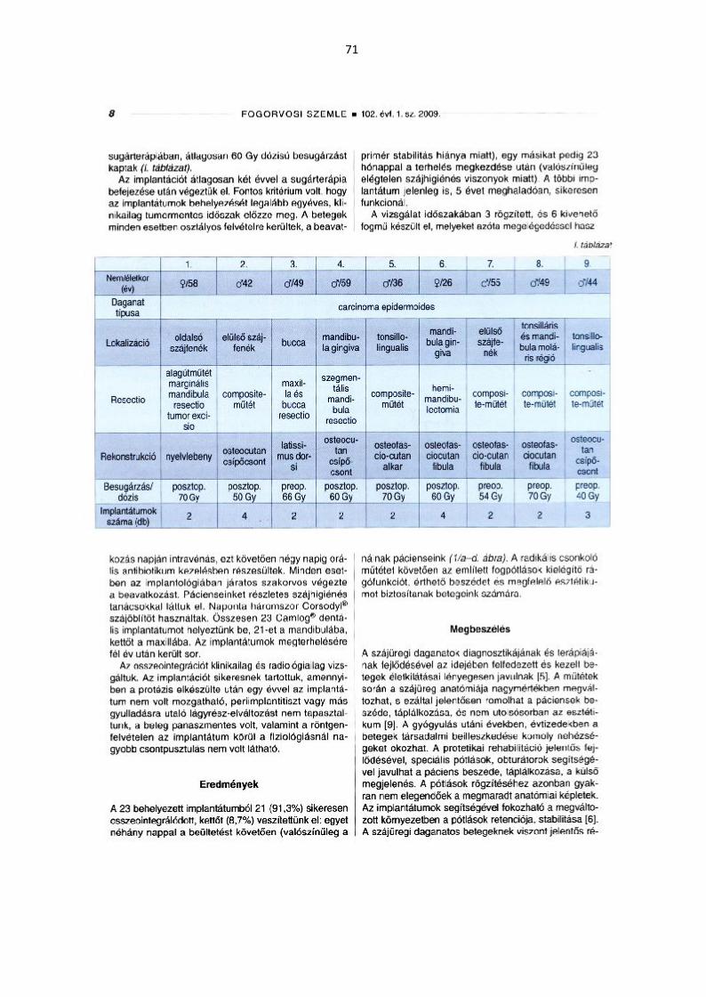
71

72

73