Embed Size (px)
Citation preview
8/4/2019 QM MM Calculations in Drug Discovery a Useful Method for Studying Binding
http://slidepdf.com/reader/full/qm-mm-calculations-in-drug-discovery-a-useful-method-for-studying-binding 1/8
QM/MM Calculations in Drug Discovery: A Useful Method for Studying Binding
Phenomena?
M. Paul Gleeson*,† and Duangkamol Gleeson‡
Computational & Structural Chemistry, GlaxoSmithKline Medicines Research Centre, Gunnels Wood Road,Stevenage, Hertfordshire SG1 2NY, United Kingdom, and Department of Chemistry, Faculty of Science, King
Mongkut’s Institute of Technology Ladkrabang, Bangkok 10520, Thailand
Received November 13, 2008
Herein we investigate whether QM/MM could prove useful as a tool to study the often subtle bindingphenomena found within pharmaceutical drug discovery programs. The goal of this investigation is todetermine whether it is possible to employ high level QM/MM calculations to answer specific questionsaround a binding event in a cycle time that is aligned with medicinal chemistry synthesis. To this endQM/MM calculations have been performed on four protein kinase-ligand complexes using five differentlevels of theory, using standard hardware, in an effort to assess their utility. We conclude that the accuracyand turnaround time of such calculations mean they could prove valuable to (1) probe the subtle nature of the interactions within protein active sites, (2) facilitate the interpretation of poorly resolved electron density,and (3) study the impact of substituent changes on the binding conformation or in the assessment of alternatescaffolds. In practice, the successful application of such methods will be limited by the size of the systemunder investigation, the level of theory used, and whether there is a need for conformational sampling.
1. INTRODUCTION
Structure-based drug design (SBDD) is an importantcomponent of the drug discovery process which sees theoptimization of a lead molecule in a rational, structurallyenabled manner, to realize a more potent, developablemolecule in the fewest number of chemistry iterations.1-4
Determination of high resolution protein-ligand complexes,via X-ray crystallography or NMR, means computational
chemistry methods can be used to virtually design smallnumbers of molecules to probe the existing interactions foundin the protein-ligand complex and also explore the activesite for any additional hydrophilic or hydrophobic interactionsto increase affinity.5-8
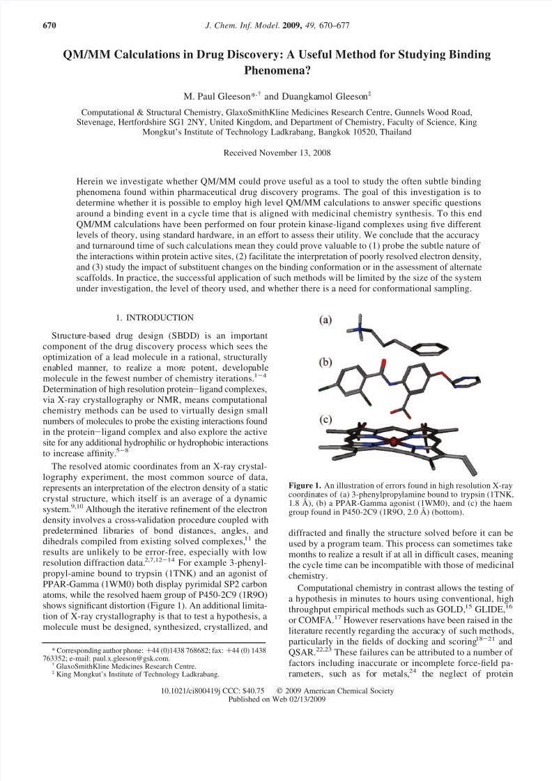
The resolved atomic coordinates from an X-ray crystal-lography experiment, the most common source of data,represents an interpretation of the electron density of a staticcrystal structure, which itself is an average of a dynamicsystem.9,10 Although the iterative refinement of the electrondensity involves a cross-validation procedure coupled withpredetermined libraries of bond distances, angles, anddihedrals compiled from existing solved complexes,11 theresults are unlikely to be error-free, especially with lowresolution diffraction data.2,7,12-14 For example 3-phenyl-propyl-amine bound to trypsin (1TNK) and an agonist of PPAR-Gamma (1WM0) both display pyrimidal SP2 carbonatoms, while the resolved haem group of P450-2C9 (1R9O)shows significant distortion (Figure 1). An additional limita-tion of X-ray crystallography is that to test a hypothesis, amolecule must be designed, synthesized, crystallized, and
diffracted and finally the structure solved before it can be
used by a program team. This process can sometimes takemonths to realize a result if at all in difficult cases, meaningthe cycle time can be incompatible with those of medicinalchemistry.
Computational chemistry in contrast allows the testing of a hypothesis in minutes to hours using conventional, highthroughput empirical methods such as GOLD,15 GLIDE,16
or COMFA.17 However reservations have been raised in theliterature recently regarding the accuracy of such methods,particularly in the fields of docking and scoring18-21 andQSAR.22,23 These failures can be attributed to a number of factors including inaccurate or incomplete force-field pa-rameters, such as for metals,24 the neglect of protein
* Corresponding author phone: +44 (0)1438 768682; fax: +44 (0) 1438763352; e-mail: [email protected].
† GlaxoSmithKline Medicines Research Centre.‡ King Mongkut’s Institute of Technology Ladkrabang.
Figure 1. An illustration of errors found in high resolution X-raycoordinates of (a) 3-phenylpropylamine bound to trypsin (1TNK,1.8 Å), (b) a PPAR-Gamma agonist (1WM0), and (c) the haemgroup found in P450-2C9 (1R9O, 2.0 Å) (bottom).
J. Chem. Inf. Model. 2009, 49, 670–677670
10.1021/ci800419j CCC: $40.75 2009 American Chemical SocietyPublished on Web 02/13/2009
8/4/2019 QM MM Calculations in Drug Discovery a Useful Method for Studying Binding
http://slidepdf.com/reader/full/qm-mm-calculations-in-drug-discovery-a-useful-method-for-studying-binding 2/8
flexibility or ensemble averaging,9,10 or due to deficiencieswith the empirical docking score. This is essentially a globalQSAR model that will suffer if the validation procedure is
not rigorous25,26
or because of the neglect of the domain of applicability,27 a concept not considered in docking andscoring.
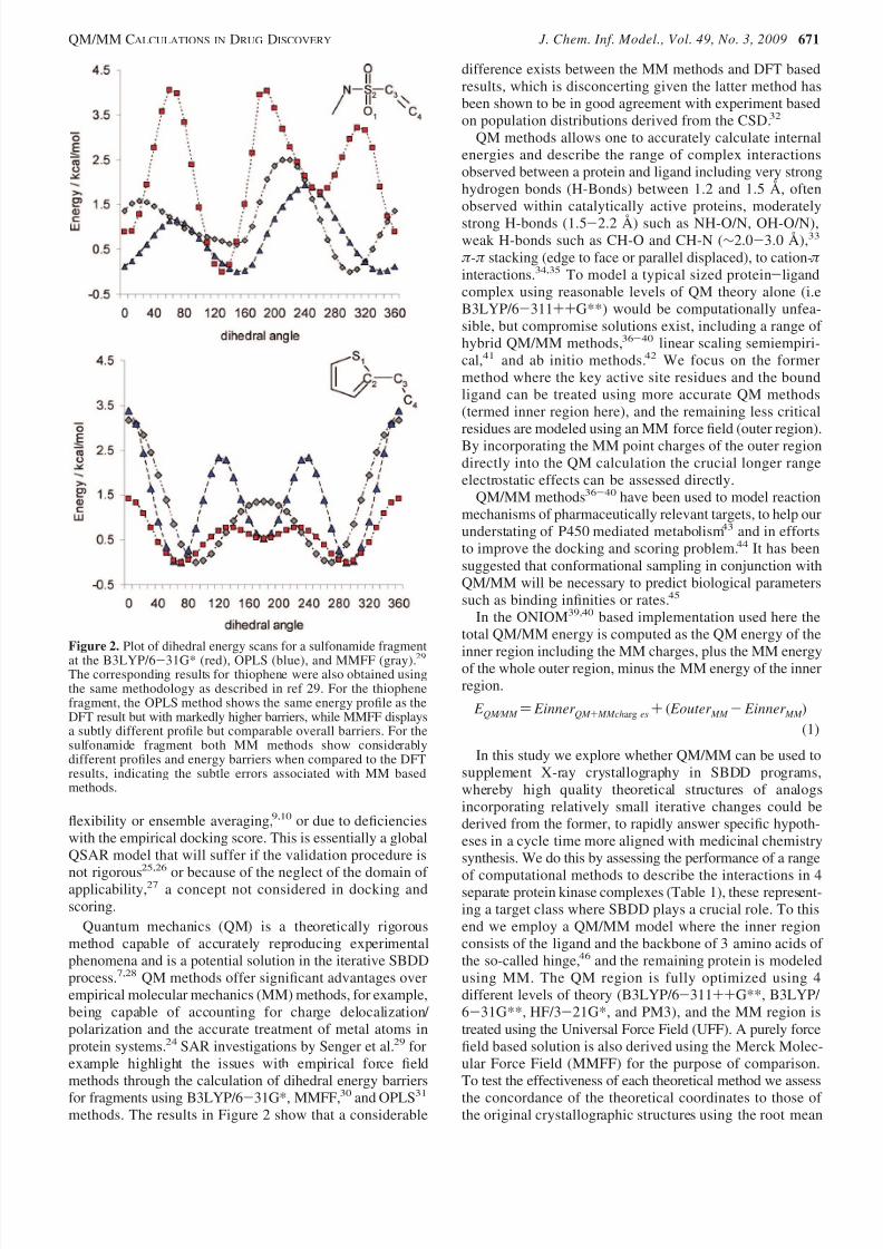
Quantum mechanics (QM) is a theoretically rigorousmethod capable of accurately reproducing experimentalphenomena and is a potential solution in the iterative SBDDprocess.7,28 QM methods offer significant advantages overempirical molecular mechanics (MM) methods, for example,being capable of accounting for charge delocalization/ polarization and the accurate treatment of metal atoms inprotein systems.24 SAR investigations by Senger et al.29 forexample highlight the issues with empirical force fieldmethods through the calculation of dihedral energy barriers
for fragments using B3LYP/6-31G*, MMFF,30 and OPLS31
methods. The results in Figure 2 show that a considerable
difference exists between the MM methods and DFT basedresults, which is disconcerting given the latter method hasbeen shown to be in good agreement with experiment basedon population distributions derived from the CSD.32
QM methods allows one to accurately calculate internalenergies and describe the range of complex interactionsobserved between a protein and ligand including very stronghydrogen bonds (H-Bonds) between 1.2 and 1.5 Å, oftenobserved within catalytically active proteins, moderately
strong H-bonds (1.5-2.2 Å) such as NH-O/N, OH-O/N),weak H-bonds such as CH-O and CH-N (∼2.0-3.0 Å),33
π -π stacking (edge to face or parallel displaced), to cation-π interactions.34,35 To model a typical sized protein-ligandcomplex using reasonable levels of QM theory alone (i.eB3LYP/6-311++G**) would be computationally unfea-sible, but compromise solutions exist, including a range of hybrid QM/MM methods,36-40 linear scaling semiempiri-cal,41 and ab initio methods.42 We focus on the formermethod where the key active site residues and the boundligand can be treated using more accurate QM methods(termed inner region here), and the remaining less criticalresidues are modeled using an MM force field (outer region).By incorporating the MM point charges of the outer regiondirectly into the QM calculation the crucial longer rangeelectrostatic effects can be assessed directly.
QM/MM methods36-40 have been used to model reactionmechanisms of pharmaceutically relevant targets, to help ourunderstating of P450 mediated metabolism43 and in effortsto improve the docking and scoring problem.44 It has beensuggested that conformational sampling in conjunction withQM/MM will be necessary to predict biological parameterssuch as binding infinities or rates.45
In the ONIOM39,40 based implementation used here thetotal QM/MM energy is computed as the QM energy of the
inner region including the MM charges, plus the MM energyof the whole outer region, minus the MM energy of the innerregion.
E QM ⁄ MM ) Einner QM + MMcharg es+ ( Eouter MM - Einner MM )(1)
In this study we explore whether QM/MM can be used tosupplement X-ray crystallography in SBDD programs,whereby high quality theoretical structures of analogsincorporating relatively small iterative changes could bederived from the former, to rapidly answer specific hypoth-eses in a cycle time more aligned with medicinal chemistrysynthesis. We do this by assessing the performance of a range
of computational methods to describe the interactions in 4separate protein kinase complexes (Table 1), these represent-ing a target class where SBDD plays a crucial role. To thisend we employ a QM/MM model where the inner regionconsists of the ligand and the backbone of 3 amino acids of the so-called hinge,46 and the remaining protein is modeledusing MM. The QM region is fully optimized using 4different levels of theory (B3LYP/6-311++G**, B3LYP/ 6-31G**, HF/3-21G*, and PM3), and the MM region istreated using the Universal Force Field (UFF). A purely forcefield based solution is also derived using the Merck Molec-ular Force Field (MMFF) for the purpose of comparison.To test the effectiveness of each theoretical method we assess
the concordance of the theoretical coordinates to those of the original crystallographic structures using the root mean
Figure 2. Plot of dihedral energy scans for a sulfonamide fragmentat the B3LYP/6-31G* (red), OPLS (blue), and MMFF (gray).29The corresponding results for thiophene were also obtained usingthe same methodology as described in ref 29. For the thiophenefragment, the OPLS method shows the same energy profile as theDFT result but with markedly higher barriers, while MMFF displaysa subtly different profile but comparable overall barriers. For thesulfonamide fragment both MM methods show considerablydifferent profiles and energy barriers when compared to the DFTresults, indicating the subtle errors associated with MM basedmethods.
QM/MM CALCULATIONS IN DRUG DISCOVERY J. Chem. Inf. Model., Vol. 49, No. 3, 2009 671
8/4/2019 QM MM Calculations in Drug Discovery a Useful Method for Studying Binding
http://slidepdf.com/reader/full/qm-mm-calculations-in-drug-discovery-a-useful-method-for-studying-binding 3/8
squared deviation (RMSD), the computed H-bond distances,and the electron density where available.
2. COMPUTATIONAL PROCEDURESCrystal structures of 4 protein-kinases (2UVX, 1WCC,
1O9U, 17wh) containing small molecule ligands were down-loaded from the RCSB protein databank47 (Table 1). As iscommon in many theoretical studies,19 cofactors, ions, or watermolecules are removed from the protein-ligand complexes forcomputational efficiency, and the impact of this will bediscussed later. Each protein was subsequently prepared usingthe protein preparation wizard in Maestro.48 Hydrogen atomswere added to the system, and ionizable amino acid side chainswere protonated assuming a pH of 7.4. The system thenunderwent restrained minimization using the IMPREF utility,
only to optimize hydrogen atoms and to remove any high energycontacts or distorted bonds, angles, and dihedrals. Thesecoordinates were used for the subsequent QM/MM calcula-tions.
All QM/MM calculations were performed in Gaussian 0349
using the ONIOM methodology developed by Morokumaand co-workers.39,40 Our strategy is to use the highest levelsof QM possible for a reasonable computational resource, tomirror how one might use the methodology in a programenvironment where rapid turnaround is required. To this endwe have employed the smallest acceptable QM regionconsisting of the bound ligand and the backbone of the 3amino acids of the hinge, with the point charges of the outer
MM region being electrically embedded. Where bonds crossthe QM and MM interface, the valences were satisfied byhydrogen link atoms. In this case the outer region is keptfixed, and the charges are assigned using the chargeequalization method.50 The latter method has been demon-strated to produce molecular charges that correlate well withmore expensive QM calculations. The inner QM region wastreated using a range of different levels of theory: B3LYP/ 6-311++G**, B3LYP/6-31G**, HF/3-21G*, and PM3.Electrical embedding was used in all cases apart from PM3.Mechanical embedding was used as the former is notsupported (Gaussian 03 enables both mechanical and electri-cal embedding to be used in ONIOM calculations, unlike
earlier versions where only the former is supported). Thevan der Waals contribution to the protein-ligand complexes
was treated classically using the universal force field (UFF).51
While this combination has been successfully applied in thepast for calculations on large representations of 3-dimensional
zeolites catalysts,52-54 some concerns have been raised withQM/MM methods, including the use of nonideal empiricalfunctions (i.e., Lennard-Jones potentials) to compute interac-tions across the QM and MM interface and the issue of overpolarization of the QM region.55 With these issues inmind, we assess the utility of the method to protein kinasesby comparison of the theoretical coordinates to those fromthe original X-ray experiment.
All Gaussian 03 calculations were submitted as singleprocessor jobs on a Linux cluster and took no more than 2days to converge.
For the purpose of comparison the prepared protein-ligandcomplexes were also assessed using force field methodsalone. In this case the whole protein was fixed, and theligands alone were optimized using MMFF as implementedin MOE.56 More conventional force field methods forsimulating protein-ligand complexes were not considereddue to the relatively time-consuming setup required toprepare each ligand correctly.57,58
3. RESULTS
The RMSDs and H-bond distances obtained from the fivedifferent calculations on each of the four protein-ligandcomplexes are reported in Table 2. We first discuss thisoutput obtained in terms of the RMS deviation between the
experimental and theoretical coordinates as this is the mostcommonly used parameter in such studies. The results in
Table 1. List of Protein-Kinase-Inhibitor Complexes Used inThis Study
Table 2. QM/MM and MM Results for the 4 OptimizedProtein-Inhibitor Complexesa
ID model RMSDHB1
(center)HB2
(inner)
PKA/B 2UVX XRAY (2.0 Å Res.) - 1.98 1.93B3LYP/6-311++G** 0.19 1.99 1.81B3LYP/6-31G** 0.19 1.97 1.80HF/3-21G* 0.19 1.92 1.76PM3 0.20 1.87 1.81MMFF 0.26 1.82 1.65
CDK2 1WCC XRAY (2.2 Å Res.) - 2.11 -B3LYP/6-311++G** 0.31 2.08 -B3LYP/6-31G** 0.33 2.06 -HF/3-21G* 0.34 1.98 -PM3 0.34 1.89 -MMFF 0.34 2.41 -
GSK3 1O9U XRAY (2.4 Å Res.) - 2.24 1.82B3LYP/6-311++G** 0.77 2.04 1.76B3LYP/6-31G** 0.78 2.01 1.74HF/3-21G* 0.78 2.01 1.72PM3 0.77 1.86 1.82MMFF 0.74 2.11 1.76
P38 1W7H XRAY (2.2 Å Res.) - 2.06 2.04B3LYP/6-311++G** 0.26 2.07 1.82B3LYP/6-31G** 0.24 2.04 1.81
HF/3-21G* 0.41 1.95 1.81PM3 0.30 2.35 1.90MMFF 0.61 1.88 1.48
a Listed are the heavy atom RMSD to the original crystal-lographic coordinates and the H-bond distance between the inhibitoracceptor and the central hinge donor (HB1), and between theinhibitor donor and the inner hinge acceptor (HB2). Hydrogenatoms were added to the original X-ray structure using theOPLS-AA force field as implemented in Maestro.
672 J. Chem. Inf. Model., Vol. 49, No. 3, 2009 GLEESON AND GLEESON
8/4/2019 QM MM Calculations in Drug Discovery a Useful Method for Studying Binding
http://slidepdf.com/reader/full/qm-mm-calculations-in-drug-discovery-a-useful-method-for-studying-binding 4/8
Table 2 unsurprisingly show that the lower the resolution of the X-ray structure the larger the RMSD, irrespective of thetheoretical model. The lowest resolution structure, 1O9U, issolved to 2.4 Å resolution, and the average RMSD of theoretical models is ∼0.8 Å. In contrast 2UVX, the highestresolution structure at 2.0 Å, displays a mean RMSD of ∼0.2Å. In between these, 1WCC and 1W7H are solved to aresolution of 2.2 Å, and the RMSDs of the theoreticalstructures are ∼0.3 Å on average. It is reported that the
experimental atomic positional errors for complexes withresolutions between 1.8 and 2.0 Å will range from∼0.2-0.3Å.12,14
The highest level B3LYP/6-311++G**//UFF results donot show demonstrably different RMSDs than any of theother theoretical models. This however may be a reflectionof the fact that the 1-dimensional RMSD value captures anaverage effect and because the X-ray coordinates themselvesare not error free. In fact, the focus on this parameter indocking studies has come in for criticism recently as theythemselves are a fitted atomic model derived from theelectron density.59 We therefore conclude that starting fromthe X-ray coordinates, all methods give qualitatively the sameminima. However, as we shall see later, the HF and DFTmethods more effectively reproduce the H-bond distancesand orientations than the more approximate PM3 and MMFFbased methods.
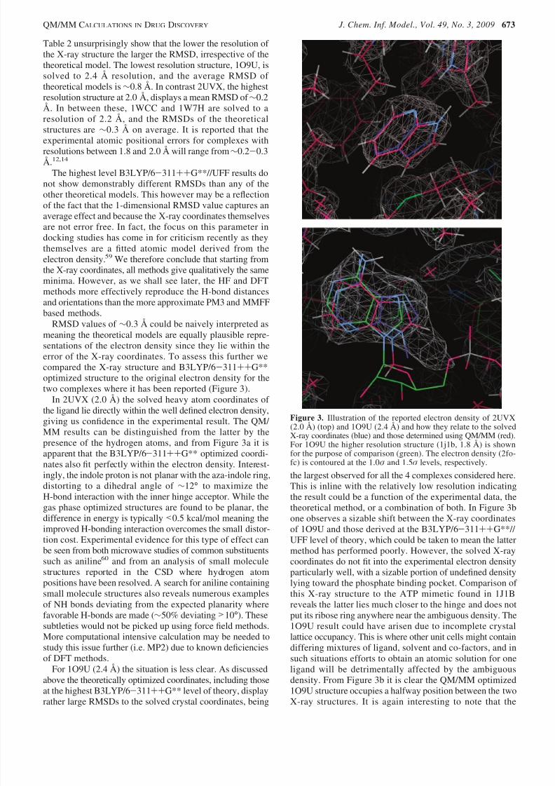
RMSD values of ∼0.3 Å could be naively interpreted asmeaning the theoretical models are equally plausible repre-sentations of the electron density since they lie within theerror of the X-ray coordinates. To assess this further wecompared the X-ray structure and B3LYP/6-311++G**optimized structure to the original electron density for thetwo complexes where it has been reported (Figure 3).
In 2UVX (2.0 Å) the solved heavy atom coordinates of
the ligand lie directly within the well defined electron density,giving us confidence in the experimental result. The QM/ MM results can be distinguished from the latter by thepresence of the hydrogen atoms, and from Figure 3a it isapparent that the B3LYP/6-311++G** optimized coordi-nates also fit perfectly within the electron density. Interest-ingly, the indole proton is not planar with the aza-indole ring,distorting to a dihedral angle of ∼12° to maximize theH-bond interaction with the inner hinge acceptor. While thegas phase optimized structures are found to be planar, thedifference in energy is typically <0.5 kcal/mol meaning theimproved H-bonding interaction overcomes the small distor-tion cost. Experimental evidence for this type of effect can
be seen from both microwave studies of common substituentssuch as aniline60 and from an analysis of small moleculestructures reported in the CSD where hydrogen atompositions have been resolved. A search for aniline containingsmall molecule structures also reveals numerous examplesof NH bonds deviating from the expected planarity wherefavorable H-bonds are made (∼50% deviating >10°). Thesesubtleties would not be picked up using force field methods.More computational intensive calculation may be needed tostudy this issue further (i.e. MP2) due to known deficienciesof DFT methods.
For 1O9U (2.4 Å) the situation is less clear. As discussedabove the theoretically optimized coordinates, including those
at the highest B3LYP/6-311++G** level of theory, displayrather large RMSDs to the solved crystal coordinates, being
the largest observed for all the 4 complexes considered here.This is inline with the relatively low resolution indicatingthe result could be a function of the experimental data, thetheoretical method, or a combination of both. In Figure 3bone observes a sizable shift between the X-ray coordinatesof 1O9U and those derived at the B3LYP/6-311++G**// UFF level of theory, which could be taken to mean the latter
method has performed poorly. However, the solved X-raycoordinates do not fit into the experimental electron densityparticularly well, with a sizable portion of undefined densitylying toward the phosphate binding pocket. Comparison of this X-ray structure to the ATP mimetic found in 1J1Breveals the latter lies much closer to the hinge and does notput its ribose ring anywhere near the ambiguous density. The1O9U result could have arisen due to incomplete crystallattice occupancy. This is where other unit cells might containdiffering mixtures of ligand, solvent and co-factors, and insuch situations efforts to obtain an atomic solution for oneligand will be detrimentally affected by the ambiguousdensity. From Figure 3b it is clear the QM/MM optimized
1O9U structure occupies a halfway position between the twoX-ray structures. It is again interesting to note that the
Figure 3. Illustration of the reported electron density of 2UVX(2.0 Å) (top) and 1O9U (2.4 Å) and how they relate to the solvedX-ray coordinates (blue) and those determined using QM/MM (red).For 1O9U the higher resolution structure (1j1b, 1.8 Å) is shownfor the purpose of comparison (green). The electron density (2fo-fc) is contoured at the 1.0σ and 1.5σ levels, respectively.
QM/MM CALCULATIONS IN DRUG DISCOVERY J. Chem. Inf. Model., Vol. 49, No. 3, 2009 673
8/4/2019 QM MM Calculations in Drug Discovery a Useful Method for Studying Binding
http://slidepdf.com/reader/full/qm-mm-calculations-in-drug-discovery-a-useful-method-for-studying-binding 5/8
aromatic-amine substituent of the QM/MM optimized struc-ture is nonplanar, one hydrogen being ∼4° and the other∼13° to the plane of the ring to maximize the interactionwith the hinge acceptor.
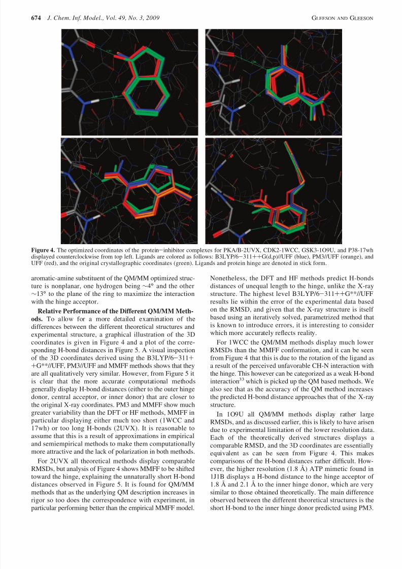
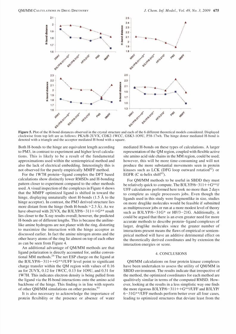
Relative Performance of the Different QM/MM Meth-
ods. To allow for a more detailed examination of thedifferences between the different theoretical structures andexperimental structure, a graphical illustration of the 3Dcoordinates is given in Figure 4 and a plot of the corre-sponding H-bond distances in Figure 5. A visual inspectionof the 3D coordinates derived using the B3LYP/6-311++G**//UFF, PM3//UFF and MMFF methods shows that theyare all qualitatively very similar. However, from Figure 5 itis clear that the more accurate computational methodsgenerally display H-bond distances (either to the outer hinge
donor, central acceptor, or inner donor) that are closer tothe original X-ray coordinates. PM3 and MMFF show muchgreater variability than the DFT or HF methods, MMFF inparticular displaying either much too short (1WCC and17wh) or too long H-bonds (2UVX). It is reasonable toassume that this is a result of approximations in empiricaland semiempirical methods to make them computationallymore attractive and the lack of polarization in both methods.
For 2UVX all theoretical methods display comparableRMSDs, but analysis of Figure 4 shows MMFF to be shiftedtoward the hinge, explaining the unnaturally short H-bonddistances observed in Figure 5. It is found for QM/MMmethods that as the underlying QM description increases in
rigor so too does the correspondence with experiment, inparticular performing better than the empirical MMFF model.
Nonetheless, the DFT and HF methods predict H-bondsdistances of unequal length to the hinge, unlike the X-raystructure. The highest level B3LYP/6-311++G**//UFFresults lie within the error of the experimental data basedon the RMSD, and given that the X-ray structure is itself based using an iteratively solved, parametrized method thatis known to introduce errors, it is interesting to considerwhich more accurately reflects reality.
For 1WCC the QM/MM methods display much lowerRMSDs than the MMFF conformation, and it can be seenfrom Figure 4 that this is due to the rotation of the ligand asa result of the perceived unfavorable CH-N interaction withthe hinge. This however can be categorized as a weak H-bondinteraction33 which is picked up the QM based methods. Wealso see that as the accuracy of the QM method increases
the predicted H-bond distance approaches that of the X-raystructure.In 1O9U all QM/MM methods display rather large
RMSDs, and as discussed earlier, this is likely to have arisendue to experimental limitation of the lower resolution data.Each of the theoretically derived structures displays acomparable RMSD, and the 3D coordinates are essentiallyequivalent as can be seen from Figure 4. This makescomparisons of the H-bond distances rather difficult. How-ever, the higher resolution (1.8 Å) ATP mimetic found in1J1B displays a H-bond distance to the hinge acceptor of 1.8 Å and 2.1 Å to the inner hinge donor, which are verysimilar to those obtained theoretically. The main difference
observed between the different theoretical structures is theshort H-bond to the inner hinge donor predicted using PM3.
Figure 4. The optimized coordinates of the protein-inhibitor complexes for PKA/B-2UVX, CDK2-1WCC, GSK3-1O9U, and P38-17whdisplayed counterclockwise from top left. Ligands are colored as follows: B3LYP/6-311++G(d,p)//UFF (blue), PM3//UFF (orange), andUFF (red), and the original crystallographic coordinates (green). Ligands and protein hinge are denoted in stick form.
674 J. Chem. Inf. Model., Vol. 49, No. 3, 2009 GLEESON AND GLEESON
8/4/2019 QM MM Calculations in Drug Discovery a Useful Method for Studying Binding
http://slidepdf.com/reader/full/qm-mm-calculations-in-drug-discovery-a-useful-method-for-studying-binding 6/8
Both H-bonds to the hinge are equivalent length accordingto PM3, in contrast to experiment and higher level calcula-tions. This is likely to be a result of the fundamentalapproximations used within the semiempirical method andalso the lack of electrical embedding. Interestingly this isnot observed for the purely empirically MMFF method.
For the 1W7H protein-ligand complex the DFT basedcalculations show distinctly lower RMSDs and H-bondingpattern closer to experiment compared to the other methodsused. A visual inspection of the complexes in Figure 4 showsthat the MMFF optimized ligand is shifted in toward thehinge, displaying unnaturally short H-bonds (1.5 Å to thehinge acceptor). In contrast, the PM3 derived structure liesmore distant from the hinge (both H-bonds >2.3 Å). As wehave observed with 2UVX, the B3LYP/6-311++G** resultlies closer to the X-ray results overall; however, the predictedH-bonds are of different lengths. This is because the aniline-like amine hydrogens are not planer with the ring, distortingto maximize the interaction with the hinge acceptor as
discussed earlier. In fact the amine nitrogen atoms and theother heavy atoms of the ring lie almost on top of each otheras can be seen from Figure 4.
An additional advantage of QM/MM methods are thatligand polarization is directly accounted for, unlike conven-tional MM methods.24 The net ESP charge on the ligand atthe B3LYP/6-311++G**//UFF level point to significantcharge transfer within the QM region with values of 0.16au for 2UVX, 0.12 for 1WCC, 0.13 for 1O9U, and 0.31 for1W7H. This indicates electron density is being pulled fromthe ligand via the H-bond interactions onto the amino acidbackbone of the hinge. This finding is in line with reportsof other QM/MM simulations on other proteins.61
It is also necessary to acknowledge the importance of protein flexibility or the presence or absence of water
mediated H-bonds on these types of calculations. A largerrepresentation of the QM region, coupled with flexible activesite amino acid side chains in the MM region, could be used;however, this will be more time-consuming and will notproduce the more substantial movements seen in proteinkinases such as LCK (DFG loop outward rotation62) or
EGFR (C R-helix shift63
).For QM/MM methods to be useful in SBDD they must
be relatively quick to compute. The B3LYP/6-311++G**// UFF calculations performed here took no more than 2 daysto complete as single processors jobs. Even though theligands used in this study were fragmentlike in size, studieson more druglike molecules would be feasible if submittedas multiprocessor jobs or run at a more modest level of theorysuch as B3LYP/6-31G* or HF/3-21G. Additionally, itcould be argued that there is an even greater need for moreaccurate methods to describe protein-ligand complexes of larger, druglike molecules since the greater number of interactions present means the flaws of empirical or semiem-
pirical method will have an additive detrimental effect onthe theoretically derived coordinates and by extension theinteraction energies or score.
4. CONCLUSIONS
QM/MM calculations on four protein kinase complexeshave been undertaken to assess the utility of QM/MM inSBDD environment. The results indicate that irrespective of the method, the optimized coordinates for each method arequalitively similar in terms of the computed RMSD. How-ever, looking at the results in a less simplistic way one findsthe more rigorous B3LYP/6-311++G**//UFF and B3LYP/
6-31G**/UFF methods perform better over all four cases,leading to optimized structures that deviate least from the
Figure 5. Plot of the H-bond distances observed in the crystal structure and each of the 6 different theoretical models considered. Displayedclockwise from top left are as follows: PKA/B-2UVX, CDK2-1WCC, GSK3-1O9U, P38-17wh. The hinge donor mediated H-bond isdenoted with a triangle and the acceptor mediated H-bond with a square.
QM/MM CALCULATIONS IN DRUG DISCOVERY J. Chem. Inf. Model., Vol. 49, No. 3, 2009 675
8/4/2019 QM MM Calculations in Drug Discovery a Useful Method for Studying Binding
http://slidepdf.com/reader/full/qm-mm-calculations-in-drug-discovery-a-useful-method-for-studying-binding 7/8
8/4/2019 QM MM Calculations in Drug Discovery a Useful Method for Studying Binding
http://slidepdf.com/reader/full/qm-mm-calculations-in-drug-discovery-a-useful-method-for-studying-binding 8/8
(39) Dapprich, S.; Komaromi, I.; Byun, K. S.; Morokuma, K.; Frisch, M. J.A new ONIOM implementation in Gaussian98. J. Mol. Struct. 1999,1-21, 461–462.
(40) Vreven, T.; Byun, K. S.; Komaromi, I.; Dapprich, S.; Montgomery,J. A.; Morokuma, K.; Frisch, M. J. Combining Quantum MechanicsMethods with Molecular Mechanics Methods in ONIOM. J. Chem.Theory Comput. 2006, 2, 815–826.
(41) Dixon, S. L.; Merz, K. M., Jr. Semiempirical molecular orbitalcalculations with linear system size scaling. J. Chem. Phys. 1996, 104,6643–6649.
(42) Claeyssens, F.; Harvey, J. N.; Manby, F. R.; Mata, R. A.; Mulholland,
A. J.; Ranaghan, K. A.; Schutz, M.; Thiel, S.; Thiel, W.; Werner, H.High-Accuracy Computation of Reaction Barriers in Enzymes. Angew.Chem., Int. Ed. 2006, 45, 6856–6859.
(43) Mulholland, A. J. Modelling enzyme reaction mechanisms, specificityand catalysis. Drug DiscoVery Today 2005, 10, 1393–1402.
(44) Cho, A. E.; Guallar, V.; Berne, B.; Friesner, R. A. Importance of Accurate Charges in Molecular Docking: Quantum Mechanical/ Molecular Mechanical (QM/MM) Approach. J. Comput. Chem. 2005,26 , 915–931.
(45) Bowman, A. L.; Ridder, L.; Rietjens, I. M. C. M.; Vervoort, J.;Mulholland, A. J. Molecular Determinants of Xenobiotic Metabolism:QM/MM Simulation of the Conversion of 1-Chloro-2,4-dinitrobenzeneCatalyzed by M1-1 Glutathione S-Transferase. Biochemistry 2007,46 , 6353–6363.
(46) Liao, J. J.-L. Molecular recognition of protein kinase binding pocketsfor design of potent and selective kinase inhibitors. J. Med. Chem.2007, 50, 1–16.
(47) RCSB Protein databank. http://www.rcsb.org/ (accessed Nov 20, 2008).(48) Maestro; Schrodinger, Dynamostrasse 13, D-68165 Mannheim, Ger-
many. www.schrodinger.com (accessed Nov 20, 2008).(49) Frisch, M. J. ; Trucks, G. W.; Schlegel, H. B.; Scuseria, G. E.;
Robb, M. A.; Cheeseman, J. R.; Montgomery, J. A., Jr.; Vreven,T.; Kudin, K. N.; Burant, J. C.; Millam, J. M.; Iyengar, S. S.;Tomasi, J.; Barone, V.; Mennucci, B.; Cossi, M.; Scalmani, G.;Rega, N.; Petersson, G. A.; Nakatsuji, H.; Hada, M.; Ehara, M.;Toyota, K.; Fukuda, R.; Hasegawa, J.; Ishida, M.; Nakajima, T.;Honda, Y.; Kitao, O.; Nakai, H.; Klene, M.; Li, X.; Knox, J. E.;Hratchian, H. P.; Cross, J. B.; Bakken, V.; Adamo, C.; Jaramillo,J.; Gomperts, R.; Stratmann, R. E.; Yazyev, O.; Austin, A. J.;Cammi, R.; Pomelli, C.; Ochterski, J. W.; Ayala, P. Y.; Morokuma,K.; Voth, G. A.; Salvador, P.; Dannenberg, J. J.; Zakrzewski, V. G.;Dapprich, S.; Daniels, A. D.; Strain, M. C.; Farkas, O.; Malick,D. K.; Rabuck, A. D.; Raghavachari, K.; Foresman, J. B.; Ortiz,J. V.; Cui, Q.; Baboul, A. G.; Clifford, S.; Cioslowski, J.; Stefanov,
B. B.; Liu, G.; Liashenko, A.; Piskorz, P.; Komaromi, I.; Martin,R. L.; Fox, D. J.; Keith, T.; Al-Laham, M. A.; Peng, C. Y.;Nanayakkara, A.; Challacombe, M.; Gill, P. M. W.; Johnson, B.;Chen, W.; Wong, M. W.; Gonzalez, C.; Pople, J. A.; Gaussian 03,
ReVision C.02; Gaussian, Inc.: Wallingford, CT, 2004.(50) Bultinck, P.; Langenaeker, W.; Lahorte, P.; De Proft, F.; Geerlings,
P.; Waroquier, M.; Tollenaere, J. P. The Electronegativity EqualizationMethod I: Parametrization and Validation for Atomic Charge Calcula-tions. J. Phys. Chem. A 2002, 106 , 7887–7894.
(51) Rappe, A. K.; Casewit, C. J.; Colwell, K. S.; Goddard, W. A., III.;Skiff, W. M. UFF, a Full Periodic Table Force Field for MolecularMechanics and Molecular Dynamics Simulations. J. Am. Chem. Soc.1992, 114, 10024–10035.
(52) Namuangruk, S.; Khongpracha, P.; Pantu, P.; Limtrakul, J. Structuresand Reaction Mechanisms of Propene Oxide Isomerization on H-ZSM-5: An ONIOM Study. J. Phys. Chem. B 2006, 110, 25950–25957.
(53) Namuangruk, S.; Tantanak, D.; Limtrakul, J. Application of ONIOMcalculations in the study of the effect of the zeolite framework on theadsorption of alkenes to ZSM-5. J. Mol. Catal. A: Chem. 2006, 256 ,113–121.
(54) Jungsuttiwong, S.; Limtrakul, J.; Truong, T. N. Theoretical Study of Modes of Adsorption of Water Dimer on H-ZSM-5 and H-FaujasiteZeolites. J. Phys. Chem. B 2005, 109, 13342–13351.
(55) Senthilkumar, K.; Mujika, J. I.; Ranaghan, K. E.; Manby, F. R.;Mulholland, A. J.; Harvey, J. N. Analysis of polarization in QM/MM
modelling of biologically relevant hydrogen. J. R. Soc. Interface 2008,5, S207–S216.
(56) Molecular Operating EnVironment (MOE); Chemical ComputingGroup, 1010 Sherbrooke St. W, Suite 910 Montreal, Quebec, CanadaH3A 2R7. www.chemcomp.com (accessed Nov 20, 2008).
(57) Cornell, W. D.; Cieplak, P.; Bayly, C. I.; Gould, I. R., Jr.; Ferguson,D. M.; Spellmeyer, D. C.; Fox, T.; Caldwell, J. W.; Kollman, P. A. ASecond Generation Force Field for the Simulation of Proteins, NucleicAcids, and Organic Molecules. J. Am. Chem. Soc. 1995, 117 , 5179–5197.
(58) MacKerell, A. D., Jr.; Bashford, D.; Bellott, M.; Dunbrack, R. L., Jr.;Evanseck, J. D.; Field, M. J.; Fischer, S.; Gao, J.; Guo, H.; Ha, S.;Joseph-McCarthy, D.; Kuchnir, L.; Kuczera, K.; Lau, F. T. K.; Mattos,C.; Michnick, S.; Ngo, T.; Nguyen, D. T.; Prodhom, B.; Reiher, W. E.,III.; Roux, B.; Schlenkrich, M.; Smith, J. C.; Stote, R.; Straub, J.;Watanabe, M.; Wiorkiewicz-Kuczera, J.; Yin, D.; Karplus, M. All-Atom Empirical Potential for Molecular Modeling and Dynamics
Studies of Proteins. J. Phys. Chem. B 1998, 102, 3586–3616.(59) Yusuf, D.; Davis, A. M.; Kleywegt, G. J.; Schmitt, S. An AlternativeMethod for the Evaluation of Docking Performance: RSR vs RMSD.
J. Chem. Inf. Model 2008, 48, 1411–1422.(60) Lister, D. G.; Tylern, J. K. Non-planarity of the Aniline Molecule.
Chem. Commun. 1966, 6 , 152–153.(61) Raha, K.; Merz, K. M., Jr. Large scale validation of a quantum
mechanics scoring function: predicting the binding affinity and thebinding mode of a diverse set of protein-ligand complexes. J. Med.Chem. 2005, 48, 4558–2475.
(62) Yun, C.; Mengwasser, K. E.; Toms, A. V.; Woo, M. S.; Greulich, H.;Wong, K.; Meyerson, M.; Eck, M. J. The T790M mutation in EGFRkinase causes drug resistance by increasing the affinity for ATP. Proc.
Natl. Acad. Sci. U.S.A. 2008, 105, 2070–2075.(63) Pargellis, C.; Tong, L.; Churchill, L.; Cirillo, P. F.; Gilmore, T.;
Graham, A. G.; Grob, P. M.; Hickey, E. R.; Moss, N.; Pav, S.; Regan,J. Inhibition of p38 MAP kinase by utilizing a novel allosteric binding
site. Nat. Struct. Biol. 2002, 9, 268–272.(64) Brameld, K. A.; Kuhn, B.; Reuter, D. C.; Stahl, M. Small MoleculeConformational Preferences Derived from Crystal Structure Data. AMedicinal Chemistry Focused Analysis. J. Chem. Inf. Model. 2008,48, 1–24.
(65) Carr, R.; Jhoti, H. Structure-based screening of low-affinity compounds. Drug DiscoVery Today 2002, 2, 522–527.
(66) Leach, A. R.; Hann, M. M.; Burrows, J. N.; Griffen, E. J. Fragmentscreening: an introduction. Mol. BioSyst. 2006, 2, 430–446.
(67) Hann, M. M.; Leach, A. R.; Harper, G. Molecular complexity and itsimpact on the probability of finding leads for drug discovery. J. Chem.
Inf. Comput. Sci. 2001, 41, 856–864.
CI800419J
QM/MM CALCULATIONS IN DRUG DISCOVERY J. Chem. Inf. Model., Vol. 49, No. 3, 2009 677