Embed Size (px)
Citation preview
Chapter 19 Oral Growths and Tumors.
147
Chapter 19: Oral Growths and TumorsThere are many growths that may occur in the mouth. Some are very malignant while others are considered benign. Even the benign lesions can be a big problem. Many are locally invasive and inclined to recur and their physical presence in the mouth can lead to traumatic occlusion and entrapment of debris, predisposing to infection. Therefore, all oral growths should be taken seriously and treated aggressively.
Though clinical and radiographic assessment are important to evaluating oral masses, nothing can replace the value of microscopic examination. Therefore, any tissue removed from the mouth should be sent to a pathologist for evaluation. Careful evaluation of region lymph nodes (palpation, biopsy) and thoracic radiographs are also indicated.
What follows is a discussion of the surgical options for various oral growths. Radiation and chemotherapy may also be appropriate for some of these conditions and when dealing with malignant oral tumors, consultation with an oncologist would be in order.
Gingival Hyperplasia
Gingival hyperplasia is a benign overgrowth of the gingival tissue. It may be focal or generalized.
Focal gingival hyperplasia appears as an area of relatively normal-looking redundant gingiva. Typically there is inflammation in the pseudopocket between the excess gingiva and the tooth. Whether this inflammation is the cause or a result of the hyperplasia is rarely clear.
Generalized gingival hyperplasia is most commonly seen in the boxer dog but there is also a breed predisposition in huskies and collies. It also seems to have a propensity for affecting large breeds such as wolf hounds and St. Bernards. In advanced cases, teeth may be completely engulfed in the excess tissue and/or may be displaced. The hyperplastic tissue is at risk of trauma from the teeth of the opposing jaw and affected animals often have pain and oral bleeding when eating.
As the gingiva grows to engulf the crowns, deep pseudopockets form and accumulate food, hair, debris and bacteria. This can lead to the development of periodontal disease.
Treatment for gingival hyperplasia is preceded by intra-oral radiographs and probing to evaluate the status of the tooth roots and periodontal structures. Seriously diseased teeth may require extraction. If there are no significant findings then the gingiva is contoured back to a normal height and width using cold steel, electro or radio surgery or laser. A thorough oral hygiene procedure follows.
Though the exact cause of generalized gingival hyperplasia is not known, the trigger seems to be gingival inflammation. Therefore, in an effort to delay or prevent recurrence of the hyperplasia, a strict plaque-control program (daily tooth brushing, plaque-fighting diets and chew treats, periodic professional treatments) should be instituted. Even with these measures, owners should be prepared that the problem will likely recur eventually and will require further surgery.
Figure #19.1 Before and after views of the left maxilla of a dog with generalized hyperplasia. In the top photo, the crowns of many of the premolars and molars are completely engulfed in hyperplastic gingiva. In the bottom photo the third premolar has been extracted and the excess gingiva removed with electrosurgery.

Chapter 19 Oral Growths and Tumors.
148
Canine Viral Papillomas
Viral papillomas may occur in the mouths of young dogs. They tend to be self-limiting and often resolve spontaneously by two-years-of-age. Lesions may be anywhere on the lips, gingiva, oral mucosa, tongue, pharynx or even the esophagus. If the size and/or location of the lesions leads to occlusal interference (dog chews on lesions), then surgical removal is indicated. This can often be done by just debulking at the base.
Epulis
The term epulis comes from the Greek root for “on the gum” and as such really only describes the location of the lesion. None-the-less, there are three oral masses that have traditionally been termed epulids. They are fibromatous, ossifying and acanthomatous epulis. There is a movement among some pathologists and veterinary dentists to reclassify these lesions based on their histology and behaviour. Under this system, fibromatous and ossifying epulids are classified as peripheral odontogenic fibromas and acanthomatous epulis is reclassified as peripheral acanthomatous ameloblastoma. I prefer and will be using these newer terms.
Regardless of the type of ‘epulis’ the tissue of origin is the epithelial rest cells of Malassez, which are remnants of Hertwig’s epithelial root sheath. Once the tooth has finished erupting, the sheath goes dormant, with islands of cells remaining in the periodontal ligament space. Occasionally these cells will become neoplastic leading to the development of either a peripheral odontogenic fibroma or a peripheral acanthomatous ameloblastoma.
Peripheral odontogenic fibromas are considered to be benign and are not very locally invasive. They often appear as a rough-surfaced mass protruding from the gingiva. Radiographically and histologically, there may be dystrophic calcification within the mass. There may or may not be any apparent changes to the alveolar bone. There may also be disruption of the dental alignment as the growing mass applies orthodontic forces which may push teeth aside or extrude them from their alveoli.
Figure #19.3. A typical peripheral odontogenic fibroma affecting the left lower canine tooth. Radiographically, there is calcification within the mass as well as continuity with the alveolar crestal bone on the buccal side of the tooth.
Figure #19.2. Viral papilloma.
Chapter 19 Oral Growths and Tumors.
149
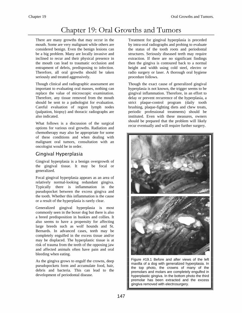
As the mass is growing from the periodontal ligament, cure is obtained by extracting the tooth from which the mass is growing along with its periodontal ligament. This often involves removal of the alveolar bone that supported the tooth. In order to create a wound that will be conducive to tension-free-closure, it is frequently necessary to remove one or more teeth on either side of the mass.
Complete resection of the mass is curative whereas merely debulking the mass ensures its continued growth. Collecting a superficial incisional biopsy through a debulking procedure may be indicated to arrive at a diagnosis prior to
definite surgery, but always assume that more aggressive surgery will be required for a resolution of the problem.
Peripheral acanthomatous ameloblastomas are also considered benign, but they are much more locally invasive and inclined to recur if surgical margins are less than 1 cm. Some authors have suggested that these masses are responsive to radiation therapy, but recurrence with malignant transformation and osteoradionecrosis are potential outcomes. Therefore, aggressive surgical resection remains the treatment of choice for most of these masses.
Odontomas
Odontomas are considered hamartomas in that they are composed of normal tissue arranged in an abnormal fashion. They occur in young animals during development of the permanent teeth. The exception to this is the rat, in which odontomas may occur at any age.
Complex odontomas are composed of dental tissues (enamel, dentin, cementum, pulp) without any organized architecture. Radiographically they appear as an amorphous radiodense mass in the mandible or maxilla.
Compound odontomas are also composed of normal dental tissues arranged into recognizable tooth-like structures known as denticles. Compound odontomas are typically centred on a deformed but recognizable permanent tooth which may be unerupted. There may be a few to several dozen denticles surrounding the central tooth. If unerupted, the teeth are surrounded by dense fibrovascular tissue.
Treatment for either form of odontoma is surgical enucleation including all denticles and soft tissue which might give rise to more denticles. It may be necessary to extract adjacent permanent teeth to gain access to all of the denticles.
Odontoma should be one of the rule-outs for all oral swellings in young animals.
Figure #19.4. Postoperative radiograph and photograph of the mass in Figure #19.2. The canine tooth has been removed along with most of its alveolus as well as the left incisors, the fractured right intermediate incisor and the first and second premolars. The wound has been sutured without tension.
Chapter 19 Oral Growths and Tumors.
150
.
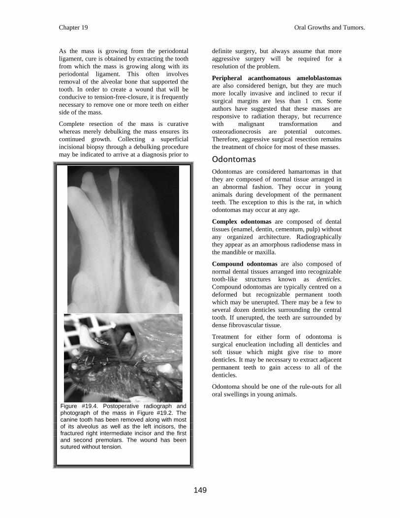
Figure #19.6. Compound Odontoma. Top photo is of a mandibular “swelling” in a 6-month-old spaniel. Centre is the pre-operative radiograph showing dozens of denticles surrounding the under-developed mandibular first molar tooth. Bottom photo is the collection of denticles removed from this mandible. The deformed mandibular first molar is seen top-left in the bottom photo.
Figure #19.5. The preoperative view of this caudal mandibular mass indicates a proliferative lesion centred on the fourth premolar. The preoperative radiograph shows how the mass is extruding the premolar. There is also evidence of bone involvement extending deeply into the bone. The owners opted for a conservative resection rather than segemental mandibulectomy. Fortunately the biopsy diagnosis was peripheral acanthomatous ameloblastoma and this surgery was curative.
Figure #19.7. A compound odontoma in the maxilla of a six-month-old shepherd dog. The lesion is centred on a deformed right maxillary canine tooth situated within the nasal cavity, the crown of which can be seen at the left of the radiograph.
Chapter 19 Oral Growths and Tumors.
151
Oral Malignancies
There are several malignancies which may occur in and around the oral cavity. The most common of these are squamous cell carcinoma, fibrosarcoma, melanoma and osteosarcoma.
Since lesions growing in the oral cavity are often not detected until they are quite large and possibly because of the excellent blood supply to oral structures, oral malignancies tend to have a poor prognosis. The more rostral the tumor location, the more favourable the prognosis. This may be due to earlier detection compared to masses hidden in the deeper recesses of the oral cavity. Also, more rostral tumors lend themselves to more complete surgical resection while still allowing for the maintenance of a functional mouth. In general, however, oral malignancies bear a poor prognosis.
Squamous cell carcinoma may affect the cutaneous tissues of the lip, but more commonly affects tissues within the oral cavity. Though non-tonsillar forms may be locally invasive, they tend to be slow to metastisize.
Gingival squamous cell carcinoma is centred on a tooth and may appear as a soft tissue proliferation above the gingival margin or may extend down into the alveolus, destroying the periodontal support for the tooth. In this second case, the process can look much like chronic periodontal disease. Whenever a tooth seems very mobile and easy to extract, especially if most of the other teeth in the mouth are periodontally sound, then gingival squamous cell carcinoma should be suspected. Following extraction of the mobile tooth, soft tissue from the alveolus should be harvested for submission to a pathologist. If gingival squamous cell carcinoma is diagnosed, then surgical resection of the tumor with at least 1 cm margins and tension-free closure is indicated.
Sublingual squamous cell carcinoma is particularly prevalent in cats. Surgical resection with adequate margins is often not possible as it would involve removal of a large portion of the tongue. Various chemotherapeutic regimens may be of palliative benefit, but long-term survival rates are poor.
Papillary squamous cell carcinoma is a form of the tumor that has been reported in young animals and occurs on the papillary gingiva. Though rapidly growing, they do not appear to metastasize and they tend to respond well to
surgery. Resection often involves removal of both deciduous teeth and developing permanent tooth buds depending on the age of onset.
Tonsillar squamous cell carcinoma is typically a unilateral lesion. It is the most aggressive form of squamous cell carcinoma with early metastasis to region lymph nodes and beyond. These masses are usually not detected until their size is causing problems with swallowing or vocalization and by that point, the prognosis is grave even with tonsillectomy and region radiotherapy.
Fibrosarcoma tends to metastasize slowly, but it is so aggressively invasive that complete surgical cure is rare. Early and radical surgical removal of the mass with at least 2-cm margins may provide some months of palliation, but recurrence is to be expected. Palatal masses may require complete removal of the palate and replacement with a prosthetic obturator to recreate a physical separation between the oral and nasal cavities.
Fibrosarcomas may appear as proliferative masses growing into the oral cavity or may be growing inwardly. In this latter case, there is often dramatic destruction of bone seen radiographically and the teeth may be very mobile.
Melanoma may be pigmented or amelanotic. There is a higher incidence in dogs with pigmented oral epithelium. Melanomas metastasize early and so have often spread to regional lymph nodes, bone or the lungs by the time the primary oral mass becomes evident. Even with aggressive surgery and ancillary treatments (chemotherapy and radiation), the prognosis is poor.
Osteosarcoma may affect the bones of the mandible or maxilla. As with fibrosarcomas, there may be a proliferative soft tissue mass evident in the oral cavity or there may be a subtle swelling with massive destruction of bone evident radiographically. As with the other oral malignancies, wide surgical resection is indicated, provided there is no evidence of metastasis. In general, oral osteosarcoma metastasizes slower than the appendicular version and so is more responsive to surgical resection.
Chapter 19 Oral Growths and Tumors.
152
Other oral malignancies include lymphosarcoma, mastocytoma and hemangiosacroma. In looking at the photos and radiographs in this chapter, you may have noticed that it would be virtually impossible to tell one type of mass from another based on gross appearance and radiographs alone. Deep incisional or excisional biopsy is needed for accurate diagnosis and appropriate treatment planning.
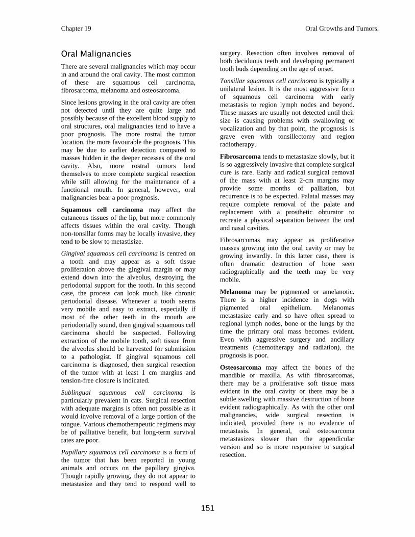
Figure #19.8. This osteosarcoma presented clinically as a diffuse mandibular swelling. There was loss of soft tissue around the left mandibular canine and premolars. The radiograph revealed extensive changes in the bone from the canine to the fourth premolar.
Figure #19.9. An acrylic obturator used to recreate a physical separation between the oral and nasal cavities following several failed attempts to close the surgical defect created by the resection of a large caudal palatal fibrosarcoma.
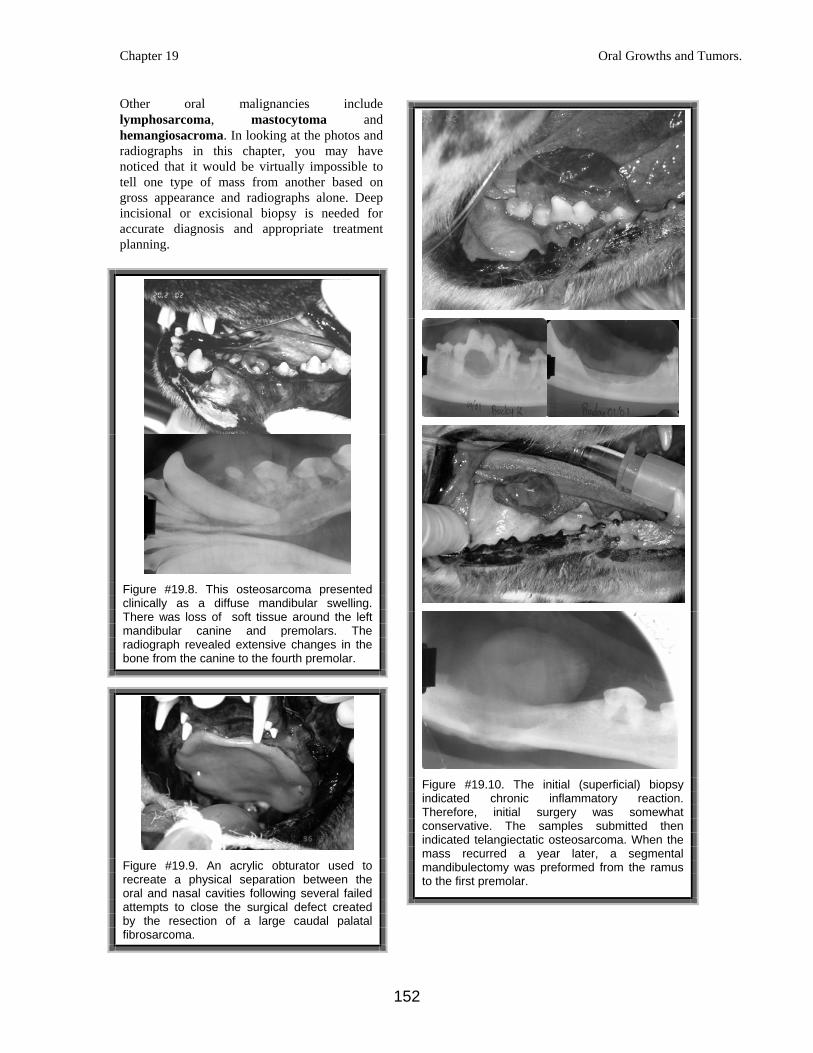
Figure #19.10. The initial (superficial) biopsy indicated chronic inflammatory reaction. Therefore, initial surgery was somewhat conservative. The samples submitted then indicated telangiectatic osteosarcoma. When the mass recurred a year later, a segmental mandibulectomy was preformed from the ramus to the first premolar.