Embed Size (px)
Citation preview
Imaging ofSoft Tissue Tumors
Leanne L Seeger, MD, FACRProfessor & Chief, Musculoskeletal ImagingDavid Geffen School of Medicine at UCLA
Department of Radiology
Objectives1. Apply understanding and awareness of
the role imaging and image-guided intervention play to improve the interdisciplinary diagnosis of musculoskeletal tumors.
2. Develop open lines of communication between Pathology and Radiology to optimize diagnostic sensitivity and accuracy.
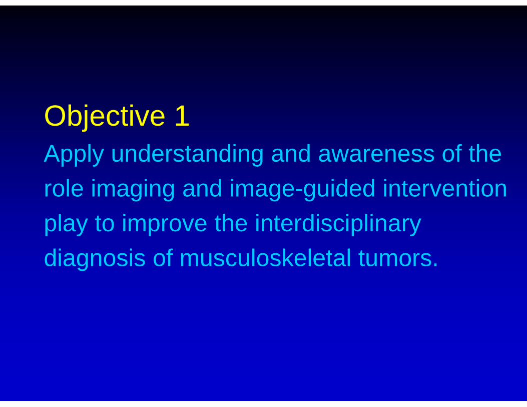
Objective 1Apply understanding and awareness of the role imaging and image-guided intervention play to improve the interdisciplinary diagnosis of musculoskeletal tumors.
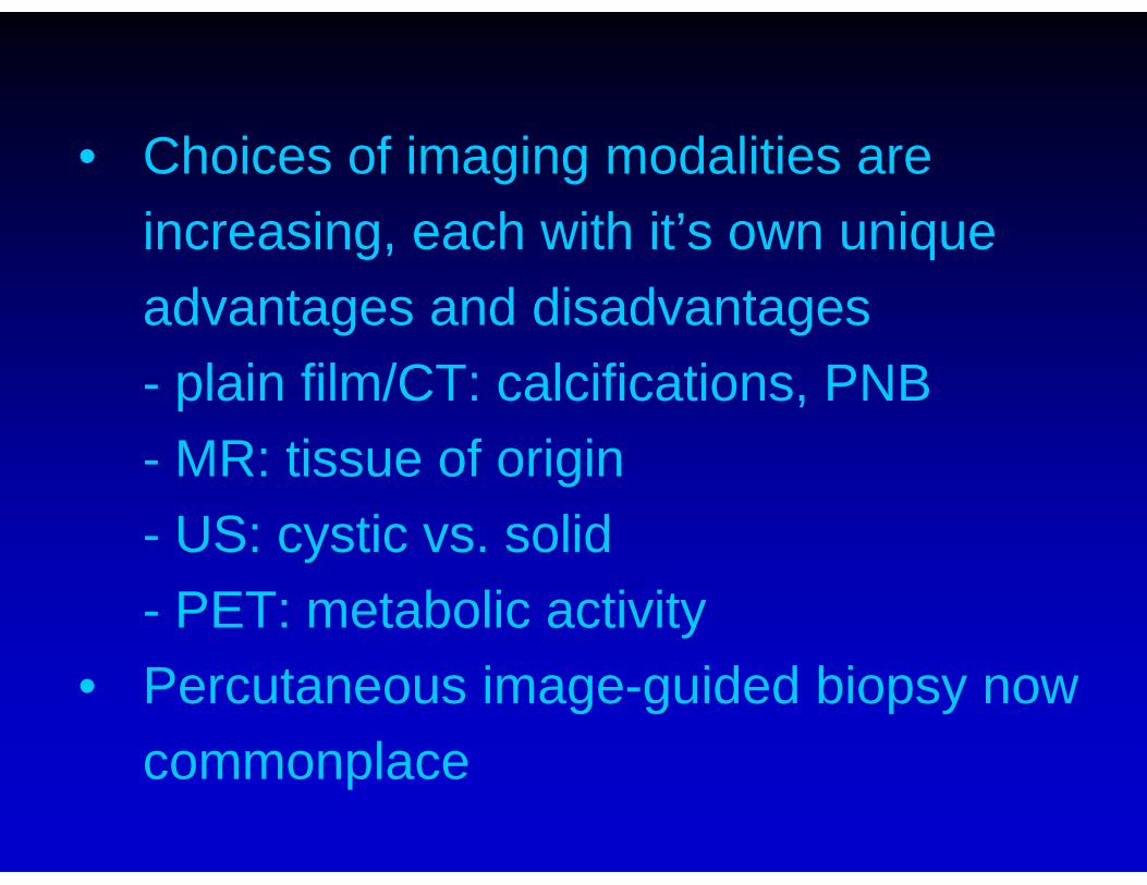
• Choices of imaging modalities are increasing, each with it’s own unique advantages and disadvantages- plain film/CT: calcifications, PNB- MR: tissue of origin- US: cystic vs. solid- PET: metabolic activity
• Percutaneous image-guided biopsy now commonplace
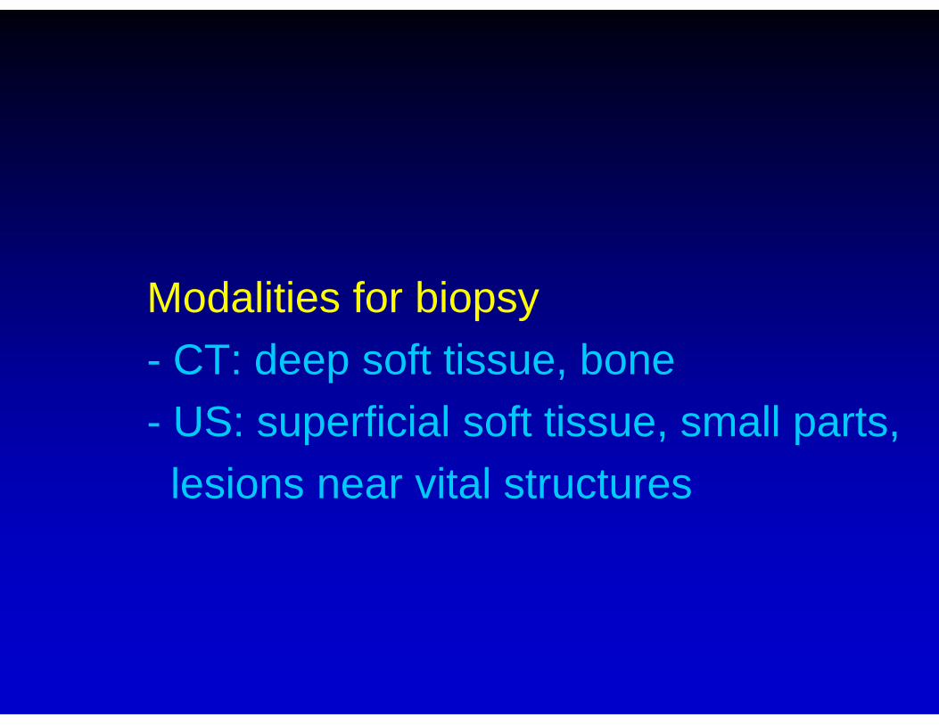
Modalities for biopsy- CT: deep soft tissue, bone- US: superficial soft tissue, small parts, lesions near vital structures
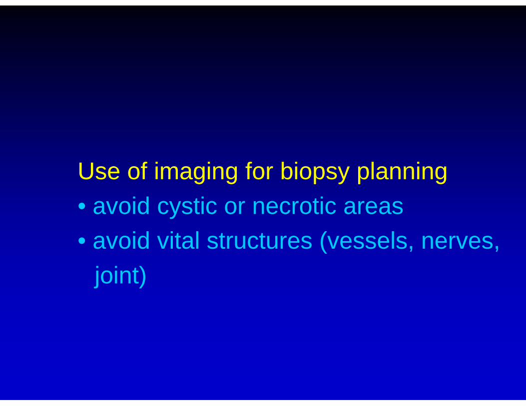
Use of imaging for biopsy planning• avoid cystic or necrotic areas• avoid vital structures (vessels, nerves,
joint)
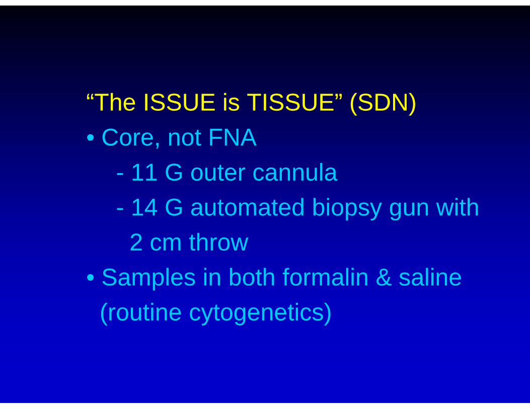
“The ISSUE is TISSUE” (SDN)• Core, not FNA
- 11 G outer cannula- 14 G automated biopsy gun with2 cm throw
• Samples in both formalin & saline (routine cytogenetics)
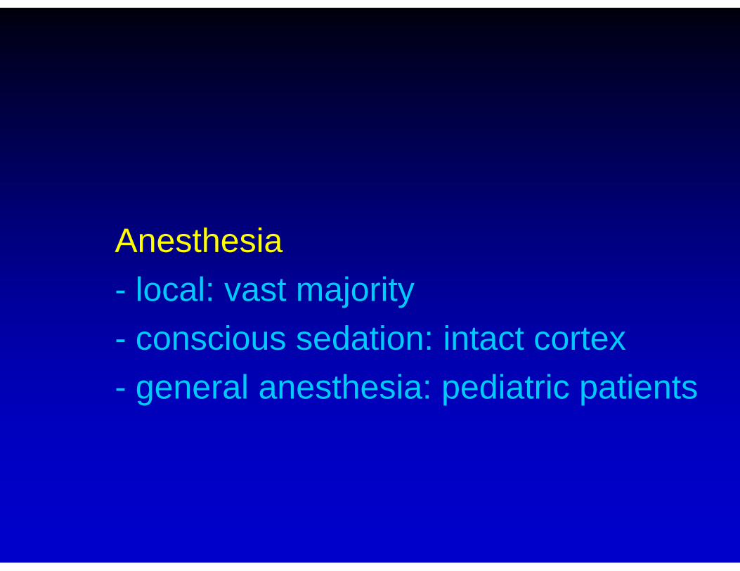
Anesthesia- local: vast majority- conscious sedation: intact cortex- general anesthesia: pediatric patients
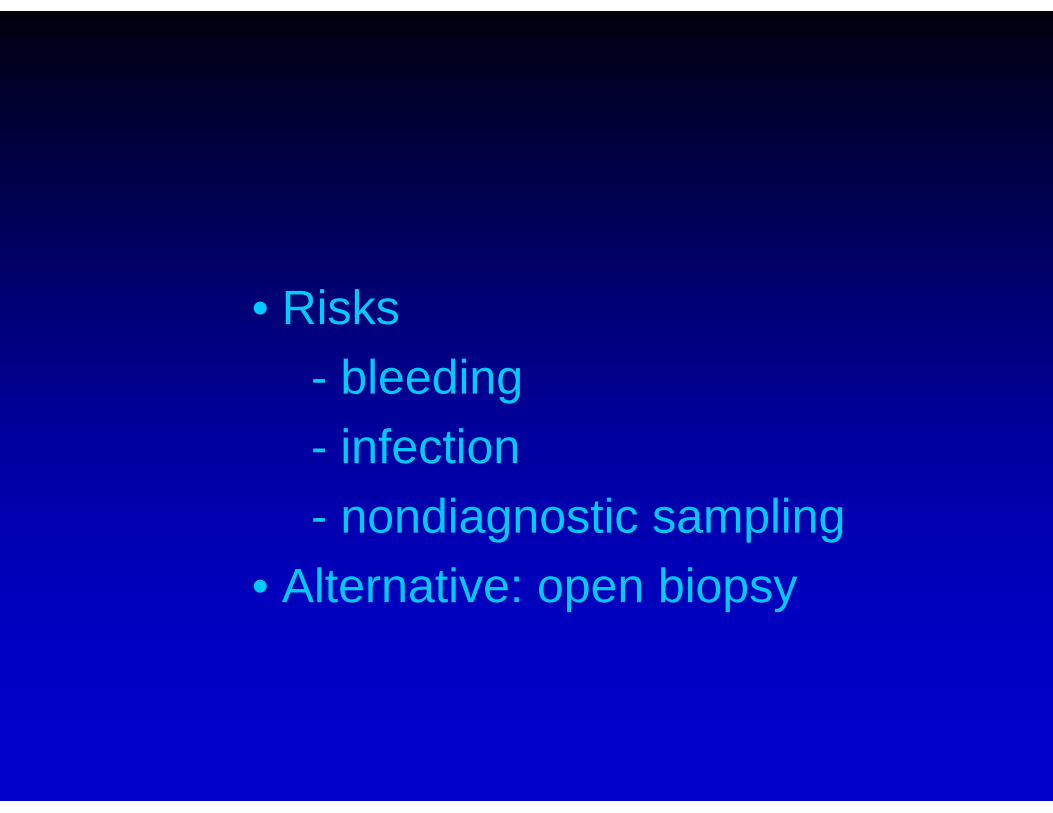
• Risks- bleeding- infection- nondiagnostic sampling
• Alternative: open biopsy
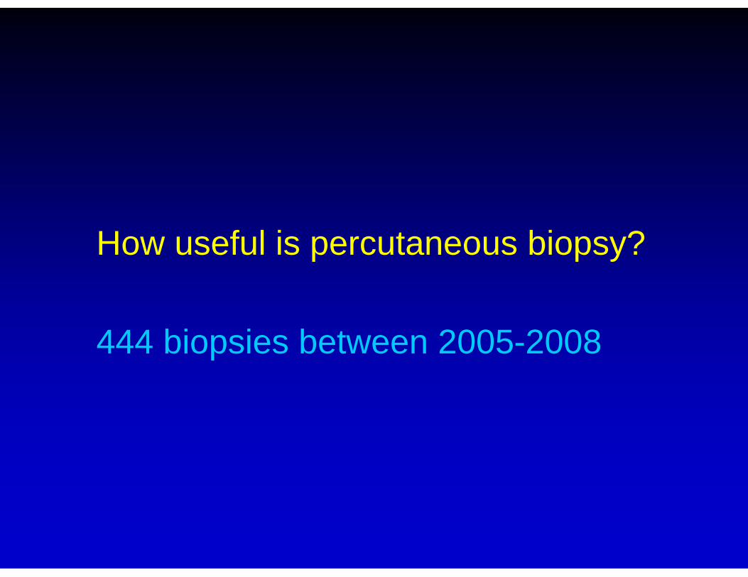
How useful is percutaneous biopsy?
444 biopsies between 2005-2008
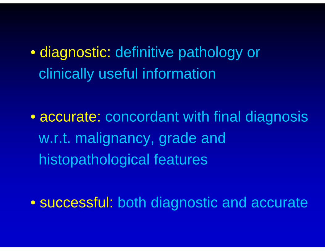
• diagnostic: definitive pathology or clinically useful information
• accurate: concordant with final diagnosis w.r.t. malignancy, grade and histopathological features
• successful: both diagnostic and accurate
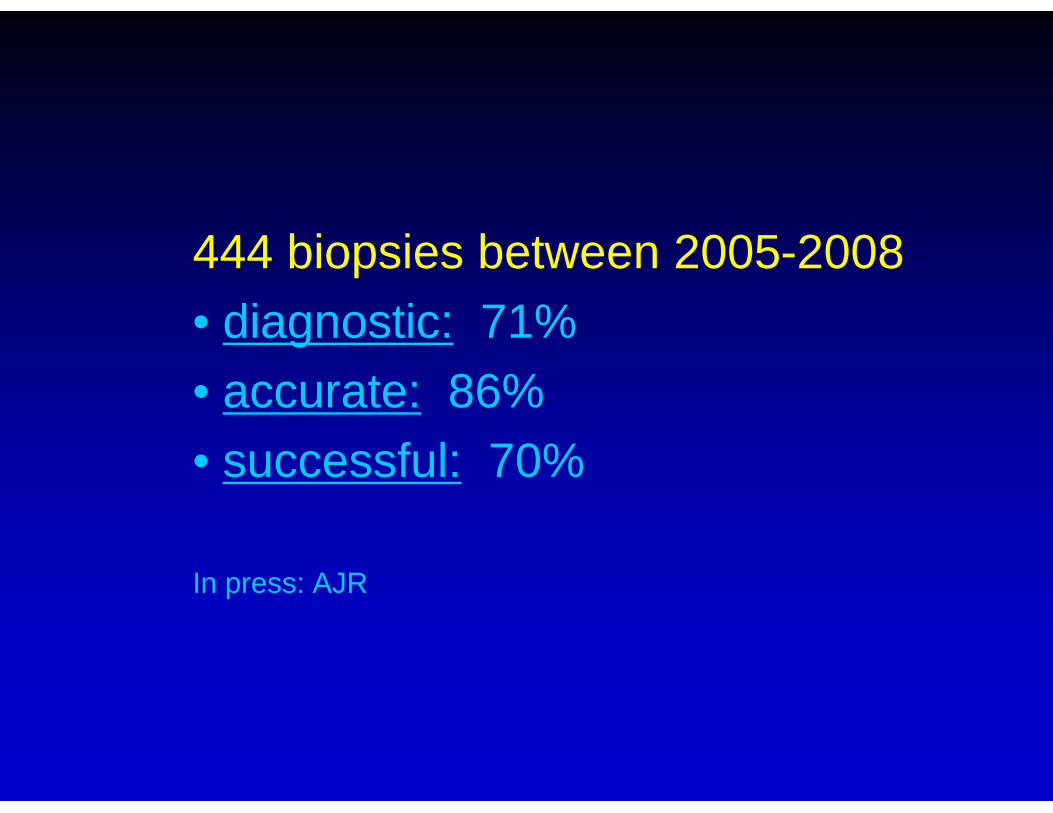
444 biopsies between 2005-2008• diagnostic: 71%• accurate: 86%• successful: 70%
In press: AJR
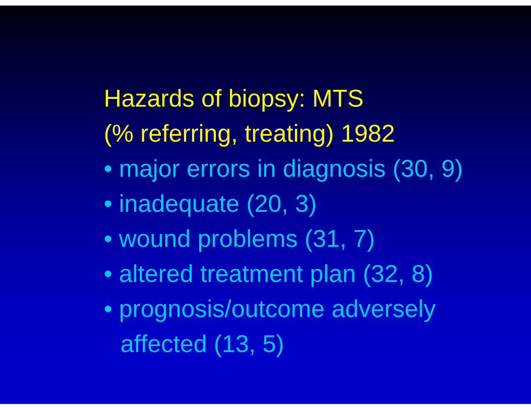
Hazards of biopsy: MTS (% referring, treating) 1982• major errors in diagnosis (30, 9)• inadequate (20, 3)• wound problems (31, 7)• altered treatment plan (32, 8)• prognosis/outcome adversely
affected (13, 5)
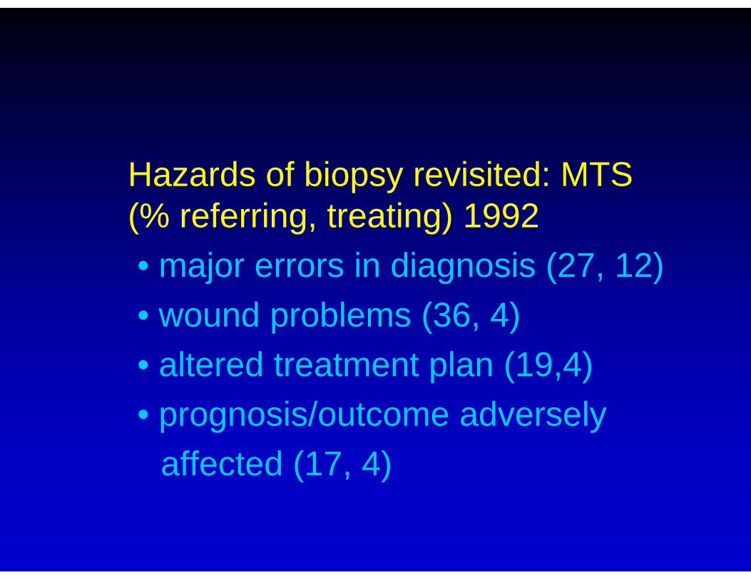
Hazards of biopsy revisited: MTS (% referring, treating) 1992• major errors in diagnosis (27, 12)• wound problems (36, 4)• altered treatment plan (19,4)• prognosis/outcome adversely
affected (17, 4)
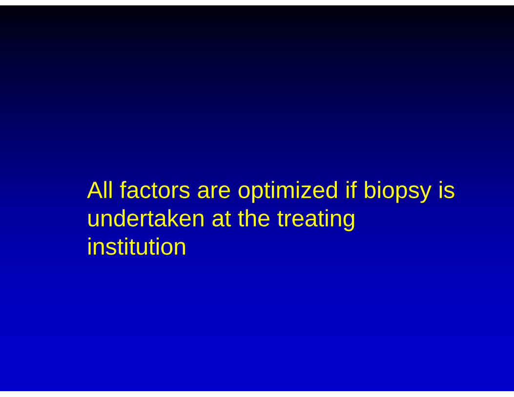
All factors are optimized if biopsy is undertaken at the treating institution
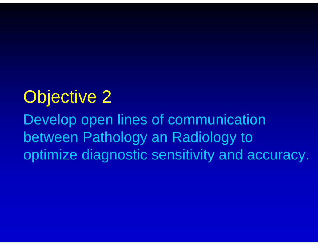
Objective 2Develop open lines of communication between Pathology an Radiology to optimize diagnostic sensitivity and accuracy.

This one is up to you…
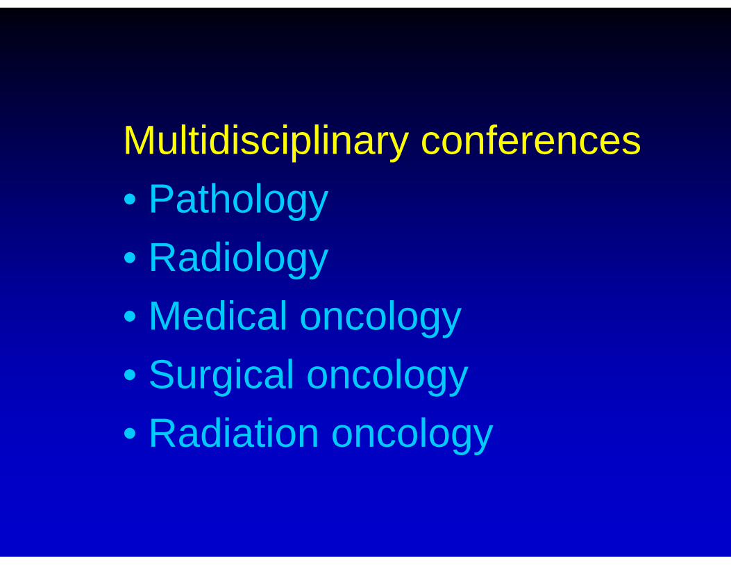
Multidisciplinary conferences• Pathology• Radiology• Medical oncology• Surgical oncology• Radiation oncology
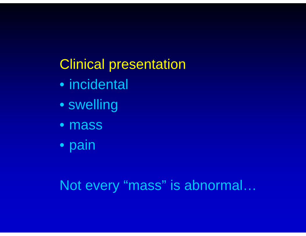
Clinical presentation • incidental• swelling• mass• pain
Not every “mass” is abnormal…
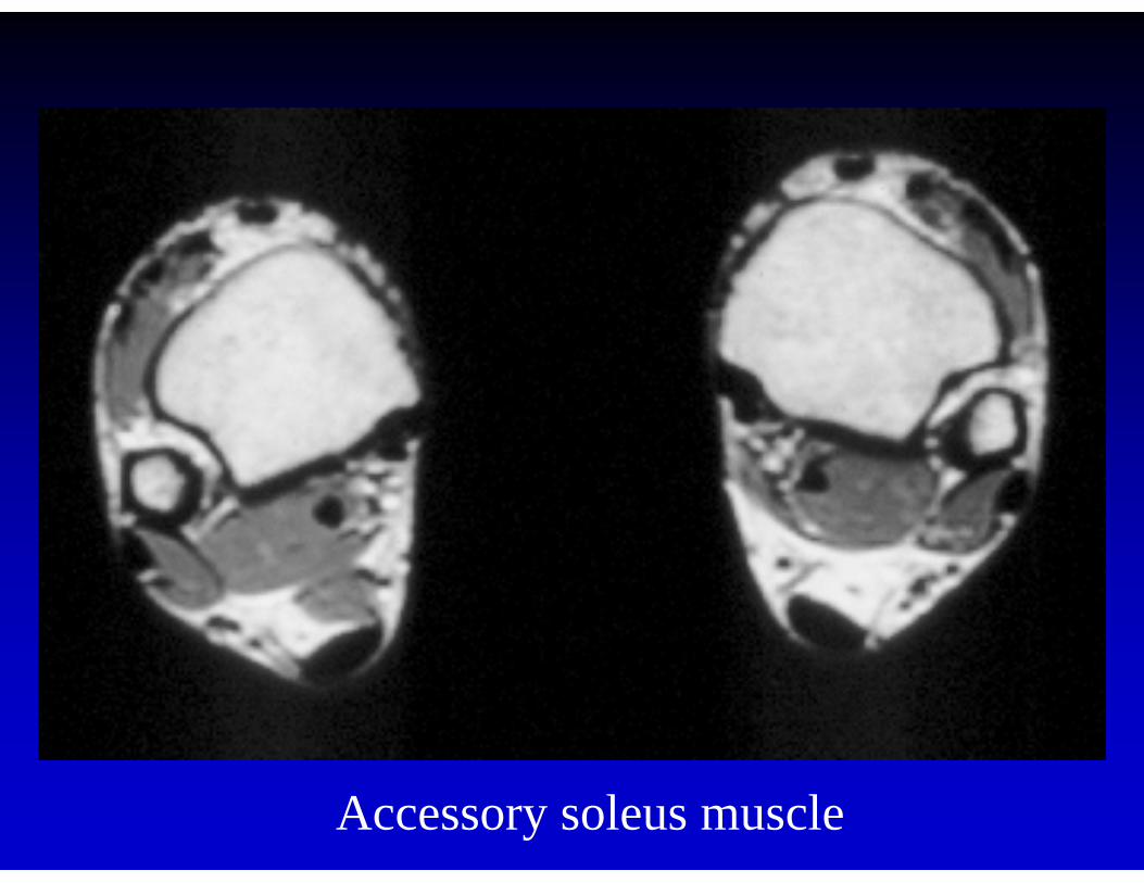
Accessory soleus muscle
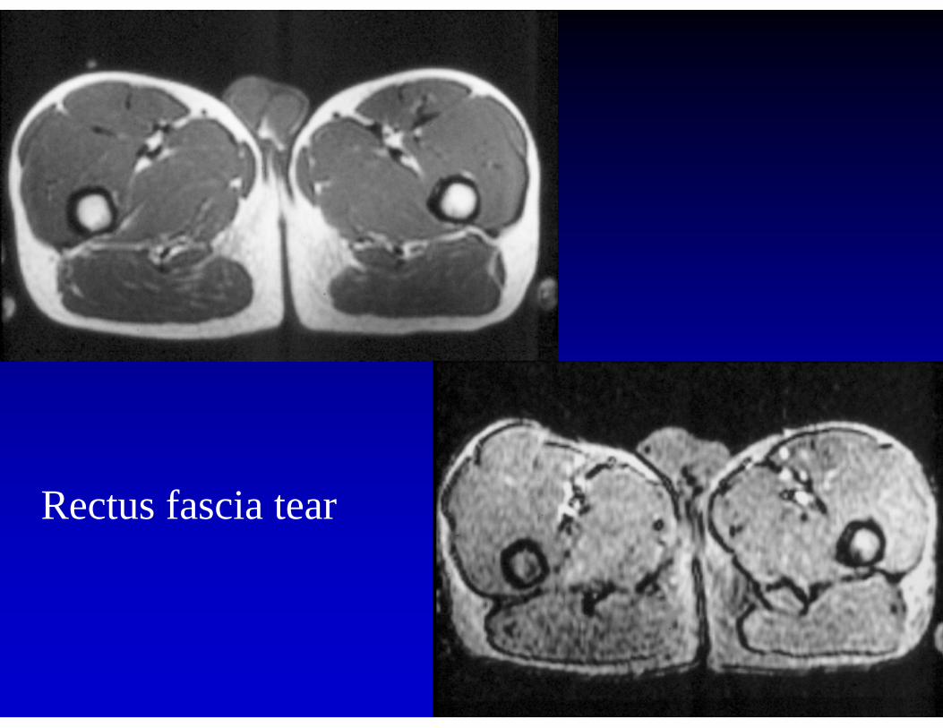
Rectus fascia tear
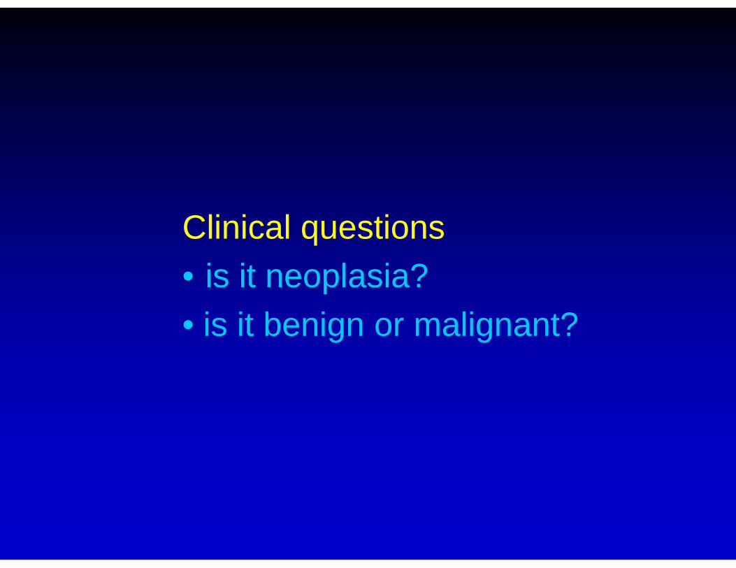
Clinical questions• is it neoplasia?• is it benign or malignant?
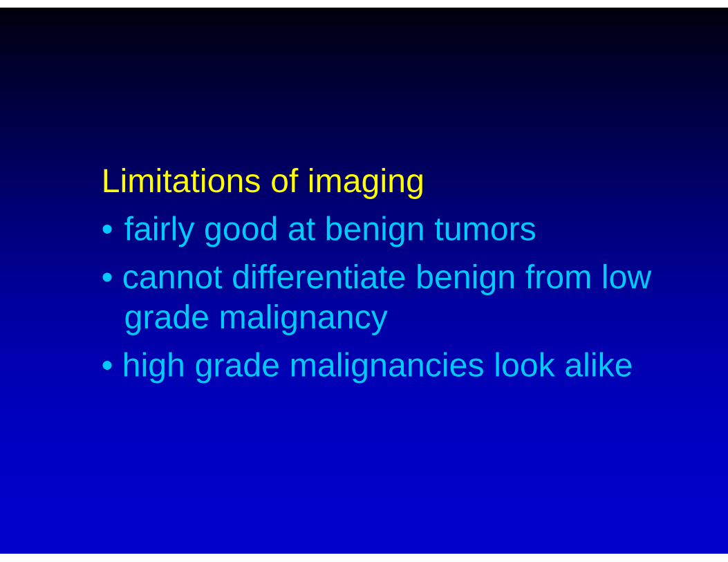
Limitations of imaging• fairly good at benign tumors• cannot differentiate benign from low
grade malignancy• high grade malignancies look alike
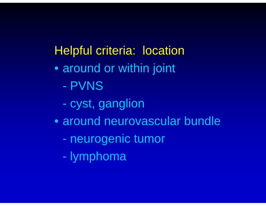
Helpful criteria: location• around or within joint
- PVNS- cyst, ganglion
• around neurovascular bundle- neurogenic tumor- lymphoma
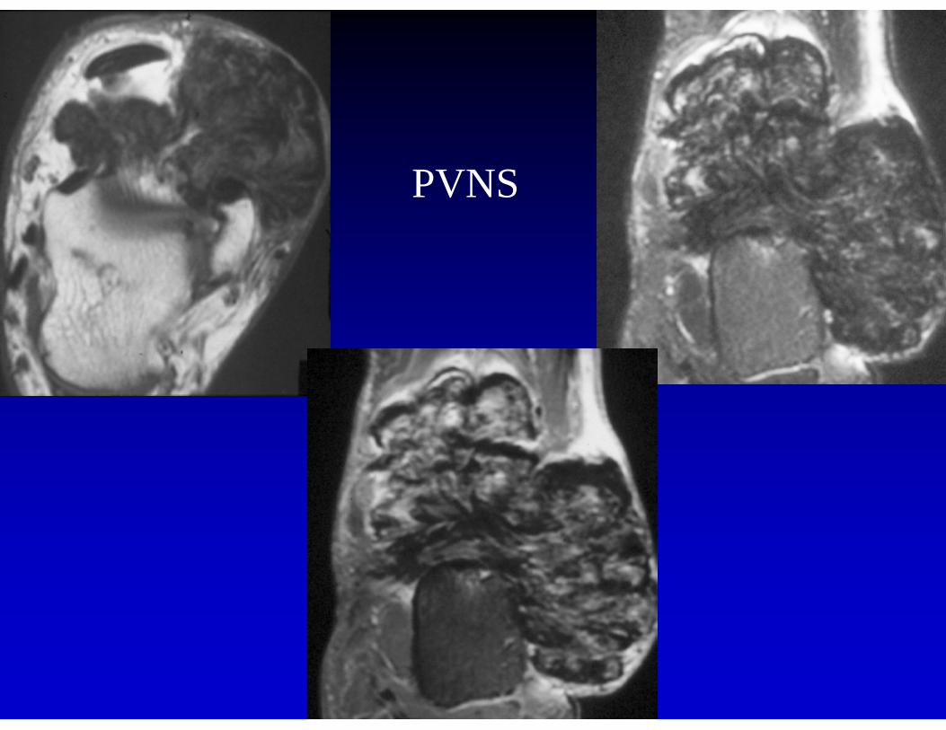
PVNS
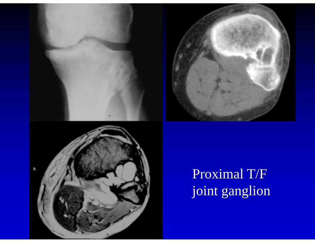
Proximal T/F joint ganglion
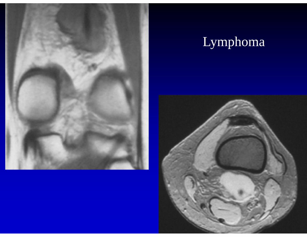
Lymphoma
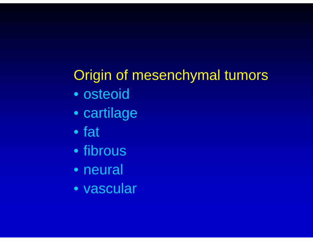
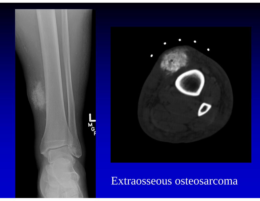
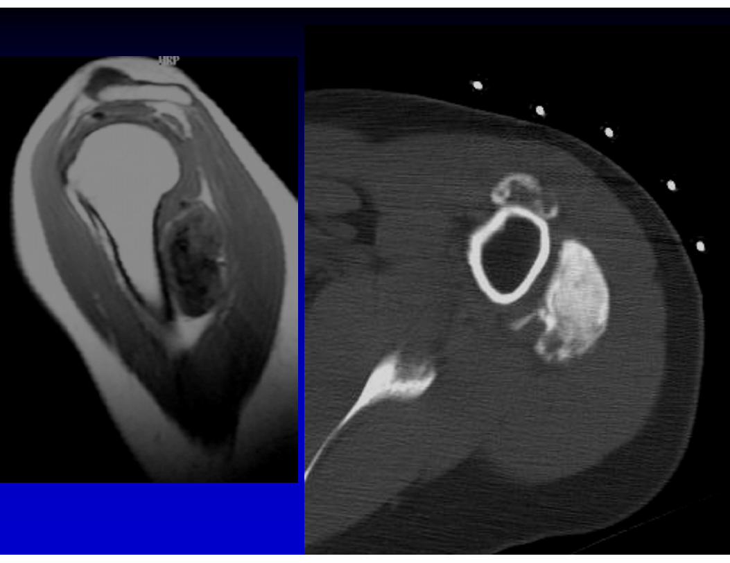
Origin of mesenchymal tumors• osteoid• cartilage• fat• fibrous• neural• vascular
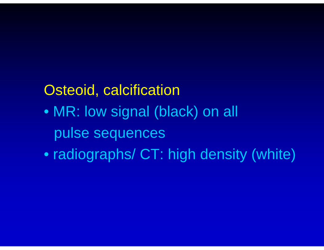
Osteoid, calcification• MR: low signal (black) on all
pulse sequences• radiographs/ CT: high density (white)
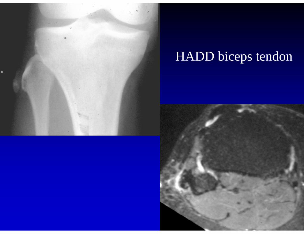
HADD biceps tendon
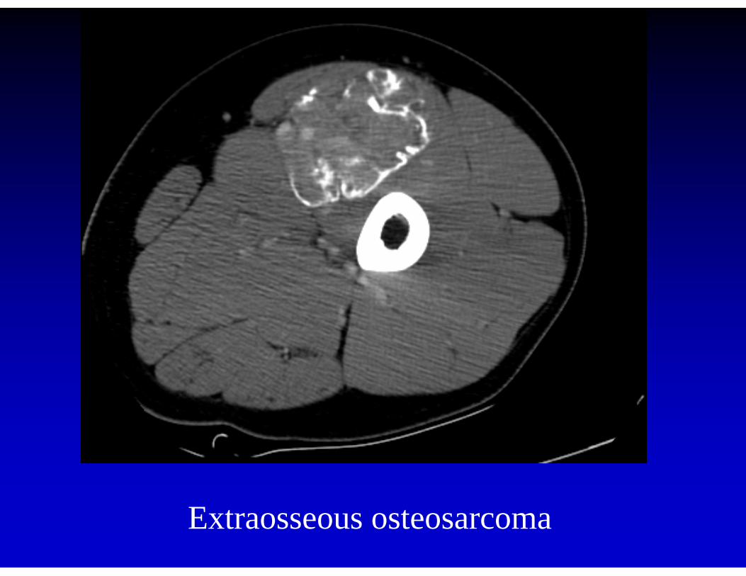
Extraosseous osteosarcoma
Extraosseous osteosarcoma
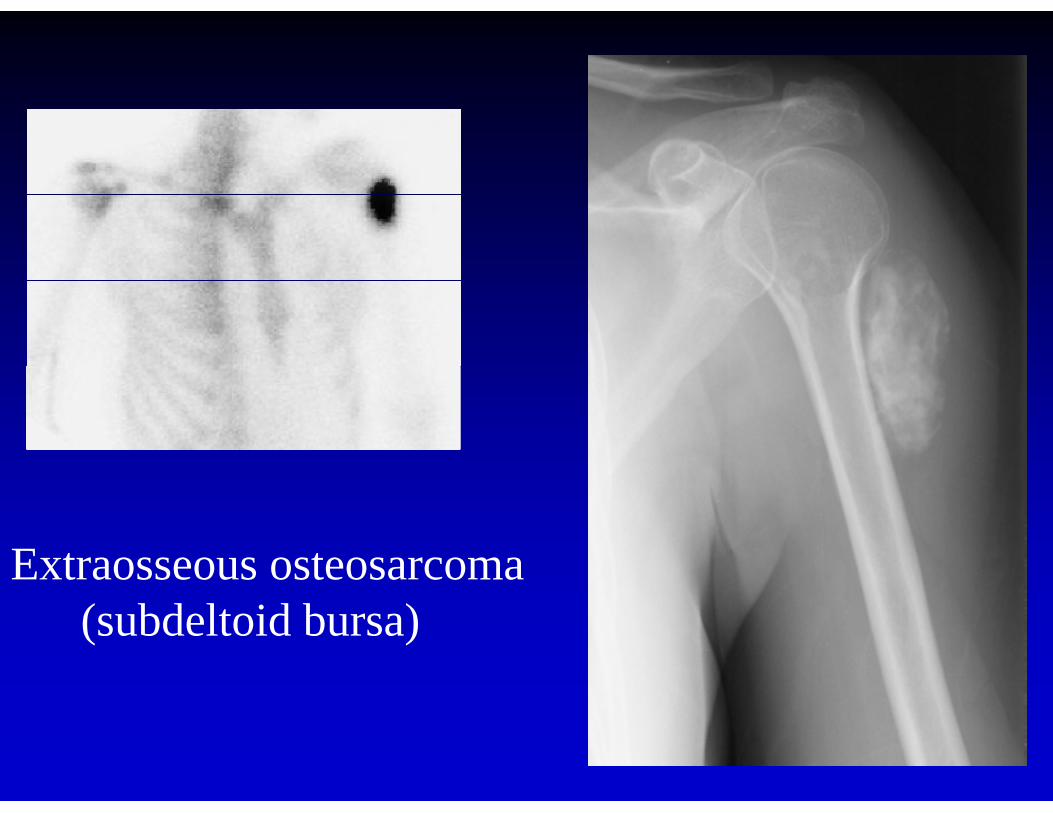
Extraosseous osteosarcoma (subdeltoid bursa)
Cartilage• lobulated• MR: T2’ cartilage very high and
calcification very low signal• CT: cartilage lower density than
muscle, calcifications dense
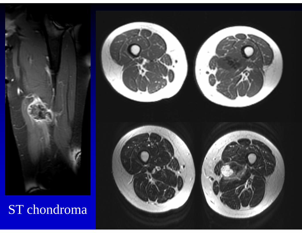
ST chondroma
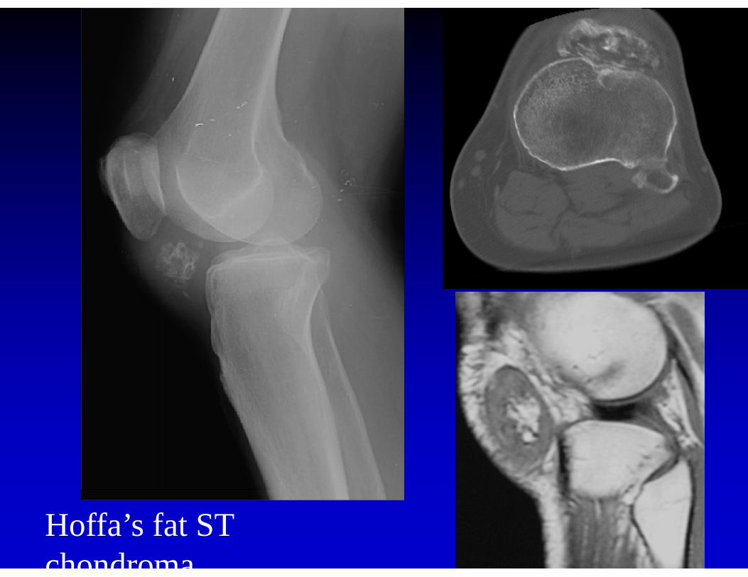
Hoffa’s fat ST chondroma
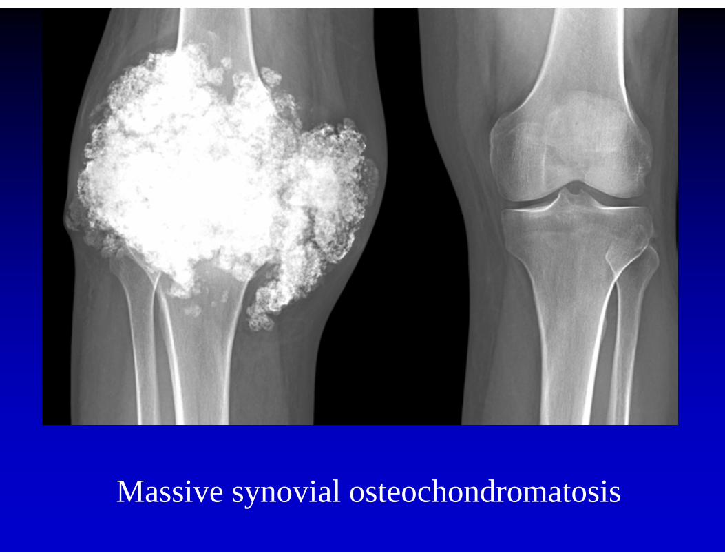
Massive synovial osteochondromatosis
Fat• MR: high signal T1, uniform fat suppression• CT: low density
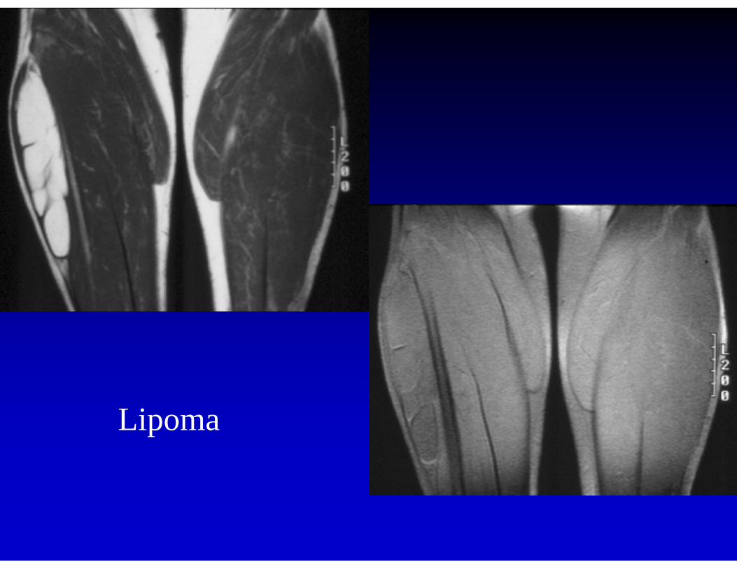
Lipoma
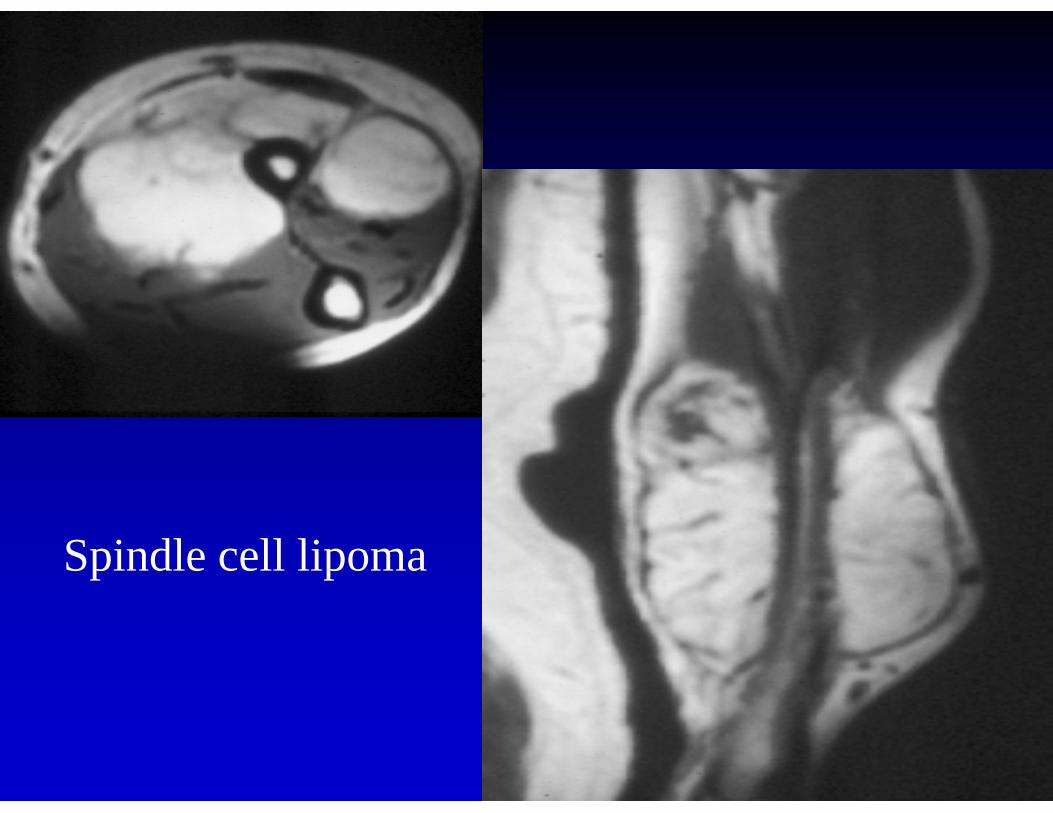
Spindle cell lipoma
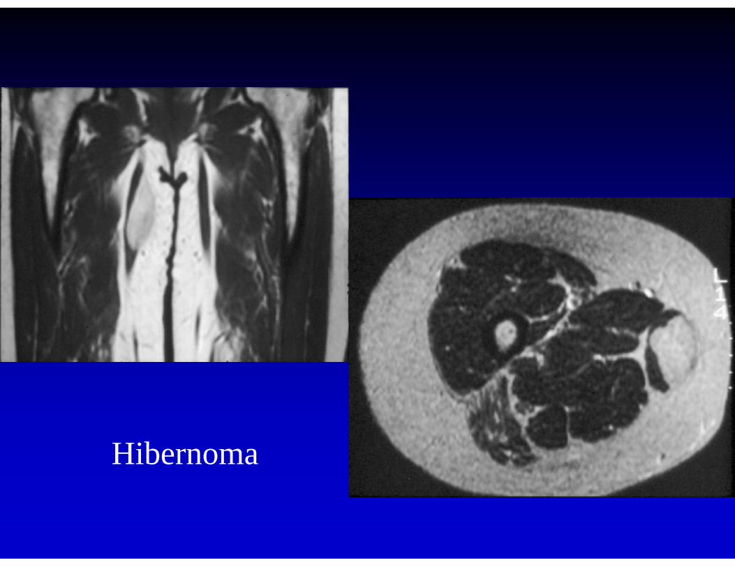
Hibernoma
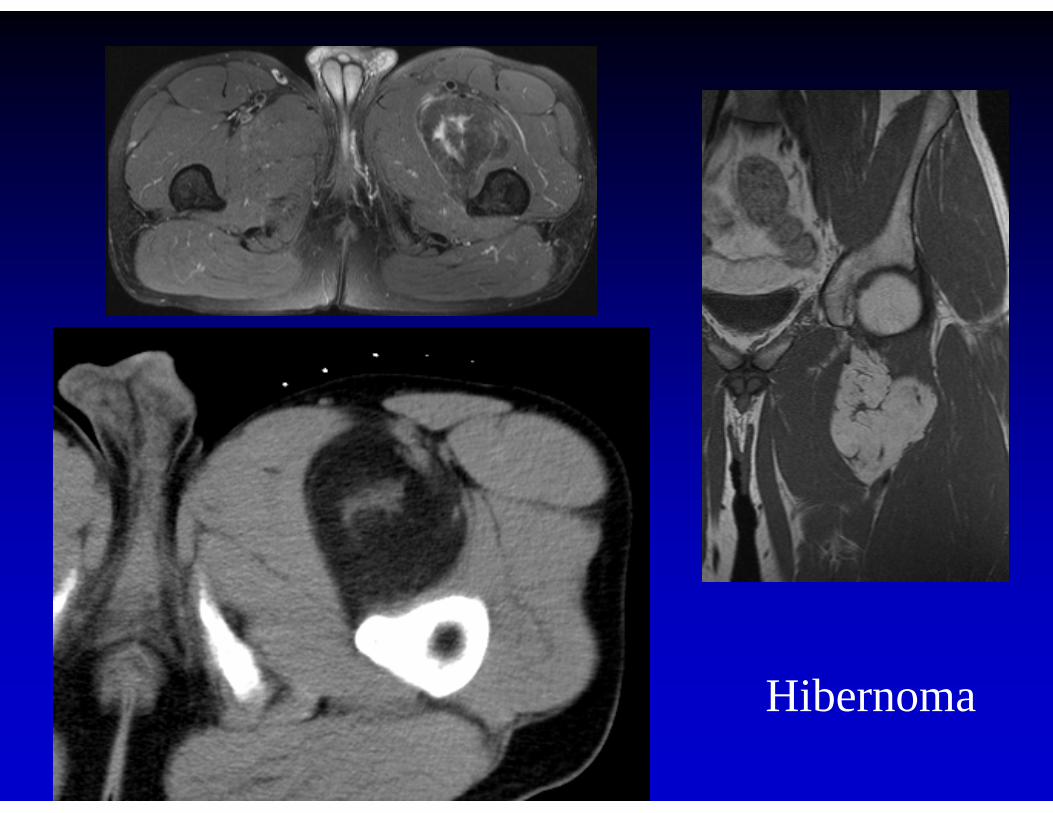
Hibernoma
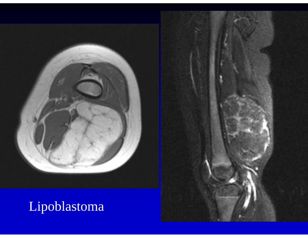
Lipoblastoma
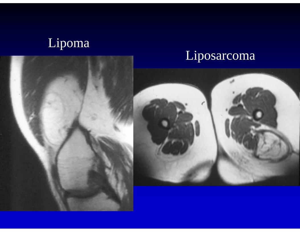
LipomaLiposarcoma
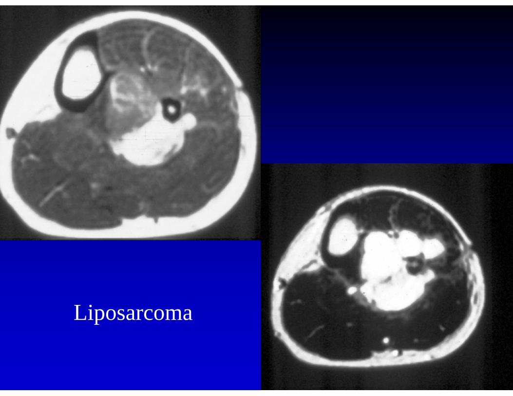
Liposarcoma
Fibrous• low signal T2 • signal varies depending on amount of
fibrous tissue, hemosiderin, foamy histiocytes
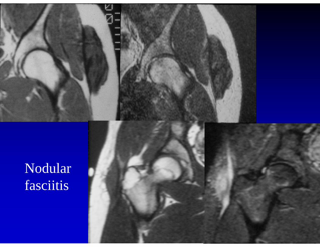
Nodularfasciitis
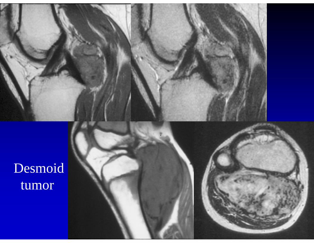
Desmoidtumor
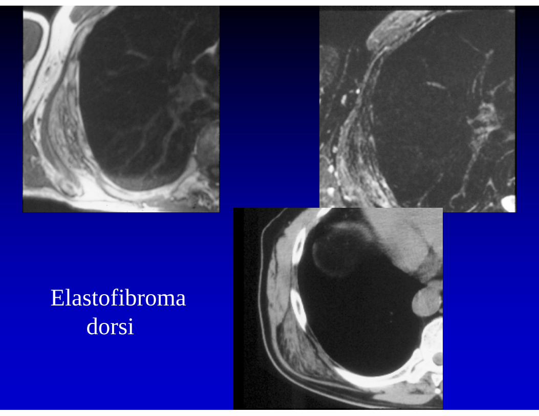
Elastofibroma dorsi
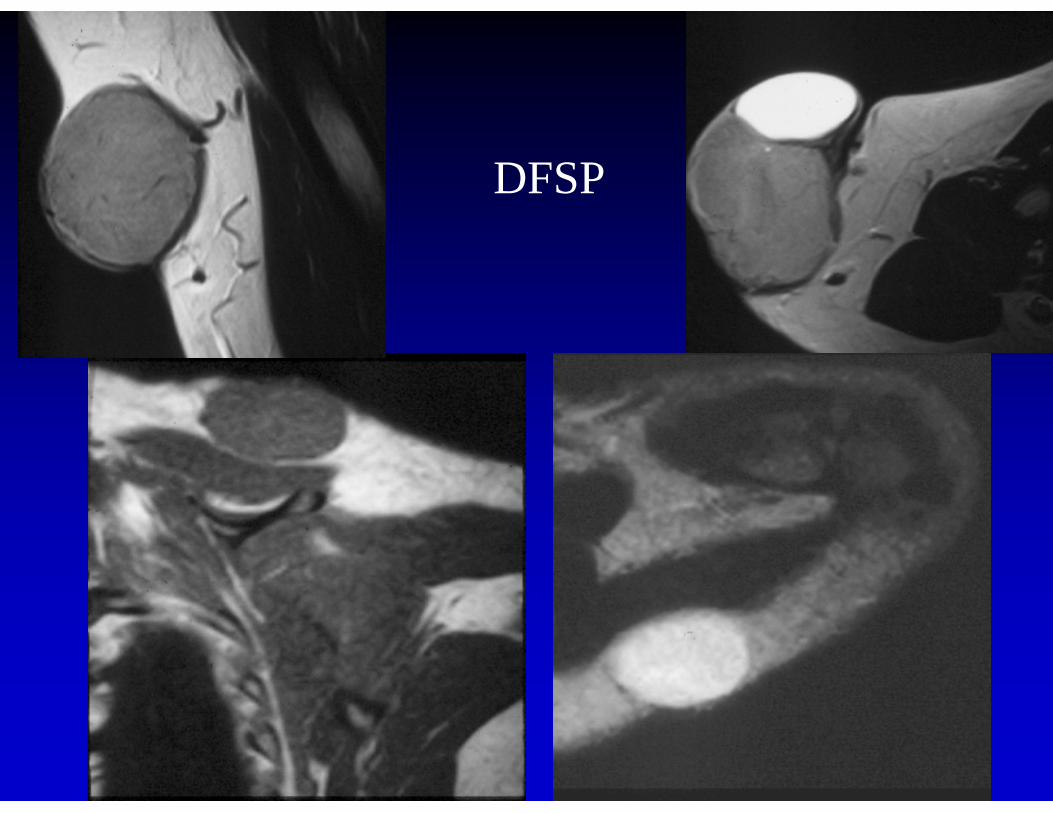
DFSP
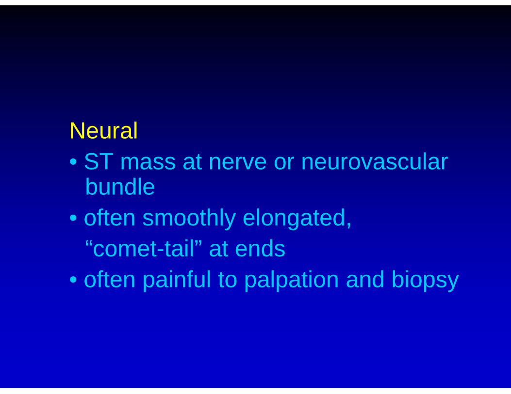
Neural • ST mass at nerve or neurovascular
bundle• often smoothly elongated,
“comet-tail” at ends• often painful to palpation and biopsy
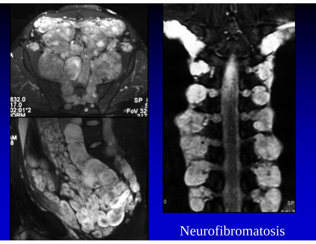
Neurofibromatosis
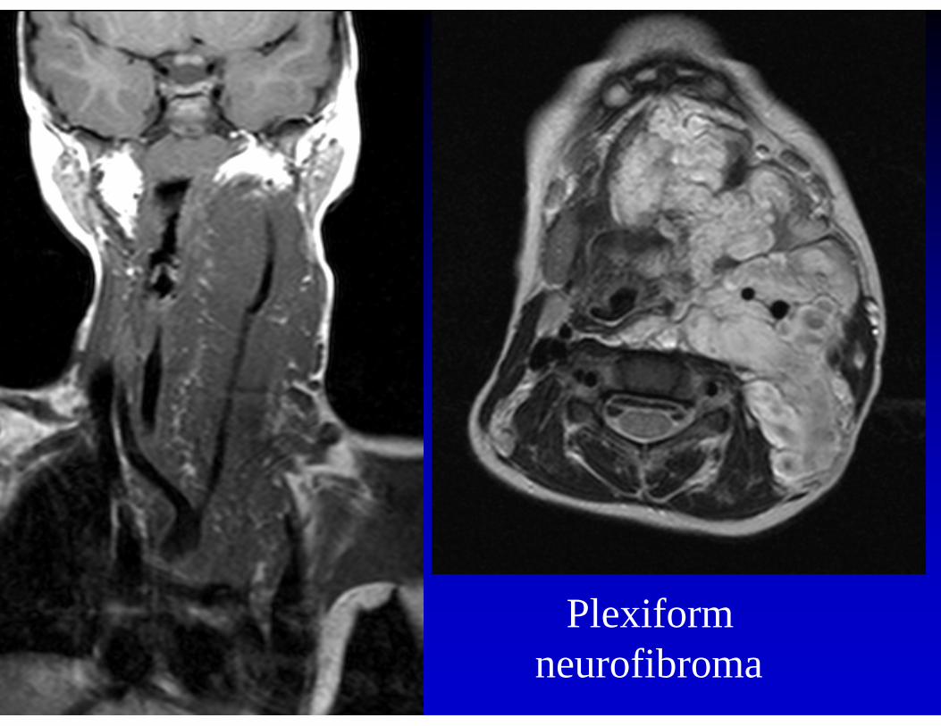
Plexiform neurofibroma
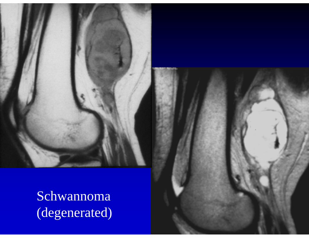
Schwannoma (degenerated)
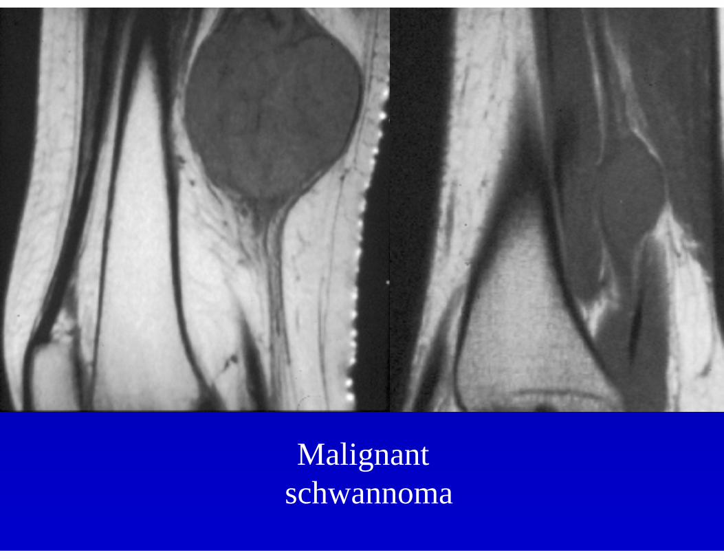
Malignantschwannoma
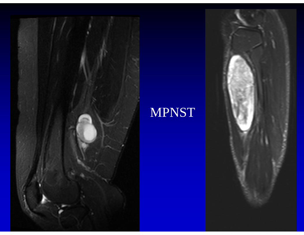
MPNST
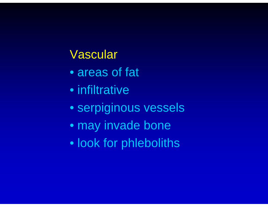
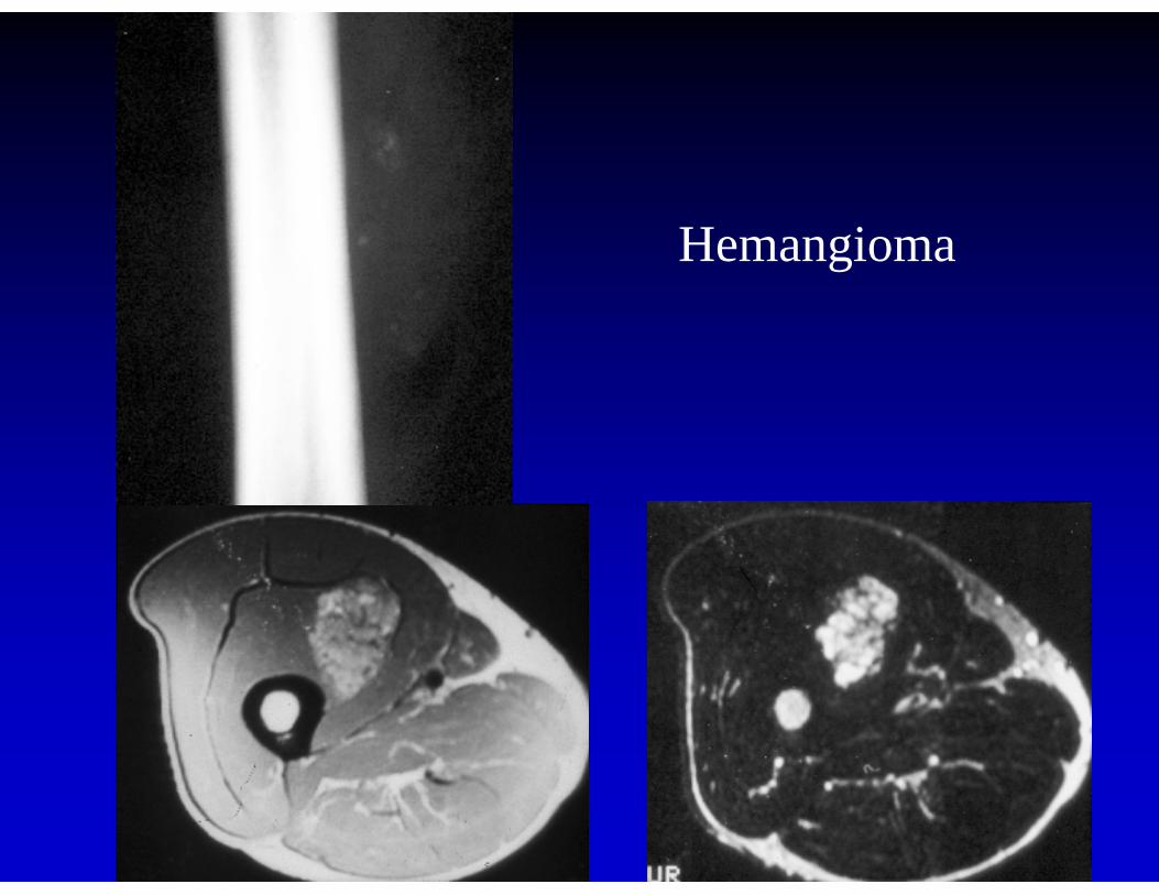
Vascular• areas of fat• infiltrative• serpiginous vessels• may invade bone• look for phleboliths
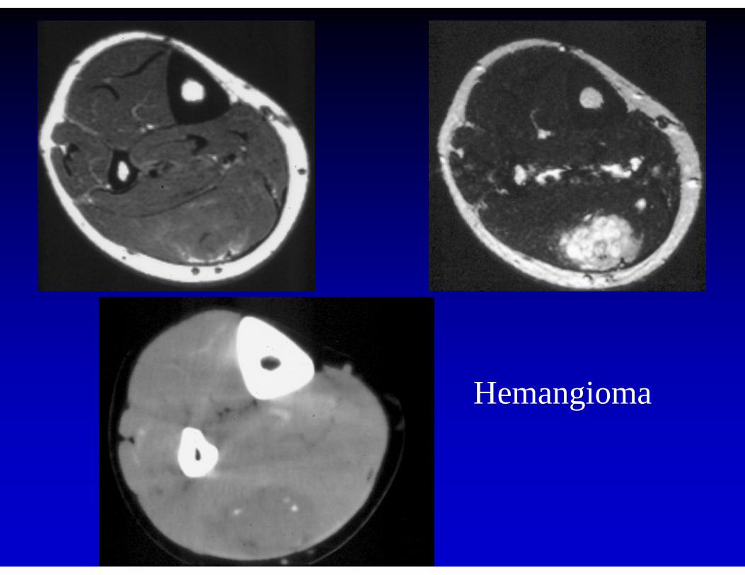
Hemangioma
Hemangioma
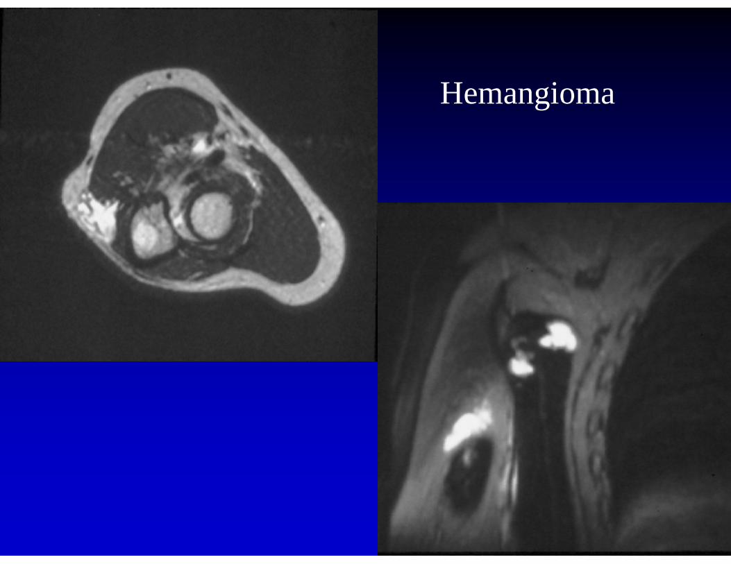
Hemangioma
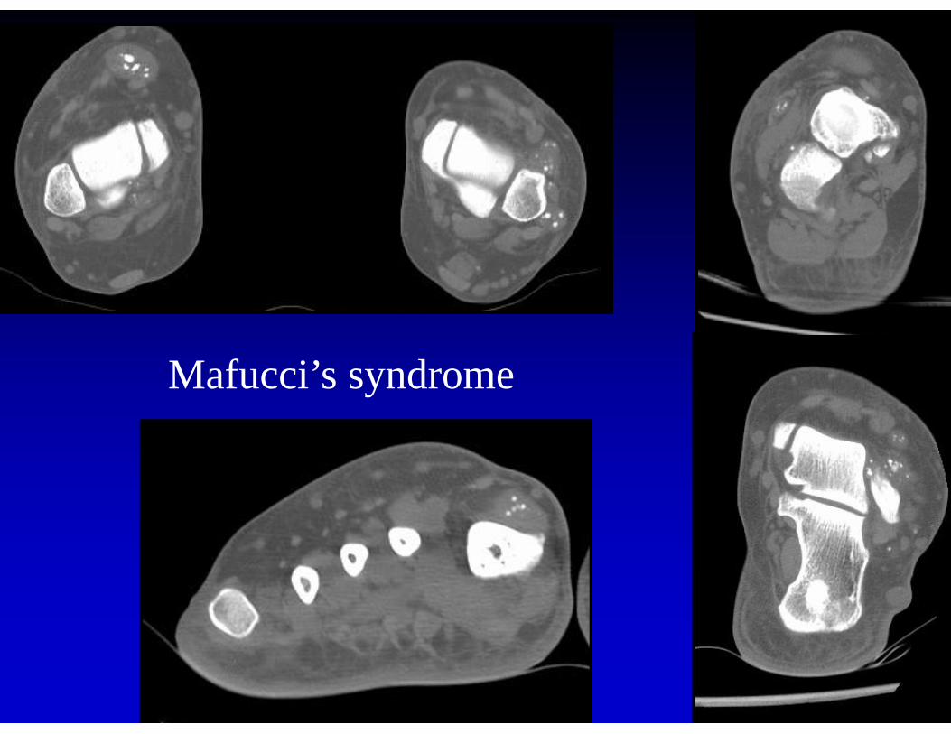
Mafucci’s syndrome
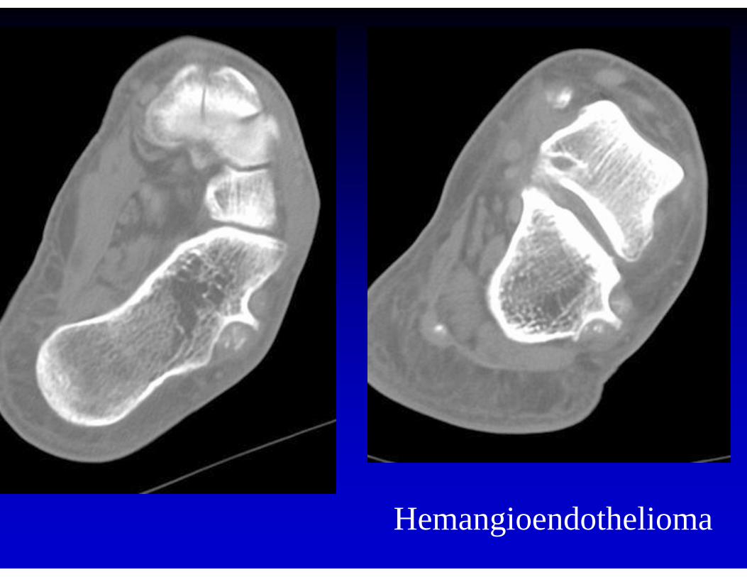
Hemangioendothelioma
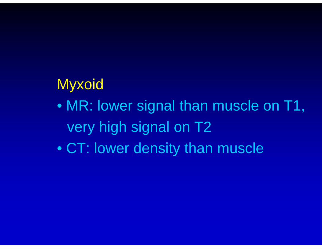
Myxoid• MR: lower signal than muscle on T1,
very high signal on T2• CT: lower density than muscle
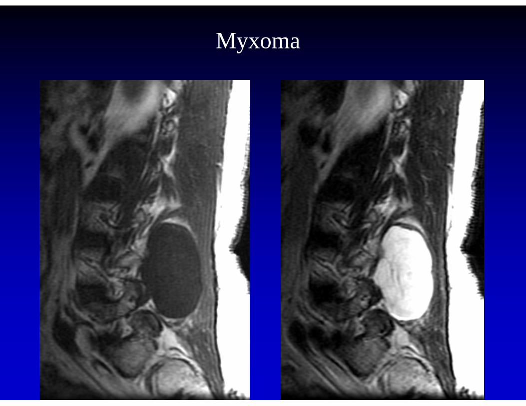
Myxoma
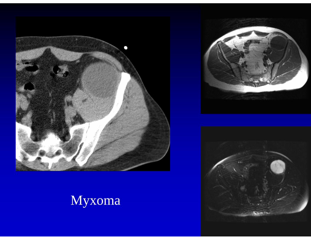
Myxoma
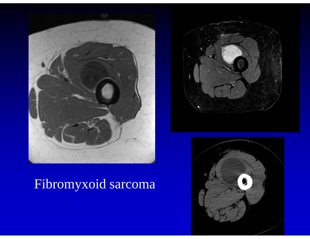
Fibromyxoid sarcoma
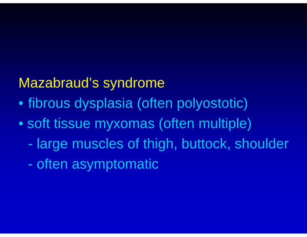
Mazabraud’s syndrome• fibrous dysplasia (often polyostotic)• soft tissue myxomas (often multiple)
- large muscles of thigh, buttock, shoulder- often asymptomatic
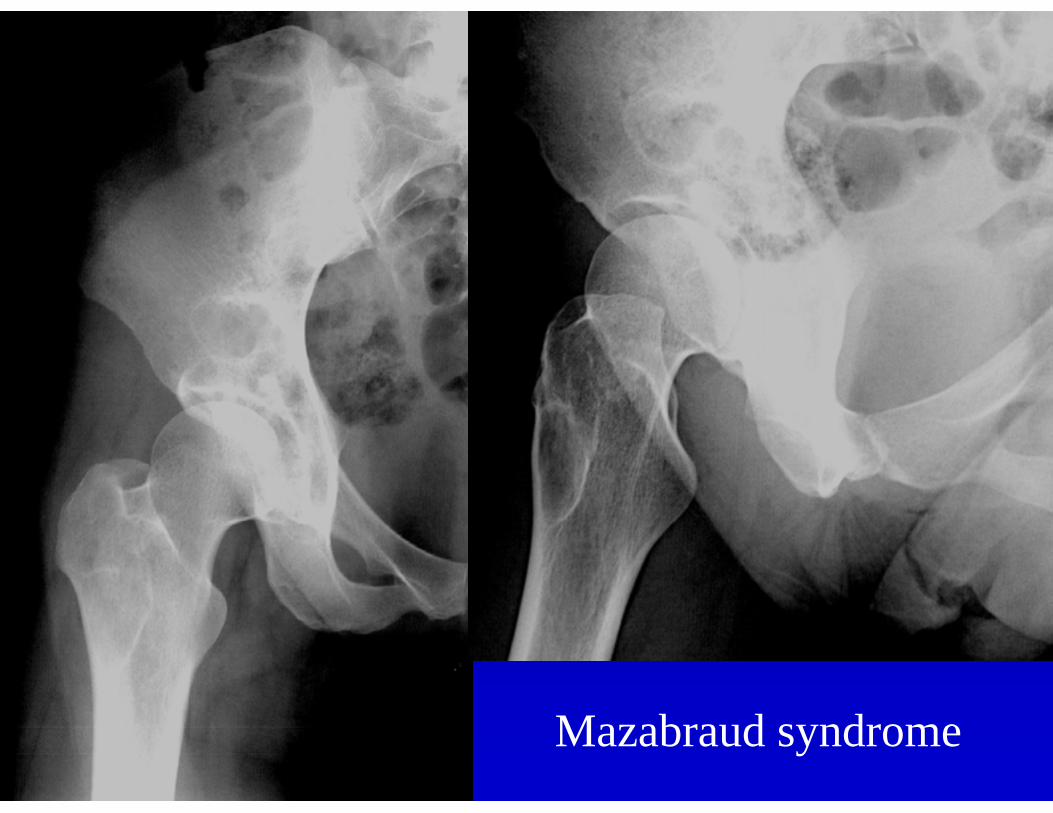
Mazabraud syndrome
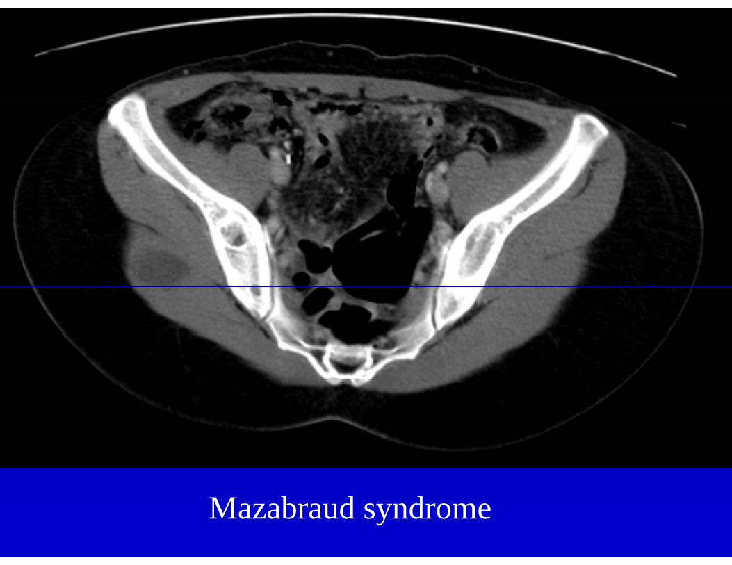
Mazabraud syndrome
Conclusions1. Imaging plays an important role in
diagnosis and treatment planning of soft tissue tumors
2. A close working relationship between Pathology and Radiology will optimize patient care
Conclusion3. Benign lesions cannot be
differentiated from low grade malignancy with imaging