Embed Size (px)
Citation preview

DOI 101167tvst454
Article
Automated Analysis of Vitreous Inflammation UsingSpectral-Domain Optical Coherence Tomography
Pearse A Keane1 Konstantinos Balaskas2 Dawn A Sim1 Kiran Aman2 Alastair KDenniston345 Tariq Aslam267 and for the EQUATOR Study Group1 NIHR Biomedical Research Centre for Ophthalmology Moorfields Eye Hospital NHS Foundation Trust and UCL Institute ofOphthalmology London UK2 Manchester Royal Eye Hospital Central Manchester University Hospitals NHS Foundation Trust Manchester Academic Health ScienceCentre Manchester UK3 Queen Elizabeth Hospital Birmingham University Hospitals Birmingham NHS Foundation Trust Birmingham UK4 Centre for Translational Inflammation Research College of Medical and Dental Sciences University of Birmingham EdgbastonBirmingham UK5 Birmingham amp Midland Eye Centre Sandwell and West Birmingham NHS Trust Birmingham UK6 Faculty of Medical and Human Sciences University of Manchester UK7 School of Built Environment Herriot-Watt University UK
Correspondence Alastair K Den-niston Queen Elizabeth HospitalBirmingham University HospitalsBirmingham NHS Foundation TrustBirmingham United Kingdom adennistonbhamacuk
Received 18 March 2015Accepted 27 April 2015Published 16 September 2015
Keywords vitreous inflammationoptical coherence tomographyuveitis automated segmentation
Citation Keane PA Balaskas K SimDA et al Automated Analysis ofVitreous Inflammation Using Spec-tral Domain Optical Coherence To-mography 20154(5)4 doi101167tvst454
Purpose To develop an automated method for quantifying vitreous signal intensityon optical coherence tomography (OCT) with particular application for use in theassessment of vitreous inflammation
Methods This retrospective observational case-control series comprised 30patients (30 eyes) with vitreous haze secondary to intermediate posterior orpanuveitis 12 patients (12 eyes) with uveitis without evidence of vitreous haze and18 patients (18 eyes) without intraocular inflammation or vitreoretinal disease Thepresence and severity of vitreous haze was classified according to the National EyeInstitute system other inflammatory indices and clinical parameters were alsodocumented Spectral-domain OCT images were analyzed using custom VITreousANalysis software (termed lsquoVITANrsquo) which is fully automated and avoids the need formanual segmentation
Results VITAN performed accurate segmentation in all scans Automated measure-ments of the vitreousretinal pigment epithelium (RPE) signal ratio showed amoderate correlation with clinical vitreous haze scores (r frac14 0585 P 0001)comparable to that reported using manual segmentation in our previous study (r frac140566 P frac14 00001) The novel parameter of vitreousRPE textural ratio showed amarginally stronger correlation (r frac14 0604 P 0001) with clinical vitreous haze scoresthan the VitreousRPE signal ratio
Conclusions The custom OCT image analysis software (VITAN) allows rapid andautomated measurement of vitreous parameters that is comparable to our previouslyreported vitreousRPE index and correlates with clinically measured disease activitySuch OCT-based indices may provide the much needed objective markers of vitreousactivity which may be used in both clinical assessment and as outcome measures inclinical trials for intermediate posterior and panuveitis
Translational Relevance We describe a rapid automated method for quantifyingvitreous signal intensity on optical coherence tomography (OCT) and show that thiscorrelates with clinical assessment of vitreous inflammation Such OCT-based indicesmay provide the much needed objective markers of vitreous activity which may beused both in routine clinical assessment and as outcome measures in clinical trials forintermediate posterior and panuveitis
1 TVST j 2015 j Vol 4 j No 5 j Article 4
Introduction
Uveitis a group of diseases characterized byintraocular inflammation is a significant cause ofblindness worldwide1ndash4 One of the greatest challengesin managing this disorder is the lack of sensitive andobjective markers of disease activity56 This is both anissue in day-to-day clinical management but alsohampers all clinical trials in the field Critically themajor disease activity endpoint for trials in posteriorsegment-involving uveitis that is recognized by theUnited States Food and Drug Administration (FDA)and European Medicines Agency (EMA) is theNational Eye Institute (NEI) system for grading ofvitreous haze78 Unfortunately this is limited bybeing (1) subjective (2) noncontinuous (3) poorlydiscriminatory at lower levels of inflammation and(4) poorly sensitive in a clinical trial context569
We recently reported the results of an exploratorystudy where optical coherence tomography (OCT)imaging was used to provide objective measurementof vitreous inflammation10 In this study a method forquantitative analysis of vitreous signal intensity onOCT was described and was correlated with clinicalvitreous haze scores in patients with uveitis and inhealthy volunteers This method produces a continuousmeasure of vitreous inflammation and thus hasconsiderable potential for adoption as a biomarker ofuveitic disease activity Despite this a significantlimitation of our study was the need for manualsegmentation of OCT image sets by human gradersWhile such grading is relatively straightforward andhighly reproducible1112 the requirement for humaninput nonetheless introduces a subjective element to theprocess Perhaps more importantly manual gradingtypically requires 5 to 10 minutes for completion aconsiderable barrier to any future adoption in clinicalpractice Our preliminary study also assessed OCTvitreous signal intensity as a whole such an intensityassessment would not necessarily encompass the varietyof different vitritis patterns seen on the OCTs whichoften for example have areas of clumping and othervarieties of hyperintense inflammatory pixel textures
To address these concerns we undertook todevelop a software algorithm that can provide bothautomated analysis of vitreous signal intensity onOCT and other potentially useful OCT measures ofvitreous inflammation In this report we present anew dedicated software application for vitreousanalysis and assess its use for assessment of patientswith uveitis The application has been designed purely
for the purpose of assessment of vitreous activity fromspectral-domain (SD) OCT and is fully automatedwith no subjective steps It incorporates a new texturemeasure designed to improve measurement validityand reliability Finally it automatically assessesbroader aspects of vitreous inflammation not mea-surable by simple intensity such as vitreous clumping
Materials and Methods
Study Population
Detailed information about the study populationand data collected has previously been reported10 Inbrief data was collected retrospectively from subjectsat Moorfields Eye Hospital and the Birmingham ampMidland Eye Centre in three categories (1) eyes withvitreous haze secondary to intermediate posterior orpanuveitis (2) eyes with uveitis but with no evidenceof vitreous haze and (3) eyes without evidence ofintraocular inflammation or vitreoretinal diseaseApproval for data collection and analysis wasobtained from a UK National Health Service (NHS)research ethics committee and adhered to the tenetsset forth in the Declaration of Helsinki
Data Collection
Information about age sex diagnosis and anatomictype of uveitis were gathered before clinical ophthalmicexamination and OCT image acquisition Best-correct-ed visual acuity (VA) was measured using EarlyTreatment Diabetic Retinopathy Study (ETDRS)charts Clinical ophthalmic parameters assessed includ-ed (1) visually significant keratic precipitates (KPs) (2)posterior synechiae (3) cataract and (4) posteriorcapsular opacification (in pseudophakic eyes) In eacheye the anterior chamber (AC) was graded for cellularactivity and flare according to standardized protocolsby experienced observers The presence and severity ofvitreous haze was also classified by the same gradersaccording to the NEI system in which clinical exami-nation of the posterior pole is compared against astandard set of photographs7 For each eye OCT imagesets were obtained using a SD-OCT system (SpectralisOCT Heidelberg Engineering Germany) In each casevolume scans centered on the fovea were obtained
Automated OCT Image Analysis
Previous experience informed the development ofthe new dedicated software application presented inthis article termed lsquolsquoVITANrsquorsquo (VITreous ANalysis)
2 TVST j 2015 j Vol 4 j No 5 j Article 4
Keane et al
VITAN is based upon the MATLAB image-proces-sing platform (MathWorks Natick MA USA) andhas been designed developed and coded to work withHeidelberg Spectralis OCT images
Once the specific OCT scan required for analysis isloaded into the software several automated algorithmsare initiated The first stage of the automated proces-sing steps is the application of an image processingmorphological technique known as lsquolsquoopeningrsquorsquo This isperformed with a small disc-shaped lsquolsquostructuringelementrsquorsquo and is followed by subtraction of the resultingarea from the original image The image then has athreshold applied to segment it into a binary form Thisalgorithm sequence allows for reliable segmentation ofthe retinal and pigment epithelial layers It also allowsthe computer software to precisely define a constructedrectangular area comprising only vitreous tissue justanterior to the macula for analysis This constructedarea is outlined for the user who is asked to confirmthat it is indeed appropriate vitreous and that theanalysis should proceed without interference Uponconfirmation the analysis is completed without inter-vention The software also allows for manual localiza-tion of the constructed area if necessary
Image processing and analysis of this vitreous areathen involves three steps each of which are fullyautomated Firstly as in our previous research paperthe system assesses the mean intensity of pixels in thevitreous relative to that of the previously segmentedretinal and pigment epithelial layers This is performedin order to compensate for overall variations in imagegain (eg in cases where there is generalized reductionin signal strength from media opacities such as
cataract) Measures other than mean intensity mightactually be more robust and valid indicators forinflammation We therefore also developed algo-rithms using mathematical descriptors of texturerelative to retinal pigment intensity as an alternativeapproach Lastly the system applies algorithms toautomatically quantify any tendencies toward clump-ing of vitreous cells in inflammation the assessmentrectangle area is scanned for high intensity pixels thatare adjoining each other These groups of pixels arelabelled by the computer software and subsequentlyassessed for their number mean and maximum sizeThis process is repeated with a higher gain of sensitivity(after a morphological dilation algorithm was applied)to provide two potential scores for vitreous clumping
All results are automatically transferred to aMicrosoft Excel spreadsheet (Microsoft Corp SeattleWA) The process is fully automated and thecompletion of all testing takes less than 2 secondsThe process pauses only to allow the user to verifycorrect segmentation for the purposes of this investi-gation but no subjective procedures are required Thecomputer outputs the mean pixel intensity score afteradjustment for retinal pigment epithelium (RPE) levelsand texture analysis indicators of smoothness alsomodified according to RPE intensity Finally data onvitreous clumping is output
To produce a final score for each image threeOCT images were analyzed from each eye (the OCTB-scan passing through the foveal center and theimmediately adjacent B-scans) and mean valuescalculated An example of the sequence of analysisis given in Figure 1 and in Table 1
Figure 1 (A) Example original image (B) Binary image of OCT scan automatically segmented to highlight retinalRPE layers and
cropped to isolate central areas (C) Final automated area of capture overlaid onto original image for user approval
3 TVST j 2015 j Vol 4 j No 5 j Article 4
Keane et al
Reproducibility of Vitreous Analysis
Results were generated by a single researcher(KA) For all images the software interacted withthe user only to request confirmation to proceed withthe displayed segmented areas for analysis For allimages analyzed we recorded whether the displayedareas were deemed correct and whether any suchsubjective input was required for area correction Asecond researcher also assessed whether additionaluser input was required for any image (TA) For allimage processing and analysis we recorded thenumber of patients for whom segmentation wasdeemed correct thereby avoiding any subjectivehuman intervention
Data Analysis and Statistical Methods
Two image processing outcome measures wereassessed for each eye (1) the mean intensity adjusted(MIA) for RPE and (2) the texture intensity withadjustment (TIA) for RPE The difference in thesevalues across clinical groupings was first assessedusing Kruskal-Wallis one-way analysis of variance byranks (ANOVA) for nonparametric data Spearmanrsquosrank correlation was then used to assess the relation-ship between each of these values and the clinicalgrading of vitreous haze Other outputs from theanalyses were not statistically analyzed in this paperincluding vitreous clumping scores for which num-bers of patients were low
P values less than 005 were considered statisticallysignificant Statistical analysis was performed usingcommercially available software (SPSS Version 180SPSS Inc Chicago IL)
Results
Baseline Characteristics
In total 60 patients (60 eyes) were analyzed In themain study group (patients with uveitis and evidenceof vitreous haze) 30 patients (30 eyes) were analyzedIn the control group 30 patients (30 eyes) were alsoevaluated consisting of 12 patient (12 eyes) withuveitis but without vitreous haze and 18 patients (18eyes) without any evidence of uveitis or vitreoretinaldisease A detailed description of the clinical charac-teristics of the study cohort has been presentedpreviously10 Within the main study group of patientswith uveitis and evidence of vitreous haze four eyeswere graded asthorn05 13 eyes asthorn1 10 eyes asthorn2 andthree eyes as thorn3 vitreous haze score
Accuracy of Automated VitreousSegmentation
The software analysis system was easy to use andrapid with no malfunctions
Out of 180 total images (three per patient) the userconfirmed correct segmentation for all A secondexperimenter independently confirmed this findingFor any individual image analysis therefore thealgorithms are completely automatic with no oppor-tunity for bias or human error and completelyidentical results would be repeatedly obtained
Adjusted Vitreous Mean Intensity andTexture Intensity Values
The TIA values for each clinical category ofpatient are demonstrated in Figure 2 The distribu-tions were similar enough to proceed to KruskalWallis testing This showed a significant difference forboth adjusted means and texture analysis betweeneyes with uveitis uveitis with no haze and healthy eyes(P 0001 v2 statistic 226) Spearmanrsquos rankcorrelations between VITAN scores and clinicalgrading of vitreous haze are presented in Table 2There was a significant correlation between MIAvalues and clinical vitreous haze score (coefficient0585 P 0001) A marginally stronger correlationwas found between texture analysis of the vitreousand clinical vitreous haze scores (coefficient 0604 P
Table 1 Objective Outcome Measures From Analysisof Image in Figure 1
Maths adjusted mean 199Maths adjusted texture 668Number of instances of regions of pixel of
greater than 1 pixel 1Mean area of each region identified above 7Maximum area of any region identified as
above 7Total number of regions detected of any
size 1Number of instances of regions of pixel
greater than 2 pixel 4Mean area of each region identified by
above rules 11Maximum area of any region identified by
above rules 3Total number of regions detected of any
size 458
4 TVST j 2015 j Vol 4 j No 5 j Article 4
Keane et al
0001) Finally Figure 3 demonstrates a detailedbreakdown of MIA and TIA scores for each clinicalgrade of uveitis
Discussion
In this report we describe the development andapplication of custom OCT image analysis softwaretermed VITAN that allows automated measurementof vitreous parameters in patients with vitreouspathology and its potential application in theassessment of uveitis involving the posterior segment
Patients with these forms of uveitis (intermediateposterior and panuveitis) typically develop perme-ation of the vitreous with a protein-rich exudatewhich may be assessed by the NEI Vitreous Hazescore7 Our previous study demonstrated the potentialfor OCT measurement of vitreous to replace thesubjective NEI Vitreous Haze score with an objectivequantitative tool the VITRPE index10 In this studywe demonstrate that this process can be improvedfurther by moving from manual segmentation andquantification of images to a rapid automatedquantification process
Automated measurements of mean vitreous signal
Figure 2 VITAN Texture Scores (TIA) according to clinical category of patient
Table 2 Correlations
ClinicalScore
VITAN MeanScore
VITAN TextureScore
Spearmanrsquos rho Clinical score Correlation coefficient 585 604sig (two-tailed) 000 000
VITAN texture score Correlation coefficient 604 935sig (two-tailed) 000 000
VITAN mean score Correlation coefficient 585 935sig (two-tailed) 000 000
Correlation is significant at the 001 level (two-tailed)
5 TVST j 2015 j Vol 4 j No 5 j Article 4
Keane et al
Figure 3 TIA (A) and MIA (B) scores for the detailed different clinical grades of uveitis
6 TVST j 2015 j Vol 4 j No 5 j Article 4
Keane et al
intensity adjusted for mean RPE signal intensityshowed a moderate but significant correlation withclinical vitreous haze scores (r frac14 0585 P 0001)This result was comparable to that reported usingmanual segmentation in our previous study (rfrac140566P frac14 00001)10 We also analyzed the novel parameterof mean vitreous textural intensity again adjusted forRPE textural intensity (image texture refers to thespatial variation in its pixel intensities and allowsquantification of intuitive features such as thelsquolsquoroughnessrsquorsquo or lsquolsquobumpinessrsquorsquo of the image) Usingthis approach a marginally superior correlation wasfound with clinical vitreous haze scores (r frac14 0604 P 0001) Finally we assessed the parameter ofvitreous lsquolsquoclumpingrsquorsquo although the number of eyesthat demonstrated this feature was too low to performa detailed statistical analysis Taken together theseresults show considerable promise particularly giventhat they are obtained in a fraction of the timerequired for manual grading (2 seconds vs 5ndash10minutes) and with greatly increased reproducibility
As with our original study the correlationsdetected with clinical vitreous haze scores are moder-ate It should be recognized that clinical vitreous hazescores are not themselves perfect measures of vitreousinflammation and therefore any discordance betweenclinical and OCT analysis of the vitreous may reflectthe limitations of either1314 Our method of vitreoustextural analysis represents our first attempt at furtheroptimization of our quantitative OCT-derived tech-nique The dataset utilized in this study was obtainedunder normal macular scanning conditions It may bepossible to further optimize vitreous OCT scanacquisition using two approaches (1) lsquolsquoextramacularscanningrsquorsquo to maximize the volume of vitreouscaptured OCT volume scans may be obtained
systematically from peripheral locations15 and (2)lsquolsquoenhanced depth vitreous scanningrsquorsquo to maximize thedepth of vitreous captured the OCT operator can pullback on the joystick during image acquisitionAdvances in OCT technology enabling greater volumeof vitreous sampling as standard may also facilitatethis process For example the next generation oflsquolsquoswept-sourcersquorsquo OCT technology will allow for greatlyincreased scanning ranges with the introduction ofvertical cavity surface-emitting lasers (VCSEL) offer-ing the potential to allow greater visualization of thevitreous16ndash18 It will be interesting to compare theresults of these optimization techniquesnew technol-ogies with the both conventional vitreous haze scoresand newer approaches such as the nine-point gradedphotograph-to-photograph technique utilized in theMUST trial131419 Given the well-documented limi-tations of clinical vitreous haze scores56919 it mayalso be useful to perform more detailed analyses ofspecific cases where there is a disparity with vitreousOCT parameters For example in the example patientshown in Figure 4 increased vitreous reflectivity andclumping of vitreous aggregates is seen Accordinglythe VITAN scores were high (TIA 71 MIA 216)Despite this the NEI clinical vitreous haze score inthis example was 0
Our results suggest that some patients withoutclinical evidence of uveitis can have moderatelyelevated vitreous intensity measures on OCT Thismay be related to age-related condensations of thevitreous 20 or to intrinsic limitations of our softwareor of OCT imaging itself It is also conceivable thatvitreous OCT intensities could be elevated in a varietyof retinal diseases for example increased vitreousconcentrations of vascular endothelial growth factor(VEGF) may result in increased level of proteinaceousexudation within the vitreous This hypothesis isbeing explored by our group in a separate studyTherefore the software application described in thisreport is as of yet intended as a tool for measuringdisease activity and response to treatment rather thanas a diagnostic tool per se As such we have notattempted to calculate diagnostic sensitivities orspecificities It must also be borne in mind that thecomplications of uveitis (eg vitreous hemorrhage inthe context of retinal vasculitis) or coexistent pathol-ogy may also increase the reflectivity of the vitreousspace and that this must be considered wheninterpreting such scans
It is hard to over-emphasize the need for betteroutcome measures in uveitis for use in both routineclinical practice and in the evaluation of emerging
Figure 4 OCT demonstrating extensive vitreous inflammationand clumping The corresponding VITAN scores are high (TIA 71and MIA 216) Despite this the clinical vitreous haze score in thisexample was 0
7 TVST j 2015 j Vol 4 j No 5 j Article 4
Keane et al
therapies569 OCT provides an exciting opportunityto quantify vitreous changes in an objective mannerusing noninvasive technology that is already used asstandard in ophthalmic clinics Automated quantifi-cation such as VITAN demonstrates that this can beachieved in routine practice and not just as a researchtool Ideally for such a marker to be considered foradoption by trialists and regulatory authorities as asurrogate endpoint in clinical trials of this disease itwould first be shown to correlate with diseaseoutcome in patients with uveitis21ndash23 This is likelyto be challenging and indeed validation studies forboth the NEI Vitreous Haze score and the nine-pointgraded photograph-to-photograph technique focusedon issues of inter- and intraobserver reliability ratherthan visual function Indeed in their validation studyusing the nine-point photographic study to analyzebaseline data in the MUST trial Madow et al 14 notethat lsquolsquoIt is unknown whether changes in the vitreoushaze score over time will correlate with outcomes ofknown significance such as vision so that haze can beconsidered a legitimate outcome measure for clinicaltrialsrsquorsquo Due to the multiple ways in which uveitis (andsometimes its treatment) impacts the ocular tissuesand consequently impairs visual function6 com-pounded by the irreversible nature of some of thesechanges it is not surprising that both trials andclinical experience demonstrate that there is imperfectcorrelation between markers of disease activity (suchas vitreous haze) and measures of visual function(such as visual acuity)914
The applications of automated quantification ofvitreous intensity may also extend beyond noninfec-tious uveitis to other forms of ocular pathology inwhich vitreous changes are relevant either as anoutcome measure or where more detailed character-ization may inform us of the natural history of thedisease ranging from the severe (such as endophthal-mitis) to the more benign (eg asteroid hyalosisvitreous syneresis etc) Within uveitis future studieswill assess the implications of vitreous clumpinganalyses and study varying locations of texture andmean intensity vitreous analysis considering whetherassessments centrally may vary compared withassessments nearer the uveal tract A key clinicalapplication will be to study progression in uveitis andother vitreous-involving pathologies to aid determi-nation of clinical change Studies are underway toassess this Although many further exploratory andclinical assessments are needed it is hoped that thecontinued development of this application will lead toimportant clinical benefits in aiding those involved in
the complex task of assessing and managing patientswith intraocular inflammation and uveitis
Acknowledgments
This research was funded by National Institute ofHealth Research and facilitated by the ManchesterBiomedical Research Centre the Greater ManchesterComprehensive Local Research Network and a FightFor Sight UKOliviarsquos Vision Uveitis Small GrantAward (principal investigator Dr Denniston) DrsKeane and Sim have received a proportion of theirfunding from the Department of Healthrsquos NIHRBiomedical Research Centre for Ophthalmology atMoorfields Eye Hospital and UCL Institute ofOphthalmology The views expressed in the publica-tion are those of the author and not necessarily thoseof the Department of Health
Extended OCT-Quantification of Uveitis Activ-ity for Trial Outcomes and Reporting (EQUATOR)For full list of contributing authors and affiliations ofauthors from the EQUATOR Consortium please seeAppendix
Disclosure PA Keane member of the AllerganEuropean Retina panel educational presentations forNovartis Bayer Allergan Topcon Heidelberg K
Balaskas received educational and travel grants fromBayer and Novartis DA Sim member of theAllergan European Retina panel received travelgrants from Allergan K Aman none AK Dennistonnone T Aslam received educational and travelgrants from Novartis Bayer Thea Bausch amp Lomband research grants from Bayer and Thea
References
1 Durrani OM Meads CA Murray PI Uveitis apotentially blinding disease Ophthalmologica2004218223ndash236
2 Wakefield D Chang JH Epidemiology of uveitisInt Ophthalmol Clin 2005451ndash13
3 Nussenblatt RB The natural history of uveitisInt Ophthalmol 199014303ndash308
4 Rothova A Suttorp-van Schulten MS FritsTreffers W Kijlstra A Causes and frequency ofblindness in patients with intraocular inflamma-tory disease Br J Ophthalmol 199680332ndash336
8 TVST j 2015 j Vol 4 j No 5 j Article 4
Keane et al
5 Lin P Suhler EB Rosenbaum JT The future ofuveitis treatment Ophthalmology 2014121365ndash376
6 Denniston AK Dick AD Systemic therapies forinflammatory eye disease past present andfuture BMC Ophthalmol 20131318
7 Nussenblatt RB Palestine AG Chan CC Ro-berge F Standardization of vitreal inflammatoryactivity in intermediate and posterior uveitisOphthalmology 198592467ndash471
8 Jabs DA Nussenblatt RB Rosenbaum JTStandardization of Uveitis Nomenclature Work-ing G Standardization of uveitis nomenclaturefor reporting clinical data Results of the FirstInternational Workshop Am J Ophthalmol 2005140509ndash516
9 Kempen JH Ganesh SK Sangwan VS Rathi-nam SR Interobserver agreement in gradingactivity and site of inflammation in eyes ofpatients with uveitis Am J Ophthalmol 2008146813ndash818 e1
10 Keane PA Karampelas M Sim DA et alObjective measurement of vitreous inflammationusing optical coherence tomography Ophthal-mology 20141211706ndash1714
11 Joeres S Tsong JW Updike PG et al Repro-ducibility of quantitative optical coherence to-mography subanalysis in neovascular age-relatedmacular degeneration Invest Ophthalmol Vis Sci2007484300ndash43097
12 Sadda SR Joeres S Wu Z et al Error correctionand quantitative subanalysis of optical coherencetomography data using computer-assisted grad-ing Invest Ophthalmol Vis Sci 200748839ndash848
13 Davis JL Madow B Cornett J et al Scale forphotographic grading of vitreous haze in uveitisAm J Ophthalmol 2010150637ndash641 e1
14 Madow B Galor A Feuer WJ et al Validationof a photographic vitreous haze grading tech-nique for clinical trials in uveitis Am J Oph-thalmol 2011152170ndash176 e1
15 Keane PA Allie M Turner SJ et al Character-ization of birdshot chorioretinopathy using ex-tramacular enhanced depth optical coherencetomography JAMA Ophthalmol 2013131341ndash350
16 Keane PA Sadda SR Retinal imaging in thetwenty-first century state of the art and futuredirections Ophthalmology 20141212489ndash2500
17 Grulkowski I Liu JJ Potsaid B et al High-precision high-accuracy ultralong-range swept-
source optical coherence tomography using ver-tical cavity surface emitting laser light source OptLett 201338673ndash675
18 Grulkowski I Liu JJ Zhang JY et al Repro-ducibility of a long-range swept-source opticalcoherence tomography ocular biometry systemand comparison with clinical biometers Ophthal-mology 20131202184ndash2190
19 Hornbeak DM Payal A Pistilli M et alInterobserver agreement in clinical grading ofvitreous haze using alternative grading scalesOphthalmology 20141211643ndash1648
20 Itakura H Kishi S Aging changes of vitreomac-ular interface Retina 2011311400ndash1404
21 Csaky KG Richman EA Ferris FL III Reportfrom the NEIFDA ophthalmic clinical trialdesign and endpoints symposium Invest Oph-thalmol Vis Sci 200849479ndash489
22 Lloyd R Harris J Wadhwa S Chambers WFood and Drug Administration approval processfor ophthalmic drugs in the US Curr OpinOphthalmol 200819190ndash194
23 Taylor RS Elston J The use of surrogateoutcomes in model-based cost-effectiveness anal-yses a survey of UK Health Technology Assess-ment reports Health Technol Assess 200913iiiix-xi 1-50
Appendix
EQUATOR Consortium members who contribut-ed to this study comprise
Tariq Aslam123 Alastair K Denniston456 An-drew D Dick78 Michael Karampelas79 Pearse AKeane7 Richard W Lee78 Philip I Murray56Robert B Nussenblatt10 Carlos E Pavesio7 SrinivasR Sadda11 H Nida Sen10 Dawn A Sim7 AdnanTufail71Manchester Royal Eye Hospital Central ManchesterUniversity Hospitals NHS Foundation Trust Man-chester Academic Health Science Centre Manches-ter UK
2Faculty of Medical and Human Sciences Universityof Manchester UK
3School of Built Environment Herriot-Watt Univer-sity UK
4Queen Elizabeth Hospital Birmingham UniversityHospitals Birmingham NHS Foundation TrustBirmingham UK
5Academic Unit of Ophthalmology University ofBirmingham Birmingham UK
9 TVST j 2015 j Vol 4 j No 5 j Article 4
Keane et al
6Birmingham amp Midland Eye Centre Sandwell andWest Birmingham NHS Trust Birmingham UK
7NIHR Biomedical Research Centre for Ophthalmol-ogy Moorfields Eye Hospital NHS FoundationTrust and UCL Institute of Ophthalmology UK
8Academic Unit of Ophthalmology University ofBristol Bristol UK
9Hinchingbrooke Hospital Hinchingbrooke Health
Care NHS Trust UK10National Eye Institute National Institutes of
Health Bethesda Maryland USA11Doheny Eye Institute University of California Los
Angeles (UCLA) USA
10 TVST j 2015 j Vol 4 j No 5 j Article 4
Keane et al
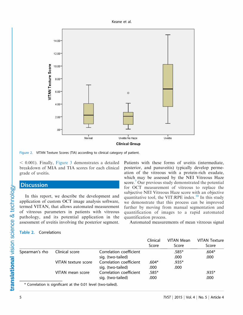