Embed Size (px)
Citation preview

Korean J Radiol 9(1), February 2008 87
Pulmonary Paraganglioma Manifesting asan Endobronchial Mass
Thoracic paragangliomas comprise only 1 2% of all paragangliomas, includ-ing the adrenal pheochromocytomas, and these tumors are mostly found in themediastinal compartments (1). To the best of our knowledge, there is only onecase report in the pathology literature of endobronchial involvement by a primarypulmonary paraganglioma (2). We report here on the CT and bronchoscopic find-ings of a case of pathologically proven endobronchial paraganglioma in a 37-year-old woman. In our case, bronchoscopy and CT demonstrated an endo-bronchial hypervascular mass, which indicated the presence of carcinoid orhypervascular metastasis based on the known incidence of such tumors.
he term “paraganglioma” is a generic term that’s applied to tumorsarising from the paraganglia, regardless of their location. This tumor canbe found in any part of the body where healthy paraganglia are known to
occur. However, the radiologic manifestations of endobronchial paragangliomas havenot been well documented on account of the tumor’s rarity (3 5). We present here avery rare case of primary endobronchial paraganglioma.
CASE REPORT
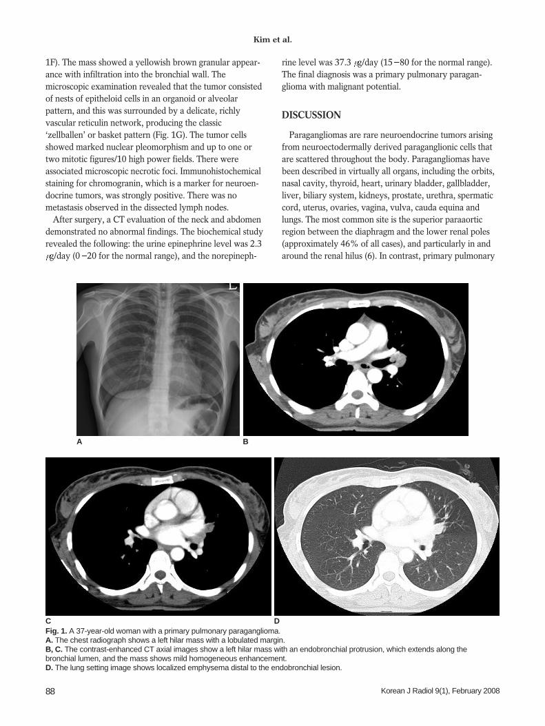
A 37-year-old woman presented with a 3-month history of exertional dyspnea,cough and one recent episode of hemoptysis. At the time of admission, she showed nohypertension or systemic symptoms. A chest radiograph showed a left hilar mass witha lobulated margin (Fig. 1A). The axial and coronal CT scans were obtained with using16-channel multidetector CT after IV administration of contrast media. A mildlyenhancing left hilar mass with an endobronchial protrusion and an extension along theleft lingular segmental bronchus was noted (Figs. 1B, C). The lung setting imageshowed localized emphysema distal to the endobronchial lesion (Fig. 1D).
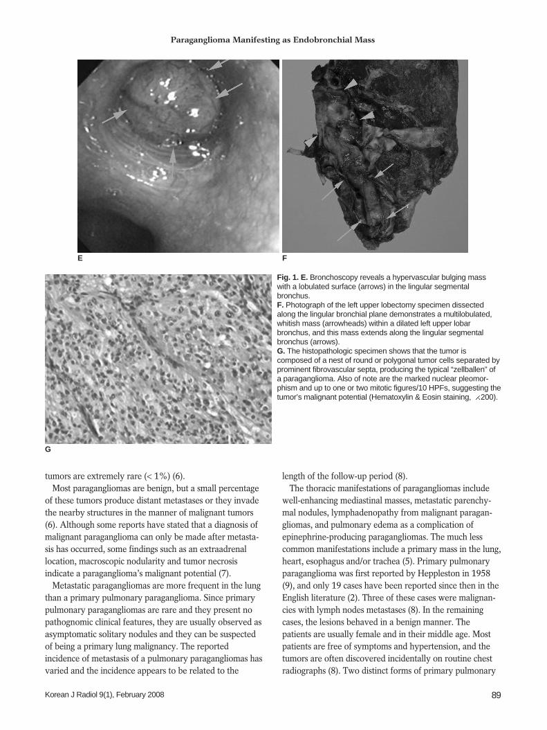
Bronchoscopy revealed a mutilobular, hypervasacular mass obstructing the lumen ofthe left lingular segmental bronchus (Fig. 1E). A careful biopsy was taken from theperipheral portion because the tumor tended to bleed. This tumor was believed to beeither an endobronchial bronchogenic carcinoma or another type of hypervasculartumor. The pathologic diagnosis was chronic inflammation with granulation tissue,which was not in accordance with the radiologic findings.
We subsequently performed a left upper sleeve lobectomy with dissection of themediastinal lymph nodes. The patient suffered no hypertensive crisis during or aftersurgery. The gross examination showed an endobronchially growing solid mass alongthe bronchial lumen, the so-called toothpaste figure, and this mass measured 7 3 cmin dimension. The remaining lung parenchyma showed no remarkable change (Fig.
Ki Nam Kim, MD1
Ki-Nam Lee, MD1
Mee Sook Roh, MD2
Pil Jo Choi, MD3
Doo Kyung Yang, MD4
Ki Nam Kim, MD1
Ki-Nam Lee, MD1
Mee Sook Roh, MD2
Pil Jo Choi, MD3
Doo Kyung Yang, MD4
Index terms:Lung neoplasms, CT Paraganglioma
DOI:10.3348/kjr.2008.9.1.87
Korean J Radiol 2008;9:87-90Received October 16, 2006; accepted after revision January 4, 2007.
1Department of Radiology, 2Department ofPathology, 3Department of Thoracic andCardiovascular Surgery, and 4InternalMedicine, College of Medicine, Dong-AUniversity, Pusan 602-103, Korea
Address reprint requests to:Ki-Nam Lee, MD, Department ofRadiology, College of Medicine, Dong-AUniversity, 3-ga, Dongdaesin-dong, Seo-gu, Pusan 602-103, Korea.Tel. (8251) 240-5367Fax. (8251) 253-4931e-mail: [email protected]
T
1F). The mass showed a yellowish brown granular appear-ance with infiltration into the bronchial wall. Themicroscopic examination revealed that the tumor consistedof nests of epitheloid cells in an organoid or alveolarpattern, and this was surrounded by a delicate, richlyvascular reticulin network, producing the classic‘zellballen’ or basket pattern (Fig. 1G). The tumor cellsshowed marked nuclear pleomorphism and up to one ortwo mitotic figures/10 high power fields. There wereassociated microscopic necrotic foci. Immunohistochemicalstaining for chromogranin, which is a marker for neuroen-docrine tumors, was strongly positive. There was nometastasis observed in the dissected lymph nodes.
After surgery, a CT evaluation of the neck and abdomendemonstrated no abnormal findings. The biochemical studyrevealed the following: the urine epinephrine level was 2.3g/day (0 20 for the normal range), and the norepineph-
rine level was 37.3 g/day (15 80 for the normal range).The final diagnosis was a primary pulmonary paragan-glioma with malignant potential.
DISCUSSION
Paragangliomas are rare neuroendocrine tumors arisingfrom neuroectodermally derived paraganglionic cells thatare scattered throughout the body. Paragangliomas havebeen described in virtually all organs, including the orbits,nasal cavity, thyroid, heart, urinary bladder, gallbladder,liver, biliary system, kidneys, prostate, urethra, spermaticcord, uterus, ovaries, vagina, vulva, cauda equina andlungs. The most common site is the superior paraaorticregion between the diaphragm and the lower renal poles(approximately 46% of all cases), and particularly in andaround the renal hilus (6). In contrast, primary pulmonary
Kim et al.
88 Korean J Radiol 9(1), February 2008
Fig. 1. A 37-year-old woman with a primary pulmonary paraganglioma.A. The chest radiograph shows a left hilar mass with a lobulated margin.B, C. The contrast-enhanced CT axial images show a left hilar mass with an endobronchial protrusion, which extends along thebronchial lumen, and the mass shows mild homogeneous enhancement. D. The lung setting image shows localized emphysema distal to the endobronchial lesion.
C D
A B
tumors are extremely rare (< 1%) (6).Most paragangliomas are benign, but a small percentage
of these tumors produce distant metastases or they invadethe nearby structures in the manner of malignant tumors(6). Although some reports have stated that a diagnosis ofmalignant paraganglioma can only be made after metasta-sis has occurred, some findings such as an extraadrenallocation, macroscopic nodularity and tumor necrosisindicate a paraganglioma’s malignant potential (7).
Metastatic paragangliomas are more frequent in the lungthan a primary pulmonary paraganglioma. Since primarypulmonary paragangliomas are rare and they present nopathognomic clinical features, they are usually observed asasymptomatic solitary nodules and they can be suspectedof being a primary lung malignancy. The reportedincidence of metastasis of a pulmonary paragangliomas hasvaried and the incidence appears to be related to the
length of the follow-up period (8).The thoracic manifestations of paragangliomas include
well-enhancing mediastinal masses, metastatic parenchy-mal nodules, lymphadenopathy from malignant paragan-gliomas, and pulmonary edema as a complication ofepinephrine-producing paragangliomas. The much lesscommon manifestations include a primary mass in the lung,heart, esophagus and/or trachea (5). Primary pulmonaryparaganglioma was first reported by Heppleston in 1958(9), and only 19 cases have been reported since then in theEnglish literature (2). Three of these cases were malignan-cies with lymph nodes metastases (8). In the remainingcases, the lesions behaved in a benign manner. Thepatients are usually female and in their middle age. Mostpatients are free of symptoms and hypertension, and thetumors are often discovered incidentally on routine chestradiographs (8). Two distinct forms of primary pulmonary
Paraganglioma Manifesting as Endobronchial Mass
Korean J Radiol 9(1), February 2008 89
E F
Fig. 1. E. Bronchoscopy reveals a hypervascular bulging masswith a lobulated surface (arrows) in the lingular segmentalbronchus.F. Photograph of the left upper lobectomy specimen dissectedalong the lingular bronchial plane demonstrates a multilobulated,whitish mass (arrowheads) within a dilated left upper lobarbronchus, and this mass extends along the lingular segmentalbronchus (arrows). G. The histopathologic specimen shows that the tumor iscomposed of a nest of round or polygonal tumor cells separated byprominent fibrovascular septa, producing the typical “zellballen” ofa paraganglioma. Also of note are the marked nuclear pleomor-phism and up to one or two mitotic figures/10 HPFs, suggesting thetumor’s malignant potential (Hematoxylin & Eosin staining, 200).
G

paragangliomas have been reported (4, 8). The first andmore common form consists of multiple minute tumors, inproximity to the pulmonary veins. The second and lesscommon form consists of large solid tumors. The reportedincidence of malignancy for a pulmonary paraganglioma isapproximately 18%, which is lower than that for paragan-glioma in other locations (20 50%). However, there arereports of metastases after a long time (10). Therefore, alifetime of follow-up with careful, long-term observationby checking the urinary catecholamines and performingimaging studies is essential.
While pulmonary involvement by an adrenal or extraa-drenal pheochromocytoma is uncommon, endobronchialinvolvement like that seen in our case is very rare. Onlyone case of hilar and subcarinal lymphadenopathies withendobronchial metastases from recurrent adrenalpheochromocytoma and another case of a 0.9 cm primaryendobronchial paraganglioma of the lung have beenreported (2, 5).
Functioning extra-adrenal paragangliomas representmore than 10% of all pheochromocytomas (4). Among allthe reported cases of pulmonary paraganglioma, two casesdeveloped hypertension and the patients died from acardiac disorder that was believed to be associated withthe functional tumors. There was a report of a case offunctioning metastases of a nonfunctioning paraganglioma(10). They assumed that a hypothetical phenotypic hetero-geneity of the primary tumor could explain the differencein the biological behavior of the primary tumor and itsmetastases (10).
The endobronchial paraganglioma in our patient
manifested on CT as a hypervascular endobronchial mass,and this manifestation was similar to that of anendobronchial carcinoid or bronchogenic carcinoma.Therefore, we think paraganglioma should be consideredwhen making the differential diagnosis of an enhancingendobronchial mass.
References1. Aravot DJ, Banner NR, Cantor AM, Theodoropoulos S, Yacoub
MH. Location, localization and surgical treatment of cardiacpheochromocytoma. Am J Cardiol 1992;69:283-285
2. Aubertine CL, Flieder DB. Primary paraganglioma of the lung.Ann Diagn Pathol 2004;8:237-241
3. Rosai J. Ackerman’s surgical pathology, 8th ed. New York:Mosby, 1996:1015-1058
4. Saeki T, Akiba T, Joh K, Inoue K, Doi N, Kanai M, et al. Anextremely large solitary primary paraganglioma of the lung:report of a case. Surg Today 1999;29:1195-1200
5. Sandur S, Dasgupta A, Shapiro JL, Arroliga AC, Mehta AC.Thoracic involvement with pheochromocytoma: a review. Chest1999;115:511-521
6. Whalen RK, Althausen AF, Daniels GH. Extra-adrenalpheochromocytoma. J Urol 1992;147:1-10
7. Linnoila RI, Keiser HR, Steinberg SM, Lack EE. Histopathologyof benign versus malignant sympathoadrenal paragangliomas:clinicopathologic study of 120 cases including unusual histologicfeatures. Hum Pathol 1990;21:1168-1180
8. Lemonick DM, Pai PB, Hines GL. Malignant primarypulmonary paragagnlioma with hilar metastasis. J ThoracCardiovasc Surg 1990;99:563-564
9. Hangartner JR, Loosemore TM, Burke M, Pepper JR. Malignantprimary pulmonary paraganglioma. Thorax 1989;44:154-156
10. Fenandez-Llamazares J, Sabria-Leal M, Armengol-Carrasco M,Garsia-Bonafe M, Salca-Lacombe JA. Functioning metastases ofa nonfunctioning paraganglioma. J Surg Oncol 1988;37:213-214
Kim et al.
90 Korean J Radiol 9(1), February 2008