Embed Size (px)
Citation preview
Pulmonary Bacteriophage Therapy on Pseudomonasaeruginosa Cystic Fibrosis Strains: First Steps TowardsTreatment and PreventionEric Morello1, Emilie Saussereau1, Damien Maura1, Michel Huerre2, Lhousseine Touqui3,4, Laurent
Debarbieux1*
1 Molecular Biology of the Gene in Extremophiles Unit, Department of Microbiology, Institut Pasteur, Paris, France, 2 Histotechnology and Pathology Unit, Department of
Infection and Epidemiology, Institut Pasteur, Paris, France, 3 Innate Host Defense and Inflammation Unit, Department of Infection and Epidemiology, Institut Pasteur, Paris,
France, 4 INSERM, U874, Paris, France
Abstract
Multidrug-resistant bacteria are the cause of an increasing number of deadly pulmonary infections. Because there iscurrently a paucity of novel antibiotics, phage therapy—the use of specific viruses that infect bacteria—is now morefrequently being considered as a potential treatment for bacterial infections. Using a mouse lung-infection model caused bya multidrug resistant Pseudomonas aeruginosa mucoid strain isolated from a cystic fibrosis patient, we evaluatedbacteriophage treatments. New bacteriophages were isolated from environmental samples and characterized. Bacteria andbacteriophages were applied intranasally to the immunocompetent mice. Survival was monitored and bronchoalveolarfluids were analysed. Quantification of bacteria, bacteriophages, pro-inflammatory and cytotoxicity markers, as well ashistology and immunohistochemistry analyses were performed. A curative treatment (one single dose) administrated 2 hafter the onset of the infection allowed over 95% survival. A four-day preventive treatment (one single dose) resulted in a100% survival. All of the parameters measured correlated with the efficacy of both curative and preventive bacteriophagetreatments. We also showed that in vitro optimization of a bacteriophage towards a clinical strain improved both its efficacyon in vivo treatments and its host range on a panel of 20 P. aeruginosa cystic fibrosis strains. This work provides an incentiveto develop clinical studies on pulmonary bacteriophage therapy to combat multidrug-resistant lung infections.
Citation: Morello E, Saussereau E, Maura D, Huerre M, Touqui L, et al. (2011) Pulmonary Bacteriophage Therapy on Pseudomonas aeruginosa Cystic FibrosisStrains: First Steps Towards Treatment and Prevention. PLoS ONE 6(2): e16963. doi:10.1371/journal.pone.0016963
Editor: Ramy Aziz, Cairo University, Egypt
Received November 8, 2010; Accepted January 15, 2011; Published February 15, 2011
Copyright: � 2011 Morello et al. This is an open-access article distributed under the terms of the Creative Commons Attribution License, which permitsunrestricted use, distribution, and reproduction in any medium, provided the original author and source are credited.
Funding: Funding was provided by Institut Pasteur PTR Nu255, Vaincre la Mucoviscidose grant IC0704. The funders had no role in study design, data collectionand analysis, decision to publish, or preparation of the manuscript.
Competing Interests: The authors have declared that no competing interests exist.
* E-mail: [email protected]
Introduction
Pseudomonas aeruginosa is the second most common pathogen
responsible for hospital-acquired bacterial pneumonia as well as
ventilator-associated pneumonia, and the first causative agent of
morbidity and mortality in cystic fibrosis (CF) patients [1,2].
Although antibiotics are still an effective means of treating
bacterial lung infections, the alarming rise of multidrug-resistant
bacteria in hospitals has highlighted the need for new therapies
[3,4,5]. Bacteriophages —viruses infecting bacteria— have been
proposed to treat human bacterial infections since their discovery
in the early 20th century [6,7]. However, after a short period of
development, the advent of antibiotics led to this therapeutic
approach being abandoned, except in Eastern Europe where
bacteriophages are still used today to treat patients [8,9,10].
During the past 20 years, studies in animal models have
demonstrated the potential of bacteriophages [11,12,13,14,15].
Recently the first phase II clinical trial performed under European
regulations on bacteriophage treatments of chronic otitis was
published, and demonstrated the interest of using bacteriophages
on multidrug resistant infections [16]. The effects of bacteriophage
therapy on lung infections has only very recently been addressed in
animal models [14,17]. On the one hand, a proof of concept with
a bioluminescent strain of P. aeruginosa showed that bacteriophages
administrated intranasally had a rapid efficacy with respect to
preventing and curing deadly lung infections [14]. On the other
hand, a clinical strain of Burkholderia cenocepacia isolated from a CF
patient was used to show that the intraperitoneal administration of
bacteriophages was more effective than intranasal applications in a
non-deadly infectious model [17]. Here we report an in-depth
evaluation of the efficacy of curative and preventive bacteriophage
treatments of lung infections using a multidrug resistant mucoid P.
aeruginosa strain isolated from a CF patient of Grenoble hospital,
France [18,19]. For this study we specifically optimized the
virulence of a bacteriophage of our collection towards this clinical
strain and studied its efficacy both in vitro and in vivo.
Results
Characterization of a lung infection by a P. aeruginosastrain isolated from a cystic fibrosis patient
To investigate bacteriophage treatments on a mouse lung-
infection caused by a clinical P. aeruginosa strain named CHA
[18,19], we first established the conditions in which an intranasal
administration of this strain was lethal. We found that inoculation
of 36106 bacteria was sufficient to induce death in 100% of
PLoS ONE | www.plosone.org 1 February 2011 | Volume 6 | Issue 2 | e16963
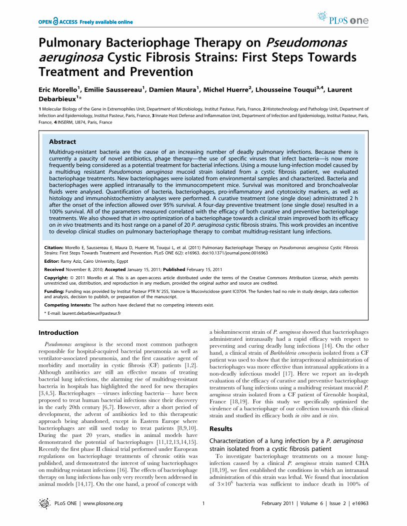
animals within 2 days (Figure 1A). Progress of the infection was
assessed by quantification of bacteria, inflammatory markers, and
cytotoxicity levels at 20 h post-infection (Figures 1 and 2). The
number of bacteria in the lungs had increased at least by two
orders of magnitude compared with the initial infectious dose (over
46108 cfu were found in the broncho-alveolar lavages [BAL] form
each infected mouse compared with the infectious dose of
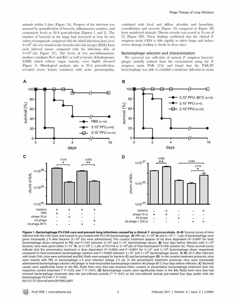
36106 cfu; Figure 1C). The levels of two pro-inflammatory
markers (cytokines IL-6 and KC) as well as lactate dehydrogenase
(LDH) which reflects organ toxicity, were highly elevated
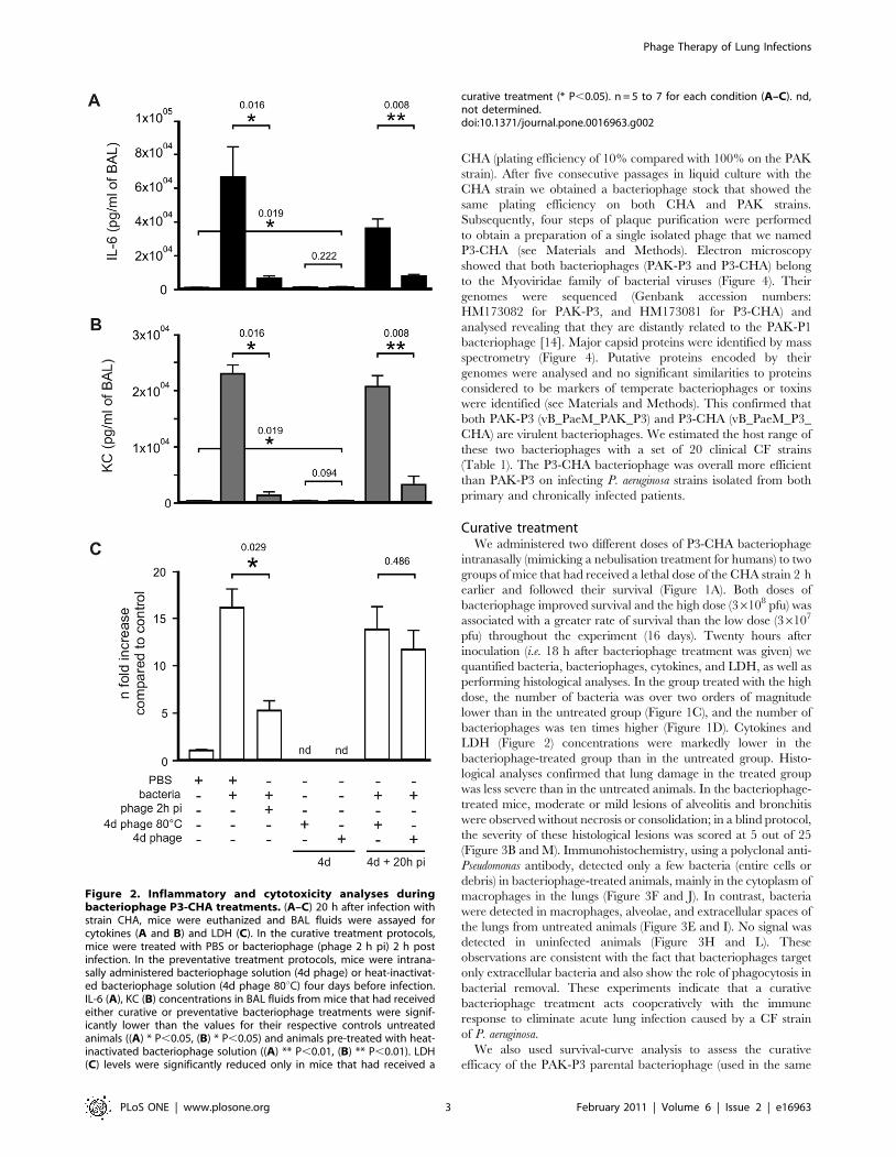
(Figure 2). Histological analysis, also at 20 h post-infection,
revealed severe lesions consistent with acute pneumopathy,
combined with focal and diffuse alveolitis and bronchitis,
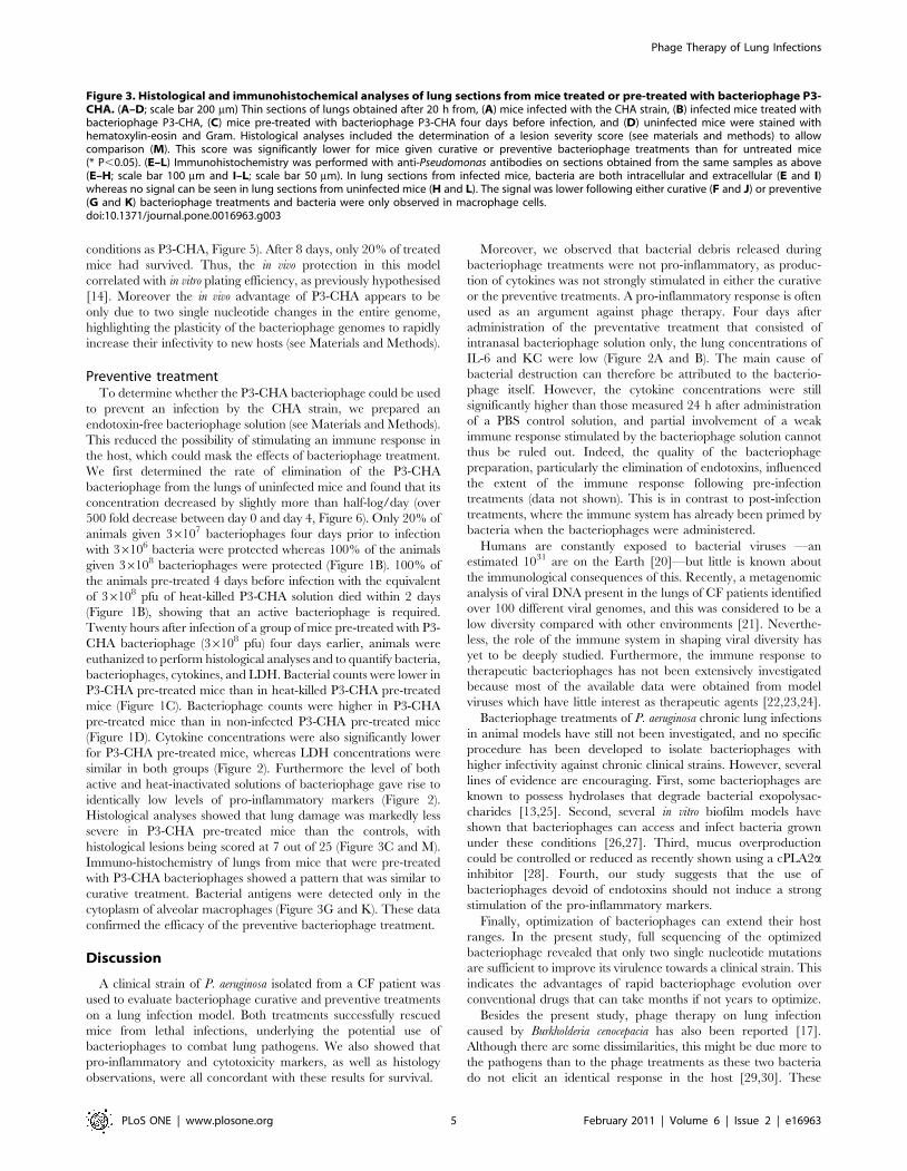
consolidation and necrosis (Figure 3A compared to Figure 3D
from uninfected animals). Disease severity was scored at 16 out of
25 (Figure 3M). These findings confirmed that the clinical P.
aeruginosa strain CHA is able rapidly to infect lungs and induce
severe damage leading to death in these mice.
Bacteriophage selection and characterizationWe screened our collection of natural P. aeruginosa bacterio-
phages (initially isolated from the environment using the P.
aeruginosa strain PAK [14]) and found that the PAK-P3
bacteriophage was able to establish a moderate infection in strain
Figure 1. Bacteriophage P3-CHA cure and prevent lung infections caused by a clinical P. aeruginosa strain. (A–B) Survival curves of miceinfected with the CHA strain and treated or pre-treated with P3-CHA bacteriophage. (A) PBS (¤), 36106 (N) and 36107 (%) pfu of bacteriophage weregiven intranasally 2 h after bacteria (36106 cfu) were administered. This curative treatment appears to be dose dependent (P,0.0001 for bothbacteriophage doses compared to PBS and P,0.01 between 36106 and 36107 bacteriophage doses). (B) Four days before infection with 36106
bacteria, mice were given either 36107 (N), or 36108 (%) pfu of P3-CHA or 36108 pfu of heat-inactivated P3-CHA solution (¤). These survival curvesindicate that the preventative treatment is dose dependent (P,0.0005 and P,0.0001 for 36107 and 36108 bacteriophage doses respectivelycompared to heat-inactivated bacteriophage solution and P,0.0005 between 36107 and 36108 bacteriophage doses). (C–D) 20 h after infectionwith strain CHA, mice were euthanized and BAL fluids were assayed for bacteria (C) and bacteriophages (D). In the curative treatment protocols, micewere treated with PBS or bacteriophage 2 h post infection (phage 2 h pi). In the preventative treatment protocols, mice were intranasallyadministered bacteriophage solution (4d phage) or heat-inactivated bacteriophage solution (4d phage 80uC) four days before infection. (C) Bacterialcounts were significantly lower in the BAL fluids from mice that had received either curative or preventative bacteriophage treatment than therespective control treatment (* P,0.05, and ** P,0.01). (D) Bacteriophage counts were significantly lower in the BAL fluids from mice that hadreceived bacteriophage treatment than the non-infected animals (** P,0.01) or the non-infected animals pre-treated four days earlier with thebacteriophage P3-CHA (* P,0.05).doi:10.1371/journal.pone.0016963.g001
Phage Therapy of Lung Infections
PLoS ONE | www.plosone.org 2 February 2011 | Volume 6 | Issue 2 | e16963
CHA (plating efficiency of 10% compared with 100% on the PAK
strain). After five consecutive passages in liquid culture with the
CHA strain we obtained a bacteriophage stock that showed the
same plating efficiency on both CHA and PAK strains.
Subsequently, four steps of plaque purification were performed
to obtain a preparation of a single isolated phage that we named
P3-CHA (see Materials and Methods). Electron microscopy
showed that both bacteriophages (PAK-P3 and P3-CHA) belong
to the Myoviridae family of bacterial viruses (Figure 4). Their
genomes were sequenced (Genbank accession numbers:
HM173082 for PAK-P3, and HM173081 for P3-CHA) and
analysed revealing that they are distantly related to the PAK-P1
bacteriophage [14]. Major capsid proteins were identified by mass
spectrometry (Figure 4). Putative proteins encoded by their
genomes were analysed and no significant similarities to proteins
considered to be markers of temperate bacteriophages or toxins
were identified (see Materials and Methods). This confirmed that
both PAK-P3 (vB_PaeM_PAK_P3) and P3-CHA (vB_PaeM_P3_
CHA) are virulent bacteriophages. We estimated the host range of
these two bacteriophages with a set of 20 clinical CF strains
(Table 1). The P3-CHA bacteriophage was overall more efficient
than PAK-P3 on infecting P. aeruginosa strains isolated from both
primary and chronically infected patients.
Curative treatmentWe administered two different doses of P3-CHA bacteriophage
intranasally (mimicking a nebulisation treatment for humans) to two
groups of mice that had received a lethal dose of the CHA strain 2 h
earlier and followed their survival (Figure 1A). Both doses of
bacteriophage improved survival and the high dose (36108 pfu) was
associated with a greater rate of survival than the low dose (36107
pfu) throughout the experiment (16 days). Twenty hours after
inoculation (i.e. 18 h after bacteriophage treatment was given) we
quantified bacteria, bacteriophages, cytokines, and LDH, as well as
performing histological analyses. In the group treated with the high
dose, the number of bacteria was over two orders of magnitude
lower than in the untreated group (Figure 1C), and the number of
bacteriophages was ten times higher (Figure 1D). Cytokines and
LDH (Figure 2) concentrations were markedly lower in the
bacteriophage-treated group than in the untreated group. Histo-
logical analyses confirmed that lung damage in the treated group
was less severe than in the untreated animals. In the bacteriophage-
treated mice, moderate or mild lesions of alveolitis and bronchitis
were observed without necrosis or consolidation; in a blind protocol,
the severity of these histological lesions was scored at 5 out of 25
(Figure 3B and M). Immunohistochemistry, using a polyclonal anti-
Pseudomonas antibody, detected only a few bacteria (entire cells or
debris) in bacteriophage-treated animals, mainly in the cytoplasm of
macrophages in the lungs (Figure 3F and J). In contrast, bacteria
were detected in macrophages, alveolae, and extracellular spaces of
the lungs from untreated animals (Figure 3E and I). No signal was
detected in uninfected animals (Figure 3H and L). These
observations are consistent with the fact that bacteriophages target
only extracellular bacteria and also show the role of phagocytosis in
bacterial removal. These experiments indicate that a curative
bacteriophage treatment acts cooperatively with the immune
response to eliminate acute lung infection caused by a CF strain
of P. aeruginosa.
We also used survival-curve analysis to assess the curative
efficacy of the PAK-P3 parental bacteriophage (used in the same
Figure 2. Inflammatory and cytotoxicity analyses duringbacteriophage P3-CHA treatments. (A–C) 20 h after infection withstrain CHA, mice were euthanized and BAL fluids were assayed forcytokines (A and B) and LDH (C). In the curative treatment protocols,mice were treated with PBS or bacteriophage (phage 2 h pi) 2 h postinfection. In the preventative treatment protocols, mice were intrana-sally administered bacteriophage solution (4d phage) or heat-inactivat-ed bacteriophage solution (4d phage 80uC) four days before infection.IL-6 (A), KC (B) concentrations in BAL fluids from mice that had receivedeither curative or preventative bacteriophage treatments were signif-icantly lower than the values for their respective controls untreatedanimals ((A) * P,0.05, (B) * P,0.05) and animals pre-treated with heat-inactivated bacteriophage solution ((A) ** P,0.01, (B) ** P,0.01). LDH(C) levels were significantly reduced only in mice that had received a
curative treatment (* P,0.05). n = 5 to 7 for each condition (A–C). nd,not determined.doi:10.1371/journal.pone.0016963.g002
Phage Therapy of Lung Infections
PLoS ONE | www.plosone.org 3 February 2011 | Volume 6 | Issue 2 | e16963
Phage Therapy of Lung Infections
PLoS ONE | www.plosone.org 4 February 2011 | Volume 6 | Issue 2 | e16963
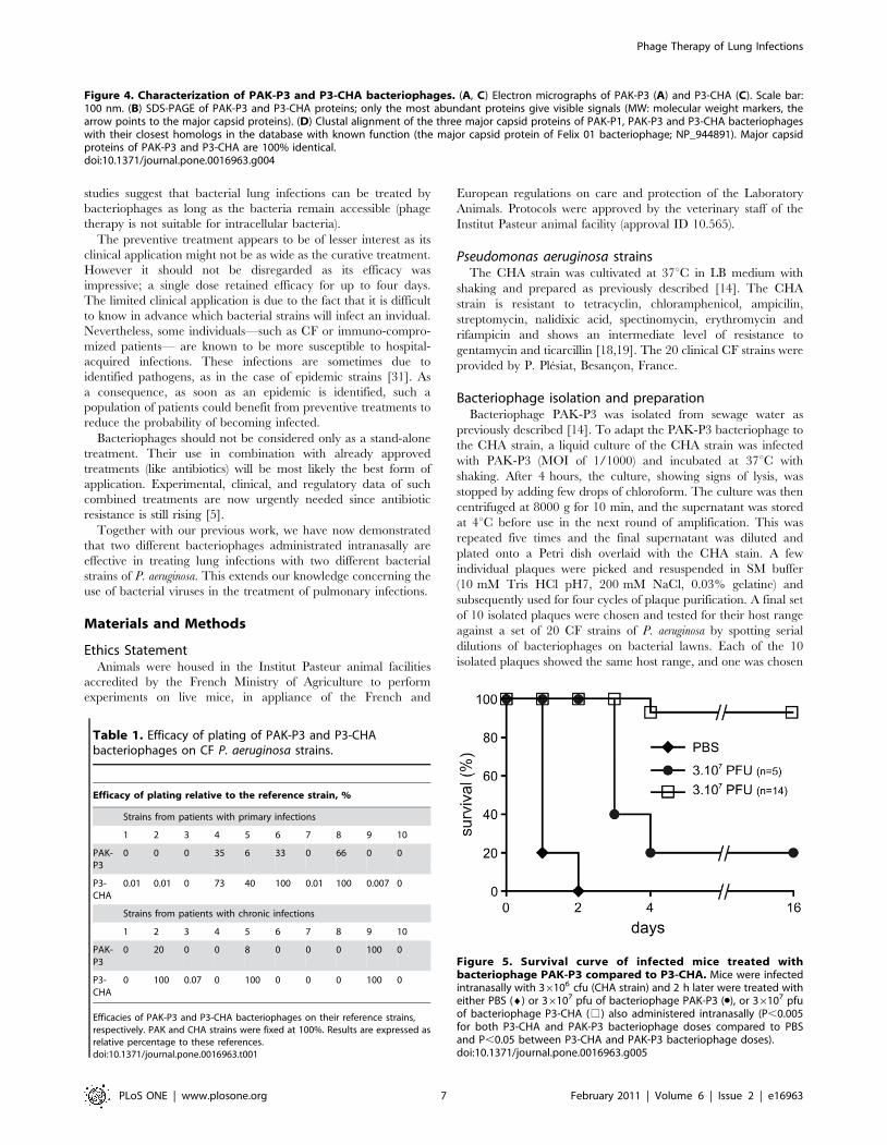
conditions as P3-CHA, Figure 5). After 8 days, only 20% of treated
mice had survived. Thus, the in vivo protection in this model
correlated with in vitro plating efficiency, as previously hypothesised
[14]. Moreover the in vivo advantage of P3-CHA appears to be
only due to two single nucleotide changes in the entire genome,
highlighting the plasticity of the bacteriophage genomes to rapidly
increase their infectivity to new hosts (see Materials and Methods).
Preventive treatmentTo determine whether the P3-CHA bacteriophage could be used
to prevent an infection by the CHA strain, we prepared an
endotoxin-free bacteriophage solution (see Materials and Methods).
This reduced the possibility of stimulating an immune response in
the host, which could mask the effects of bacteriophage treatment.
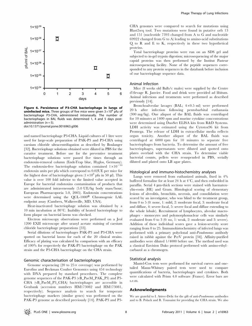
We first determined the rate of elimination of the P3-CHA
bacteriophage from the lungs of uninfected mice and found that its
concentration decreased by slightly more than half-log/day (over
500 fold decrease between day 0 and day 4, Figure 6). Only 20% of
animals given 36107 bacteriophages four days prior to infection
with 36106 bacteria were protected whereas 100% of the animals
given 36108 bacteriophages were protected (Figure 1B). 100% of
the animals pre-treated 4 days before infection with the equivalent
of 36108 pfu of heat-killed P3-CHA solution died within 2 days
(Figure 1B), showing that an active bacteriophage is required.
Twenty hours after infection of a group of mice pre-treated with P3-
CHA bacteriophage (36108 pfu) four days earlier, animals were
euthanized to perform histological analyses and to quantify bacteria,
bacteriophages, cytokines, and LDH. Bacterial counts were lower in
P3-CHA pre-treated mice than in heat-killed P3-CHA pre-treated
mice (Figure 1C). Bacteriophage counts were higher in P3-CHA
pre-treated mice than in non-infected P3-CHA pre-treated mice
(Figure 1D). Cytokine concentrations were also significantly lower
for P3-CHA pre-treated mice, whereas LDH concentrations were
similar in both groups (Figure 2). Furthermore the level of both
active and heat-inactivated solutions of bacteriophage gave rise to
identically low levels of pro-inflammatory markers (Figure 2).
Histological analyses showed that lung damage was markedly less
severe in P3-CHA pre-treated mice than the controls, with
histological lesions being scored at 7 out of 25 (Figure 3C and M).
Immuno-histochemistry of lungs from mice that were pre-treated
with P3-CHA bacteriophages showed a pattern that was similar to
curative treatment. Bacterial antigens were detected only in the
cytoplasm of alveolar macrophages (Figure 3G and K). These data
confirmed the efficacy of the preventive bacteriophage treatment.
Discussion
A clinical strain of P. aeruginosa isolated from a CF patient was
used to evaluate bacteriophage curative and preventive treatments
on a lung infection model. Both treatments successfully rescued
mice from lethal infections, underlying the potential use of
bacteriophages to combat lung pathogens. We also showed that
pro-inflammatory and cytotoxicity markers, as well as histology
observations, were all concordant with these results for survival.
Moreover, we observed that bacterial debris released during
bacteriophage treatments were not pro-inflammatory, as produc-
tion of cytokines was not strongly stimulated in either the curative
or the preventive treatments. A pro-inflammatory response is often
used as an argument against phage therapy. Four days after
administration of the preventative treatment that consisted of
intranasal bacteriophage solution only, the lung concentrations of
IL-6 and KC were low (Figure 2A and B). The main cause of
bacterial destruction can therefore be attributed to the bacterio-
phage itself. However, the cytokine concentrations were still
significantly higher than those measured 24 h after administration
of a PBS control solution, and partial involvement of a weak
immune response stimulated by the bacteriophage solution cannot
thus be ruled out. Indeed, the quality of the bacteriophage
preparation, particularly the elimination of endotoxins, influenced
the extent of the immune response following pre-infection
treatments (data not shown). This is in contrast to post-infection
treatments, where the immune system has already been primed by
bacteria when the bacteriophages were administered.
Humans are constantly exposed to bacterial viruses —an
estimated 1031 are on the Earth [20]—but little is known about
the immunological consequences of this. Recently, a metagenomic
analysis of viral DNA present in the lungs of CF patients identified
over 100 different viral genomes, and this was considered to be a
low diversity compared with other environments [21]. Neverthe-
less, the role of the immune system in shaping viral diversity has
yet to be deeply studied. Furthermore, the immune response to
therapeutic bacteriophages has not been extensively investigated
because most of the available data were obtained from model
viruses which have little interest as therapeutic agents [22,23,24].
Bacteriophage treatments of P. aeruginosa chronic lung infections
in animal models have still not been investigated, and no specific
procedure has been developed to isolate bacteriophages with
higher infectivity against chronic clinical strains. However, several
lines of evidence are encouraging. First, some bacteriophages are
known to possess hydrolases that degrade bacterial exopolysac-
charides [13,25]. Second, several in vitro biofilm models have
shown that bacteriophages can access and infect bacteria grown
under these conditions [26,27]. Third, mucus overproduction
could be controlled or reduced as recently shown using a cPLA2ainhibitor [28]. Fourth, our study suggests that the use of
bacteriophages devoid of endotoxins should not induce a strong
stimulation of the pro-inflammatory markers.
Finally, optimization of bacteriophages can extend their host
ranges. In the present study, full sequencing of the optimized
bacteriophage revealed that only two single nucleotide mutations
are sufficient to improve its virulence towards a clinical strain. This
indicates the advantages of rapid bacteriophage evolution over
conventional drugs that can take months if not years to optimize.
Besides the present study, phage therapy on lung infection
caused by Burkholderia cenocepacia has also been reported [17].
Although there are some dissimilarities, this might be due more to
the pathogens than to the phage treatments as these two bacteria
do not elicit an identical response in the host [29,30]. These
Figure 3. Histological and immunohistochemical analyses of lung sections from mice treated or pre-treated with bacteriophage P3-CHA. (A–D; scale bar 200 mm) Thin sections of lungs obtained after 20 h from, (A) mice infected with the CHA strain, (B) infected mice treated withbacteriophage P3-CHA, (C) mice pre-treated with bacteriophage P3-CHA four days before infection, and (D) uninfected mice were stained withhematoxylin-eosin and Gram. Histological analyses included the determination of a lesion severity score (see materials and methods) to allowcomparison (M). This score was significantly lower for mice given curative or preventive bacteriophage treatments than for untreated mice(* P,0.05). (E–L) Immunohistochemistry was performed with anti-Pseudomonas antibodies on sections obtained from the same samples as above(E–H; scale bar 100 mm and I–L; scale bar 50 mm). In lung sections from infected mice, bacteria are both intracellular and extracellular (E and I)whereas no signal can be seen in lung sections from uninfected mice (H and L). The signal was lower following either curative (F and J) or preventive(G and K) bacteriophage treatments and bacteria were only observed in macrophage cells.doi:10.1371/journal.pone.0016963.g003
Phage Therapy of Lung Infections
PLoS ONE | www.plosone.org 5 February 2011 | Volume 6 | Issue 2 | e16963
Phage Therapy of Lung Infections
PLoS ONE | www.plosone.org 6 February 2011 | Volume 6 | Issue 2 | e16963
studies suggest that bacterial lung infections can be treated by
bacteriophages as long as the bacteria remain accessible (phage
therapy is not suitable for intracellular bacteria).
The preventive treatment appears to be of lesser interest as its
clinical application might not be as wide as the curative treatment.
However it should not be disregarded as its efficacy was
impressive; a single dose retained efficacy for up to four days.
The limited clinical application is due to the fact that it is difficult
to know in advance which bacterial strains will infect an invidual.
Nevertheless, some individuals—such as CF or immuno-compro-
mized patients— are known to be more susceptible to hospital-
acquired infections. These infections are sometimes due to
identified pathogens, as in the case of epidemic strains [31]. As
a consequence, as soon as an epidemic is identified, such a
population of patients could benefit from preventive treatments to
reduce the probability of becoming infected.
Bacteriophages should not be considered only as a stand-alone
treatment. Their use in combination with already approved
treatments (like antibiotics) will be most likely the best form of
application. Experimental, clinical, and regulatory data of such
combined treatments are now urgently needed since antibiotic
resistance is still rising [5].
Together with our previous work, we have now demonstrated
that two different bacteriophages administrated intranasally are
effective in treating lung infections with two different bacterial
strains of P. aeruginosa. This extends our knowledge concerning the
use of bacterial viruses in the treatment of pulmonary infections.
Materials and Methods
Ethics StatementAnimals were housed in the Institut Pasteur animal facilities
accredited by the French Ministry of Agriculture to perform
experiments on live mice, in appliance of the French and
European regulations on care and protection of the Laboratory
Animals. Protocols were approved by the veterinary staff of the
Institut Pasteur animal facility (approval ID 10.565).
Pseudomonas aeruginosa strainsThe CHA strain was cultivated at 37uC in LB medium with
shaking and prepared as previously described [14]. The CHA
strain is resistant to tetracyclin, chloramphenicol, ampicilin,
streptomycin, nalidixic acid, spectinomycin, erythromycin and
rifampicin and shows an intermediate level of resistance to
gentamycin and ticarcillin [18,19]. The 20 clinical CF strains were
provided by P. Plesiat, Besancon, France.
Bacteriophage isolation and preparationBacteriophage PAK-P3 was isolated from sewage water as
previously described [14]. To adapt the PAK-P3 bacteriophage to
the CHA strain, a liquid culture of the CHA strain was infected
with PAK-P3 (MOI of 1/1000) and incubated at 37uC with
shaking. After 4 hours, the culture, showing signs of lysis, was
stopped by adding few drops of chloroform. The culture was then
centrifuged at 8000 g for 10 min, and the supernatant was stored
at 4uC before use in the next round of amplification. This was
repeated five times and the final supernatant was diluted and
plated onto a Petri dish overlaid with the CHA stain. A few
individual plaques were picked and resuspended in SM buffer
(10 mM Tris HCl pH7, 200 mM NaCl, 0.03% gelatine) and
subsequently used for four cycles of plaque purification. A final set
of 10 isolated plaques were chosen and tested for their host range
against a set of 20 CF strains of P. aeruginosa by spotting serial
dilutions of bacteriophages on bacterial lawns. Each of the 10
isolated plaques showed the same host range, and one was chosen
Figure 4. Characterization of PAK-P3 and P3-CHA bacteriophages. (A, C) Electron micrographs of PAK-P3 (A) and P3-CHA (C). Scale bar:100 nm. (B) SDS-PAGE of PAK-P3 and P3-CHA proteins; only the most abundant proteins give visible signals (MW: molecular weight markers, thearrow points to the major capsid proteins). (D) Clustal alignment of the three major capsid proteins of PAK-P1, PAK-P3 and P3-CHA bacteriophageswith their closest homologs in the database with known function (the major capsid protein of Felix 01 bacteriophage; NP_944891). Major capsidproteins of PAK-P3 and P3-CHA are 100% identical.doi:10.1371/journal.pone.0016963.g004
Table 1. Efficacy of plating of PAK-P3 and P3-CHAbacteriophages on CF P. aeruginosa strains.
Efficacy of plating relative to the reference strain, %
Strains from patients with primary infections
1 2 3 4 5 6 7 8 9 10
PAK-P3
0 0 0 35 6 33 0 66 0 0
P3-CHA
0.01 0.01 0 73 40 100 0.01 100 0.007 0
Strains from patients with chronic infections
1 2 3 4 5 6 7 8 9 10
PAK-P3
0 20 0 0 8 0 0 0 100 0
P3-CHA
0 100 0.07 0 100 0 0 0 100 0
Efficacies of PAK-P3 and P3-CHA bacteriophages on their reference strains,respectively. PAK and CHA strains were fixed at 100%. Results are expressed asrelative percentage to these references.doi:10.1371/journal.pone.0016963.t001
Figure 5. Survival curve of infected mice treated withbacteriophage PAK-P3 compared to P3-CHA. Mice were infectedintranasally with 36106 cfu (CHA strain) and 2 h later were treated witheither PBS (¤) or 36107 pfu of bacteriophage PAK-P3 (N), or 36107 pfuof bacteriophage P3-CHA (%) also administered intranasally (P,0.005for both P3-CHA and PAK-P3 bacteriophage doses compared to PBSand P,0.05 between P3-CHA and PAK-P3 bacteriophage doses).doi:10.1371/journal.pone.0016963.g005
Phage Therapy of Lung Infections
PLoS ONE | www.plosone.org 7 February 2011 | Volume 6 | Issue 2 | e16963
and named bacteriophage P3-CHA. Liquid cultures of 1 litre were
used for large-scale preparation of PAK-P3 and P3-CHA using
caesium chloride ultracentrifugation as described by Boulanger
[32]. Bacteriophage solutions obtained were diluted in PBS for the
curative treatment. Before use for the preventive treatment
bacteriophage solutions were passed five times through an
endotoxin-removal column (EndoTrap blue, Hyglos, Germany).
The endotoxin-free bacteriophage solution contained 1610210
endotoxin units per pfu which correspond to 0.02UE per mice for
the highest dose of bacteriophage given (16108 pfu in 50 ml). This
value is over 100 fold inferior to the limited value accepted in
Europe for bacterial endotoxins contamination of products that
are administrated intraveneously (5.0 UE/kg body mass/hour;
European Pharmacopoeia 5.0, 2005). Endotoxin concentrations
were determined using the QCL-1000H Chromogenic LAL
endpoint assay (Cambrex, Walkersville, MD, USA).
Heat-inactivated bacteriophage solution was obtained by a
10 min incubation at 80uC. Inability of heated bacteriophage to
form plaque on bacterial lawns was checked.
Electron microscopy observations were performed on a Jeol
1200 EXII microscope after uranyl acetate staining of caesium
chloride bacteriophage preparations [33].
Serial dilutions of bacteriophages PAK-P3 and P3-CHA were
spotted on bacterial lawns for each of the 20 clinical strains.
Efficacy of plating was calculated by comparison with an efficacy
of 100% for respectively the PAK-P3 bacteriophage on the PAK
strain and the P3-CHA bacteriophage on the CHA strain.
Genomic characterisation of bacteriophagesGenome sequencing (20 to 256 coverage) was performed by
Eurofins and Beckman Coulter Genomics using 454 technology
with DNA prepared by standard procedures. The complete
genome sequences of the PAK-P3 (vB_PaeM_PAK_P3) and P3-
CHA (vB_PaeM_P3_CHA) bacteriophages are accessible in
Genbank (accession numbers HM173082 and HM173081,
respectively). Sequence analysis to search for temperate
bacteriophage markers (similar genes) was performed on the
PAK-P3 genome as described previously [14]. PAK-P3 and P3-
CHA genomes were compared to search for mutations using
Blast2seq tool. Two mutations were found in putative orfs 13
and 151 (nucleotide 7393 changed from A to G and nucleotide
69922 changed from G to A) leading to amino-acid substitutions
Q to R and E to K, respectively in these two hypothetical
proteins.
Total bacteriophage proteins were run on an SDS gel and
subjected to in-gel trypsin digestion; microsequencing of the major
capsid proteins was then performed by the Institut Pasteur
microsequencing facility. None of the peptide sequences corre-
sponded to any protein sequences in the databank before inclusion
of our bacteriophage sequence data.
Animal InfectionMice (8 weeks old Balb/c males) were supplied by the Centre
d’elevage R. Janvier. Food and drink were provided ad libitum.
Animal infections and treatments were performed as described
previously [14].
Bronchoalveolar lavages (BAL; 460.5 ml) were performed
20 h after infection following pentobarbital euthanasia
(300 mg/kg). One aliquot of the BAL fluids was centrifuged
for 10 minutes at 1400 rpm and murine cytokine concentrations
were determined using DuoSet ELISA kits from R&D Systems.
LDH activity was estimated using the Cytotox96 kit from
Promega. The release of LDH in extracellular media reflects
organ toxicity. Another aliquot of the BAL fluids was
centrifuged at 6000 rpm for 10 minutes to separate free
bacteriophages from bacteria. To determine the amount of free
bacteriophages, supernatants were diluted and spotted onto
plates overlaid with the CHA strain. To determine viable
bacterial counts, pellets were resuspended in PBS, serially
diluted and plated onto LB agar plates.
Histological and immuno-histochemistry analysesLungs were removed from euthanized animals, fixed in 4%
buffered formaline for at least 24 h at 4uC, and then embedded in
paraffin. Serial 4 mm-thick sections were stained with haematox-
ylin-eosin (HE) and Gram. Histological scoring of elementary
lesions of alveolitis, bronchitis, necrosis, and consolidation were
scored by an investigator, who was blind to the treatment group,
from 0 to 5 (0: none, 1: mild, 2: moderate focal, 3: moderate focal
and diffuse, 4: severe focal, 5: severe focal and diffuse extending to
the whole lobule). Recruitment of lymphocytes, alveolar macro-
phages - monocytes and polymorphonuclear cells was similarly
evaluated from 0 to 3 (0: no, 1: weak, 2: moderate and 3: severe).
Addition of these individual scores gave a lesion-severity score
ranging from 0 to 25. Immunohistochemistry of infected lungs was
performed with a primary polyclonal anti-Pseudomonas antibody
raised in rabbit against the PcrV protein [34]. Affinity-purified
antibodies were diluted 1/4000 before use. The method used was
a classical Envision Dako protocol performed with amino-ethyl–
carbazol as a chromogen.
Statistical analysisMantel-Cox tests were performed for survival curves and one-
tailed Mann-Whitney paired tests were used to compare
quantifications of bacteria, bacteriophages and cytokines. Both
were calculated with Prism 4.0 software (France). Error bars are
s.e.m.
Acknowledgments
We are grateful to I. Attree-Delic for the gift of anti-Pseudomonas antibodies
and to B. Polack and B. Toussaint for providing the CHA strain. We also
Figure 6. Persistence of P3-CHA bacteriophage in lungs ofuninfected mice. Three groups of five mice were given 36108 pfu ofbacteriophage P3-CHA, administered intranasally. The number ofbacteriophages in BAL fluids was determined 1, 4 and 5 days post-administration (n = 5).doi:10.1371/journal.pone.0016963.g006
Phage Therapy of Lung Infections
PLoS ONE | www.plosone.org 8 February 2011 | Volume 6 | Issue 2 | e16963
acknowledge the help of A. Criscuolo for bioinformatic analyses and D.
Leduc for cytokine and LDH assays. We thank the BMGE team for its
support and are grateful to the staff of the animal facility of Institut Pasteur.
Author Contributions
Conceived and designed the experiments: LT LD. Performed the
experiments: EM ES DM MH. Analyzed the data: MH LT LD. Wrote
the paper: LD.
References
1. Jones RN (2010) Microbial etiologies of hospital-acquired bacterial pneumoniaand ventilator-associated bacterial pneumonia. Clin Infect Dis 51(Suppl 1):
S81–87.
2. Gomez MI, Prince A (2007) Opportunistic infections in lung disease:Pseudomonas infections in cystic fibrosis. Curr Opin Pharmacol 7: 244–251.
3. Nordmann P, Naas T, Fortineau N, Poirel L (2007) Superbugs in the comingnew decade; multidrug resistance and prospects for treatment of Staphylococcus
aureus, Enterococcus spp. and Pseudomonas aeruginosa in 2010. Curr Opin
Microbiol 10: 436–440.4. Gould IM (2008) The epidemiology of antibiotic resistance. Int J Antimicrob
Agents 32(Suppl 1): S2–9.5. Kumarasamy KK, Toleman MA, Walsh TR, Bagaria J, Butt F, et al. (2010)
Emergence of a new antibiotic resistance mechanism in India, Pakistan, and theUK: a molecular, biological, and epidemiological study. Lancet Infect Dis 10:
597–602.
6. Merril CR, Scholl D, Adhya SL (2003) The prospect for bacteriophage therapyin Western medicine. Nat Rev Drug Discov 2: 489–497.
7. Summers WC (2001) Bacteriophage therapy. Annu Rev Microbiol 55: 437–451.8. Kutateladze M, Adamia R (2008) Phage therapy experience at the Eliava
Institute. Med Mal Infect 38: 426–430.
9. Gorski A, Miedzybrodzki R, Borysowski J, Weber-Dabrowska B, Lobocka M,et al. (2009) Bacteriophage therapy for the treatment of infections. Curr Opin
Investig Drugs 10: 766–774.10. Kutateladze M, Adamia R (2010) Bacteriophages as potential new therapeutics
to replace or supplement antibiotics. Trends Biotechnol.
11. O’Flaherty S, Ross RP, Coffey A (2009) Bacteriophage and their lysins forelimination of infectious bacteria. FEMS Microbiol Rev 33: 801–819.
12. Sulakvelidze A, Kutter E (2005) Bacteriophage therapy in humans. In: Kutter E,Sulakvelidze A, eds. Bacteriophages: biology and applications. Boca Raton, FL:
CRC Press. pp 381–436.13. Donlan RM (2009) Preventing biofilms of clinically relevant organisms using
bacteriophage. Trends Microbiol 17: 66–72.
14. Debarbieux L, Leduc D, Maura D, Morello E, Criscuolo A, et al. (2010)Bacteriophages can treat and prevent Pseudomonas aeruginosa lung infections.
J Infect Dis 201: 1096–1104.15. Smith HW, Huggins MB (1983) Effectiveness of phages in treating experimental
Escherichia coli diarrhoea in calves, piglets and lambs. J Gen Microbiol 129:
2659–2675.16. Wright A, Hawkins CH, Anggard EE, Harper DR (2009) A controlled clinical
trial of a therapeutic bacteriophage preparation in chronic otitis due toantibiotic-resistant Pseudomonas aeruginosa; a preliminary report of efficacy.
Clin Otolaryngol 34: 349–357.17. Carmody LA, Gill JJ, Summer EJ, Sajjan US, Gonzalez CF, et al. (2009)
Efficacy of bacteriophage therapy in a model of Burkholderia cenocepacia
pulmonary infection. J Infect Dis 2010: 264–271.18. Dacheux D, Attree I, Schneider C, Toussaint B (1999) Cell death of human
polymorphonuclear neutrophils induced by a Pseudomonas aeruginosa cysticfibrosis isolate requires a functional type III secretion system. Infect Immun 67:
6164–6167.
19. Delic-Attree I, Toussaint B, Froger A, Willison JC, Vignais PM (1996) Isolation
of an IHF-deficient mutant of a Pseudomonas aeruginosa mucoid isolate and
evaluation of the role of IHF in algD gene expression. Microbiology 142 (Pt 10):
2785–2793.
20. Bergh O, Borsheim KY, Bratbak G, Heldal M (1989) High abundance of viruses
found in aquatic environments. Nature 340: 467–468.
21. Willner D, Furlan M, Haynes M, Schmieder R, Angly FE, et al. (2009)
Metagenomic analysis of respiratory tract DNA viral communities in cystic
fibrosis and non-cystic fibrosis individuals. PLoS One 4: e7370.
22. Clark JR, March JB (2004) Bacterial viruses as human vaccines? Expert Rev
Vaccines 3: 463–476.
23. Kurzepa A, Dabrowska K, Skaradzinski G, Gorski A (2009) Bacteriophage
interactions with phagocytes and their potential significance in experimental
therapy. Clin Exp Med 9: 93–100.
24. Miedzybrodzki R, Fortuna W, Weber-Dabrowska B, Gorski A (2005) Bacterial
viruses against viruses pathogenic for man? Virus Res 110: 1–8.
25. Glonti T, Chanishvili N, Taylor PW (2010) Bacteriophage-derived enzyme that
depolymerizes the alginic acid capsule associated with cystic fibrosis isolates of
Pseudomonas aeruginosa. J Appl Microbiol 108: 695–702.
26. Fu W, Forster T, Mayer O, Curtin JJ, Lehman SM, et al. (2010) Bacteriophage
cocktail for the prevention of biofilm formation by Pseudomonas aeruginosa on
catheters in an in vitro model system. Antimicrob Agents Chemother 54:
397–404.
27. Knezevic P, Petrovic O (2008) A colorimetric microtiter plate method for
assessment of phage effect on Pseudomonas aeruginosa biofilm. J Microbiol
Methods 74: 114–118.
28. Dif F, Wu YZ, Burgel PR, Ollero M, Leduc D, et al. (2010) Critical role of
cytosolic phospholipase a2 in bronchial mucus hyper-secretion in CFTR-
deficient mice. Eur Respir J.
29. Ramphal R, Balloy V, Huerre M, Si-Tahar M, Chignard M (2005) TLRs 2 and
4 are not involved in hypersusceptibility to acute Pseudomonas aeruginosa lung
infections. J Immunol 175: 3927–3934.
30. Ventura GM, Balloy V, Ramphal R, Khun H, Huerre M, et al. (2009) Lack of
MyD88 protects the immunodeficient host against fatal lung inflammation
triggered by the opportunistic bacteria Burkholderia cenocepacia. J Immunol
183: 670–676.
31. Armstrong D, Bell S, Robinson M, Bye P, Rose B, et al. (2003) Evidence for
spread of a clonal strain of Pseudomonas aeruginosa among cystic fibrosis clinics.
J Clin Microbiol 41: 2266–2267.
32. Boulanger P (2009) Purification of Bacteriophages and SDS-PAGE Analysis of
Phage Structural Proteins from Ghost Particles. Methods Mol Biol 502:
227–238.
33. Ackermann HW (2009) Basic phage electron microscopy. Methods Mol Biol
501: 113–126.
34. Goure J, Pastor A, Faudry E, Chabert J, Dessen A, et al. (2004) The V antigen of
Pseudomonas aeruginosa is required for assembly of the functional PopB/PopD
translocation pore in host cell membranes. Infect Immun 72: 4741–4750.
Phage Therapy of Lung Infections
PLoS ONE | www.plosone.org 9 February 2011 | Volume 6 | Issue 2 | e16963