Embed Size (px)
Citation preview
To cite this article: Neuroendocrinol Lett 2017; 38(6):101–107
CA
SE
R
EP
OR
TNeuroendocrinology Letters Volume 38 No. 6 2017
ISSN: 0172-780X; ISSN-L: 0172-780X; Electronic/Online ISSN: 2354-4716Web of Knowledge / Web of Science: Neuroendocrinol Lett
Pub Med / Medline: Neuro Endocrinol Lett
Complete objective response, stable for 5 years, with the Di Bella Method, of multiple-metastatic carcinoma of the breast Giuseppe Di Bella 1, Biagio Colori 2, Rosilde Toscano 1
1 Di Bella Foundation, Via Guglielmo Marconi 51 Bologna, 40122 Italy2 Rizzoli Scientific Research and Care Institute, Via Giulio Cesare Pupilli, 40136 Bologna, Italy
Correspondence to: Dr. Giuseppe Di BellaDi Bella FoundationVia Marconi 51, Post code 40122, Bologna, Italy.tel: +39 051 239662; +39 051 230369; e-mail: [email protected]
Submitted: 2017-06-24 Accepted: 2017-08-28 Published online: 2017-00-00
Key words: breast cancer; somatostatin; octreotide; melatonin; retinoids; vitamin D3; vitamin E; Di Bella method; D2R agonists; estrogenic deprivation
neuroendocrinol Lett 2017; 38(6):101–107 PMID: ----- NEL380617AXX © 2017 Neuroendocrinology Letters • www.nel.edu
Abstract Breast cancer is the most common cancer and the leading cause of cancer death among women. Despite all efforts, about 11,939 deaths and 50,000 new diagnoses for breast cancer were estimated among Italian women in 2016. Therefore new approaches are needed to improve the survival and higher remission rates. We present a case of a woman with carcinoma of the breast and multiple metastases after right mastectomy, axillary dissection, repeated cycles of chemo and radio-therapy, and estrogen block. Biological method formulated by Prof. L. Di Bella (DBM) produced a complete and stable objective response without toxicity. The DBM includes antiproliferative molecules, such as somatostatin, prolactin and estrogen inhibitors together with differentiating and apoptotic molecules such as melatonin (MLT), Retinoids, Vitamin E, D3, Vit. C, Calcium, Amino sugars, associated with metronomic microdoses of chemotherapy drugs. The blood tests did not show any damage but a progressive reduction of Prolactin, Estradiol, and IGF1, and continuing low levels of GH. The objective result of this case, in the absence of toxicity, demonstrates the efficacy of the treatment and is in agreement with the positive results already published on the use of the DBM. Not requiring hospital or day hospital admission, and with no significant toxicity, the DBM avoided the significant side effects of chemo- and radiotherapy. We believe that this case can encourage more interest and more in-depth studies on the possibili-ties that have been opened up in oncology by the DBM treatment of the metastatic breast cancer.
INTRODUCTION
The estimate in Italy dating year 2016, shows 50,000 new cases of breast cancer confirming that this type of tumour is the most diagnosed in woman. In 2013, the breast cancer represented the first absolute cause of death in all the world
for tumour in woman, with 11,939 deaths (Fonte ISTAT 2016). Breast cancer features 29% of death cause before 50 years old, 21 % between 50 and 69 and 16 % after 70 years old. The survival at 5 years is 85.5% in Italy, little bit more than the percentage in Europe ( 84.7%). (I numeri del cancro in Italia, 2016)
102 Copyright © 2017 Neuroendocrinology Letters ISSN 0172–780X • www.nel.edu
Giuseppe Di Bella, Biagio Colori, Rosilde Toscano
Current approaches could be different and depend from the type of cancer. The procedure includes conser-vative or radical surgery (mastectomy) associated with radiotherapy and/or chemotherapy, which also have variations according to the type and stage of the cancer (I numeri del cancro in Italia, 2016).
CASE REPORT
We present 35-year-old woman with carcinoma of the breast detected during pregnancy, terminated at the 34th week of gestation by Cesarean section due to the diagnosis of the breast carcinoma. After the needle biopsy which showed an infiltrating carcinoma (G2), the patient underwent right mastectomy with axillary dissection and plastic reconstruction on 03.12.2009. Histological examination showed: “Ductal carcinoma with an intermediate degree of differentiation, solid type intraductal focal component and widespread peritumoral vascular invasion. Tumor-free mammary parenchyma, with gravidic type modifications. Duct intraepithelial neoplasia areas (DIN2) with extension to the retroareo-lar ducts and extending close to the retroareolar margin. Lymph node: peri lymph node metastases of carcinoma with two sentinel lymph nodes and one II level lymph node. Histiocytosis of the breasts in the remaining twenty-four lymph nodes examined”, and pT2 staging (2.5cm) pN1a(3/27) MX G2. Extensive vascular invasion; ER 90%, PgR 10%, Ki67 15%”, c-erbB2: weak complete in 70%.”
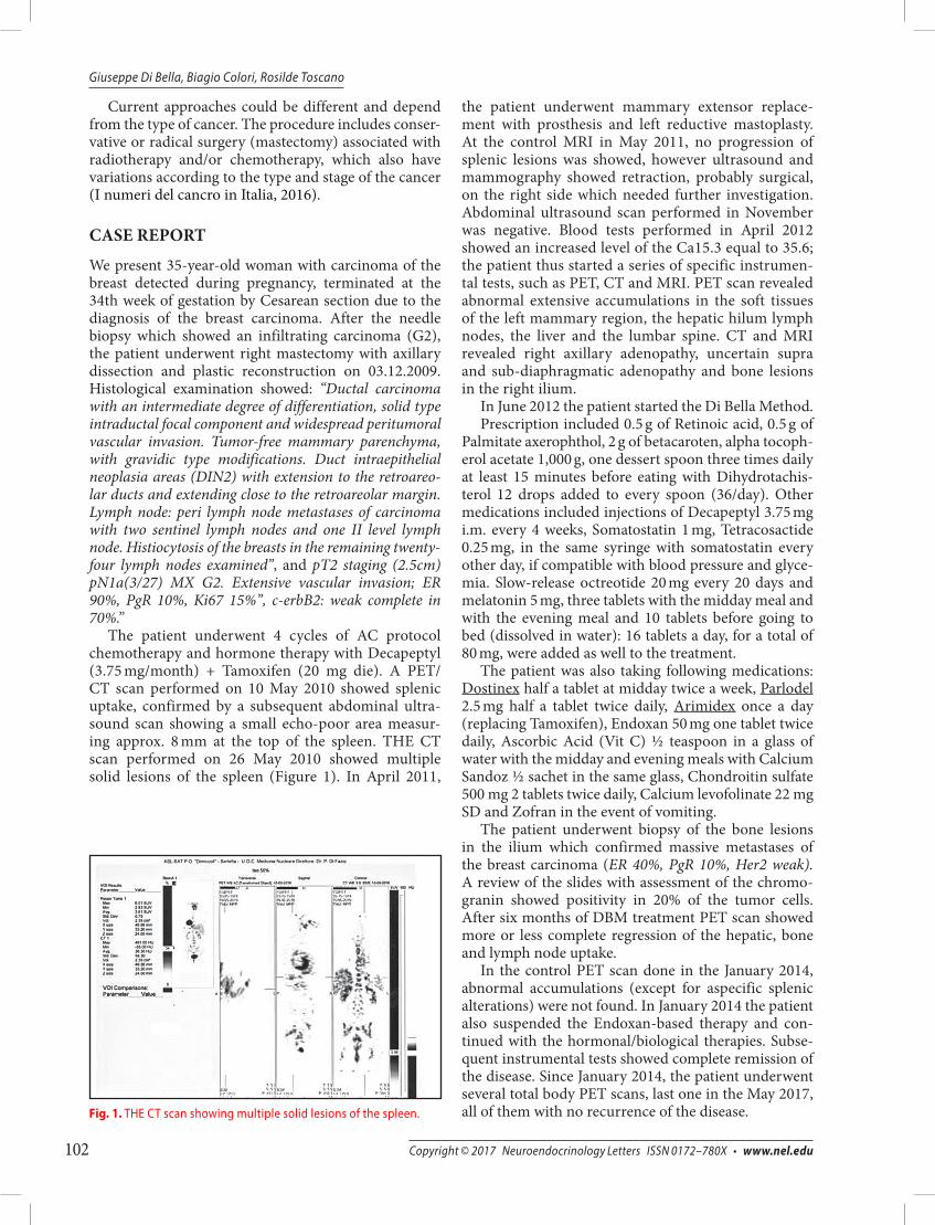
The patient underwent 4 cycles of AC protocol chemotherapy and hormone therapy with Decapeptyl (3.75 mg/month) + Tamoxifen (20 mg die). A PET/CT scan performed on 10 May 2010 showed splenic uptake, confirmed by a subsequent abdominal ultra-sound scan showing a small echo-poor area measur-ing approx. 8 mm at the top of the spleen. THE CT scan performed on 26 May 2010 showed multiple solid lesions of the spleen (Figure 1). In April 2011,
the patient underwent mammary extensor replace-ment with prosthesis and left reductive mastoplasty. At the control MRI in May 2011, no progression of splenic lesions was showed, however ultrasound and mammography showed retraction, probably surgical, on the right side which needed further investigation. Abdominal ultrasound scan performed in November was negative. Blood tests performed in April 2012 showed an increased level of the Ca15.3 equal to 35.6; the patient thus started a series of specific instrumen-tal tests, such as PET, CT and MRI. PET scan revealed abnormal extensive accumulations in the soft tissues of the left mammary region, the hepatic hilum lymph nodes, the liver and the lumbar spine. CT and MRI revealed right axillary adenopathy, uncertain supra and sub-diaphragmatic adenopathy and bone lesions in the right ilium.
In June 2012 the patient started the Di Bella Method.Prescription included 0.5 g of Retinoic acid, 0.5 g of
Palmitate axerophthol, 2 g of betacaroten, alpha tocoph-erol acetate 1,000 g, one dessert spoon three times daily at least 15 minutes before eating with Dihydrotachis-terol 12 drops added to every spoon (36/day). Other medications included injections of Decapeptyl 3.75 mg i.m. every 4 weeks, Somatostatin 1 mg, Tetracosactide 0.25 mg, in the same syringe with somatostatin every other day, if compatible with blood pressure and glyce-mia. Slow-release octreotide 20 mg every 20 days and melatonin 5 mg, three tablets with the midday meal and with the evening meal and 10 tablets before going to bed (dissolved in water): 16 tablets a day, for a total of 80 mg, were added as well to the treatment.
The patient was also taking following medications: Dostinex half a tablet at midday twice a week, Parlodel 2.5 mg half a tablet twice daily, Arimidex once a day (replacing Tamoxifen), Endoxan 50 mg one tablet twice daily, Ascorbic Acid (Vit C) ½ teaspoon in a glass of water with the midday and evening meals with Calcium Sandoz ½ sachet in the same glass, Chondroitin sulfate 500 mg 2 tablets twice daily, Calcium levofolinate 22 mg SD and Zofran in the event of vomiting.
The patient underwent biopsy of the bone lesions in the ilium which confirmed massive metastases of the breast carcinoma (ER 40%, PgR 10%, Her2 weak). A review of the slides with assessment of the chromo-granin showed positivity in 20% of the tumor cells. After six months of DBM treatment PET scan showed more or less complete regression of the hepatic, bone and lymph node uptake.
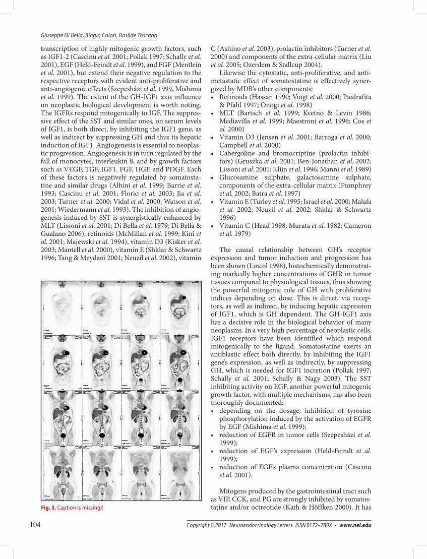
In the control PET scan done in the January 2014, abnormal accumulations (except for aspecific splenic alterations) were not found. In January 2014 the patient also suspended the Endoxan-based therapy and con-tinued with the hormonal/biological therapies. Subse-quent instrumental tests showed complete remission of the disease. Since January 2014, the patient underwent several total body PET scans, last one in the May 2017, all of them with no recurrence of the disease.Fig. 1. THE CT scan showing multiple solid lesions of the spleen.

103Neuroendocrinology Letters Vol. 38 No. 5 2017 • Article available online: http://node.nel.edu
Di Bella method
THE THERAPY AND THE CLINICAL COURSE
In June 2012, due to recurrence of the disease and having refused a proposed second cycle of chemother-apy, the patient asked to be treated with the Di Bella Method, which consisted of the synergic use of mol-ecules with differentiating, cytostatic, apoptotic, and antiproliferative activity, with an increase in immuni-tary activity. Due to the myeloprotective, antidegenera-tive and trophic action on the parenchyma and tissues, above all of MLT and the high doses of Vitamin E, retinoids, vitamins C and D3, the continuous adminis-tration for approx. 6 months of 100 milligrams of cyclo-phosphamide per day, together with all the components of the DBM, did not cause any medullary, hepatorenal, metabolic, cardiocirculatory or neurological toxicity or immunitary depression. It did not lead to any signifi-cant alteration in blood values and marrow dynamics. The Di Bella therapy includes Endoxan (cyclophos-phamide) at doses varying from 50 to 100 mg a day for apoptotic purposes, not cytolytic as in oncological pro-tocols. Comparing the 100 mg/day cyclophosphamide of the DBM with the intravenous 10 to 12 g in the pre-transplant therapies of lymphoproliferative diseases gives a ratio of 1 to 100. 100 mg, instead of 10 g, does
not have a cytolytic or cytotoxic effect, but apoptotic. In the time of six months, having the treatment at home, the patient achieved partial remission, afterwards com-plete remission, allowing her to return to work.
DISCUSSION RATIONALE OF THE THERAPY
The loss of differentiation and proliferation, even if to different extents, are common denominators of all neo-plasms. The ubiquitous receptor expression of prolactin (Ben-Jonathan et al. 2002; Hooghe et al. 1998) and GH (Lincoln et al. 1998; De Souza et al. 1974) are one of the confirmations of the direct and generalized mitogenic role of this molecule. Cellular proliferation is highly dependent on prolactin and GH, both powerful growth factors, and on GH dependent mitogenic molecules which are positively regulated by it, such as EGF, FGF, HGF, IGF1-2, NGF, PDGF, VEGF, and TGF in addition to growth factors produced by the gastrointestinal tract, such as VIP, CCK, and PG. Both physiological as well as neoplastic cellular proliferation take place by means of these same molecules, which the neoplastic cells use to an exponential extent compared to healthy ones. Biological antidotes of GH such as somatostatine and similar compounds, reduce not only the expression and
Fig. 2. Caption is missing!!
104 Copyright © 2017 Neuroendocrinology Letters ISSN 0172–780X • www.nel.edu
Giuseppe Di Bella, Biagio Colori, Rosilde Toscano
transcription of highly mitogenic growth factors, such as IGF1-2 (Cascinu et al. 2001; Pollak 1997; Schally et al. 2001), EGF (Held-Feindt et al. 1999), and FGF (Mentlein et al. 2001), but extend their negative regulation to the respective receptors with evident anti-proliferative and anti-angiogenic effects (Szepesházi et al. 1999, Mishima et al. 1999). The extent of the GH-IGF1 axis influence on neoplastic biological development is worth noting. The IGFRs respond mitogenically to IGF. The suppres-sive effect of the SST and similar ones, on serum levels of IGF1, is both direct, by inhibiting the IGF1 gene, as well as indirect by suppressing GH and thus its hepatic induction of IGF1. Angiogenesis is essential to neoplas-tic progression. Angiogenesis is in turn regulated by the fall of monocytes, interleukin 8, and by growth factors such as VEGF, TGF, IGF1, FGF, HGF, and PDGF. Each of these factors is negatively regulated by somatosta-tine and similar drugs (Albini et al. 1999; Barrie et al. 1993; Cascinu et al. 2001; Florio et al. 2003; Jia et al. 2003; Turner et al. 2000; Vidal et al. 2000; Watson et al. 2001; Wiedermann et al. 1993). The inhibition of angio-genesis induced by SST is synergistically enhanced by MLT (Lissoni et al. 2001; Di Bella et al. 1979; Di Bella & Gualano 2006), retinoids (McMillan et al. 1999; Kini et al. 2001; Majewski et al. 1994), vitamin D3 (Kisker et al. 2003; Mantell et al. 2000), vitamin E (Shklar & Schwartz 1996; Tang & Meydani 2001; Neuzil et al. 2002), vitamin
C (Ashino et al. 2003), prolactin inhibitors (Turner et al. 2000) and components of the extra-cellular matrix (Liu et al. 2005; Ozerdem & Stallcup 2004).
Likewise the cytostatic, anti-proliferative, and anti-metastatic effect of somatostatine is effectively syner-gized by MDB’s other components: • Retinoids (Hassan 1990; Voigt et al. 2000; Piedrafita
& Pfahl 1997; Onogi et al. 1998) • MLT (Bartsch et al. 1999; Kvetno & Levin 1986;
Mediavilla et al. 1999; Maestroni et al. 1996; Cos et al. 2000)
• Vitamin D3 (Jensen et al. 2001; Barroga et al. 2000; Campbell et al. 2000)
• Cabergoline and bromocriptine (prolactin inhibi-tors) (Gruszka et al. 2001; Ben-Jonathan et al. 2002; Lissoni et al. 2001; Klijn et al. 1996; Manni et al. 1989)
• Glucosamine sulphate, galactosamine sulphate, components of the extra-cellular matrix (Pumphrey et al. 2002; Batra et al. 1997)
• Vitamin E (Turley et al. 1995; Israel et al. 2000; Malafa et al. 2002; Neuzil et al. 2002; Shklar & Schwartz 1996)
• Vitamin C (Head 1998; Murata et al. 1982; Cameron et al. 1979)
The causal relationship between GH’s receptor expression and tumor induction and progression has been shown (Lincol 1998), histochemically demonstrat-ing markedly higher concentrations of GHR in tumor tissues compared to physiological tissues, thus showing the powerful mitogenic role of GH with proliferative indices depending on dose. This is direct, via recep-tors, as well as indirect, by inducing hepatic expression of IGF1, which is GH dependent. The GH-IGF1 axis has a decisive role in the biological behavior of many neoplasms. In a very high percentage of neoplastic cells, IGF1 receptors have been identified which respond mitogenically to the ligand. Somatostatine exerts an antiblastic effect both directly, by inhibiting the IGF1 gene’s expression, as well as indirectly, by suppressing GH, which is needed for IGF1 incretion (Pollak 1997; Schally et al. 2001; Schally & Nagy 2003). The SST inhibiting activity on EGF, another powerful mitogenic growth factor, with multiple mechanisms, has also been thoroughly documented: • depending on the dosage, inhibition of tyrosine
phosphorylation induced by the activation of EGFR by EGF (Mishima et al. 1999);
• reduction of EGFR in tumor cells (Szepesházi et al. 1999);
• reduction of EGF’s expression (Held-Feindt et al. 1999);
• reduction of EGF’s plasma concentration (Cascinu et al. 2001).
Mitogens produced by the gastrointestinal tract such as VIP, CCK, and PG are strongly inhibited by somatos-tatine and/or octreotide (Kath & Höffken 2000). It has Fig. 3. Caption is missing!!
105Neuroendocrinology Letters Vol. 38 No. 5 2017 • Article available online: http://node.nel.edu
Di Bella method
been shown that breast tumors express SSTR1, SSTR2, and SSTR3, and less frequently SSTR5 (Albérini et al. 2000; Schaer et al. 1997),
Complete objective response to biological therapy of plurifocal breast carcinoma which in at least 50% of cases is scintigraphically visible, while in over half of the negative scintigraphs histochemical examinations revealed the presence of SSTR. Therefore the presence of SSTR (Barnett 2003; Pinzani et al. 2001; van Eijck et al. 1998), and of neuro-endocrine receptors in a signifi-cant percentage of these carcinomas constitutes a fur-ther rational indication for using SST, which in any case has already been extensively justified by the above-cited negative effect of SST on GH, GH-correlated oncogenes and angiogenesis.
Angiogenesis and neoangiogenesis, necessary con-ditions for tumour progression, as well as the cascade of monocytes, the paracrine release of interleukin 8 (IL-8) and the contribution of GFs (whose synergism is essen-tial), are specific molecular targets negatively regulated by Somatostatin and its analogues (Ruscica et al. 2012). The inhibition of angiogenesis induced by SST is syn-ergically reinforced by MLT, Retinoids and vitamin D3 (Kim et al. 2013; Lissoni et al. 2001; Sogno et al. 2009; Picotto et al. 2012). Furthermore, the local conditions of hypoxia/ anoxia and acidosis promote angiogenesis, and are mostly corrected by the improvement in the bloodtissue exchanges induced by the differentiating components of the DBM. At the same time, the cyto-static, antiproliferative, and antimetastatic effects of Somatostatin are synergically increased by the other components of the DBM. An additional contribution is provided by the daily administration of low doses (50–100 mg/die per os) of Cyclophosphamide (Endoxan®). As well as drastically reducing the known anitblastic/ myelosuppressive effects, this dosage induces a marked turnaround of its mechanisms of action: triggering of the mitochondrial-dependent apoptotic cascade, anti-angiogenetic action by drastically down-regulating the VEGF gene expression (Loven et al. 2013). Numerous preclinical investigations have also demonstrated the mechanisms of action of MLT. The use of such indole extends to all histotypes of breast cancer due to its high membrane receptorial/ nuclear distribution (Oprea-Ilies et al. 2012; Rögelsperger et al. 2011). Since the molecule is associated with the signalling pathways of both the physiological and neoplastic epithelial devel-opment, this substance has the properties to selectively neutralize the proliferative signals of estrogens and negatively modulate their local biosynthesis (Hill et al. 2011; Girgert et al. 2009). The administration of low doses of second generation aromatase inhibitors (Anas-trozole©), already used in clinical practice, combined with MLT, SST and Retinoids, negatively regulates the hormone-dependent processes of proliferation of breast tumours (Alvarez-Garcia et al. 2013; Margheri et al. 2012; Wang et al. 2012; Knower et al. 2012; Ciolino et al. 2011).
The biological DBM therapy slowly and progres-sively achieved a complete objective response, without toxicity, through a receptorial, differentiating, apop-totic and antiproliferative mechanism of action, with criteria, aims and mechanisms of action totally differ-ing from the usual cytotoxic and cytolytic therapies.
The objective response to the DBM (Di Bella Method) extended to resolution of the hepatic, lesions, thoracic lymphadenopathies, axillary adenopathies, supra- and sub-diaphragmatic adenopathies, and bone and splenic lesions which were no longer detected. Thanks to the progressive reduction, and complete elimination, of the metastatic lesions, the objective non-toxic result shows the efficacy and tolerability of this treatment.
Competing Interest. All the authors declare that they have no competing interest.Autor’s Contribution. GDB were responsible for concep-tion and interpretation for the provision of study mate-rial and manuscript writing. RT and BC responsible for conception. All authors read and approved the final manuscript
REFERENCES
1 Albérini JL, Meunier B, Denzler B, Devillers A, Tass P, Dazord L, et al. (2000). Somatostatin receptor in breast cancer and axillary nodes: study with scintigraphy, histopathology and receptor autoradiography. Breast Cancer Res Treat. 61(1): 21–32.
2 Albini A, Florio T, Giunciuglio D, Masiello L, Carlone S, Corsaro A, et al. (1999). Somatostatin controls Kaposi©s sarcoma tumor growth through inhibition of angiogenesis. FASEB J. 13(6): 647–655.
3 Alvarez-García V, González A, Martínez-Campa C, Alonso- González C and Cos S (2013). Melatonin modulates aromatase activity and expression in endothelial cells. Oncol Rep. 29(5). 2058–64.
4 Ashino H, Shimamura M, Nakajima H, Dombou M, Kawanaka S, Oikawa T, et al. (2003). Novel function of ascorbic acid as an angiostatic factor. Angiogenesis. 6(4): 259–269.
5 Associazione Italiana di Oncologia Medica, Airtum. “I numeri del cancro in Italia 2016” [(The numbers of cancer in Italy 2016) (in Italian)] . pp. 75–82.
6 Barnett P (2003). Somatostatin and somatostatin receptor physi-ology. Endocrine. 20(3): 255–264.
7 Barrie R, Woltering EA, Hajarizadeh H, Mueller C, Ure T, Fletcher WS (1993). Inhibition of angiogenesis by somatostatin and somatostatin-like compounds is structurally dependent. J Surg Res. 55(4): 446–450.
8 Barroga EF, Kadosawa T, Okumura M, Fujinaga T (2000). Inhibi-tory effects of 22-oxa-calcitriol and all- trans retinoic acid on the growth of a canine osteosarcoma derived cell-line in vivo and its pulmonary metastasis in vivo. Res Vet Sci. 68(1): 79–87.
9 Bartsch C, Bartsch H, Buchberger A, Stieglitz A, Effenberger-Klein A, Kruse-Jarres JD, et al. (1999). Serial transplants of DMBA-induced mammary tumors in Fischer rats as a model system for human breast cancer. VI. The role of different forms of tumor-associated stress for the regulation of pineal melatonin secre-tion. Oncology. 56(2): 169–176.
10 Batra RK, Olsen JC, Hoganson DK, Caterson B, Boucher RC (1997). Retroviral gene transfer is inhibited by chondroitin sulfate pro-teoglycans/glycosaminoglycans in malignant pleural effusions. J Biol Chem. 272(18): 11736–43.
106 Copyright © 2017 Neuroendocrinology Letters ISSN 0172–780X • www.nel.edu
Giuseppe Di Bella, Biagio Colori, Rosilde Toscano
11 Ben-Jonathan N, Liby K, McFarland M, Zinger M (2002). Prolactin as an autocrine/paracrine growth factor in human cancer. Trends Endocrinol Metab. 13(6): 245–250.
12 Cameron E, Pauling L, Leibovitz B (1979). Ascorbic acid and cancer: a review. Cancer Res. 39(3): 663–681.
13 Campbell MJ, Gombart AF, Kwok SH, Park S, Koeffler HP (2000). The anti-proliferative effects of 1alpha,25(OH)2D3 on breast and prostate cancer cells are associated with induction of BRCA1 gene expression. Oncogene. 19(44): 5091–7.
14 Cascinu S, Del Ferro E, Ligi M, Staccioli MP, Giordani P, Catalano V, et al. (2001). Inhibition of vascular endothelial growth factor by octreotide in colorectal cancer patients. Cancer Invest. 19(1): 8–12.
15 Ciolino HP, Dai Z and Nair V (2011). Retinol inhibits aromatase activity and expression in vitro. J Nutr Biochem. 22: 522–6.
16 Cos S, Sánchez-Barceló EJ (2000). Melatonin and mammary pathological growth. Front Neuroendocrinol. 21(2): 133–170.
17 De Souza I, Morgan L, Lewis UL, Raggatt PR, Salih H, Hobbs JR (1974). Growth-hormone dependence among human breast cancers. Lancet. 2(7874): 182–184.
18 Di Bella L, Gualano L (2006). Key aspects of melatonin physiol-ogy: thirty years of research. Neuro Endocrinol Lett. 27(4): 425–432.
19 Di Bella L, Rossi MT, Scalera G (1979). Perspectives in pineal func-tions. Prog Brain Res. 52: 475–478.
20 Florio T, Morini M, Villa V, Arena S, Corsaro A, Thellung S, et al. (2003). Somatostatin inhibits tumor angiogenesis and growth via somatostatin receptor-3-mediated regulation of endothelial nitric oxide synthase and mitogen-activated protein kinase activities. Endocrinology. 144(4): 1574–1584.
21 Girgert R, Hanf V, Emons G and Gründker C (2009). Mem-branebound melatonin receptor MT1 down-regulates estrogen responsive genes in breast cancer cells. J Pineal Res. 47: 23–31.
22 Grande E, Inghelmann R, Francisci S, Verdecchia A, Micheli A, Baili P, Capocaccia R, De Angelis R (2007). Regional estimates of breast cancer burden in Italy Tumori. 93(4): 374–9. Erratum in: Tumori. 2007; 93(6): 649. MISSING IN THE TEXT
23 Gruszka A, Pawlikowski M, Kunert-Radek J (2001). Anti-tumoral action of octreotide and bromocriptine on the experimental rat prolactinoma: anti-proliferative and pro-apoptotic effects. Neuro Endocrinol Lett. 22(5): 343–348.
24 Hassan HT, Rees J (1990). Triple combination of retinoic acid plus actinomycin D plus dimethylformamide induces differentiation of human acute myeloid leukaemic blasts in primary culture. Cancer Chemother Pharmacol. 26(1): 26–30. MISSING IN THE TEXT
25 Head KA (1998). Ascorbic acid in the prevention and treatment of cancer. Altern Med Rev. 3(3): 174–186.
26 Held-Feindt J, Krisch B, Mentlein R (1999). Molecular analysis of the somatostatin receptor subtype 2 in human glioma cells. Brain Res Mol Brain Res. 64(1): 101–7.
27 Hill SM, Blask DE, Xiang S et al. (2011). Melatonin and associated signaling pathways that control normal breast epithelium and breast cancer. J Mammary Gland Biol Neoplasia. 16: 235–45.
28 Hooghe R, Merchav S, Gaidano G, Naessens F, Matera L (1998). A role for growth hormone and prolactin in leukaemia and lym-phoma? Cell Mol Life Sci. 54(10): 1095–1101.
29 Israel K, Yu W, Sanders BG, Kline K (2000). Vitamin E succinate induces apoptosis in human prostate cancer cells: role for Fas in vitamin E succinate-triggered apoptosis. Nutr Cancer. 36(1): 90–100.
30 Jensen SS, Madsen MW, Lukas J, Binderup L, Bartek J (2001). Inhibitory effects of 1alpha,25-dihydroxyvitamin D(3) on the G(1)-S phase-controlling machinery. Mol Endocrinol. 15(8): 1370–1380.
31 Jia WD, Xu GL, Xu RN, Sun HC, Wang L, Yu JH, et al. (2003). Octreotide acts as an antitumor angiogenesis compound and suppresses tumor growth in nude mice bearing human hepato-cellular carcinoma xenografts. J Cancer Res Clin Oncol. 129(6): 327–334.
32 Kath R, Höffken K (2000). The significance of somatostatin ana-logues in the antiproliferative treatment of carcinomas. Recent Results Cancer Res. 153: 23–43.
33 Kim KJ, Choi JS, Kang I et al. (2013). Melatonin suppresses tumor progression by reducing angiogenesis stimulated by HIF-1 in a mouse tumor model. J Pineal Res. 54(3): 264–70.
34 Kini AR, Peterson LA, Tallman MS, Lingen MW (2001). Angio-genesis in acute promyelocytic leukemia: induction by vascular endothelial growth factor and inhibition by all-trans retinoic acid. Blood. 97(12): 3919–3924
35 Kisker O, Onizuka S, Becker CM, Fannon M, Flynn E, D©Amato R, et al. (2003). Vitamin D binding protein-macrophage activating factor (DBP-maf ) inhibits angiogenesis and tumor growth in mice. Neoplasia. 5(1): 32–40.
36 Klijn JG, Setyono-Han B, Bontenbal M, Seynaeve C, Foekens J (1996). Novel endocrine therapies in breast cancer. Acta Oncol. 35 Suppl 5: 30–37.
37 Knower KC, To SQ, Takagi K (2012). Melatonin suppresses aro-matase expression and activity inbreast cancer associated fibro-blasts. Breast Cancer Res Treat. 132: 765–71.
38 Kvetno IM, Levin IM (1986). Melatonin and tumor growth. (In Russian with English abstract). Eksp Onkol. 8(4): 11–15.
39 Lincoln DT, Sinowatz F, Temmim-Baker L, Baker HI, Kölle S, Waters MJ (1998). Growth hormone receptor expression in the nucleus and cytoplasm of normal and neoplastic cells. Histochem Cell Biol. 109(2): 141–159.
40 Lissoni P, Rovelli F, Malugani F, Bucovec R, Conti A, Maestroni GJ (2001). Anti-angiogenic activity of melatonin in advanced cancer patients. Neuro Endocrinol Lett. 22: 45–7.
41 Liu Y, Yang H, Otaka K, Takatsuki H, Sakanishi A (2005). Effects of vascular endothelial growth factor (VEGF) and chondroitin sul-fate A on human monocytic THP-1 cell migration. Colloids Surf B Biointerfaces. 43(3–4): 216–220.
42 Loven D, Hasnis E, Bertolini F and Shaked Y (2013). Low-dose metronomic chemotherapy: from past experience to new para-digms in the treatment of cancer. Drug Discov Today. 18: 193– 201.
43 Maestroni GJ, Hertens E, Galli P, Conti A, Pedrinis E (1996). Mela-tonin-induced T-helper cell hematopoietic cytokines resembling both interleukin-4 and dynorphin. J Pineal Res. 21(3): 131–9.
44 Majewski S, Szmurlo A, Marczak M, Jablonska S, Bollag W (1994). Synergistic effect of retinoids and interferon alpha on tumor-induced angiogenesis: anti-angiogenic effect on HPV-harboring tumor-cell lines. Int J Cancer. 57(1): 81–85.
45 Malafa MP, Fokum FD, Smith L, Louis A (2002). Inhibition of angiogenesis and promotion of melanoma dormancy by vitamin E succinate. Ann Surg Oncol. 9(10): 1023–1032.
46 Manni A, Boucher AE, Demers LM, Harvey HA, Lipton A, Sim-monds MA, et al. (1989). Endocrine effects of combined soma-tostatin analog and bromocriptine therapy in women with advanced breast cancer. Breast Cancer Res Treat. 14(3): 289–298.
47 Mantell DJ, Owens PE, Bundred NJ, Mawer EB, Canfield AE (2000). 1 alpha,25-dihydroxyvitamin D(3) inhibits angiogenesis in vitro and in vivo. Circ Res. 87(3): 214–220.
48 Margheri M, Pacini N, Tani A, Nosi D, Squecco R, Dama A, Masala E, Francini F, Zecchi-Orlandini S, Formigli L. (2012). Combined effects of melatonin and all-trans retinoic acid and somatostatin on breast cancer cell proliferation and death: molecular basis for the anticancer effect of these molecules. Eur J Pharmacol. 68: 34–43.
49 McMillan K, Perepelitsyn I, Wang Z, Shapshay SM (1999). Tumor growth inhibition and regression induced by photothermal vas-cular targeting and angiogenesis inhibitor retinoic acid. Cancer Lett. 137(1): 35–44.
50 Mediavilla MD, Cos S, Sánchez-Barceló EJ (1999). Melatonin increases p53 and p21WAF1 expression in MCF-7 human breast cancer cells in vitro. Life Sci. 65(4): 415–420.
51 Mentlein R, Eichler O, Forstreuter F, Held-Feindt J (2001). Soma-tostatin inhibits the production of vascular endothelial growth factor in human glioma cells. Int J Cancer. 92(4): 545–550.
52 Mishima M, Yano T, Jimbo H, Yano N, Morita Y, Yoshikawa H, et al. (1999). Inhibition of human endometrial cancer cell growth in vitro and in vivo by somatostatin analog RC-160. Am J Obstet Gynecol. 181(3): 583–590.
107Neuroendocrinology Letters Vol. 38 No. 5 2017 • Article available online: http://node.nel.edu
Di Bella method
53 Murata A, Morishige F, Yamaguchi H (1982). Prolongation of sur-vival times of terminal cancer patients by administration of large doses of ascorbate. Int J Vitam Nutr Res Suppl. 23: 103–113.
54 Neuzil J, Kagedal K, Andera L, Weber C, Brunk UT (2002). Vitamin E analogs: a new class of multiple action agents with anti-neo-plastic and anti-atherogenic activity. Apoptosis. 7(2): 179–87.
55 Onogi N, Okuno M, Matsushima-Nishiwaki R, Fukutomi Y, Moriwaki H, Muto Y, et al. (1998). Antiproliferative effect of carotenoids on human colon cancer cells without conversion to retinoic acid. Nutr Cancer. 32(1): 20–24.
56 Oprea-Ilies G, Haus E, Sackett-Lundeen L et al. (2012). Expression of melatonin receptors in triple negative breast cancer (TNBC) in African American and Caucasian women: relation to survival. Breast Cancer Res Treat. 137(3). 677–87.
57 Ozerdem U, Stallcup WB (2004). Pathological angiogenesis is reduced by targeting pericytes via the NG2 proteoglycan. Angio-genesis. 7(3): 269–276.
58 Picotto G, Liaudat AC, Bohl L and Tolosa de Talamoni N (2012). Molecular aspects of vitamin D anticancer activity. Cancer Invest. 30: 604–14.
59 Piedrafita FJ, Pfahl M (1997). Retinoid-induced apoptosis and Sp1 cleavage occur independently of transcription and require caspase activation. Mol Cell Biol. 17(11): 6348–58.
60 Pinzani P, Orlando C, Raggi CC, Distante V, Valanzano R, Tricarico C, et al. (2001). Type-2 somatostatin receptor mRNA levels in breast and colon cancer determined by a quantitative RT-PCR assay based on dual label fluorogenic probe and the TaqMan technology. Regul Pept. 99(2–3): 79–86.
61 Pollak M (1997). The potential role of somatostatin analogues in breast cancer treatment. Yale J Biol Med. 70(5–6): 535–539.
62 Pumphrey CY, Theus AM, Li S, Parrish RS, Sanderson RD (2002). Neoglycans, carbodiimide-modified glycosaminoglycans: a new class of anticancer agents that inhibit cancer cell proliferation and induce apoptosis. Cancer Res. 62(13): 3722–8.
63 Rögelsperger O, Wlcek K, Ekmekcioglu C et al. (2011). Melatonin receptors, melatonin metabolizing enzymes and cyclin D1 in human breast cancer. J Recept Signal Transduct Res. 31: 180–7.
64 Ruscica M, Arvigo M, Steffani L, Ferone D and Magni P (2012).Somatostatin, somatostatin analogs and somatostatin receptor dynamics in the biology of cancer progression. Curr Mol Med. 13(4): 555–71.
65 Schaer JC, Waser B, Mengod G, Reubi JC (1997). Somatostatin receptor subtypes sst1, sst2, sst3 and sst5 expression in human pituitary, gastroentero-pancreatic and mammary tumors: com-parison of mRNA analysis with receptor autoradiography. Int J Cancer. 70(5): 530–537.
66 Schally AV, Comaru-Schally AM, Nagy A, Kovacs M, Szepeshazi K, Plonowski A, et al. (2001). Hypothalamic hormones and cancer. Front Neuroendocrinol. 22(4): 248–291.
67 Schally AV, Nagy A (2003). New approaches to treatment of vari-ous cancers based on cytotoxic analogs of LHRH, somatostatin and bombesin. Life Sci. 72(21): 2305–20.
68 Shklar G, Schwartz JL (1996). Vitamin E inhibits experimental carcinogenesis and tumour angiogenesis. Eur J Cancer B Oral Oncol. 32B(2): 114–119.
69 Sogno I, Vene R, Sapienza C, Ferrari N, Tosetti F, Albini A (2009). Anti-angiogenic properties of chemopreventive drugs: fen-retinide as a prototype. Recent Results Cancer Res. 181: 71–6.
70 Szepesházi K, Halmos G, Schally AV, Arencibia JM, Groot K, Vadillo-Buenfil M, et al. (1999). Growth inhibition of experimental pancreatic cancers and sustained reduction in epidermal growth factor receptors during therapy with hormonal peptide analogs. J Cancer Res Clin Oncol. 125(8–9): 444–452.
71 Tang FY, Meydani M (2001). Green tea catechins and vitamin E inhibit angiogenesis of human microvascular endothelial cells through suppression of IL-8 production. Nutr Cancer. 41(1–2): 119–125.
72 Turley JM, Funakoshi S, Ruscetti FW, Kasper J, Murphy WJ, Longo DL, et al. (1995). Growth inhibition and apoptosis of RL human B lymphoma cells by vitamin E succinate and retinoic acid: role for transforming growth factor beta. Cell Growth Differ. 6(6): 655–663.
73 Turner HE, Nagy Z, Gatter KC, Esiri MM, Harris AL, Wass JA (2000). Angiogenesis in pituitary adenomas – relationship to endocrine function, treatment and outcome. J Endocrinol. 165(2): 475–481.
74 van Eijck CH, Kwekkeboom DJ, Krenning EP (1998). Somatostatin receptors and breast cancer. Q J Nucl Med. 42(1): 18–25.
75 Vidal S, Oliveira MC, Kovacs K, Scheithauer BW, Lloyd R (2000). Immunolocalization of vascular endothelial growth factor in the GH3 cell line. Cell Tissue Res. 300(1): 83–88.
76 Voigt A, Hartmann P, Zintl F (2000). Differentiation, proliferation and adhesion of human neuroblastoma cells after treatment with retinoic acid. Cell Adhes Commun. 7(5): 423–440.
77 Wang J, Xiao X, Zhang Y et al. (2012). Simultaneous modulation of COX-2, p300, Akt, and Apaf-1 signaling by melatonin to inhibit proliferation and induce apoptosis in breast cancer cells. J Pineal Res. 53: 77–90.
78 Watson JC, Balster DA, Gebhardt BM, O’Dorisio TM, O’Dorisio MS, Espenan GD, et al. (2001). Growing vascular endothelial cells express somatostatin subtype 2 receptors. Br J Cancer. 85(2): 266–272.
79 Wiedermann CJ, Reinisch N, Braunsteiner H (1993). Stimulation of monocyte chemotaxis by human growth hormone and its deactivation by somatostatin. Blood. 82(3): 954–960.