Embed Size (px)
Citation preview
• Psychomotor development• Mental retardation
Viktor Farkas M.D. First Dept. of PediatricsSemmelweis University, Budapest
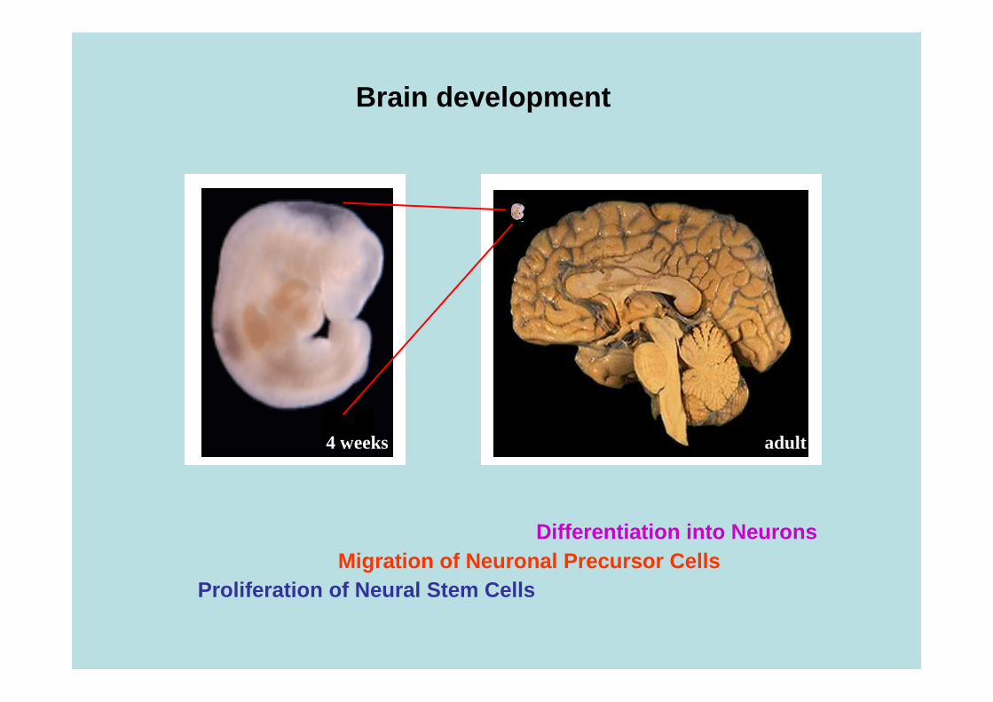
Brain development
4 weeks adult
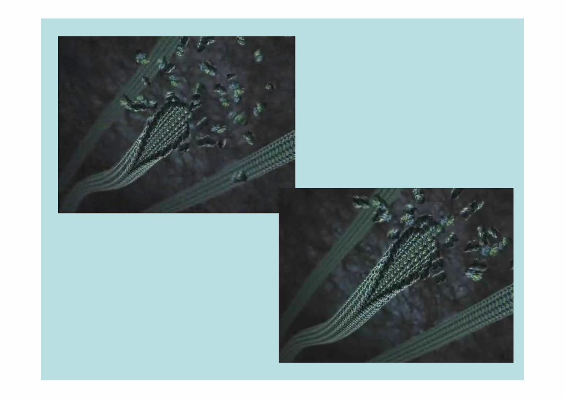
Proliferation of Neural Stem CellsMigration of Neuronal Precursor Cells
Differentiation into Neurons
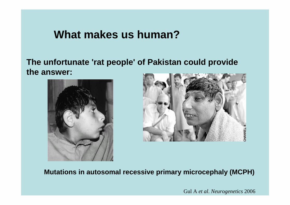
What makes us human?
The unfortunate 'rat people' of Pakistan could providethe answer:
Gul A et al. Neurogenetics 2006
Mutations in autosomal recessive primary microcephaly (MCP H)
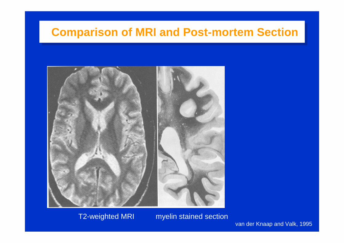
Comparison of MRI and Post-mortem SectionComparison of MRI and Post-mortem Section
van der Knaap and Valk, 1995T2-weighted MRI myelin stained section

1. Bulbus oculi2. Cavum nasi3. N. opticus4. Sinus sphenoidalis5. M. rectus med.6. Lobus temporalis
7. Art. ophthalmica8. Sella turcica9. Pons10. Ventriculus quartus11. Vermis cerebelli12. Lobus occipitalis
Csillag A, Atlas of Medical Imaging,
Koenemann, Cologne, 1999
T1 súlyozott axialis MRI felvétel
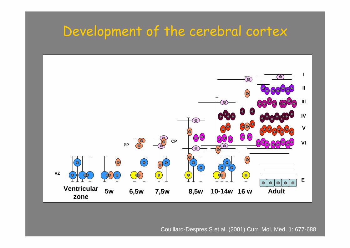
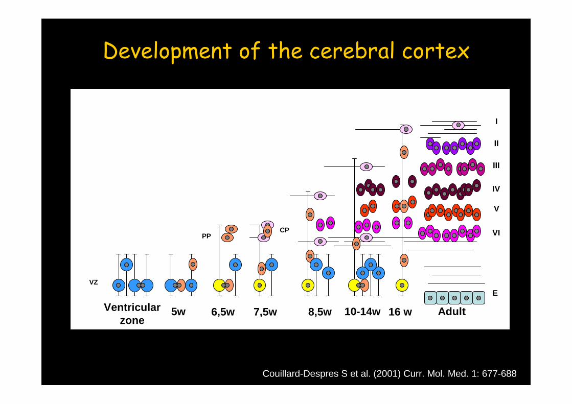
Development of the cerebral cortex
Couillard-Despres S et al. (2001) Curr. Mol. Med. 1: 677-688
I
II
V
EVZ
PPCP
5w 6,5w 7,5w 16 w8,5w Adult
III
VI
IV
Ventricularzone
10-14w
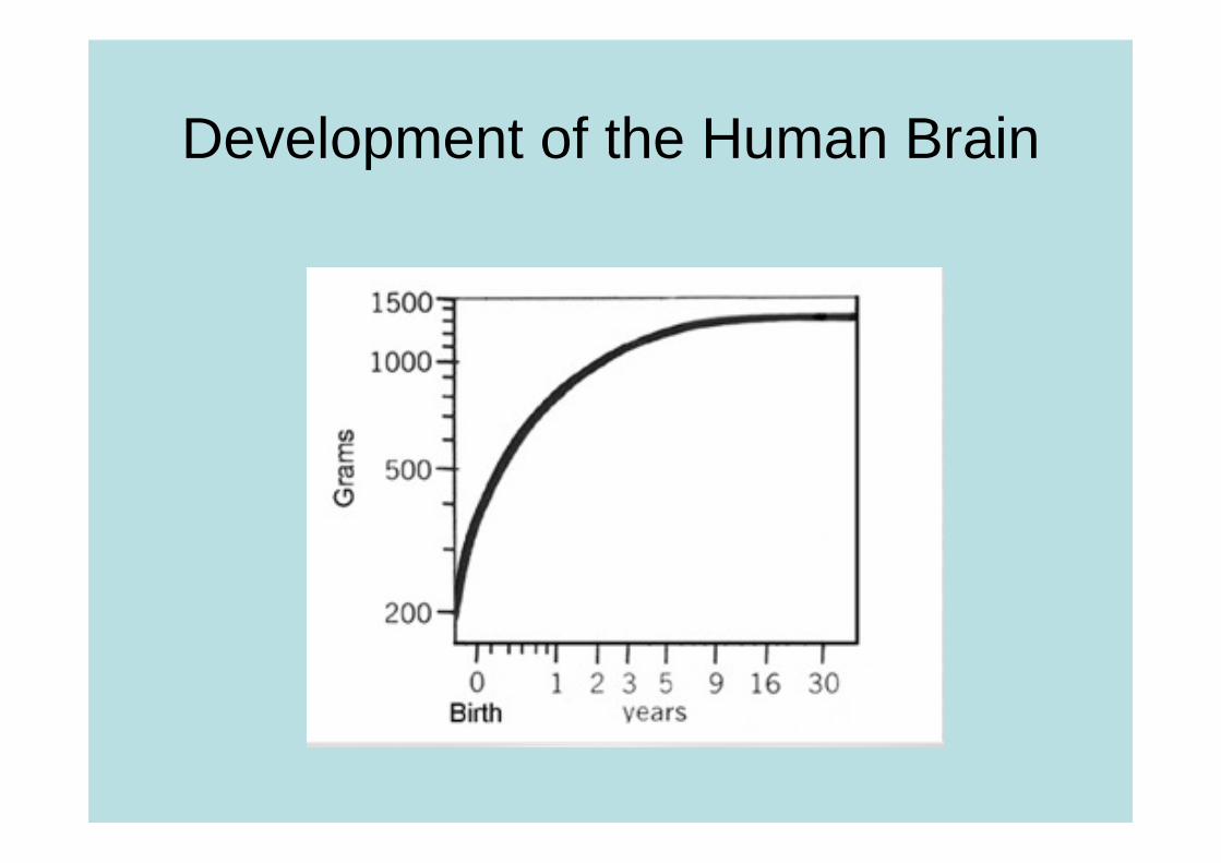
Development of the Human Brain
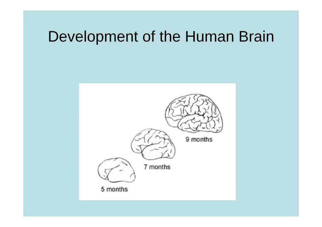
Development of the Human Brain
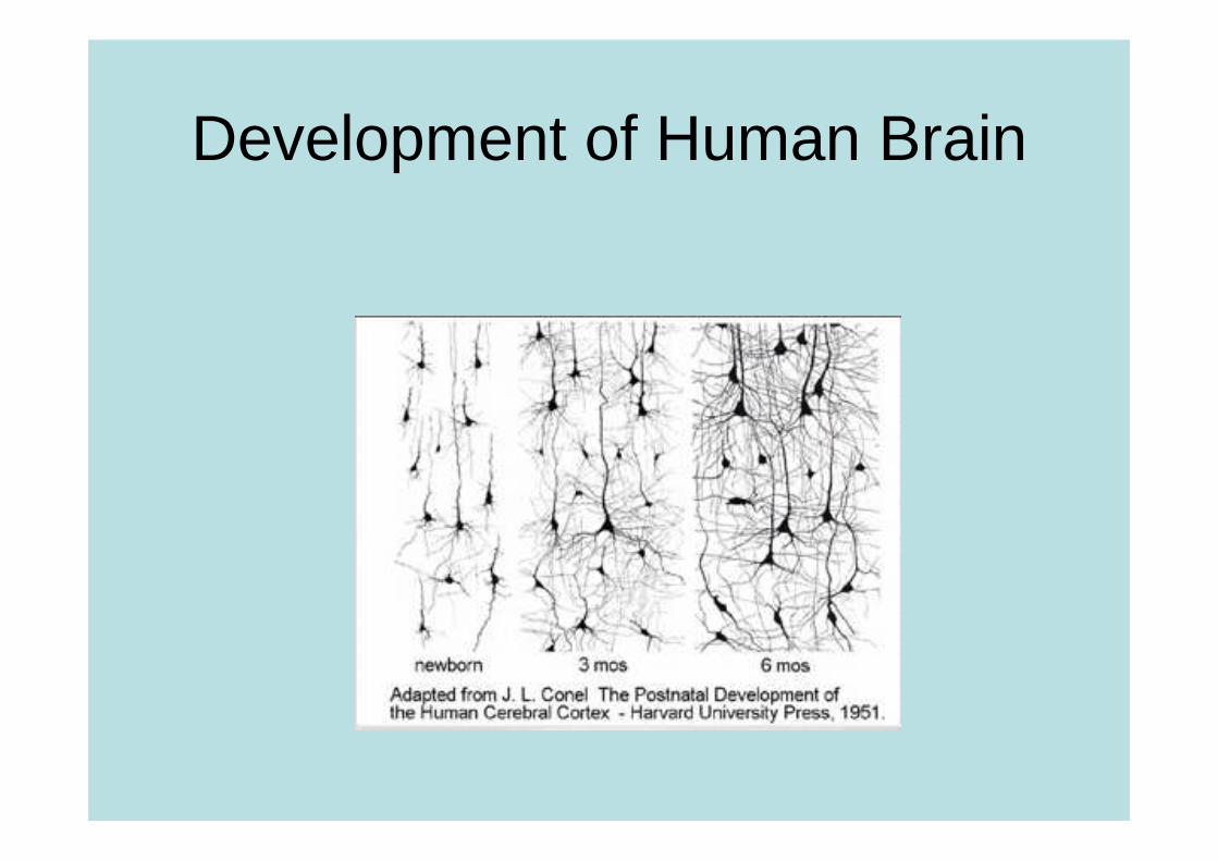
Development of Human Brain
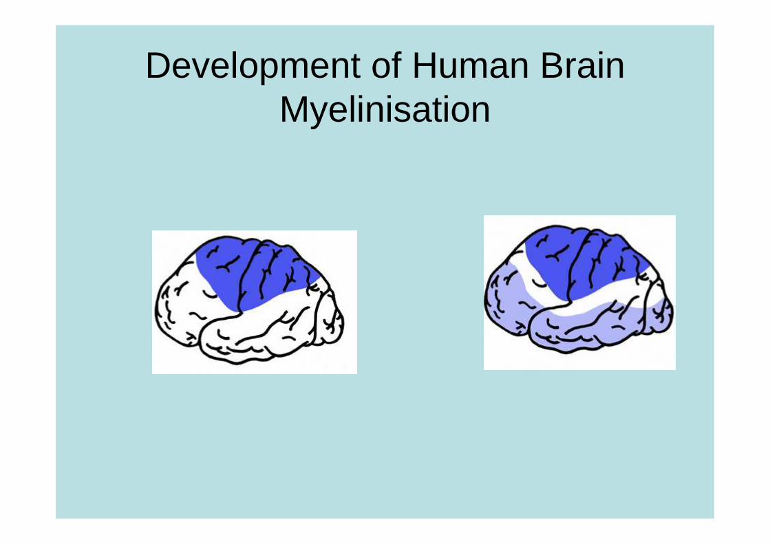
Development of Human BrainMyelinisation
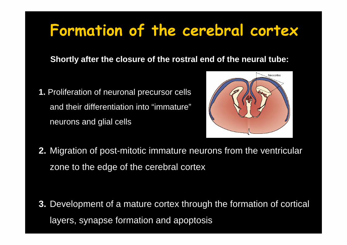
Formation of the cerebral cortex
2. Migration of post-mitotic immature neurons from the ventricular
zone to the edge of the cerebral cortex
3. Development of a mature cortex through the formation of cortical
layers, synapse formation and apoptosis
Shortly after the closure of the rostral end of the neu ral tube:
1. Proliferation of neuronal precursor cells
and their differentiation into “immature”
neurons and glial cells
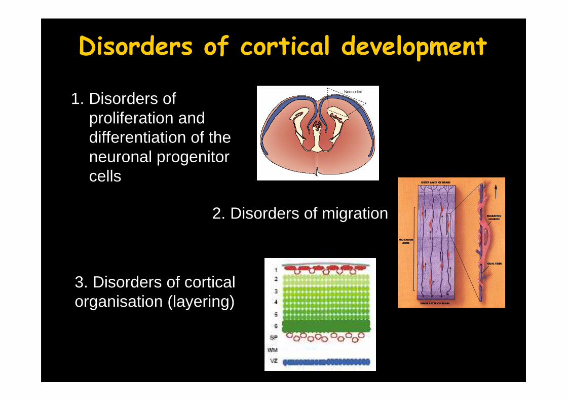
Disorders of cortical development
1. Disorders of proliferation and differentiation of theneuronal progenitorcells
2. Disorders of migration
3. Disorders of corticalorganisation (layering)
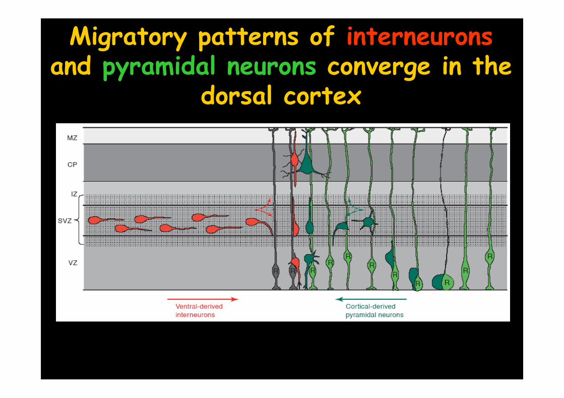
Migratory patterns of interneuronsand pyramidal neurons converge in the
dorsal cortex
Development of the cerebral cortex
Couillard-Despres S et al. (2001) Curr. Mol. Med. 1: 677-688
I
II
V
EVZ
PPCP
5w 6,5w 7,5w 16 w8,5w Adult
III
VI
IV
Ventricularzone
10-14w
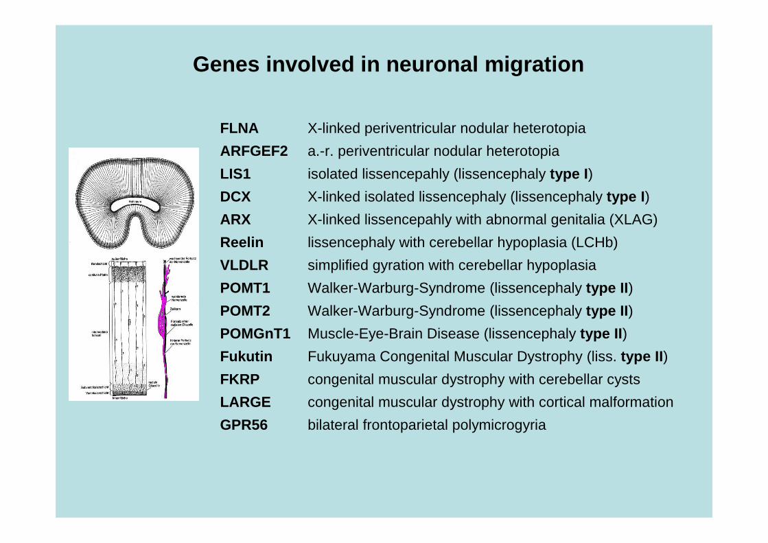
Genes involved in neuronal migration
FLNA X-linked periventricular nodular heterotopia
ARFGEF2 a.-r. periventricular nodular heterotopia
LIS1 isolated lissencepahly (lissencephaly type I )
DCX X-linked isolated lissencephaly (lissencephaly type I )
ARX X-linked lissencepahly with abnormal genitalia (XLAG)
Reelin lissencephaly with cerebellar hypoplasia (LCHb)
VLDLR simplified gyration with cerebellar hypoplasia
POMT1 Walker-Warburg-Syndrome (lissencephaly type II )
POMT2 Walker-Warburg-Syndrome (lissencephaly type II )
POMGnT1 Muscle-Eye-Brain Disease (lissencephaly type II )
Fukutin Fukuyama Congenital Muscular Dystrophy (liss. type II )
FKRP congenital muscular dystrophy with cerebellar cysts
LARGE congenital muscular dystrophy with cortical malformation
GPR56 bilateral frontoparietal polymicrogyria
PN
H
DC
S
LIS
I
LIS
II
BF
PP
LCH
b
XLA
G
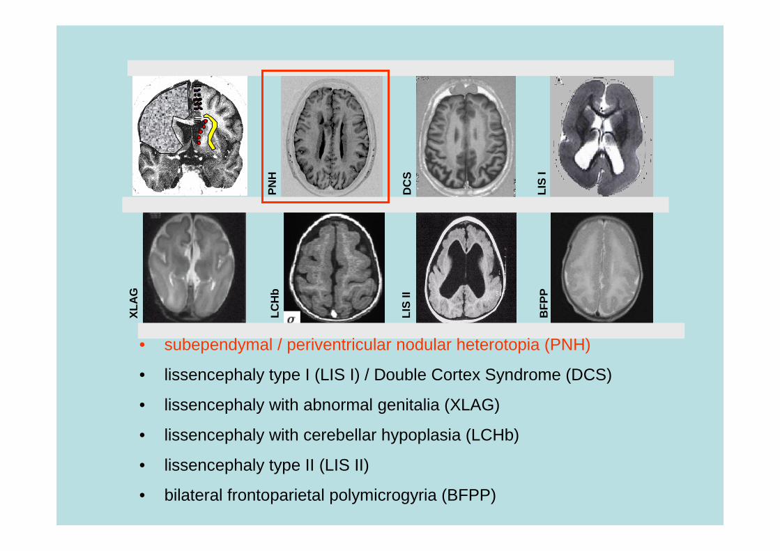
• subependymal / periventricular nodular heterotopia (PNH)
• lissencephaly type I (LIS I) / Double Cortex Syndrome (DCS)
• lissencephaly with abnormal genitalia (XLAG)
• lissencephaly with cerebellar hypoplasia (LCHb)
• lissencephaly type II (LIS II)
• bilateral frontoparietal polymicrogyria (BFPP)
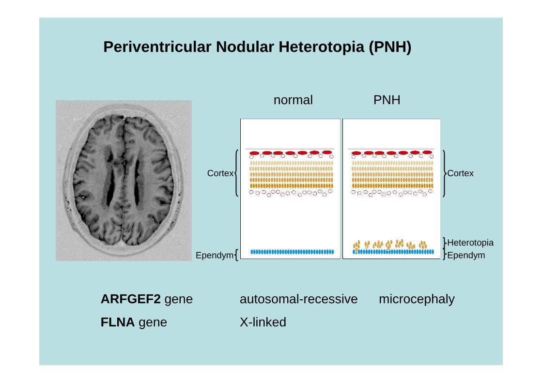
Periventricular Nodular Heterotopia (PNH)
Cortex
Ependym
Cortex
EpendymHeterotopia
normal PNH
ARFGEF2 gene autosomal-recessive microcephaly
FLNA gene X-linked
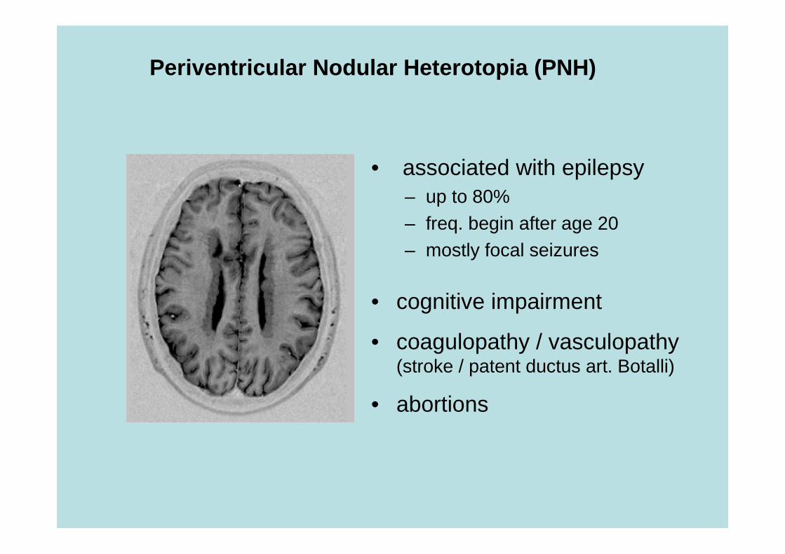
• associated with epilepsy– up to 80%– freq. begin after age 20– mostly focal seizures
• cognitive impairment
• coagulopathy / vasculopathy(stroke / patent ductus art. Botalli)
• abortions
Periventricular Nodular Heterotopia (PNH)
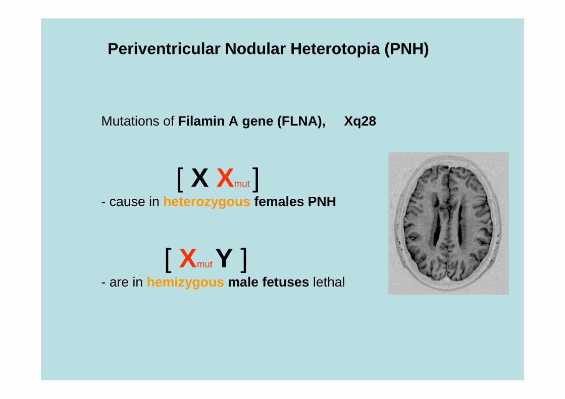
Mutations of Filamin A gene (FLNA), Xq28
[ X Xmut ]- cause in heterozygous females PNH
[ Xmut Y ]- are in hemizygous male fetuses lethal
Periventricular Nodular Heterotopia (PNH)
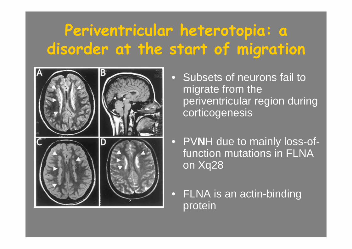
Periventricular heterotopia: a disorder at the start of migration
• Subsets of neurons fail to migrate from theperiventricular region duringcorticogenesis
• PVNH due to mainly loss-of-function mutations in FLNAon Xq28
• FLNA is an actin-bindingprotein
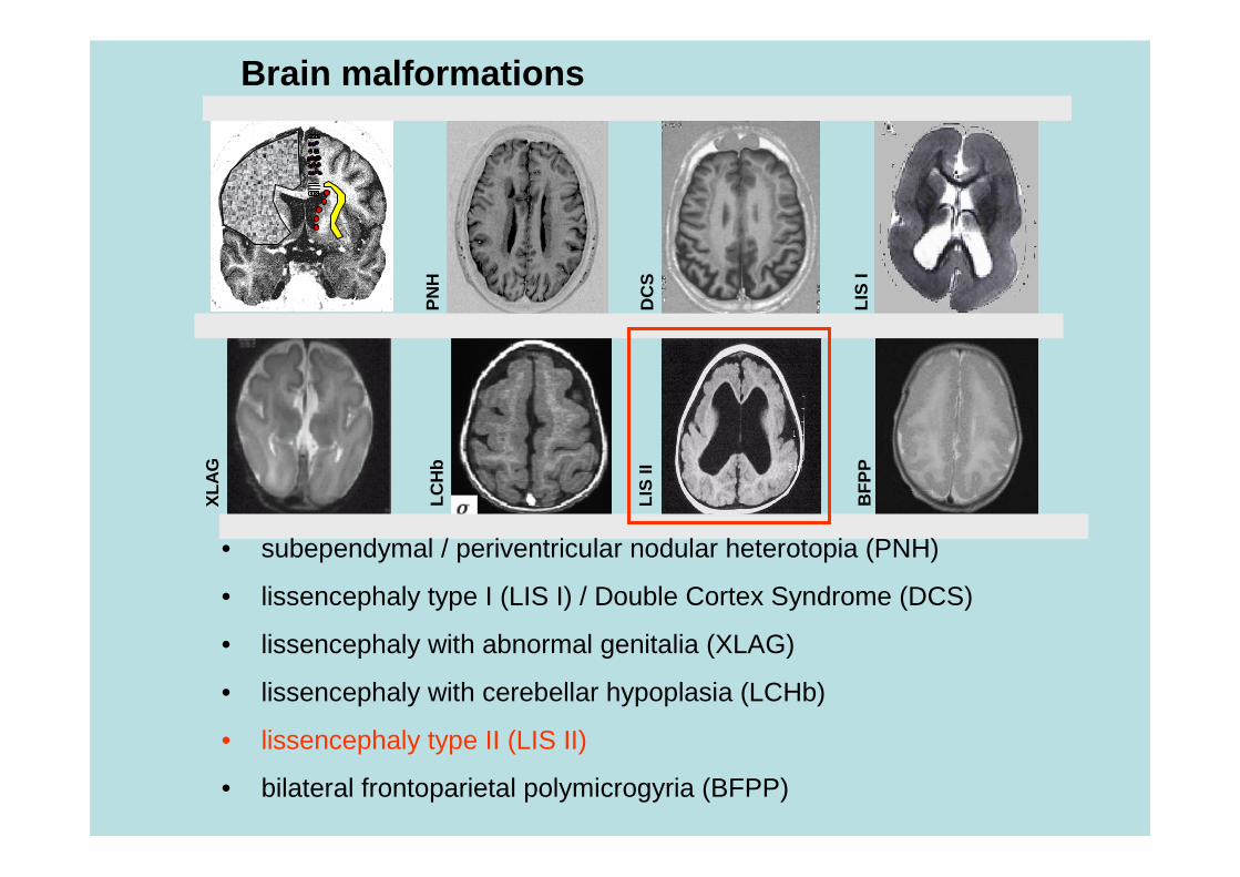
Brain malformations
PN
H
DC
S
LIS
I
LIS
II
BF
PP
LCH
b
XLA
G
• subependymal / periventricular nodular heterotopia (PNH)
• lissencephaly type I (LIS I) / Double Cortex Syndrome (DCS)
• lissencephaly with abnormal genitalia (XLAG)
• lissencephaly with cerebellar hypoplasia (LCHb)
• lissencephaly type II (LIS II)
• bilateral frontoparietal polymicrogyria (BFPP)
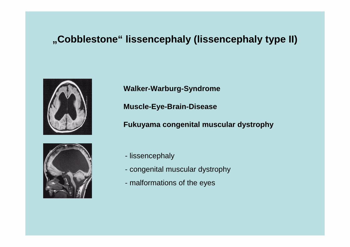
Walker-Warburg-Syndrome
Muscle-Eye-Brain-Disease
Fukuyama congenital muscular dystrophy
- lissencephaly
- congenital muscular dystrophy
- malformations of the eyes
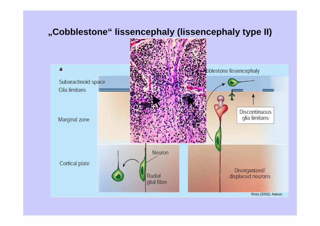
„Cobblestone“ lissencephaly (lissencephaly type II)
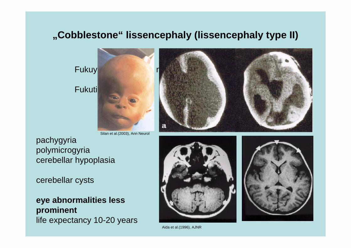
Fukuyama congenital muscular dystrophy
Fukutin gene
Silan et al.(2003), Ann Neurol
Aida et al.(1996), AJNR
pachygyriapolymicrogyriacerebellar hypoplasia
cerebellar cysts
eye abnormalities lessprominentlife expectancy 10-20 years
„Cobblestone“ lissencephaly (lissencephaly type II)
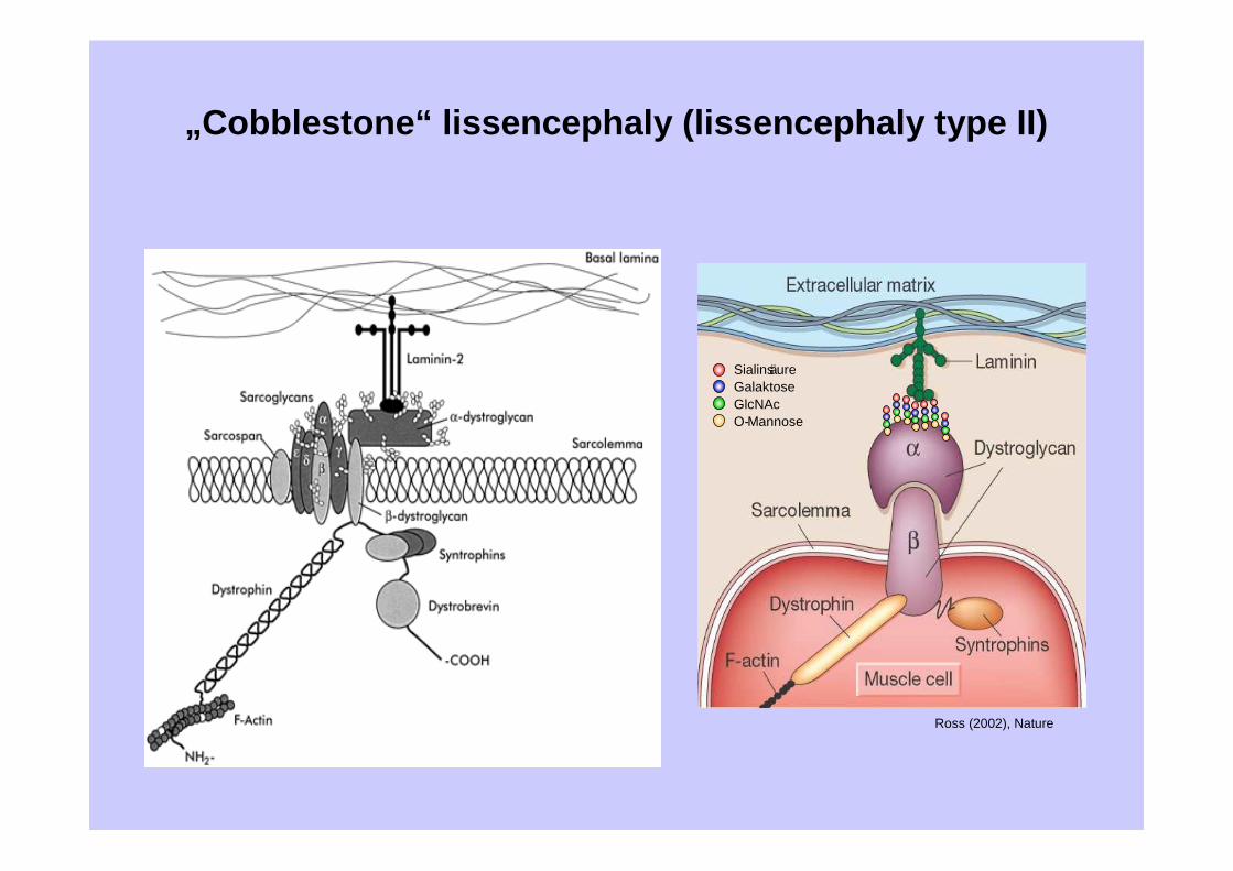
SialinsäureGalaktoseGlcNAcO-Mannose
Ross (2002), Nature
„Cobblestone“ lissencephaly (lissencephaly type II)
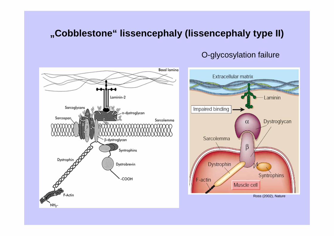
Ross (2002), Nature
O-glycosylation failure
„Cobblestone“ lissencephaly (lissencephaly type II)
Ross (2002), Nature
„Cobblestone“ lissencephaly (lissencephaly type II)
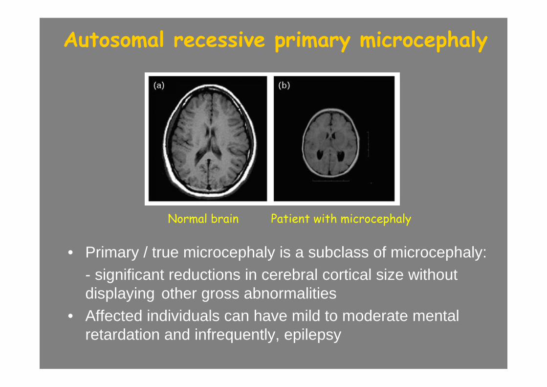
Autosomal recessive primary microcephaly
• Primary / true microcephaly is a subclass of microcephaly: - significant reductions in cerebral cortical size withoutdisplaying other gross abnormalities
• Affected individuals can have mild to moderate mental retardation and infrequently, epilepsy
Normal brain Patient with microcephaly
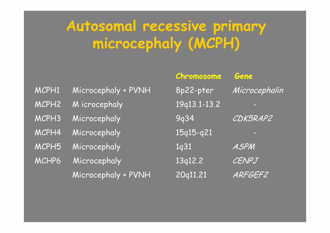
Autosomal recessive primarymicrocephaly (MCPH)
Chromosome Gene
MCPH1 Microcephaly + PVNH 8p22-pter Microcephalin
MCPH2 M icrocephaly 19q13.1-13.2 -
MCPH3 Microcephaly 9q34 CDK5RAP2
MCPH4 Microcephaly 15q15-q21 -
MCPH5 Microcephaly 1q31 ASPM
MCHP6 Microcephaly 13q12.2 CENPJ
Microcephaly + PVNH 20q11.21 ARFGEF2
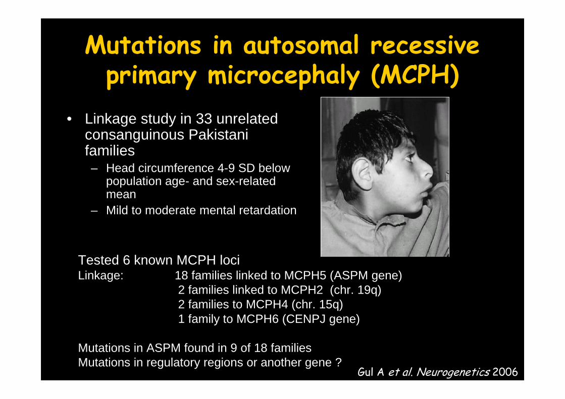
Mutations in autosomal recessiveprimary microcephaly (MCPH)
• Linkage study in 33 unrelatedconsanguinous Pakistani families– Head circumference 4-9 SD below
population age- and sex-relatedmean
– Mild to moderate mental retardation
Tested 6 known MCPH lociLinkage: 18 families linked to MCPH5 (ASPM gene)
2 families linked to MCPH2 (chr. 19q)2 families to MCPH4 (chr. 15q)1 family to MCPH6 (CENPJ gene)
Mutations in ASPM found in 9 of 18 familiesMutations in regulatory regions or another gene ?
Gul A et al. Neurogenetics 2006
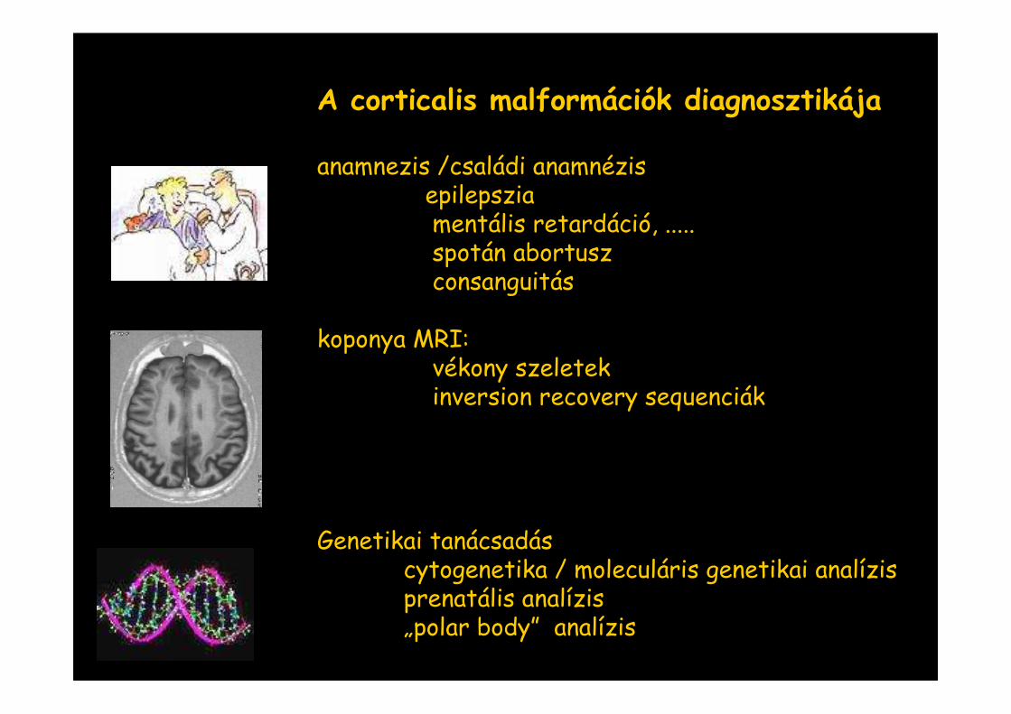
A corticalis malformációk diagnosztikája
anamnezis /családi anamnézisepilepsziamentális retardáció, .....spotán abortuszconsanguitás
koponya MRI: vékony szeletek inversion recovery sequenciák
Genetikai tanácsadáscytogenetika / moleculáris genetikai analízisprenatális analízis„polar body” analízis
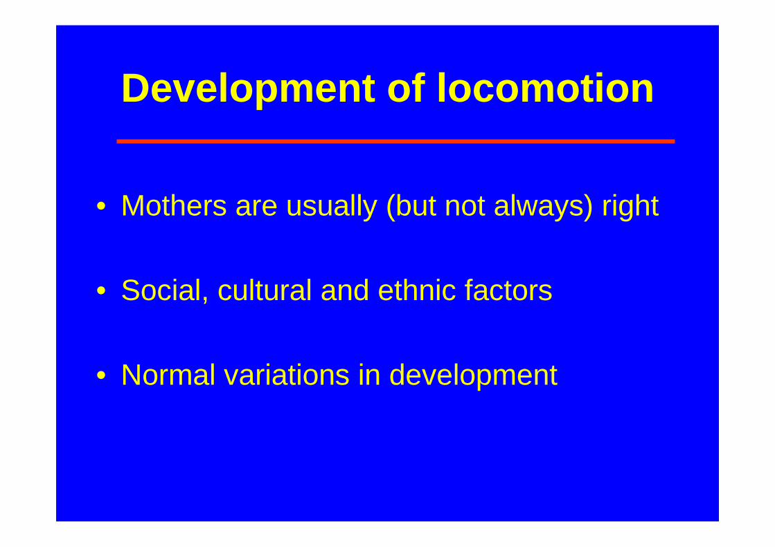
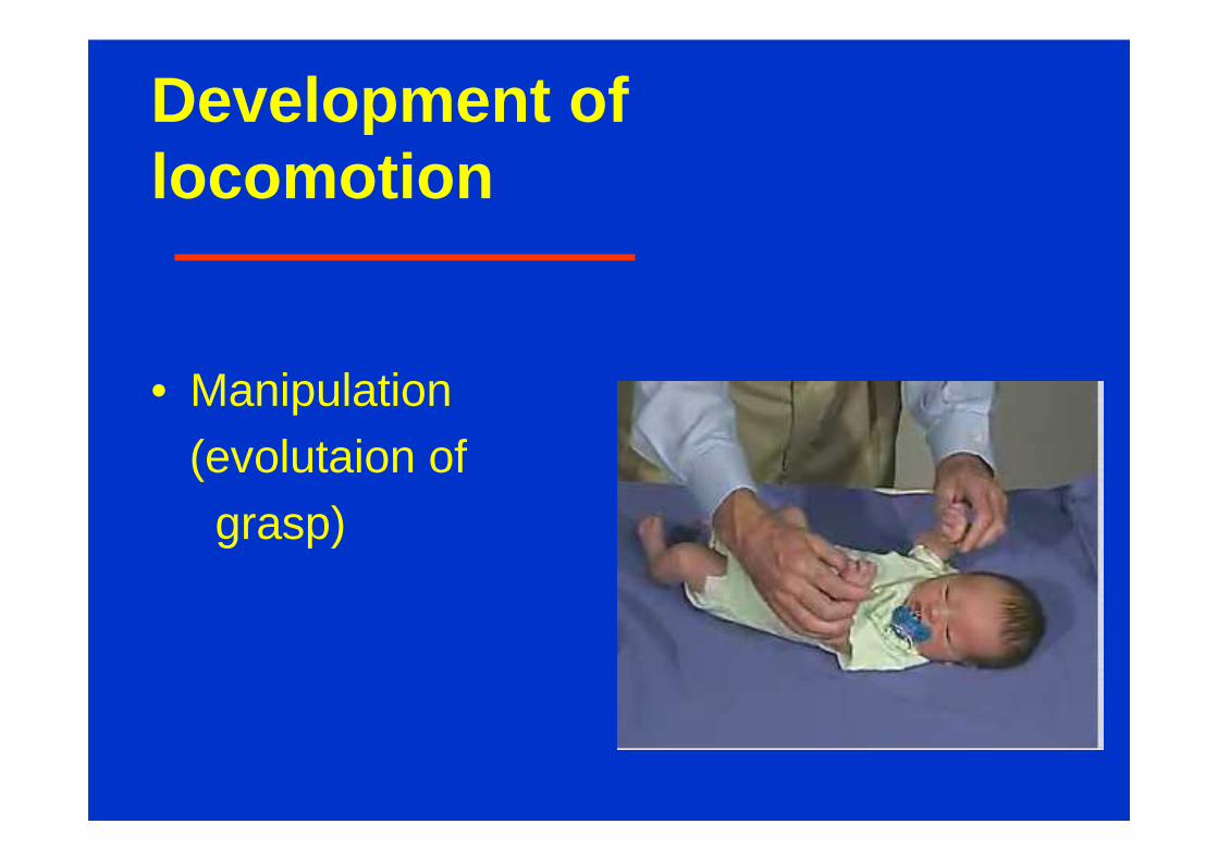
Development of locomotion
• Mothers are usually (but not always) right
• Social, cultural and ethnic factors
• Normal variations in development
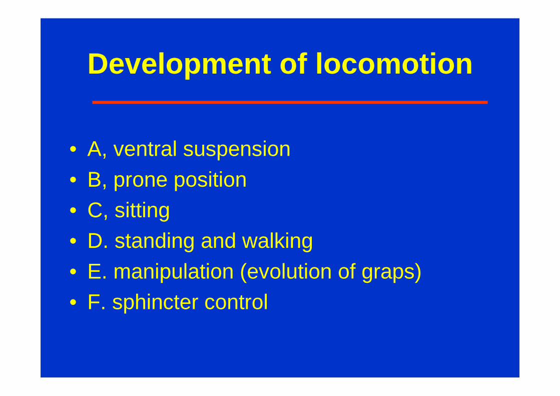
Development of locomotion
• A, ventral suspension• B, prone position• C, sitting• D. standing and walking• E. manipulation (evolution of graps)• F. sphincter control
Development of locomotion
• Moro reflex• Parachute reaction• ………
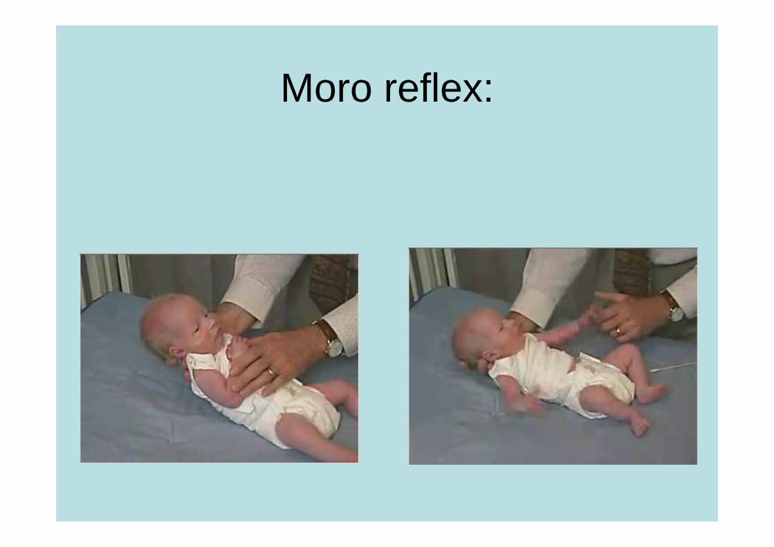
Moro reflex:
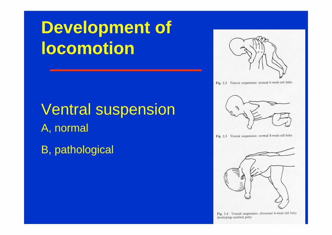
Ventral suspensionA, normal
B, pathological
Development oflocomotion
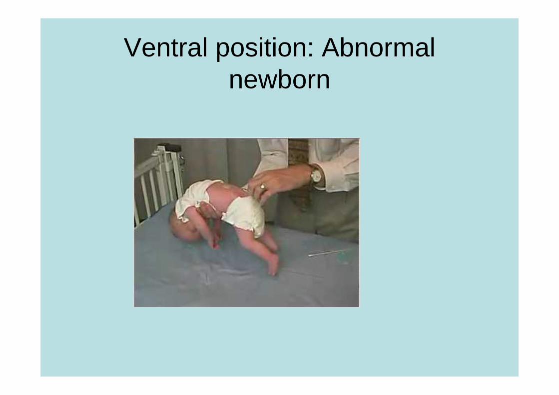
Ventral position: Abnormalnewborn
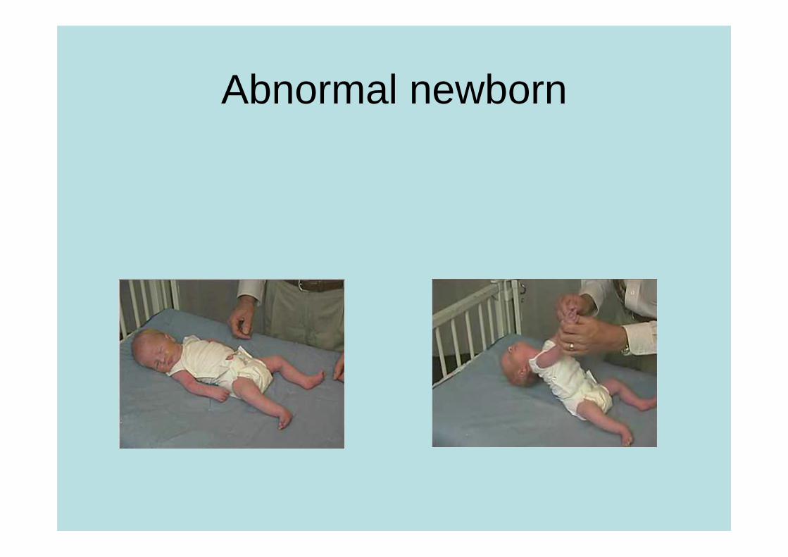
Abnormal newborn
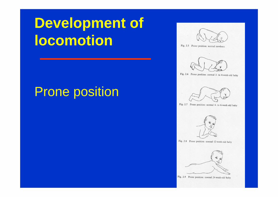
Development oflocomotion
Prone position
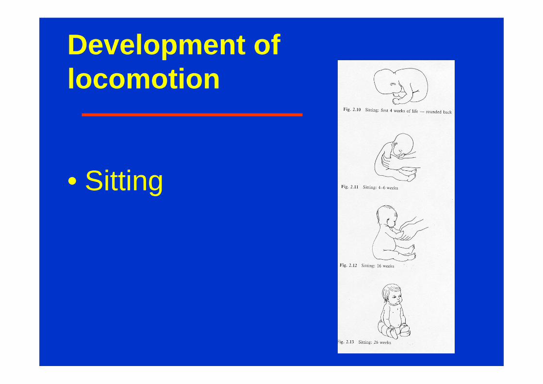
Development oflocomotion
• Sitting
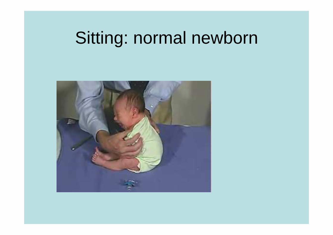
Sitting: normal newborn
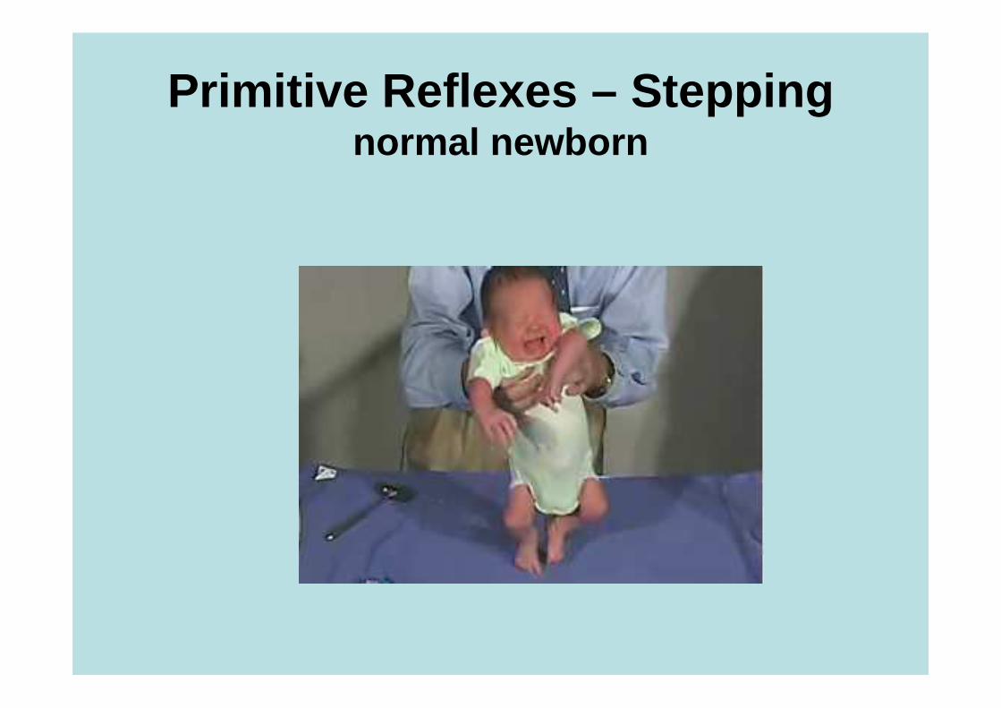
Primitive Reflexes – Steppingnormal newborn
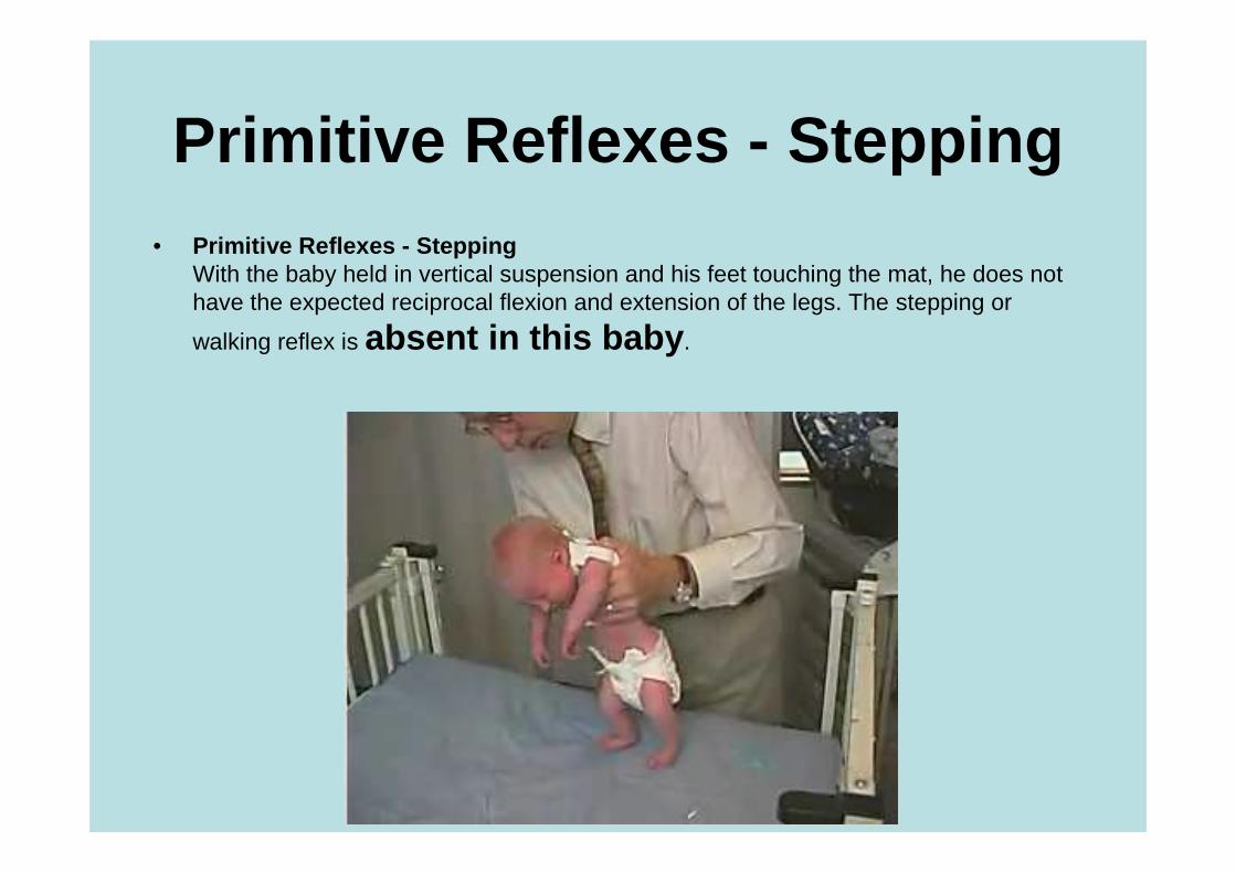
Primitive Reflexes - Stepping• Primitive Reflexes - Stepping
With the baby held in vertical suspension and his feet touching the mat, he does nothave the expected reciprocal flexion and extension of the legs. The stepping or
walking reflex is absent in this baby .
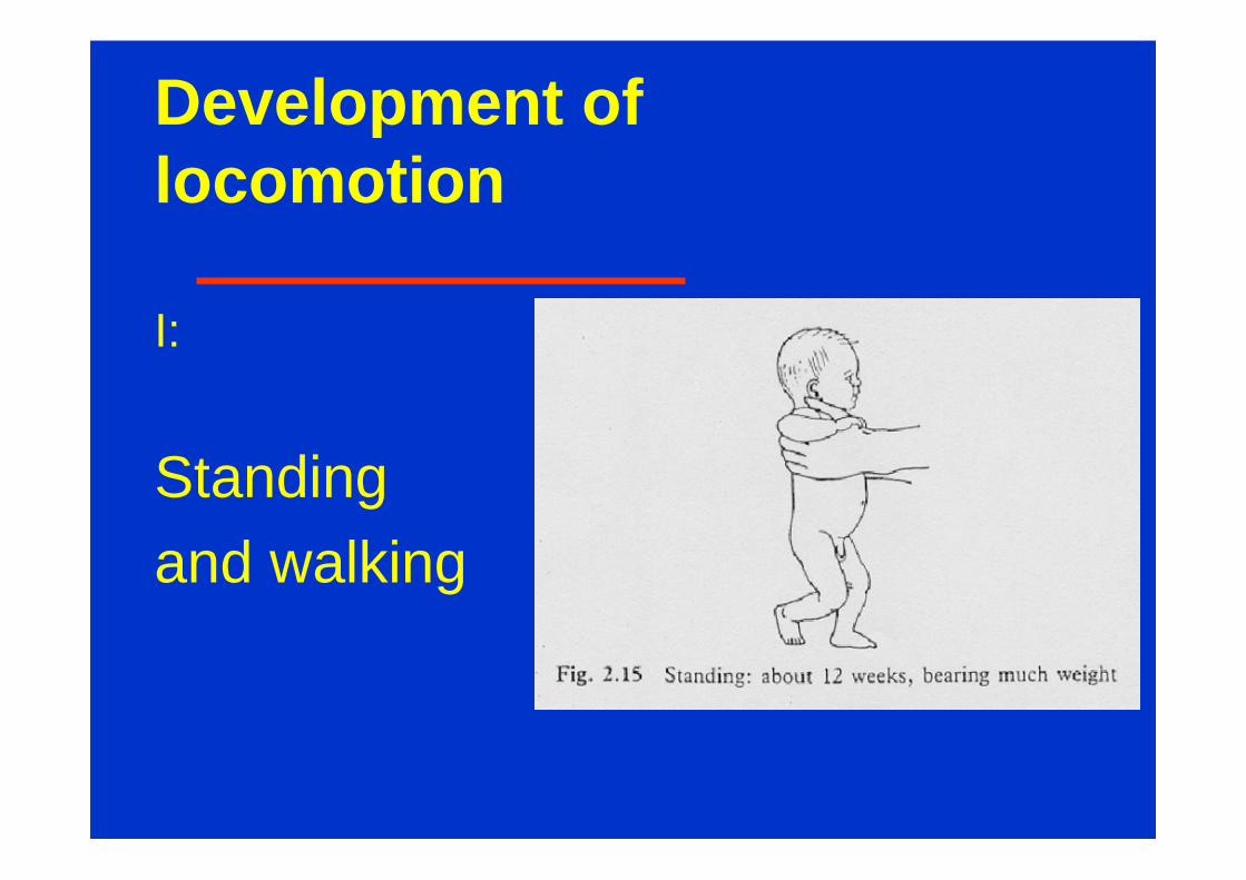
Development oflocomotion
I:
Standing and walking
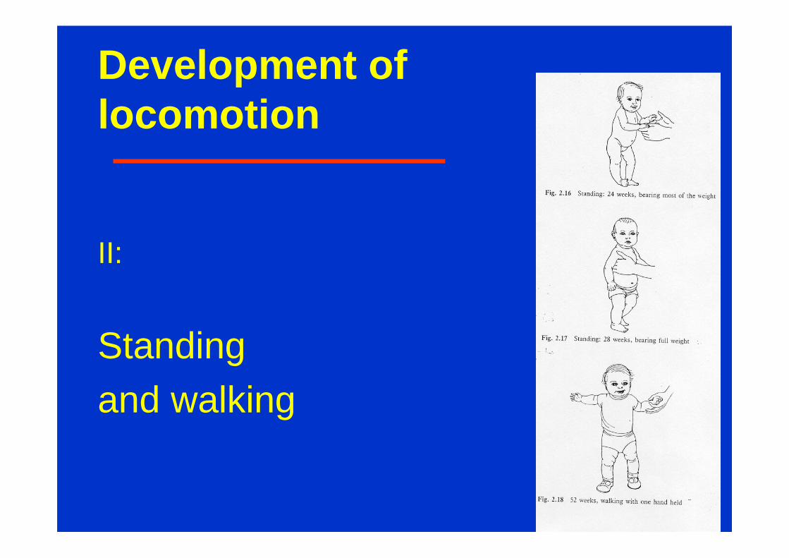
Development oflocomotion
II:
Standing and walking
Development oflocomotion
• Manipulation(evolutaion ofgrasp)
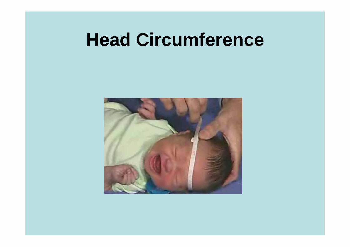
Head Circumference
• Another very important part of assessing braindevelopment is measuring the growth of the brain. Thisis accomplished by measuring the head circumference, which is an accurate reflection of brain size . The braingrows to 80% of its adult volume during the first 2 years of life so many neurological diseases that occurearly in life will impact the growth of the brain. A smallhead (microcephaly ) or a large head (macrocephaly orhydrocephalus ) can be key findings in explaining theneurological abnormalities of a child.
• It is essential to plot head circumference on a standardized head growth chart such as the Nellhauschart.
Head Circumference
SPECIAL PROGRAMSNeurodevelopment Assessment
• Attentional based disorders• Dyslexia and language related learning
difficulties• Study and organizational problems• Non-verbal learning disabilities• Emotional/Behavioral problems• Written Expression problems
Developmental Milestones
• The neurological examination of the pediatric patientmust be couched in the context of neurodevelopmentalmilestones. The normal neurological findings one wouldexpect for a newborn are certainly different than a 2, 6 or12-month-old infant. Obtaining developmentalmilestones is an important reflection of the maturation ofthe child’s nervous system and assessing developmentis an essential part of the pediatric neurologicalexamination. Delay in obtaining developmentalmilestones and abnormal patterns of development areimportant indicators of underlying neurological disease.
Diseases - Therapies• Speech Therapy• Occupational Therapy or Physical Therapy• Vision Therapy• Applied Behavioral Analysis Therapy• Neurodevelopment Therapy• Specific Educational Therapy• ADD/ADHD• The Ritalin-Free Child: Managing Hyperactivity & Attention Deficits Without Drugs by Diana Hunter (ISBN 0962833681
• Autism (also PDD)• Asperger Syndrome• Auditory Processing Dysfunction• Dysgraphia• Dyslexia• Mental Retardation• Sensory Processing Dysfunction• Speech Disorders• Vision Impaired
Cerebral Palsy
a persistent disorder of
- movement and
- posture
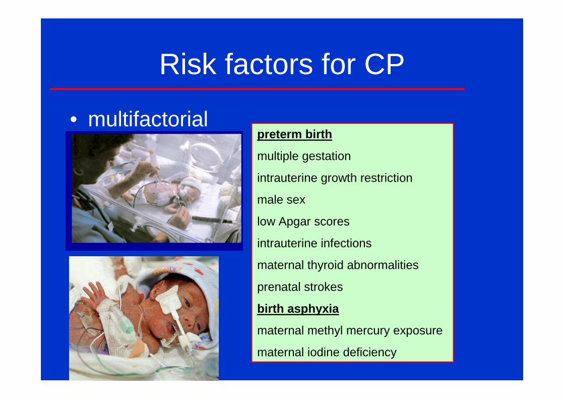
Risk factors for CP
• multifactorialpreterm birth
multiple gestation
intrauterine growth restriction
male sex
low Apgar scores
intrauterine infections
maternal thyroid abnormalities
prenatal strokes
birth asphyxia
maternal methyl mercury exposure
maternal iodine deficiency
Risk factors for CP
• prenatal factors result in 70-80% of cases of CP
• In most cases:the exact cause is unknown but is most likelymultifactorial
Clinical course of CP
CP generally is considered to be
static encephalopathy or
nonprogressive in nature !!!!
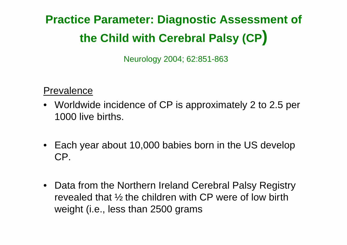
Practice Parameter: Diagnostic Assessment of
the Child with Cerebral Palsy (CP )Neurology 2004; 62:851-863
Prevalence• Worldwide incidence of CP is approximately 2 to 2.5 per
1000 live births.
• Each year about 10,000 babies born in the US develop CP.
• Data from the Northern Ireland Cerebral Palsy Registry revealed that ½ the children with CP were of low birth weight (i.e., less than 2500 grams
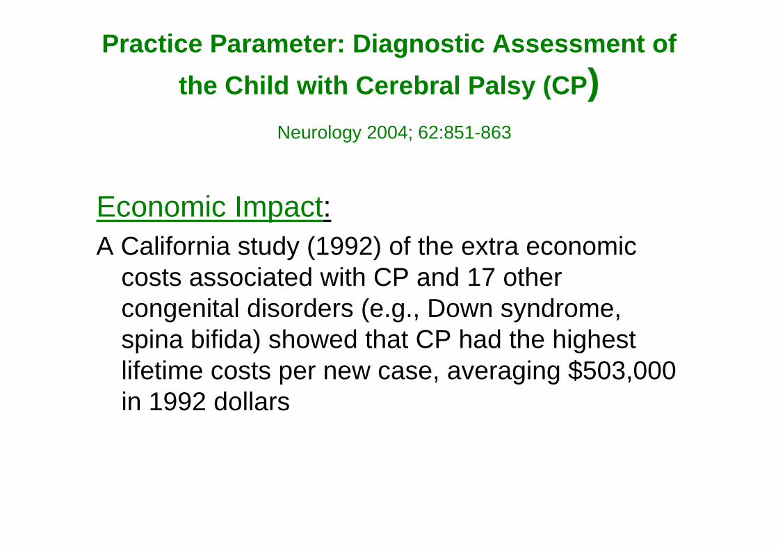
Practice Parameter: Diagnostic Assessment of
the Child with Cerebral Palsy (CP )Neurology 2004; 62:851-863
Economic Impact:A California study (1992) of the extra economic
costs associated with CP and 17 other congenital disorders (e.g., Down syndrome, spina bifida) showed that CP had the highest lifetime costs per new case, averaging $503,000 in 1992 dollars
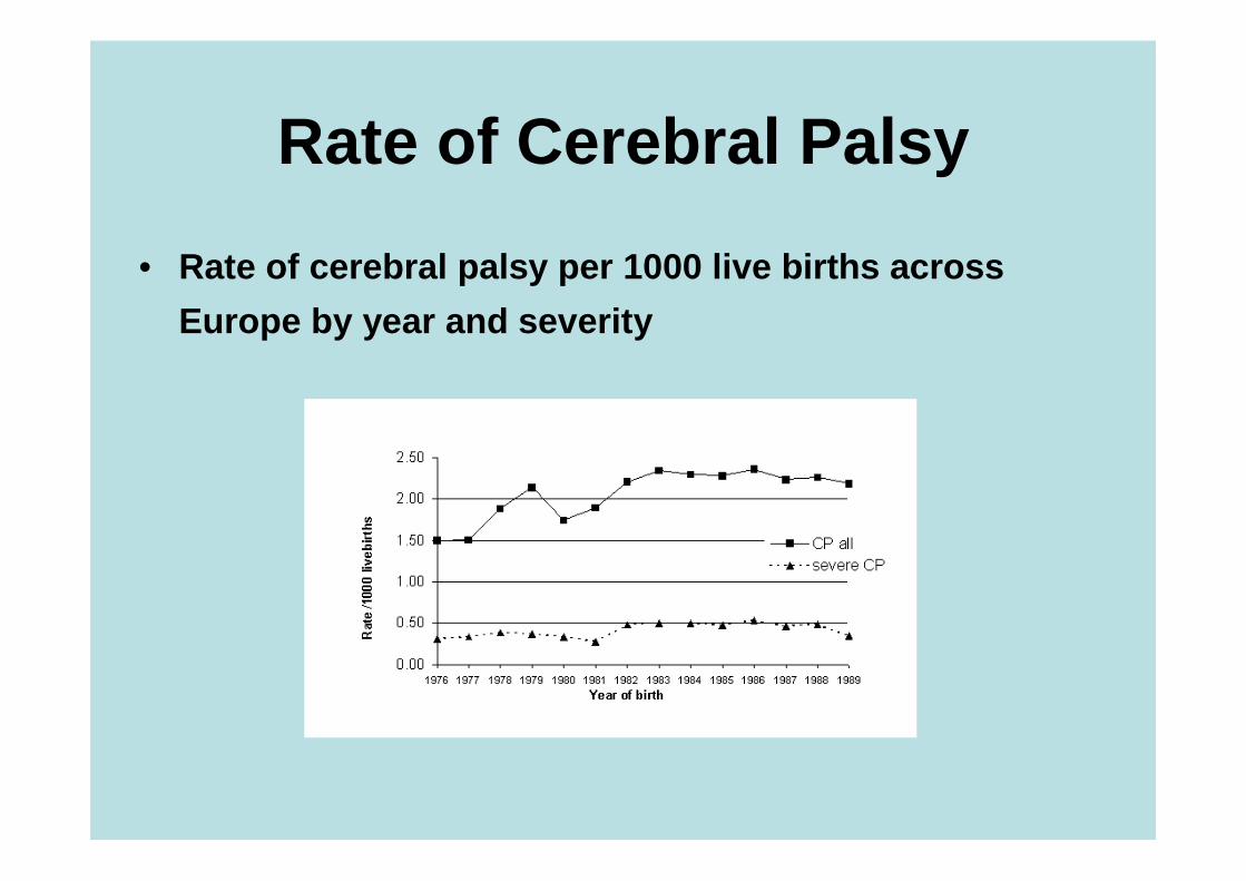
Rate of Cerebral Palsy
• Rate of cerebral palsy per 1000 live births across
Europe by year and severity