Embed Size (px)
Citation preview

Pseudoctenis cornelii nov. spec. (cycadalean foliage) from the Carnian (Upper Triassic) of Lunz, Lower Austria
By Christian POTT1, Hans KERP1 and Michael KRINGS2
(With 1 textfigure and 3 plates)
Manuskript submitted on April 12th 2006, the revised manuscript on June 7th 2006
ZusammenfassungDie Gattung Pseudoctenis (Cycadeenbeblätterung) ist ein charakteristisches Element rhätischer (oberste Trias) und jurassischer Floren in Europa. Aus der Flora von Lunz in Niederösterreich wird eine neue Art, Pseudoctenis cornelii, beschrieben, die den ältesten sicheren Nachweis der Gattung Pseudoctenis aus der europäischen Trias darstellt. Die neue Art wird auf der Basis makromorphologischer und epidermal-anatomischer Merkmale zu Pseudoctenis gestellt; besonders wichtig sind die eingesenkten, haplocheilen Spaltöffnungen, die von Papillen der umgebenden Nebenzellen überdeckt sind. Pseudoctenis cornelii weist Merkmale auf, die in ganz ähnlicher Weise bei heutigen, an ein Leben in (saisonal) trockenen Lebensräumen angepassten Pflanzen ausgebildet sind.Schlüsselwörter: Kutikularanalyse, Cycadales, Beblätterung, Karn, Triassische Flora, Paläoökologie
AbstractPseudoctenis (cycadalean foliage) is a characteristic element of Rhaetian (uppermost Triassic) and Jurassic floras from Europe. Here we describe a new species, Pseudoctenis cornelii, from the Carnian flora of Lunz in Lower Austria that provides the earliest persuasive evidence for this genus in the European Triassic. The new species is included in Pseudoctenis based on macromorphology and epidermal anatomy; a particularly significant feature is sunken, haplocheilic stomata, partially closed by overarching papillae. Pseudoctenis cornelii displays features that are consistent with those extant plants adaptated to life in (seasonally) dry environments.Keywords: Cuticular analysis, Cycadales, foliage, Carnian, Triassic flora, palaeoecology
Introduction
The famous Carnian flora from Lunz in Lower Austria is one of only a few well-preserved floras from the Alpine Triassic (CLEAL 1993; DOBRUSKINA 1998). One of the remark-able features of this flora is the abundance of compressed cycadophyte (i.e. Cycadales and Bennettitales) reproductive structures and foliage. While several detailed studies exist that focus on the fertile cycadophyte remains (KRASSER 1917, 1919; KRÄUSEL 1948,
Ann. Naturhist. Mus. Wien 109 A 1–17 Wien, September 2007
1 Forschungsstelle für Paläobotanik am Geologisch-Paläontologischen Institut, Westfälische Wilhelms-Uni-versität Münster, Hindenburgplatz 57, 48143 Münster, Germany. – e-mail: [email protected]
2 Bayerische Staatssammlung für Paläontologie und Geologie und GeoBio-CenterLMU, Richard-Wagner- Straße 10, 80333 Munich, Germany

2 Annalen des Naturhistorischen Museums in Wien 109 A
1949, 1953), the innumerable foliage specimens have not received much scholarly atten-tion to date. This is due in part to the fact that the fossils are spread among collections throughout Europe. Moreover, the various 19th and early 20th century collectors, traders, and curators, who labelled most of the fossils, along with the early scholars studying the material, unintentionally created considerable confusion with regard to the scientific names given to the different foliage types based on macromorphology. This confusion remains to date and renders establishing a trustworthy inventory of the plants that lived in the Lunz area during the Carnian a difficult task.Cuticles are known to provide a wealth of information useful in the taxonomy of com-pression seed plant fossils, but also important with regard to paleobiological and paleo-ecological considerations (e.g., KERP 1990; MCELWAIN & CHALONER 1996). Although cuticular analysis has long since proven to provide a more complete understanding of many ancient seed plants, it has largely been neglected in studies of the cycadophyte foliage from Lunz. To fill this gap, we have initiated a research project that focuses on seed plant cuticles from Lunz. In the course of the last two years, detailed information on the epidermal anatomy of most of the proposed "species" has been gathered. As a result, we now have an extensive set of heretofore largely unavailable data based on epidermal features to interpret this famous flora.Our cuticle studies of the Lunz cycadophytes revealed that the status of many of the foliage "species", which were established based exclusively on macromorphological features, is not supported by the epidermal anatomy. On the other hand, the studies also produced evidence for the occurrence in the Lunz flora of a few taxa that are new to science. We here describe the macromorphology and epidermal anatomy of Pseudoctenis cornelii, a new cycadalean foliage species from Lunz. This species provides the earliest persuasive evidence for the genus Pseudoctenis SEWARD in the European Triassic. Pseudoctenis cornelii is compared to other species in the genus, and some hypotheses with regard to the palaeoautecology are offered based on the adaptative significance of macromorphological and epidermal features.
The genus Pseudoctenis SEWARD
The cycadalean foliage genus Pseudoctenis is a common element of several Rhaetian (uppermost Triassic) and many Jurassic floras in Europe (SEWARD 1911, 1917; HARRIS 1932, 1964). SEWARD (1911) introduced the genus for Zamites-type leaves from the Jurassic of Sutherland, Great Britain. However, this author did not provide a generic diagnosis, but only a comparison to Ctenis LINDLEY et W. HUTTON (cycadalean foliage). Although Ctenis and Pseudoctenis are similar in macromorphology, SEWARD (1911) notes that they are easily distinguishable based on the occurrence of anastomoses in the venation of Ctenis. HARRIS (1950) concurs with SEWARD in that the Ctenis-Pseudoctenis series consists of two distinct groups. Nevertheless, the genus Pseudoctenis historically often was used in an arbitrary way because no valid generic diagnosis existed. As a result, through the years, numerous foliage specimens were accommodated in genera such as Pterophyllum BRONGNIART, Ctenophyllum SCHIMPER, and Zamites BRONGNIART, although they actually belonged to Pseudoctenis. However, this changed when HARRIS (1932) described epidermal features

POTT & al.: Pseudoctenis cornelii nov. spec. from the Carnian of Lunz, Lower Austria 3
of Pseudoctenis spectabilis HARRIS, P. depressa HARRIS, and P. lanei THOMAS from the Jurassic of Yorkshire. The epidermal anatomy establishes the cycadalean affinities of Pseudoctenis based on the presence of haplocheilic stomata, a feature that generally dis-criminates cycadalean from bennettitalean foliage (FLORIN 1933). Stomatal morphology is especially valuable in distinguishing Pseudoctenis leaves from those bennettitalean foliage types that are similar in macromorphology, e.g., certain taxa in Pterophyllum and Zamites. HARRIS (1932, 1964) provides a diagnosis for Pseudoctenis that includes both macromorphological and epidermal characters. Nevertheless, the validity of Pseu-doctenis SEWARD remains technically problematical because the original study (SEWARD 1911) does not include a diagnosis, and useful cuticles cannot be obtained from the type specimen (cf. HARRIS 1964).
Geological setting, material and methods
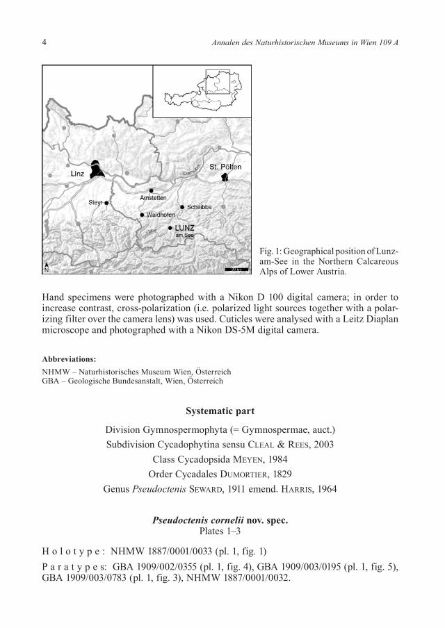
The material comes from Lunz in the Northern Calcareous Alps of Lower Austria, and was collected during the late 19th century from several coal mines in the area around Lunz-am-See, approximately 100 km west of Vienna (fig. 1). Coal mining in the Lunz area ceased after World War II., and collection of new material is today limited to the old spoil tips; however, spoil tip fossils are usually strongly weathered. The fossils occur in the "Lunzer Sandstein", which is part of the Lunz beds. The Lunz Formation (= Lunzer Schichten) consists of sandstones at basis, followed by marine marls gradually grading upwards into terrestrial sands, shales, and coal. The coal-bearing part of the sequence is overlain by marls and with a sandstone layer at the top. The plant fossils occur in the shales associated with the coal beds. The exact age of the Lunz Formation remains a problem to date since adequate biomarkers such as ammonoids and conodonts are missing. However, a recent correlation of biostratigraphically well-established sections in the Hallstatt and Reifling Intraplatform Basins (HORNUNG & BRANDNER 2005) sug-gests that the Lunz Formation may be correlated with the upper part of the Reingraben Formation (T. HORNUNG, pers. comm.). Taking this into account, the Lunz Formation is approximately late Julian (Julian 2/II) in age. Palynological studies indicate a Carnian (BHARADWAJ & SINGH 1964) and Julian age (DUNAY & FISHER 1978). The Opponitzer Limestone, the upper sub-unit of the Lunzer Schichten, has been dated as Tuvalian by DUNAY & FISHER (1978).The Pseudoctenis cornelii specimens are kept in the collections of the Natural History Museum Vienna (NHMW), and the Geological Survey of Austria (GBA), Vienna, under accession numbers NHMW 1887/0001/0032 and 1887/0001/0033, and GBA 1909/002/0355, 1909/003/0195, and 1909/003/0783. Cuticles were prepared according to procedures outlined in KERP (1990), and KERP & KRINGS (1999). Pieces of rock with identifiable plant remains were dissolved in hydro-flouridic acid (HF), and subsequently macerated according to the standard procedure using Schulze's reagent (HNO3 with a few crystals of KClO3) and 5–10 % Potassium hydroxide. Macerated cuticles were washed in distilled water, gently dehydrated in pure glycerine, and finally mounted in permanent glycerine-jelly microscope slides. Slides are deposited in the collection of the Natural History Museum Vienna (Austria); acces-sion numbers are indicated in the figure captions.
4 Annalen des Naturhistorischen Museums in Wien 109 A
Fig. 1: Geographical position of Lunz- am-See in the Northern Calcareous Alps of Lower Austria.
Hand specimens were photographed with a Nikon D 100 digital camera; in order to increase contrast, cross-polarization (i.e. polarized light sources together with a polar-izing filter over the camera lens) was used. Cuticles were analysed with a Leitz Diaplan microscope and photographed with a Nikon DS-5M digital camera.
Abbreviations:NHMW – Naturhistorisches Museum Wien, ÖsterreichGBA – Geologische Bundesanstalt, Wien, Österreich
Systematic part
Division Gymnospermophyta (= Gymnospermae, auct.)Subdivision Cycadophytina sensu CLEAL & REES, 2003
Class Cycadopsida MEYEN, 1984Order Cycadales DUMORTIER, 1829
Genus Pseudoctenis SEWARD, 1911 emend. HARRIS, 1964
Pseudoctenis cornelii nov. spec. Plates 1–3
H o l o t y p e : NHMW 1887/0001/0033 (pl. 1, fig. 1)P a r a t y p e s: GBA 1909/002/0355 (pl. 1, fig. 4), GBA 1909/003/0195 (pl. 1, fig. 5), GBA 1909/003/0783 (pl. 1, fig. 3), NHMW 1887/0001/0032.

POTT & al.: Pseudoctenis cornelii nov. spec. from the Carnian of Lunz, Lower Austria 5
T y p e l o c a l i t y : Lunz-am-See, Lower Austria.S t r a t u m t y p i c u m : Lunzer Schichten, Carnian (Upper Triassic).D i a g n o s i s : Leaves pinnate; lamina subdivided into narrow, tongue-shaped leaf segments, oppositely positioned and inserted laterally to the rachis, tapering, tips rounded; each leaf segment with several parallel veins; veins may fork once; leaves with delicate cuticles, hypostomatic, epidermal cells rectangular to isodiametric, cell walls undulate; stomata sunken, haplocheilic, occurring in intercostal fields; guard cells with prominent dorsal thickenings; subsidiary cells bear small hollow papillae that partially cover the guard cells; normal abaxial epidermal cells may bear short hollow papillae.E t y m o l o g y : The specific epithet is proposed in honour of the biologist Arne CORNELIUS (1980–2001), a friend and colleague of the first author, who was murdered during a research trip to Sabah, Borneo, Malaysia.D e s c r i p t i o n : The material consists of leaf fragments up to 14.5 cm long; based on the material at hand we estimate that adult leaves of Pseudoctenis cornelii were up to 70 cm long. The pinnate blade is elliptical, with its widest spread reaching well over 15 cm. The blade has a somewhat lax appearance because spacing of the leaf segment is rela-tively loose (pl. 1, figs 1–5). The rachis is massive and straight, up to 10 mm wide and roughly marked with longitudinal striae. Tongue-shaped leaf segments extend from the rachis at angles between 80° and 90°. They are oppositely to sub-oppositely positioned, polymorphous (size and shape strongly depend on the position in the leaf), generally oblong in outline, tapering, and with rounded tips. The largest leaf segments occur in the middle portion of the blade where they may be > 70 mm long and up to 6.5 mm wide. In the proximal portion of the blade, the leaf segments are shorter, up to 25 mm long, and 4–5.3 mm wide, and narrower in proportion to their length. Toward the leaf tip, leaf segment size gradually decreases. The segments are wholly adherent to the rachis and basiscopically decurrent. The venation is conspicuous. Seven to twelve par-allel veins enter each leaf segment from the rachis (pl. 1, figs 2, 4). The veins may fork once, shortly after entering the leaf segment. Further forking of the veins in more distal portions of the leaf segments does not occur (pl. 1, fig. 2). The leaves are hypostomatic and produce fine cuticles. Both the adaxial and abaxial epidermis are differentiated into costal and intercostals fields. The adaxial epidermal cells overlying the veins are rectangular or elongate to isodiametric in outline, and 55–140 µm long and 32.5 to 47.5 µm wide. The anticlinal walls are slightly undulated (pl. 3, figs 4–8, 10). The epidermal cells in the adaxial intercostal fields are rectangular in outline, isodiametric, and generally smaller (between 50 and 87.5 µm long and 30 to 42.5 µm wide) than those seen in the costal fields. The anticlinal cell walls are slightly undulated to sinuous (pl. 3, figs 4–8, 10). Surface ornamentations are not visible, but cuticular thickenings may occur within the individual curves of the sinuous anticlinal walls. The outer periclinal cell walls are delicate and possess a smooth surface. Papillae or trichome bases are absent from the adaxial epidermis. The abaxial costal fields are composed of 4 to 6 rows of epidermal cells. The epidermal cells over the veins possess delicate walls. The cells are narrow, rectangular or elongate to isodiametric in outline, typically ending acutely, 55–98 µm long and 20–33 µm wide. The anticlinal cell walls are slightly sinuous (pl. 2, figs 2, 9); the outer periclinal walls are smooth. The intercostal fields are broad, between 350 and 450 µm wide, and

6 Annalen des Naturhistorischen Museums in Wien 109 A
composed of polygonal to broadly rectangular, isodiametric cells (pl. 2, figs 1, 2, 9) that are up to 55 µm long and between 20 and 37.5 µm wide. Some of the cells bear a small hollow papilla (12–16 µm in diameter). Stomata are confined to the intercostal fields (pl. 2, figs 2, 9). They are haplocheilic, 40-55 µm long and 16–25 µm wide, regularly scattered across the intercostal fields, randomly oriented, and surrounded by 4 to 6 subsidiary cells forming a ?monocyclic stomatal apparatus. The exact form of the sub-sidiary cells is not discernible. Each subsidiary cell bears a small hollow papilla that overarches the pit mouth and covers the sunken guard cells (pl. 2, figs 1, 3–8, 10).R e m a r k s : A variety of names, including Pterophyllum longifolium BRONGNIART, 1828, P. riegeri STUR ex KRASSER, 1909, and "Ctenophyllum lunzense STUR", occur on the historic labels attached to the five Pseudoctenis cornelii hand specimens, and indicate that different opinions existed with regard to the affinities of the specimens. However, macromorphology and epidermal anatomy do not support placement of the specimens in either of these taxa. Rather, the new species can be referred to the genus Pseudoctenis based on a complement of macromorphological and epidermal features, including laterally inserting narrow leaf segments with parallel venation that lacks anatomoses and haplocheilic stomata with sunken guard cells, partly covered by papil-lae extending from the subsidiary cells. However, P. cornelii differs from the typical Jurassic Pseudoctenis species in that sinuous anticlinal walls characterize the epidermal cells. Moreover, heavily cutinized rings produced by the subsidiary cells typically sur-round the sunken guard cells in Pseudoctenis (cf. HARRIS 1964); such rings are not de-veloped in P. cornelii. Apart from these distinguishing features, the epidermal anatomy of P. cornelii closely resembles that seen in Pseudoctenis sp. A from the Jurassic of Yorkshire (HARRIS 1964: fig. 38). This author obtained cuticles from an isolated leaf segment, which is nearly identical in size and shape to the leaf segments of P. cornelii. HARRIS (1964) states that Pseudoctenis sp. A differs from all other Pseudoctenis spe-cies from the Jurassic of Yorkshire. As a result, this author eventually (HARRIS 1974) assignes Pseudoctenis sp. A to Eretmophyllum whitbiense THOMAS, 1913 (a member of the Ginkgoales, cf. THOMAS 1913). However, the ginkgoalean affinities of Pseudoctenis sp. A remain questionable based on the fragmentary nature of HARRIS' specimen.With regard to macromorphology, Pseudoctenis cornelii closely resembles Pterophyllum braunianum GÖPPERT, 1843 var. α from the Rhaeto-Liassic of Franconia (SCHENK 1867: pl. 38). This poorly understood taxon is characterized by loosely spaced, crescent-shaped leaf segments with slightly widened bases that insert laterally into the rachis. However, vein density of P. braunianum var. α is apparently slightly lower than that of P. cornelii (i.e. 7–12 veins in P. cornelii [see above] vs. 5–6 veins in P. braunianum var. α [according to SCHENK 1867]). Unfortunately, SCHENK (1867) does not provide a detailed description of the stomata. This aspect would be highly significant since the epidermal anatomy of P. braunianum var. α (SCHENK 1867: pl. 38, fig. 9) generally cor-responds well to that seen in P. cornelii. A second, superficially very similar form is Pseudoctenis harringtoniana BONNETTI, 1968 from the lower Carnian of the Molteno Formation of South Africa (ANDERSON & ANDERSON 1989: pls 169–172). However, documentation of the epidermal anatomy of P. harringtoniana remains incomplete to date, and hence a closer comparison of this form to P. cornelii is impossible at present.

POTT & al.: Pseudoctenis cornelii nov. spec. from the Carnian of Lunz, Lower Austria 7
The epidermal anatomy of Pseudoctenis cornelii resembles that seen in Nilssonia syllis HARRIS, 1964 and N. compta (PHILLIPS, 1829) BRONN, 1848 (HARRIS 1964: figs 19, 23). Nevertheless, P. cornelii cannot be assigned to Nilssonia BRONGNIART based primarily on the fact that the leaf segments arise laterally in P. cornelii, while Nilssonia leaf seg-ments are attached to the upper side of the rachis (cf. SCHWEITZER & KIRCHNER 1998; VAN KONIJNENBURG-VAN CITTERT et al. 2001; WATSON & CUSACK 2005).
Discussion
The discovery of a new foliage type in the Lunz flora, and subsequent assignment of this type to the cycadalean foliage morphogenus Pseudoctenis based on a complement of macromorphological and epidermal characters clearly demonstrates the value of cuticular analyses in more accurately depicting the systematic position of compression foliage fossils. The genus Pseudoctenis is a common element in several Rhaetian (uppermost Triassic) and many Jurassic floras from Europe (SEWARD 1911, 1917; HARRIS 1932, 1964; LUNDBLAD 1950; SCHWEITZER & KIRCHNER 1998), but has to date not been documented persuasively from older deposits. As a result, the new species P. cornelii from the Carnian of Lunz represents the earliest unequivocal evidence for the genus from the European Triassic, with the possible exception of Pterophyllum braunianum var. α (SCHENK, 1867). How-ever, one putative Pseudoctenis species, i.e. P. middridgensis STONELEY, 1958, comes from the Upper Permian (Thuringian) of England (STONELEY 1958). Pseudoctenis mid-dridgensis is based on a single specimen that yields only small, ill-preserved cuticle fragments from the rachis, and STONELEY (1958: p. 323) notes that, in the absence of adequate knowledge of the epidermal anatomy, assignment of this fossil to Pseudoctenis must be considered provisional. While the oldest compelling Pseudoctenis fossils in the northern Hemisphere come from the upper Carnian, the earliest evidence for this genus in the southern Hemisphere is from the lower Carnian (e.g., from the Molteno Formation, cf. ANDERSON & ANDERSON 1989). Reports of Pseudoctenis from other early Late Triassic floras (e.g., from the Santa Juan Formation, cf. LEPPE & MOISAN 2003; NIELSEN 2005) are questionable because the macromorphology of the fossils closely corresponds to that of certain Pterophyllum species, and data on the epidermal anatomy are missing. It is interesting to note that the Lunz flora contains a number of elements that are re-garded as typical for modern Mesozoic (i.e. Rhaetian, Jurassic, and Cretaceous) floras. Foremost among these are the bennettitaleans. The representatives from Lunz of the bennettitalean foliage morphogenus Pterophyllum are among the oldest in the fossil record (CLEAL 1993; KELBER 1998, 2005). Moreover, the Lunz flora contains very early representatives of the cycadalean genus Nilssonia (POTT et al., 2007a) and the bennettitalean genus Nilssoniopteris NATHORST (POTT et al., in press). The discovery of the cycadalean foliage morphogenus Pseudoctenis at Lunz adds yet another typical Jurassic element to this list, and, as a result, further substantiates the significance of the Lunz flora with regard to a more complete understanding of the vegetational changes and evolutionary innovations that occurred in this area during the Mid- to Late Trias-sic/Early Jurassic transition.

8 Annalen des Naturhistorischen Museums in Wien 109 A
The foliage macromorphology and epidermal anatomy of Pseudoctenis cornelii display several features that may have been effective as adaptations to life under conditions with (seasonal) moisture limitation. The strong linear venation of P. cornelii, along with a markedly striate rachis, may have been effective in directing rainwater (freshwater) toward the base of the frond. Unfortunately, no supporting microstructures in the form of leaf surface micro-reliefs (cf. POTT et al., 2007b) are recognizable from the cuticles. Moreover, the excellent preservation of the cuticles may suggest that the leaves of P. cornelii were coriaceous and perhaps similar to the leaves of modern cycads, oleander, sea lavender, or rubber trees, which are adapted to life in moisture limited and/or saline environments (FAHN & CUTLER 1992). The sunken stomata, partly closed by a ring of densely spaced papillae that extend from the subsidiary cells, suggest windy and dry conditions because, in extant plants, sunken stomata that are partially covered by papil-lae, are often associated with a xeromorphic epidermis, and found in plants that live under arid conditions (HUTCHINGS & SAENGER 1987). It is interesting to note in this con-text that, in P. cornelii, the stomatal apparati are characterized by papillae that overarch the stomatal pit, whereas all Jurassic Pseudoctenis species possess a heavily cutinized ring produced by the subsidiary cells around the stomatal pits. HARRIS (1964) regards this ring as a characteristic feature of the Jurassic representatives of Pseudoctenis. It is possible to envisage that the densely spaced papillae may have fused over time to form a massive ring, which was perhaps more effective as an adaptation.
AcknowledgmentsFinancial support was provided by the Deutsche Forschungsgemeinschaft (DFG grant KR 2125/3-1 to MK and HK). We are indebted to M. HARZHAUSER (NHMW) and I. DRAXLER (GBA) for making available the specimens for cuticular analysis. The critical reviews of the manuscript by E. KUSTATSCHER (Natur-museum Südtirol) and B.J. AXSMITH (University of South Alabama) are gratefully acknowledged.
ReferencesANDERSON, J.M. & ANDERSON, H.M. (1989): Molteno Formation (Triassic), Vol. II: Gymno-
sperms. – 567 pp. – Rotterdam (Balkema).BHARADWAJ, D.C. & SINGH, H.P. (1964): An Upper Triassic miospore assemblage from the coals
of Lunz, Austria. – The Palaeobotanist 12: 28-44.BONNETTI, M.I.R. (1968): Las especies del género Pseudoctenis en la flora Triásica de Barreal
(San Juan). – Ameghiniana 5: 433-446.BRONGNIART, A. (1828): Prodrome d'une histoire des végétaux fossiles. – VIII+223 pp. – Paris
(Levrault).BRONN, H.G. (1848): Index palaeontologicus oder Uebersicht der bis jetz bekannten fossilen Or-
ganismen. Erste Abtheilung, zweite Hälfte. – 604 pp. – Stuttgart (Schweizerbart).CLEAL, C.J. (1993): Gymnospermophyta. In: BENTON, M.J. (ed.): The Fossil Record, vol. 2. – p.
795-808. – London (Chapman & Hall). ––– & REES, P.M. (2003): The Middle Jurassic flora from Stonesfield, Oxfordshire, UK. –
Palaeontology 46: 739-801.DOBRUSKINA, I.A. (1998): Lunz Flora in the Austrian Alps – a standard for Carnian floras. –
Palaeogeography, Palaeoclimatology, Palaeoecology 143: 307-345.

POTT & al.: Pseudoctenis cornelii nov. spec. from the Carnian of Lunz, Lower Austria 9
DUMORTIER, B.C.J. (1829): Analyse des familles des plantes, avec l'indication des principaux genres qui s'y rattachent. – 104 pp. – Tournay (J. Casterman aîné).
DUNAY, R.F. & FISHER, M.J. (1978): The Karnian palynofloral succession in the Northern Calca-rous Alps, Lunz-am-See, Austria. – Pollen et Spores 20: 177-187.
FAHN, A. & CUTLER, D.F. (1992): Xerophytes. – IX+176 pp. – Stuttgart (Borntraeger).FLORIN, R. (1933): Studien über die Cycadales des Mesozoikums (Bennettitales, pp. 12-30).
– Kungliga Svenska Vetenskapsakademiens Handlingar 12: 4-134.GÖPPERT, H.R. (1843): Ueber die fossilen Cykadeen überhaupt, mit Rücksicht auf die in Schle-
sien vorkommenden Arten. – Uebersicht der Arbeiten und Veränderungen der schlesisch-en Gesellschaft für vaterländische Kultur: 114-144.
HARRIS, T.M. (1932): The fossil flora of Scoresby Sound East Greenland – Part 3: Caytoniales and Bennettitales. – Meddelser om Grønland 85: 1-133.
––– (1950): Notes on the Jurassic Flora of Yorkshire, 46-48. Annals and Magazine of Natural History, Series 12, 3: 1001-1030.
––– (1964): The Yorkshire Jurassic Flora II. – VIII+191 pp. – London (Trustees of the British Museum (Natural History)).
––– (1974): The Yorkshire Jurassic Flora IV. – 150 pp. – London (Trustees of the British Mu-seum (Natural History)).
HORNUNG, T. & BRANDNER, R (2005): Biochronostratigraphy of the Reingraben Turnover (Hall-statt Facies Belt): Local black shale events controlled by regional tectonics, climatic change and plate tectonics. Facies 51: 460-479.
HUTCHINGS, P. & SAENGER, P. (1987): Ecology of Mangroves. – 389 pp. – St Lucia (University of Queensland Press).
KELBER, K.-P. (1998): Phytostratigraphische Aspekte der Makrofloren des süddeutschen Keu-pers. – Documenta naturae 117: 89-115.
––– (2005): Beyond the Permian-Triassic extinction events: the highly diverse Lower Keuper flora (Ladinian, Triassic) of Southern Germany. – Workshop on Permian-Triassic Paleo-botany and Palynology Bolzano/Bozen (Italy), June 16-18, 2005.
KERP, H. (1990): The Study of fossil gymnosperms by means of cuticular analysis. – Palaios 5: 548-569.
––– & KRINGS, M. (1999): Light microscopy of cuticules. In: JONES, T. (ed.): Fossil Plants and Spores: modern techniques. – pp. 52-56 – London (Geological Society).
KRASSER, F. (1909): Zur Kenntnis der fossilen Flora der Lunzer Schichten. – Jahrbuch der kai-serlich-königlich geologischen Reichsanstalt 59: 1-26.
––– (1917): Studien über die fertile Region der Cycadophyten aus den Lunzer-Schichten: Mi-krosporophylle und männliche Zapfen. – Denkschriften der kaiserlichen Akademie der Wissenschaften (Wien), Mathematisch-Naturwissenschaftliche Klasse 94: 489-553.
––– (1919): Studien über die fertile Region der Cycadophyten aus den Lunzer Schichten: Makrosporophylle. – Denkschriften der kaiserlichen Akademie der Wissenschaften Wien 97: 1-32.
KRÄUSEL, R. (1948): Sturiella langeri nov. gen., nov. sp., eine Bennettitee aus der Trias von Lunz (Nieder-Österreich). – Senckenbergiana 29: 141-149.

10 Annalen des Naturhistorischen Museums in Wien 109 A
––– (1949): Koniferen und andere Gymnospermen aus der Trias von Lunz, Nieder-Öster-reich. – Palaeontographica Abt. B, 89: 35-82.
––– (1953): Ein neues Dioonitocarpidium aus der Trias von Lunz. – Senckenbergiana 34: 105-108.
LEPPE, M. & MOISAN, P. (2003): Nuevos registros de Cycadales y Cycadeoidales del Triásico superior del río Bíobío, Chile. – Revista Chilena de Historia Natural 76: 475-484.
LUNDBLAD, A.B. (1950): Studies in the Rhaeto-Liassic floras of Sweden. I. Pteridophyta, Pteri-dospermae and Cycadophyta from the mining district of NW Scania. – Kungliga Sven-ska Vetenskapsakademiens Handlingar 1: 1-82.
MCELWAIN, J.C. & CHALONER, W.G. (1996): The fossil cuticle as a skeletal record of environ-mental change. – Palaios 11: 376-388.
MEYEN, S.V. (1984): Basic features of gymnosperm systematics and phylogeny as evidenced by the fossil record. – Botanical Review 50: 1-111.
NIELSEN, S.N. (2005): The Triassic Santa Juana Formation at the lower Biobío River, south cen-tral Chile. – Journal of South American Earth Sciences 19: 547-562.
PHILLIPS, J. (1829): Illustrations of the Geology of Yorkshire; or, A Description of the Strata and Organic Remains of the Yorkshire Coast: Accompanied by a Geological Map, Sections, and Plates of the Fossil Plants and Animals. – 192 pp. – York (Thomas Wilson).
POTT, C., KERP, H. & KRINGS, M. (2007a): Morphology and epidermal anatomy of Nilssonia (cycadalean foliage) from the Upper Triassic of Lunz (Lower Austria). – Review of Pal-aeobotany and Palynology 143: 197-217.
––– , KRINGS, M. & KERP, H. (2007b): A surface micro-relief on the leaves of Glossophyllum florinii (?Ginkgoales) from the Upper Triassic of Lunz, Austria. – Botanical Journal of the Linnean Society 153: 87-95.
––– , KRINGS, M. & KERP, H. (in press): The first record of Nilssoniopteris (fossil Gymno-spermophyta, Bennettitales) from the Carnian (Upper Triassic) of Lunz, Lower Austria – Palaeontology.
SCHENK, A. (1867): Die fossile Flora der Grenzschichten des Keupers und Lias Frankens. – XXIV+232 pp. – Wiesbaden (C. W. Kreidel).
SCHWEITZER, H.-J. & KIRCHNER, M. (1998): Die rhäto-jurassischen Floren des Iran und Afghani-stans. 11. Pteridospermophyta und Cycadophyta, I. Cycadales. – Palaeontographica Abt. B, 248: 1-85.
SEWARD, A.C. (1911): The Jurassic flora of Sutherland. – Transactions of the Royal Society of Edinburgh 47: 643-709.
––– (1917): Fossil Plants III, (Reprint of 1969). – VIII+656 pp. – New York (Hafner).STONELEY, H.M.M. (1958): The Upper Permian flora of England. – Bulletin of the British Mu-
seum (Natural History) Geology 3: 293-337.THOMAS, H. (1913): On some new and rare Jurassic plants from Yorkshire - Eretmophyllum, a
new type of Ginkgoalean leaf. – Proceedings of the Cambridge Philosophical Society 17: 256-262.
VAN KONIJNENBURG-VAN CITTERT, J.H.A., MIKUZ, V. & PAVSIC, J. (2001): Pterophyllum (Cycadop-sida) from Carnian beds in Poljane valley (Slovenia). – Geologija 44: 317-323.

POTT & al.: Pseudoctenis cornelii nov. spec. from the Carnian of Lunz, Lower Austria 11
VERLOOP, J.H. (1908): Profil der Lunzer Schichten in der Umgebung von Lunz. – Zeitschrift der Deutschen Geologischen Gesellschaft 60: 81-88.
WATSON, J. & CUSACK, H.A. (2005): Cycadales of the English Wealden. – Monograph of the Palaeontographical Society, London – 189 pp., 10 pls. – No. 622, issued as Vol. 158 for 2004.

12 Annalen des Naturhistorischen Museums in Wien 109 A
Plate 1
Pseudoctenis cornelii nov. spec.Fig. 1: NHMW 1887/0001/0033 (Holotype)
Upper Triassic, Carnian, Lunz-am-See, Lower Austria Mid portion of a leaf – scale bar = 1 cm
Fig. 2: NHMW 1887/0001/0033 (Holotype) Upper Triassic, Carnian, Lunz-am-See, Lower Austria Detail of fig. 1 – scale bar = 5 mm
Fig. 3: GBA 1909/003/0783 Upper Triassic, Carnian, Lunz-am-See, Lower Austria Proximal portion of a leaf – scale bar = 1 cm
Fig. 4: GBA 1909/002/0355 Upper Triassic, Carnian, Lunz-am-See, Lower Austria Detail of a mid portion of a leaf, showing vein courses – scale bar = 5 mm
Fig. 5: GBA 1909/003/0195 Upper Triassic, Carnian, Lunz-am-See, Lower Austria Detail of a median portion of a leaf, showing vein courses – scale bar = 5 mm

POTT & al: Pseudoctenis cornelii nov. spec. from the Carnian of Lunz, Lower Austria Plate 1

14 Annalen des Naturhistorischen Museums in Wien 109 A
Plate 2
Pseudoctenis cornelii nov. spec. – abaxial cuticleFig. 1: NHMW 2007B0002/0005 (from holotype)
Upper Triassic, Carnian, Lunz-am-See, Lower Austria Abaxial cuticle, detail of intercostal field – scale bar = 50 µm
Fig. 2: NHMW 2007B0002/0005 (from holotype) Upper Triassic, Carnian, Lunz-am-See, Lower Austria Abaxial cuticle, overview – scale bar = 100 µm
Fig. 3: NHMW 2007B0002/0005 (from holotype) Upper Triassic, Carnian, Lunz-am-See, Lower Austria Abaxial cuticle, stoma with overarching papilla, view from inside – scale bar = 10 µm
Fig. 4: NHMW 2007B0002/0005 (from holotype) Upper Triassic, Carnian, Lunz-am-See, Lower Austria Abaxial cuticle, papillae overarching a stoma – scale bar = 10 µm
Fig. 5: NHMW 2007B0002/0005 (from holotype) Upper Triassic, Carnian, Lunz-am-See, Lower Austria Abaxial cuticle, stoma, view from the inside – scale bar = 10 µm
Fig. 6: NHMW 2007B0002/0005 (from holotype) Upper Triassic, Carnian, Lunz-am-See, Lower Austria Abaxial cuticle, two adjacent stomata with papillae – scale bar = 10 µm
Fig. 7: NHMW 2007B0002/0005 (from holotype) Upper Triassic, Carnian, Lunz-am-See, Lower Austria Abaxial cuticle, papillae overarching stoma – scale bar = 10 µm
Fig. 8: NHMW 2007B0002/0005 (from holotype) Upper Triassic, Carnian, Lunz-am-See, Lower Austria Abaxial cuticle, stoma, view from the inside – scale bar = 10 µm
Fig. 9: NHMW 2007B0002/0005 (from holotype) Upper Triassic, Carnian, Lunz-am-See, Lower Austria Abaxial cuticle, overview with costal and intercostal fields – scale bar = 50 µm
Fig. 10: NHMW 2007B0002/0005 (from holotype) Upper Triassic, Carnian, Lunz-am-See, Lower Austria Abaxial cuticle, detail of intercostal field – scale bar = 20 µm

POTT & al: Pseudoctenis cornelii nov. spec. from the Carnian of Lunz, Lower Austria Plate 2
16 Annalen des Naturhistorischen Museums in Wien 109 A
Plate 3
Pseudoctenis cornelii nov. spec. – adaxial cuticleFig. 1: NHMW 2007B0002/0005 (from holotype)
Upper Triassic, Carnian, Lunz-am-See, Lower Austria Adaxial cuticle, overview – scale bar = 100 µm
Fig. 2: NHMW 2007B0002/0005 (from holotype) Upper Triassic, Carnian, Lunz-am-See, Lower Austria Adaxial cuticle, showing the sinuous anticlinal cell walls – scale bar = 20 µm
Fig. 3: NHMW 2007B0002/0005 (from holotype) Upper Triassic, Carnian, Lunz-am-See, Lower Austria Adaxial cuticle, epidermis with sinuous anticlinal cell walls – scale bar = 20 µm
Fig. 4: NHMW 2007B0002/0005 (from holotype) Upper Triassic, Carnian, Lunz-am-See, Lower Austria Adaxial cuticle, epidermal cells with sinuous anticlinal cell walls – scale bar = 10 µm
Fig. 5: NHMW 2007B0002/0005 (from holotype) Upper Triassic, Carnian, Lunz-am-See, Lower Austria Adaxial cuticle, epidermal cell with sinuous anticlinal walls – scale bar = 10 µm
Fig. 6: NHMW 2007B0002/0005 (from holotype) Upper Triassic, Carnian, Lunz-am-See, Lower Austria Adaxial cuticle, detail with sinuous anticlinal cell walls – scale bar = 20 µm
Fig. 7: NHMW 2007B0002/0005 (from holotype) Upper Triassic, Carnian, Lunz-am-See, Lower Austria Adaxial cuticle, overview – scale bar = 20 µm

POTT & al: Pseudoctenis cornelii nov. spec. from the Carnian of Lunz, Lower Austria Plate 3