Embed Size (px)
Citation preview

98
AJCS 4(2):98-106(2010) ISSN:1835-2707
Protocols for callus and somatic embryo initiation for Hibiscus sabdariffa L. (Malvaceae): Influence of explant type, sugar, and plant growth regulators Raoul Sylvère Sié1,2*, Gilbert Charles2,5,6, Hamidou F. Sakhanokho3, Yannick Toueix2, Yao Djè1, Abdourahamane Sangaré4, Michel Branchard2 1Université d’Abobo-Adjamé, Laboratoire de Biologie et Amélioration des Productions Végétales, 02 BP 801 Abidjan 02, Côte d’Ivoire 2Université de Bretagne Occidentale, Laboratoire de Biotechnologie et Physiologie Végétales, Technopôle Brest-Iroise, 29280 Plouzané Brest, France 3USDA-ARS, Thad Cochran Southern Horticultural Laboratory, P.O. Box 287, 810 Hwy 26 West, Poplarville, MS 39470, USA 4Centre National de Recherche Agronomique, Laboratoire Central de Biotechnologie, 01 BP 1827 Abidjan 01, Côte d’Ivoire 5 Université de Bretagne Occidentale, Laboratoire d’Ecophysiologie des Halophytes et des Algues Marines, EA 3877 (LEBHAM), Institut Universitaire Européen de la Mer, Technopôle Brest-Iroise, 29280 Plouzané, France (present adress) 6Université Européenne de Bretagne, France *Corresponding author: [email protected] Abstract
A significant work about callus induction and somatic embryogenesis was realized for Hibiscus sabdariffa. Two genotypes (Hibiscus sabdariffa var. sabdariffa and Hibiscus sabdariffa var. altissima), 2 sugars (sucrose and glucose) and three concentrations (1 %, 2%, 3%) of each sugar, 3 explant types (root, hypocotyl, cotyledon) were used for tissue culture. Fourteen combinations of plant growth regulators (PGRs) in MS medium and five combinations of PGR in Driver and Kuniyuki (DKW) medium were tested on hypocotyl and cotyledon for callus and somatic embryo formation. The PGR combinations used with MS medium were naphthaleneacetic acid/kinetin (NAA/KIN), 2,4-dichlorophenoxyacetic acid/kinetin (2,4-D/KIN), and naphthaleneacetic acid/6-benzylaminopurine (NAA/BA) and those used with DKW medium were 2,4-dichlorophenoxyacetic acid/thidiazuron (2,4-D/TDZ). Callus formation was initiated on both genotypes with all concentrations of both sugars and PGRs and in all explant types. The best results for callus induction were achieved with 3% sucrose and the hypocotyl and cotyledon explants. Somatic embryos were obtained with DKW medium supplemented with 4 mg/l 2,4-D + 1 mg/l TDZ and 1 mg/l 2,4-D + 0,5 mg/l TDZ. Keywords: Malvaceae, tTCL, callogenesis, somatic embryos, plant growth regulator, sugar, explant, Hibiscus sabdariffa L. Abbreviations: ANOVA: Analysis of variance; BA: Benzyladenine; Caldiam: callus diameter; DKW: Driver and Kuniyuki medium; Embcal: Embryogenic callus; G: Glucose; KIN: Kinetin; M.S.: Murashige and Skook medium; NAA: Naphtyl-acetic acid; PGRs: Plant growth regulators; S: Sucrose; TDZ: Thidiazuron; tTCLs: Transversal thin cell layers; 2,4-D: 2,4- Dichlorophenoxyacetic acid; % Callog: Percentage of callogenesis.
Introduction
Hibiscus sabdariffa L., popularly known as roselle, is a dicotyledonous autogamous, annual or bisannual plant belonging to the Malvaceae family. It is a tetraploid species with 2n = 4x = 72 (Akpan, 2000) and is widely distributed in the tropics and subtropics of both hemispheres and in many areas of the West Indies and Central America (Morton, 1987). Roselle can be cultiva-
ted in a wide range of soils (deep, fairly fertile sandy loam) and climatic conditions (from sea-level up to 900 m with a rainfall of about 182 cm) and requires only modest labor input. Roselle is grown for nutritional, medicinal, and industrial purposes (Mizukami et al., 1988; Mizukami et al., 1989; Cissé et al., 2009). For example, the calyx is widely used for producing drinks or
99
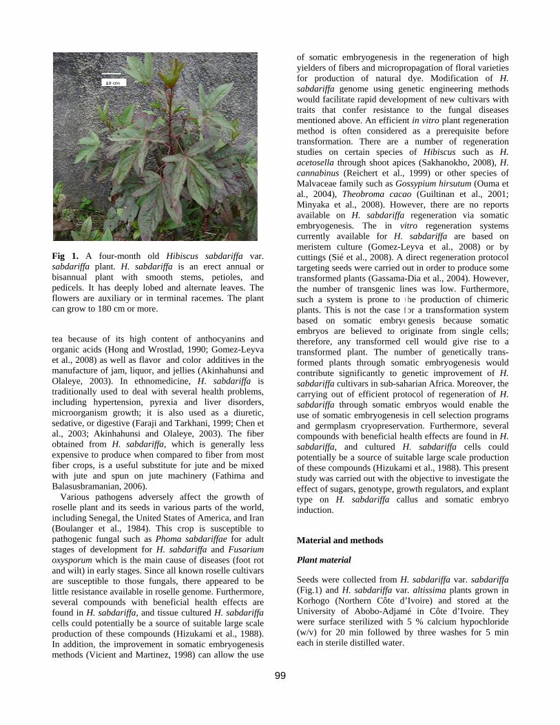
Fig 1. A four-month old Hibiscus sabdariffa var. sabdariffa plant. H. sabdariffa is an erect annual or bisannual plant with smooth stems, petioles, and pedicels. It has deeply lobed and alternate leaves. The flowers are auxiliary or in terminal racemes. The plant can grow to 180 cm or more.
tea because of its high content of anthocyanins and organic acids (Hong and Wrostlad, 1990; Gomez-Leyva et al., 2008) as well as flavor and color additives in the manufacture of jam, liquor, and jellies (Akinhahunsi and Olaleye, 2003). In ethnomedicine, H. sabdariffa is traditionally used to deal with several health problems, including hypertension, pyrexia and liver disorders, microorganism growth; it is also used as a diuretic, sedative, or digestive (Faraji and Tarkhani, 1999; Chen et al., 2003; Akinhahunsi and Olaleye, 2003). The fiber obtained from H. sabdariffa, which is generally less expensive to produce when compared to fiber from most fiber crops, is a useful substitute for jute and be mixed with jute and spun on jute machinery (Fathima and Balasusbramanian, 2006).
Various pathogens adversely affect the growth of roselle plant and its seeds in various parts of the world, including Senegal, the United States of America, and Iran (Boulanger et al., 1984). This crop is susceptible to pathogenic fungal such as Phoma sabdariffae for adult stages of development for H. sabdariffa and Fusarium oxysporum which is the main cause of diseases (foot rot and wilt) in early stages. Since all known roselle cultivars are susceptible to those fungals, there appeared to be little resistance available in roselle genome. Furthermore, several compounds with beneficial health effects are found in H. sabdariffa, and tissue cultured H. sabdariffa cells could potentially be a source of suitable large scale production of these compounds (Hizukami et al., 1988). In addition, the improvement in somatic embryogenesis methods (Vicient and Martinez, 1998) can allow the use
of somatic embryogenesis in the regeneration of high yielders of fibers and micropropagation of floral varieties for production of natural dye. Modification of H. sabdariffa genome using genetic engineering methods would facilitate rapid development of new cultivars with traits that confer resistance to the fungal diseases mentioned above. An efficient in vitro plant regeneration method is often considered as a prerequisite before transformation. There are a number of regeneration studies on certain species of Hibiscus such as H. acetosella through shoot apices (Sakhanokho, 2008), H. cannabinus (Reichert et al., 1999) or other species of Malvaceae family such as Gossypium hirsutum (Ouma et al., 2004), Theobroma cacao (Guiltinan et al., 2001; Minyaka et al., 2008). However, there are no reports available on H. sabdariffa regeneration via somatic embryogenesis. The in vitro regeneration systems currently available for H. sabdariffa are based on meristem culture (Gomez-Leyva et al., 2008) or by cuttings (Sié et al., 2008). A direct regeneration protocol targeting seeds were carried out in order to produce some transformed plants (Gassama-Dia et al., 2004). However, the number of transgenic lines was low. Furthermore, such a system is prone to the production of chimeric plants. This is not the case for a transformation system based on somatic embryogenesis because somatic embryos are believed to originate from single cells; therefore, any transformed cell would give rise to a transformed plant. The number of genetically trans- formed plants through somatic embryogenesis would contribute significantly to genetic improvement of H. sabdariffa cultivars in sub-saharian Africa. Moreover, the carrying out of efficient protocol of regeneration of H. sabdariffa through somatic embryos would enable the use of somatic embryogenesis in cell selection programs and germplasm cryopreservation. Furthermore, several compounds with beneficial health effects are found in H. sabdariffa, and cultured H. sabdariffa cells could potentially be a source of suitable large scale production of these compounds (Hizukami et al., 1988). This present study was carried out with the objective to investigate the effect of sugars, genotype, growth regulators, and explant type on H. sabdariffa callus and somatic embryo induction.
Material and methods
Plant material
Seeds were collected from H. sabdariffa var. sabdariffa (Fig.1) and H. sabdariffa var. altissima plants grown in Korhogo (Northern Côte d’Ivoire) and stored at the University of Abobo-Adjamé in Côte d’Ivoire. They were surface sterilized with 5 % calcium hypochloride (w/v) for 20 min followed by three washes for 5 min each in sterile distilled water.
100
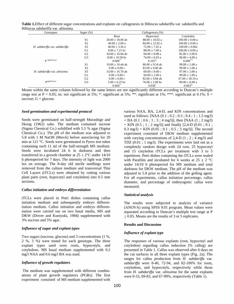
Table 1.Effect of different sugar concentrations and explants on callogenesis in Hibiscus sabdariffa var. sabdariffa and Hibiscus sabdariffa var. altissima.
Genotypes Sugar (%) Callogenesis (%) Root Hypocotyl Cotyledon S1 28.00 ± 18.00 ab 88.00 ± 10.02 a 100.00 ± 0.00 a S2 40.00 ± 2.00 a 84.00 ± 12.02 a 100.00 ± 0.00 a
H. sabdariffa var. sabdariffa S3 40.00 ± 3.50 a 72.00 ± 7.02 a 100.00 ± 0.00a G1 8.00 ± 7.21 bc 88.00 ± 7.00 a 100.00 ± 0.00 a G2 34.00 ± 16.04 ab 94.00 ± 8.08 a 82.00 ± 2.00 b G3 8.00 ± 10.58 bc 74.00 ± 6.03 a 96.00 ± 4.00 a
P significance 0.009** 0.102ns 0.000*** S1 19.00 ± 10.44 ab 80.00 ± 9.54 ab 99.00 ± 1.00 a S2 0.00 ± 0.00 c 83.00 ± 9.00 ab 99.00 ± 1.00 a
H. sabdariffa var. altissima S3 33.00 ± 16.70 ab 69.00 ± 9.00 c 97.00 ± 2.00 a G1 0.00 ± 0.00 c 94.00 ± 1.00 a 99.00 ± 1.00 a G2 0.00 ± 0.00 c 82.00 ± 9.84 ab 67.00 ± 30.19 a
Psignificance G3 5.00 ± 6.25 bc 0.004**
76.00 ± 2.00 bc 0.036*
89.00 ± 6.00 a 0.063ns
Means within the same column followed by the same letters are not significantly different according to Duncan’s multiple range test at P = 0.05; ns: not significant at 5%; *: significant at 5%; **: significant at 1%; ***: significant at 0.1%; S = sucrose; G = glucose.
Seed germination and experimental protocol
Seeds were germinated on half-strength Murashige and Skoog (1962) salts. The medium contained sucrose (Sigma Chemical Co.) solidified with 5.5 % agar (Sigma Chemical Co.). The pH of the medium was adjusted to 5.8 with 1 M NaOH (Merck) before autoclaving for 20 min at 121 °C. Seeds were germinated in Pyrex test tubes containing each 11 ml of the half-strength MS medium. Seeds were incubated 24 h in darkness and then transferred in a growth chamber at 25 ± 2°C under 14/10 h photoperiod for 7 days. The intensity of light was 2000 lux on average. The 8-day old sterile seedlings were removed from the culture medium and transversal Thin Cell Layers (tTCLs) were obtained by cutting various plant parts (root, hypocotyl and cotyledon) into 0.5 mm sections.
Callus initiation and embryo differentiation
tTCLs were placed in Petri dishes containing callus initiation medium and subsequently embryo differen- tiation medium. Callus initiation and embryo differen- tiation were carried out on two basal media, MS and DKW (Driver and Kuniyuki, 1984) supplemented with 3% sucrose and 5% agar.
Influence of sugar and explant types
Two sugars (sucrose, glucose) and 3 concentrations (1 %, 2 %, 3 %) were tested for each genotype. The three explant types used were roots, hypocotyls, and cotyledons. MS basal medium supplemented with 0.2 mg/l NAA and 0.6 mg/l BA was used.
Influence of growth regulators
The medium was supplemented with different combin- ations of plant growth regulators (PGRs). The first experiment consisted of MS medium supplemented with
various NAA, BA, 2,4-D, and KIN concentrations and used as follows: [NAA (0.1 ; 0.2 ; 0.3 ; 0.4 ; 1 ; 1.5 mg/l) + BA (0.1 ; 0.6 ; 1 ; 3 ; 4 mg/l)], then [NAA (1 ; 2 mg/l) + KIN (0.5 ; 1 ; 2 mg/l)] and finally [2,4-D (0.01 ; 0.1 ; 0.3 mg/l) + KIN (0.05 ; 0.1 ; 0.5 ; 3 mg/l)]. The second experiment consisted of DKW medium supplemented with varying concentrations of 2,4-D (1 ; 2 ; 4 mg/l) and TDZ (0.01 ; 1 mg/l). The experiments were laid out as a completely random design with 24 root, 25 hypocotyl and 15 cotyledon tTCLs per treatment with 3 to 5 repetitions. Petri dishes containing the tTCLs were sealed with Parafilm and incubated for 6 weeks at 25 ± 2 °C under 14/10 h photoperiod for MS medium and total darkness for DKW medium. The pH of the medium was adjusted to 5.8 prior to the addition of the gelling agent. For all experiments, callus initiation percentage, callus diameter, and percentage of embryogenic callus were measured.
Statistical analysis
The results were subjected to analysis of variance (ANOVA) using SPSS 9.01 program. Mean values were separated according to Duncan’s multiple test range at P < 0.05. Means are the results of 3 or 5 replicates.
Results and Discussion
Influence of explant type
The responses of various explants (root, hypocotyl and cotyledon) regarding callus induction (% callog) are presented in Table 1. Callus was observed after 6 days on the cut surfaces in all three explant types (Fig. 2a). The ranges for callus production from H. sabdariffa var. sabdariffa were 8-40, 72-94, and 82-100% for roots, cotyledons, and hypocotyls, respectively while those from H. sabdariffa var. altissima for the same explants were 0-33, 69-83, and 67-99%, respectively (Table 1).
101
Table 2. Effect of different NAA/BA combinations and explants on callus and somatic embryo initiation in Hibiscus sabdariffa var. sabdariffa and Hibiscus sabdariffa var altissima.
Genotypes PGR: NAA/BA (mg/l) Explant Callogenesis (%) Caldiam (mm) Embcal (%) MS1: 0.1/0.1 Psignificance Hypocotyl
Cotyledon
41.29 ± 41.95 b 97.89 ± 2.88 a
0.039*
6.00 ± 0.94 b 7.90 ± 0.42 a
0.007**
- -
MS2: 0/0.1 Psignificance Hypocotyl Cotyledon
37.21 ± 17.86 a 25.85 ± 10.62 a
0.316ns
3.25 ± 0.29 a 3.25 ± 0.18 a
1.000ns
- -
MS3: 1/3 Psignificance Hypocotyl Cotyledon
96.77 ± 5.59 a 98.24 ± 4.29 a
0.633ns
8.60 ± 2.63 a 11.16 ± 2.06 a
0.103ns
- -
H.sabdariffa var. altissima
MS4: 0.4/4 Psignificance Hypocotyl Cotyledon
91.61 ± 9.84 a 100.00 ± 0.00 a
0.203ns
6.90 ± 0.74 a 7.33 ± 0.76 a
0.458ns
- -
MS5: 0.3/3 Psignificance Hypocotyl Cotyledon
84.95 ± 8.12 a 86.82 ± 9.19 a
0.804ns
5.67 ± 0.76 a 5.83 ± 2.52 a
0.802ns
- -
S6: 0.2/0.6 Psignificance Hypocotyl Cotyledon
45.83 ± 7.86 a 57.57 ± 30.59 a
0.555ns
5.83 ± 2.52 a 5.83 ± 1.76 a
1.000ns
- -
MS7: 1.5/1 Psignificance Hypocotyl Cotyledon
85.05 ± 21.34 a 67.80 ± 7.62 a
0.179ns
7.12 ± 0748 a 6.25 ± 0.65 a
0.072ns
- -
MS1: 0.1/0.1 Psignificance Hypocotyl Cotyledon
96.77 ± 3.23 a 100.00 ± 0.00 a
0.056ns
6.10 ± 0.89 b 7.90 ± 0.42 a
0.004**
- -
MS2: 0/0.1 Psignificance Hypocotyl Cotyledon
38.06 ± 10.05 b 81.05 ± 13.21 a
0.000***
2.80 ±0.27 b 6.90 ± 0.74 a
0.000***
- -
MS3: 1/3 Psignificance Hypocotyl Cotyledon
97.41 ± 2.70 a 100.00 ± 0.00 a
0.099ns
7.40 ± 0.55 a 10.00 ± 1.41 a
0.007**
- -
H. sabdariffa var. sabdariffa
MS4: 0.4/4 Psignificance Hypocotyl Cotyledon
85.16 ± 8.72 b 98.50 ± 3.98 a
0.023*
6.30 ± 0.57 b 8.42 ± 0.88 a
0.001***
- -
MS5: 0.3/3 Psignificance Hypocotyl Cotyledon
91.59 ± 4.47 a 97.50 ± 5.00 a
0.129ns
8.13 ± 1.11 a 7.25 ± 0.65 a
0.130ns
- -
MS6: 0.2/0.6 Psignificance Hypocotyl Cotyledon
73.27 ± 13.65 a 92.64 ±8.82 a
0.055ns
7.87 ± 0.48 a 7.87 ± 0.63 a
1.000ns
- -
MS7: 1.5/1 Psignificance Hypocotyl Cotyledon
88.74 ± 10.60 a 95.59 ± 2.94 a
0.253ns
6.12 ± 0.85 a 7.12 ± 0.85 a
0.149ns
- -
Means within the same column followed by the same letters are not significantly different according to Duncan’s multiple range test at P = 0.05; ns: not significant at 5%; * : significant at 5%; ** : significant at 1%; *** : significant at 0.1%. Overall, more callus was produced with the hypocotyl and cotyledon tTCLs than with roots. These results reflect the existence of a large inter-explant variability in callusing responses. This inter-explant variability has also been reported in other species (Zouine and El-Hadrami, 2004; Dhar and Joshi, 2005; Zouzou et al., 2008). The high callus producing capacity of cotyledon in comparison to hypocotyl and root is probably due to the nutritive reserves and anatomical structure which are similar to leaf. Our results are not in agreement with those published by other authors who showed that hypocotyl was more callogenic compared to root and cotyledon explants (Zhang et al., 2001; Zouzou et al., 2008). Only the hypocotyl and cotyledon explants were retained in this study for the subsequent experiments on callus and somatic embryo initiation in H. sabdariffa because they produced significantly more callus than root
explants. However, it is worth mentioning that secondary medicinal metabolites such as ginsenosides have been obtained from root-derived calli in some plant species; the production of such compounds is, however, affected by several factors, such as carbohydrate source and growth regulators (Vanisree et al., 2004). Therefore, a study aimed at maximizing the production of such secondary metabolites should also focus on root-derived calli.
Influence of sugars
Both sugar types and concentration influenced callus induction. Indeed, callus mass was initiated within 6 (glucose) to 11 (sucrose) days directly on the cut surfaces in 3 types of tTCLs on MS basal medium supplemented with 0.2 mg/l NAA, 0.6 mg/l BA (Table 1). For H. sabd-
102
Table 3. Effect of different NAA/KIN combinations and explants on callus and somatic embryo initiation in Hibiscus sabdariffa var. sabdariffa and Hibiscus sabdariffa var. altissima.
Genotypes PGR: NAA/KIN (mg/l)
Explant Callogenesis (%) Caldiam (mm)
Embcal (%)
MS’1: 1/0.5 Psignificance Hypocotyl Cotyledon
96.20 ± 6.94 a 93.80 ± 11.28 a
0.696ns
5.80 ± 0.45 a 4.90 ± 1.08 a
0.087ns
- -
H.sabdariffa var. altissima
MS’2: ½ Psignificance Hypocotyl Cotyledon
94.40 ± 4.33 b 100.00 ± 0.00 a
0.045*
5.50 ± 0.35 b 8.00 ± 0.61 a
0.000***
- -
MS’3: 2/1 Psignificance Hypocotyl Cotyledon
27.33 ± 13.65 a 3.67 ± 6.35 b
0.050*
3.00 ± 0.50 a 2.89 ± 0.50 a
0.986ns
- -
MS’1: 1/0.5 Psignificance Hypocotyl
Cotyledon 76.20 ± 8.67 a
82.00 ± 22.28 a 0.602ns
5.60 ± 0.22 b 7.20 ± 0.57 a
0.000***
- -
H. sabdariffa
var. sabdariffa
MS’2 : ½ Psignificance Hypocotyl Cotyledon
95.00 ± 6.63 a 100.00 ± 0.00 a
0.130ns
5.90 ± 0.65 b 7.50 ± 0.71 a
0.006**
- -
MS’3 : 2/1 Psignificance Hypocotyl Cotyledon
31.33 ± 25.50 a 2.75 ± 5.50 a
0.075ns
3.00 ± 0.50 a 2.00 ± 0.91 a
0.152ns
- -
Means within the same column followed by the same letters are not significantly different according to Duncan’s multiple range test at P = 0.05; ns: not significant at 5%; * : significant at 5%; ** : significant at 1%; *** : significant at 0.1%.
Table 4. Effect of different 2,4-D/KIN combinations and explants on callus and somatic embryo initiation in Hibiscus sabdariffa var. sabdariffa and Hibiscus sabdariffa var. altissima.
Genotypes PGR: 2,4-D/KIN (mg/l)
Explant Callogenesis (%) Caldiam (mm) Embcal (%)
MS’’1: 0.1/05 Psignificance Hypocotyl Cotyledon
66.20 ± 28.50 a 29.60 ± 31.44 a
0.090ns
6.50 ± 0.93 a 3.90 ± 1.24 b
0.006**
- -
H.sabdariffa
MS”2: 0.01/0.05 Psignificance Hypocotyl Cotyledon
40.50 ± 16.26 a 8.75 ±5.30 b
0.011*
2.75 ±0.35 a 2.00 ± 0.00 b
0.007**
- -
var. altissima MS”3: 0.3/3 Psignificance Hypocotyl Cotyledon
73.00 ± 13.44 a 61.00 ± 15.60 a
0.288ns
8.25 ± 3.12 a 7.87 ±1.03 a
0.827ns
- -
MS”4: 0.1/0.1 Psignificance Hypocotyl Cotyledon
30.00 ± 2.83 a 5.00 ± 0.00 b
0.000***
4.00 ± 0.71 a 2.00 ± 0.87 b
0.050*
- -
MS’’1: 0.1/05 Psignificance Hypocotyl
Cotyledon 69.80 ± 29.65 a 38.60 ± 22.69 a
0.099ns
10.80 ± 1.15 a 5.70 ± 1.30 b
0.000***
- -
H. sabdariffa
MS”2: 0.01/0.05 Psignificance Hypocotyl Cotyledon
37.00 ± 40.44 a 11.25 ± 18.03 a
0.289ns
1.75 ± 1.19 a 1.12 ± 1.44 a
0.528ns
- -
var. sabdariffa
MS”3: 0.3/3 Psignificance Hypocotyl Cotyledon
50.00 ± 3.94 b 73.20 ± 4.55 a
0.000***
11.60 ± 2.68 a 8.00 ± 0.35 b
0.018*
- -
MS”4: 0.1/0.1 Psignificance Hypocotyl Cotyledon
78.50 ± 34.59 a 10.75 ± 5.91 b
0.008**
7.00 ± 0.91 a 4.25 ± 1.55 b
0.022*
- -
Means within the same column followed by the same letters are not significantly different according to Duncan’s multiple range test at P = 0.05; ns : not significant at 5%; *: significant at 5%; ** : significant at 1%;*** : significant at 0.1%.
ariffa var. sabdariffa, the percentages of callus induction in sucrose-containing media were 72% (3% sucrose) to 88% (1% sucrose) for hypocotyl explants and 100% callus induction was achieved with all three sucrose concentrations using the cotyledon explants (Table 1). For the same species, the percentages of callus production ranged from 74 to 94% (hypocotyl) and 82 to
100% (cotyledon) for the glucose-containing media. Similar results were observed with H. sabdariffa var. altissima. In general, the 3% sucrose produced more callus with both genotypes and all three explant types (Table 1). The beneficial effect of sucrose on callogenesis was also reported in many plants (Dhar and Joshi, 2005; Gopi and Vatsala, 2006). The 3% sucrose
103
Table 5. Effect of different 2,4-D/TDZ combinations and explants on callus and somatic embryo initiation in Hibiscus sabdariffa var. sabdariffa and Hibiscus sabdariffa var. altissima.
Genotypes PGR: 2,4-D/TDZ (mg/l)
Explants Callogenesis (%) Caldiam (mm) Embcal (%)
DKW1: 2/1 Psignificance Hypocotyl Cotyledon
100.00 ± 0.00 100.00 ± 0.00
-
6.00 ± 2.06 a 8.50 ± 2.55 a
0.127ns
- -
H.sabdariffa
DKW2: 1/0.01 Psignificance Hypocotyl Cotyledon
100.00 ± 0.00 100.00 ± 0.00
-
6.62 ± 2.14 a 8.12 ± 2.17 a
0.363ns
0.00 ± 0.00 a 1.78 ± 3.57 a
0.356ns var. altissima DKW3: 4/1 Psignificance Hypocotyl
Cotyledon 95.00 ± 10.00 a 100.00 ± 0.00 a
0.391ns
8.25 ± 2.78 a 10.37 ± 2.39 a
0.291ns
11.55 ± 14.06 a 1.66 ± 3.33 a
0.220ns DKW4: 1/0.5 Psignificance Hypocotyl
Cotyledon 100.00 ± 0.00 100.00 ± 0.00
-
4.34 ± 3.64 a 7.92 ± 2.69 a
0.082ns
10.83 ± 23.42 a 12.12 ± 29.69 a
0.935ns
DKW1: 2/1 Psignificance Hypocotyl
Cotyledon 100.00 ± 0.00 100.00 ± 0.00
-
8.00 ± 0.93 b 13.80 ± 3.96 a
0.013*
- -
H. sabdariffa
DKW2: 1/0.01 Psignificance Hypocotyl Cotyledon
100.00 ± 0.00 100.00 ± 0.00
-
7.90 ± 0.96 a 10.80 ± 3.60 a
0.120ns
- -
var. sabdariffa DKW3: 4/1 Psignificance Hypocotyl Cotyledon
100.00 ± 0.00 100.00 ± 0.00
-
9.92 ± 1.83 b 16.58 ± 3.14 a
0.001***
11.46 ± 28.07 a 0.00 ± 0.00 a
0.363ns DKW4: 1/0.5 Psignificance Hypocotyl
Cotyledon 88.93 ± 9.70 a 97.92 ± 5.10 a
0.072ns
7.08 ± 1.02 a 7.83 ± 2.56 a
0.520ns
0.87 ± 2.15 a 2.08 ± 5.10 a
0.605ns
Means within the same column followed by the same letters are not significantly different according to Duncan’s multiple range test at P = 0.05;ns : not significant at 5%; *** : significant at 0.1%.
concentration may have a high osmotic pressure on cytoplasm of cells, thus inducing stress and callogenesis. Similar results were recorded with Saccharum sp (Errabii et al., 2006).
Influence of PGRs
Hypocotyl and cotyledon explants were cultured on MS basal medium containing different auxin and cytokinin combinations for callus initiation, callus diameter, and somatic embryo initiation (Tables 2 and 3). The results indicated that all treatments induced callus. However, differences based on PGR regime and explant type were observed. From a total of 15 combinations of PGRs tested, high callus production was obtained in MS medium supplemented with 0.4 mg/l NAA + 4 mg/l BA (MS4 medium), 1 mg/l NAA + 3 mg/l BA (MS3 medium) and 1mg/l NAA + 2 mg/l KIN (MS’2) for H. sabdariffa var. altissima. The percentage of callus induction obtained with the cotyledon explants was 98.24, 100, and 100 % for MS3, MS4 and MS’2, respectively (Tables 2 and 3). For hypocotyl explants, callus induction was 91.61, 96.77, and 94.40 for MS4,
MS3 and MS’2, respectively for the same genotype. In H.sabdariffa var. sabdariffa, MS1 (0.1 mg/l NAA + 0.1 mg/l BA), MS5 (0.3 mg/l NAA + 3 mg/l BA) and MS’2 induced high callogenesis (Tables 2 and 3). In this genotype, callus induction obtained with cotyledon explants was 97.50, 97.89, and 100 % in MS5, MS1 and MS’2, respectively, and 91.59, 95.00, and 96.77% in MS5, MS’2 and MS1, respectively for hypocotyl eplants (Tables 2 and 3). The results obtained with the 2,4-D/NAA combinations are in agreement with the response obtained in Gossypium hirsutum (Zouzou et al., 2008). In H. sabdariffa var. altissima (Table 4), callus induction percentages were 66.20% in MS’’1 (0.1 mg/l 2,4-D + 0.5 mg/l KIN) and 73.00 % in MS’’3 (0.3 mg/l 2,4-D + 3 mg/l KIN). For H. sabdariffa var. sabdariffa, the percentage of callus induction was 69.80% in MS’’1 (0.1 mg/l 2,4-D + 0.5 mg/l KIN) and 50.00% in MS’’3 (0.3 mg/l 2,4-D + 3mg/l KIN). Compared to results obtained with the auxin NAA, the combinations involving 2,4-D seem to be unfavourable to callogenesis induction. Nevertheless, in previous works, it was reported that 2,4-D was an essential growth regulator for the induction of callogenesis in cotton (Trolinder and Goodin, 1988 ; Lee et al., 2004 ; Sun et al., 2006 ; Zouzou et al., 2008) and
104
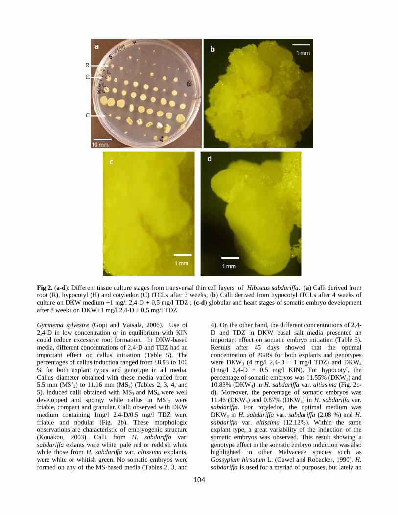
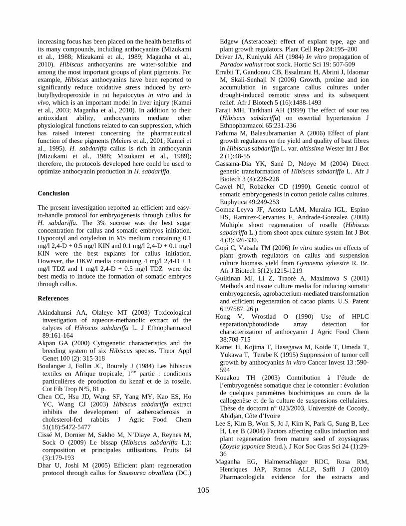
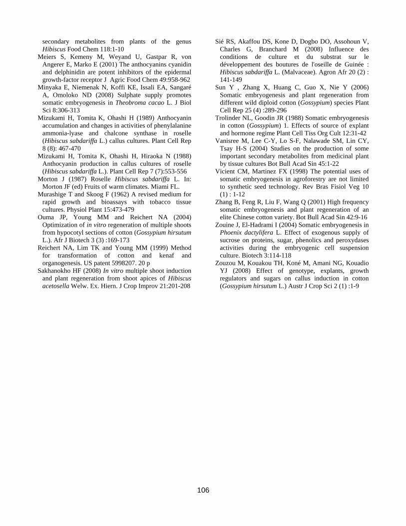
Fig 2. (a-d): Different tissue culture stages from transversal thin cell layers of Hibiscus sabdariffa. (a) Calli derived from root (R), hypocotyl (H) and cotyledon (C) tTCLs after 3 weeks; (b) Calli derived from hypocotyl tTCLs after 4 weeks of culture on DKW medium +1 mg/l 2,4-D + 0,5 mg/l TDZ ; (c-d) globular and heart stages of somatic embryo development after 8 weeks on DKW+1 mg/l 2,4-D + 0,5 mg/l TDZ Gymnema sylvestre (Gopi and Vatsala, 2006). Use of 2,4-D in low concentration or in equilibrium with KIN could reduce excessive root formation. In DKW-based media, different concentrations of 2,4-D and TDZ had an important effect on callus initiation (Table 5). The percentages of callus induction ranged from 88.93 to 100 % for both explant types and genotype in all media. Callus diameter obtained with these media varied from 5.5 mm (MS’2) to 11.16 mm (MS3) (Tables 2, 3, 4, and 5). Induced calli obtained with MS3 and MS4 were well developped and spongy while callus in MS’2 were friable, compact and granular. Calli observed with DKW medium containing 1mg/l 2,4-D/0.5 mg/l TDZ were friable and nodular (Fig. 2b). These morphologic observations are characteristic of embryogenic structure (Kouakou, 2003). Calli from H. sabdariffa var. sabdariffa exlants were white, pale red or reddish white while those from H. sabdariffa var. altissima explants, were white or whitish green. No somatic embryos were formed on any of the MS-based media (Tables 2, 3, and
4). On the other hand, the different concentrations of 2,4-D and TDZ in DKW basal salt media presented an important effect on somatic embryo initiation (Table 5). Results after 45 days showed that the optimal concentration of PGRs for both explants and genotypes were DKW3 (4 mg/l 2,4-D + 1 mg/l TDZ) and DKW4 (1mg/l 2,4-D + 0.5 mg/l KIN). For hypocotyl, the percentage of somatic embryos was 11.55% (DKW3) and 10.83% (DKW4) in H. sabdariffa var. altissima (Fig. 2c-d). Moreover, the percentage of somatic embryos was 11.46 (DKW3) and 0.87% (DKW4) in H. sabdariffa var. sabdariffa. For cotyledon, the optimal medium was DKW4 in H. sabdariffa var. sabdariffa (2.08 %) and H. sabdariffa var. altissima (12.12%). Within the same explant type, a great variability of the induction of the somatic embryos was observed. This result showing a genotype effect in the somatic embryo induction was also highlighted in other Malvaceae species such as Gossypium hirsutum L. (Gawel and Robacker, 1990). H. sabdariffa is used for a myriad of purposes, but lately an
105
increasing focus has been placed on the health benefits of its many compounds, including anthocyanins (Mizukami et al., 1988; Mizukami et al., 1989; Maganha et al., 2010). Hibiscus anthocyanins are water-soluble and among the most important groups of plant pigments. For example, Hibiscus anthocyanins have been reported to significantly reduce oxidative stress induced by tert-butylhydroperoxide in rat hepatocytes in vitro and in vivo, which is an important model in liver injury (Kamei et al., 2003; Maganha et al., 2010). In addition to their antioxidant ability, anthocyanins mediate other physiological functions related to can suppression, which has raised interest concerning the pharmaceutical function of these pigments (Meiers et al., 2001; Kamei et al., 1995). H. sabdariffa callus is rich in anthocyanin (Mizukami et al., 1988; Mizukami et al., 1989); therefore, the protocols developed here could be used to optimize anthocyanin production in H. sabdariffa.
Conclusion
The present investigation reported an efficient and easy-to-handle protocol for embryogenesis through callus for H. sabdariffa. The 3% sucrose was the best sugar concentration for callus and somatic embryos initiation. Hypocotyl and cotyledon in MS medium containing 0.1 mg/l 2,4-D + 0.5 mg/l KIN and 0.1 mg/l 2,4-D + 0.1 mg/l KIN were the best explants for callus initiation. However, the DKW media containing 4 mg/l 2,4-D + 1 mg/l TDZ and 1 mg/l 2,4-D + 0.5 mg/l TDZ were the best media to induce the formation of somatic embryos through callus.
References
Akindahunsi AA, Olaleye MT (2003) Toxicological investigation of aqueous-methanolic extract of the calyces of Hibiscus sabdariffa L. J Ethnopharmacol 89:161-164
Akpan GA (2000) Cytogenetic characteristics and the breeding system of six Hibiscus species. Theor Appl Genet 100 (2): 315-318
Boulanger J, Follin JC, Bourely J (1984) Les hibiscus textiles en Afrique tropicale, 1ère partie : conditions particulières de production du kenaf et de la roselle. Cot Fib Trop N°5, 81 p.
Chen CC, Hsu JD, Wang SF, Yang MY, Kao ES, Ho YC, Wang CJ (2003) Hibiscus sabdariffa extract inhibits the development of astherosclerosis in cholesterol-fed rabbits J Agric Food Chem 51(18):5472-5477
Cissé M, Dornier M, Sakho M, N’Diaye A, Reynes M, Sock O (2009) Le bissap (Hibiscus sabdariffa L.): composition et principales utilisations. Fruits 64 (3):179-193
Dhar U, Joshi M (2005) Efficient plant regeneration protocol through callus for Saussurea obvallata (DC.)
Edgew (Asteraceae): effect of explant type, age and plant growth regulators. Plant Cell Rep 24:195–200
Driver JA, Kuniyuki AH (1984) In vitro propagation of Paradox walnut root stock. Hortic Sci 19: 507-509
Errabii T, Gandonou CB, Essalmani H, Abrini J, Idaomar M, Skali-Senhaji N (2006) Growth, proline and ion accumulation in sugarcane callus cultures under drought-induced osmotic stress and its subsequent relief. Afr J Biotech 5 (16):1488-1493
Faraji MH, Tarkhani AH (1999) The effect of sour tea (Hibiscus sabdariffa) on essential hypertension J Ethnopharmacol 65:231-236
Fathima M, Balasubramanian A (2006) Effect of plant growth regulators on the yield and quality of bast fibres in Hibiscus sabdariffa L. var. altissima Wester Int J Bot 2 (1):48-55
Gassama-Dia YK, Sané D, Ndoye M (2004) Direct genetic transformation of Hibiscus sabdariffa L. Afr J Biotech 3 (4):226-228
Gawel NJ, Robacker CD (1990). Genetic control of somatic embryogenesis in cotton petiole callus cultures. Euphytica 49:249-253
Gomez-Leyva JF, Acosta LAM, Muraira IGL, Espino HS, Ramirez-Cervantes F, Andrade-Gonzalez (2008) Multiple shoot regeneration of roselle (Hibiscus sabdariffa L.) from shoot apex culture system Int J Bot 4 (3):326-330.
Gopi C, Vatsala TM (2006) In vitro studies on effects of plant growth regulators on callus and suspension culture biomass yield from Gymnema sylvestre R. Br. Afr J Biotech 5(12):1215-1219
Guiltinan MJ, Li Z, Traoré A, Maximova S (2001) Methods and tissue culture media for inducing somatic embryogenesis, agrobacterium-mediated transformation and efficient regeneration of cacao plants. U.S. Patent 6197587. 26 p
Hong V, Wrostlad O (1990) Use of HPLC separation/photodiode array detection for characterization of anthocyanin J Agric Food Chem 38:708-715
Kamei H, Kojima T, Hasegawa M, Koide T, Umeda T, Yukawa T, Terabe K (1995) Suppression of tumor cell growth by anthocyanins in vitro Cancer Invest 13 :590-594
Kouakou TH (2003) Contribution à l’étude de l’embryogenèse somatique chez le cotonnier : évolution de quelques paramètres biochimiques au cours de la callogenèse et de la culture de suspensions cellulaires. Thèse de doctorat n° 023/2003, Université de Cocody, Abidjan, Côte d’Ivoire
Lee S, Kim B, Won S, Jo J, Kim K, Park G, Sung B, Lee H, Lee B (2004) Factors affecting callus induction and plant regeneration from mature seed of zoysiagrass (Zoysia japonica Steud.). J Kor Soc Gras Sci 24 (1):29-36
Maganha EG, Halmenschlager RDC, Rosa RM, Henriques JAP, Ramos ALLP, Saffi J (2010) Pharmacologicla evidence for the extracts and
106
secondary metabolites from plants of the genus Hibiscus Food Chem 118:1-10
Meiers S, Kemeny M, Weyand U, Gastpar R, von Angerer E, Marko E (2001) The anthocyanins cyanidin and delphinidin are potent inhibitors of the epidermal growth-factor receptor J Agric Food Chem 49:958-962
Minyaka E, Niemenak N, Koffi KE, Issali EA, Sangaré A, Omoloko ND (2008) Sulphate supply promotes somatic embryogenesis in Theobroma cacao L. J Biol Sci 8:306-313
Mizukami H, Tomita K, Ohashi H (1989) Anthocyanin accumulation and changes in activities of phenylalanine ammonia-lyase and chalcone synthase in roselle (Hibiscus sabdariffa L.) callus cultures. Plant Cell Rep 8 (8): 467-470
Mizukami H, Tomita K, Ohashi H, Hiraoka N (1988) Anthocyanin production in callus cultures of roselle (Hibiscus sabdariffa L.). Plant Cell Rep 7 (7):553-556
Morton J (1987) Roselle Hibiscus sabdariffa L. In: Morton JF (ed) Fruits of warm climates. Miami FL.
Murashige T and Skoog F (1962) A revised medium for rapid growth and bioassays with tobacco tissue cultures. Physiol Plant 15:473-479
Ouma JP, Young MM and Reichert NA (2004) Optimization of in vitro regeneration of multiple shoots from hypocotyl sections of cotton (Gossypium hirsutum L.). Afr J Biotech 3 (3) :169-173
Reichert NA, Lim TK and Young MM (1999) Method for transformation of cotton and kenaf and organogenesis. US patent 5998207. 20 p
Sakhanokho HF (2008) In vitro multiple shoot induction and plant regeneration from shoot apices of Hibiscus acetosella Welw. Ex. Hiern. J Crop Improv 21:201-208
Sié RS, Akaffou DS, Kone D, Dogbo DO, Assohoun V, Charles G, Branchard M (2008) Influence des conditions de culture et du substrat sur le développement des boutures de l'oseille de Guinée : Hibiscus sabdariffa L. (Malvaceae). Agron Afr 20 (2) : 141-149
Sun Y , Zhang X, Huang C, Guo X, Nie Y (2006) Somatic embryogenesis and plant regeneration from different wild diploid cotton (Gossypium) species Plant Cell Rep 25 (4) :289-296
Trolinder NL, Goodin JR (1988) Somatic embryogenesis in cotton (Gossypium) 1. Effects of source of explant and hormone regime Plant Cell Tiss Org Cult 12:31-42
Vanisree M, Lee C-Y, Lo S-F, Nalawade SM, Lin CY, Tsay H-S (2004) Studies on the production of some important secondary metabolites from medicinal plant by tissue cultures Bot Bull Acad Sin 45:1-22
Vicient CM, Martinez FX (1998) The potential uses of somatic embryogenesis in agroforestry are not limited to synthetic seed technology. Rev Bras Fisiol Veg 10 (1) : 1-12
Zhang B, Feng R, Liu F, Wang Q (2001) High frequency somatic embryogenesis and plant regeneration of an elite Chinese cotton variety. Bot Bull Acad Sin 42:9-16
Zouine J, El-Hadrami I (2004) Somatic embryogenesis in Phoenix dactylifera L. Effect of exogenous supply of sucrose on proteins, sugar, phenolics and peroxydases activities during the embryogenic cell suspension culture. Biotech 3:114-118
Zouzou M, Kouakou TH, Koné M, Amani NG, Kouadio YJ (2008) Effect of genotype, explants, growth regulators and sugars on callus induction in cotton (Gossypium hirsutum L.) Austr J Crop Sci 2 (1) :1-9


![Callus formation and somatic embryogenesis in sugarcane ......new genetic variability for the determination of desired clones of sugarcane [7]. In-vitro culture technique is enriched](https://img.pdfslide.us/doc/110x75/60ee44f2791c330d4e227bb2/callus-formation-and-somatic-embryogenesis-in-sugarcane-new-genetic-variability.jpg)