Embed Size (px)
Citation preview
1616
Proteins and
Nucleic Acids
How do proteins function to maintain life? How do they grow?
How do they recognize each other? How does the structure
of DNA help explain how genetic information is encoded? Before
answering such questions, it is important to understand what pro-
teins and nucleic acids are.
PROTEIN STRUCTURE
Proteins come in many different sizes and shapes. For example,
cytochrome c, a protein that transfers electrons, has only one
polypeptide chain of 104 amino acids. Yet myosin, the protein that
makes muscles contract, has two polypeptide chains with some
2,000 amino acids each, connected by four smaller chains. It is
called a multimeric protein. No matter their size, all proteins have
a primary, secondary, and tertiary structure. Some also have
quaternary structure.
3
a
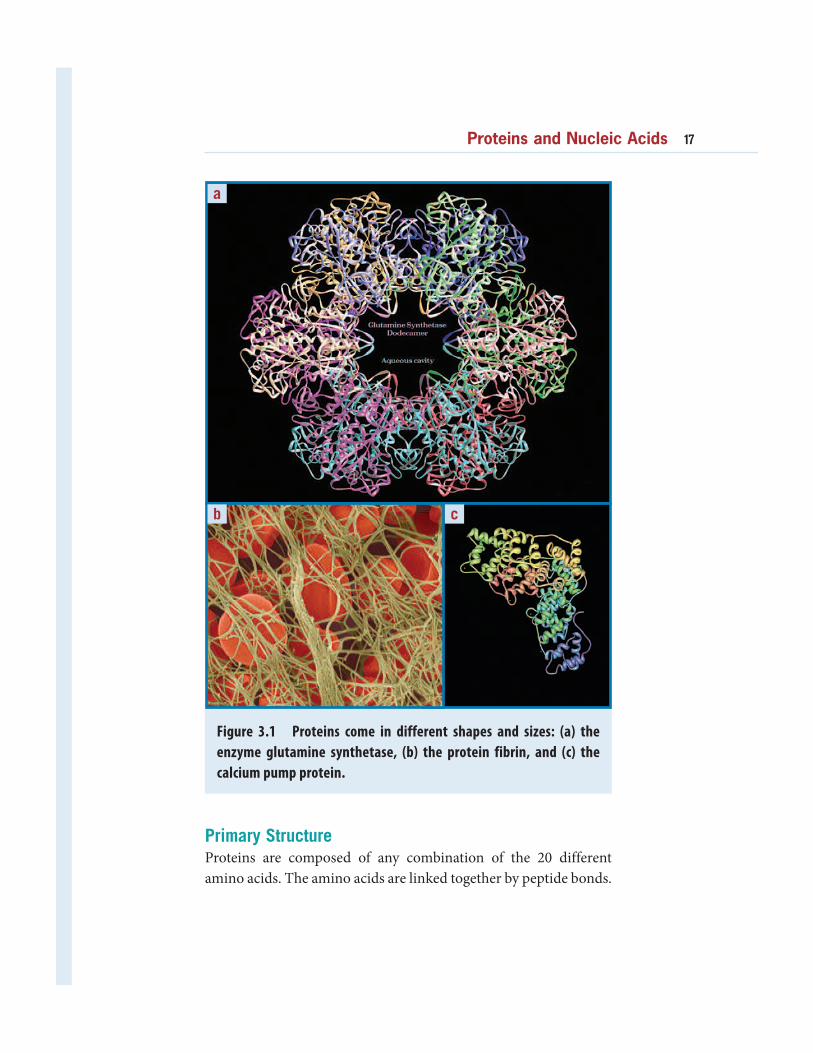
Figure 3.1 Proteins come in different shapes and sizes: (a) the
enzyme glutamine synthetase, (b) the protein fibrin, and (c) the
calcium pump protein.
cb
Primary StructureProteins are composed of any combination of the 20 different
amino acids. The amino acids are linked together by peptide bonds.
Proteins and Nucleic Acids 17
18 BIOCHEMISTRY
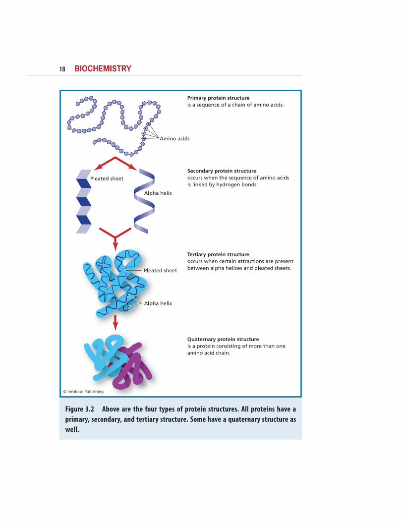
Figure 3.2 Above are the four types of protein structures. All proteins have a
primary, secondary, and tertiary structure. Some have a quaternary structure as
well.
The primary structure of a protein is the sequence of its amino
acids. For example, the first 10 amino acids in the cytochrome c
sequence are Ala-Ser-Phe-Ser-Glu-Ala-Pro-Gly-Asn-Pro, while
the first 10 amino acids in the myosin sequence are Phe-Ser-Asp-
Pro-Asp-Phe-Gln-Tyr-Leu-Ala. Therefore, the primary structure
is just the full sequence of amino acids in the polypeptide chain or
chains. Finding the primary structure of a protein is called protein
sequencing. The first protein to be sequenced was the hormone
insulin.
Secondary StructureThe polypeptide chains of proteins do not remain in a flat plane.
Instead, as a protein is formed, the polypeptide chain starts to twist
and curl up. It folds and coils like a rope that can be bundled in
many different shapes. This coiling and folding determines the pro-
tein’s secondary structure. The secondary structure is maintained
by chemical bonds between the carboxyl groups and the amino
groups in the polypeptide backbone. There are many secondary
structure patterns, but the two most common are the α−helix, and
the β−sheet.
The -helix
The α−helix (alpha helix) has a rod shape. The peptide is coiled
around an imaginary cylinder and held in shape by H-bonds
formed between components of the peptide bonds.
Because there are so many H-bonds in an α−helix, this struc-
ture is very stable and strong. Helices are common structures found
in most proteins.
The -sheet
Another folding pattern is the β−sheet (beta sheet). In this
arrangement, the amino acid chain zig-zags back and forth and
adopts the shape of a sheet of paper. Once again it is held together
by H-bonds.
Proteins and Nucleic Acids 19
20 BIOCHEMISTRY
Tertiary StructureOnce it has started folding, the protein eventually tightens into a
specific three-dimensional shape, called its tertiary structure. Just
like humans have unique sets of fingerprints, every protein has a
unique tertiary structure, which is responsible for its properties
and function. The tertiary structure is held together by bonds
between the R groups of the amino acids in the protein, and so
depends on the amino acid sequence. There are three kinds of
bonds involved in tertiary protein structure:
1. H bonds, which are weak. Since they are easy to break
and reform, they make a protein flexible.
2. Ionic bonds between R groups with positive or nega-
tive charges, which are quite strong.
a b
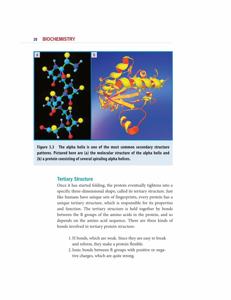
Figure 3.3 The alpha helix is one of the most common secondary structure
patterns. Pictured here are (a) the molecular structure of the alpha helix and
(b) a protein consisting of several spiraling alpha helices.
3. Disulfide bridges, the S-S bonds between two cyste-
ine amino acids, which are also strong.
Thus, the secondary structure is due to H bonds between back-
bone atoms and is independent of primary sequence; the tertiary
structure is due to bonds between R-group atoms and thus depends
on the amino acid sequence. For monomeric proteins, which have
only one amino acid chain, the tertiary structure completes the
three-dimensional description.
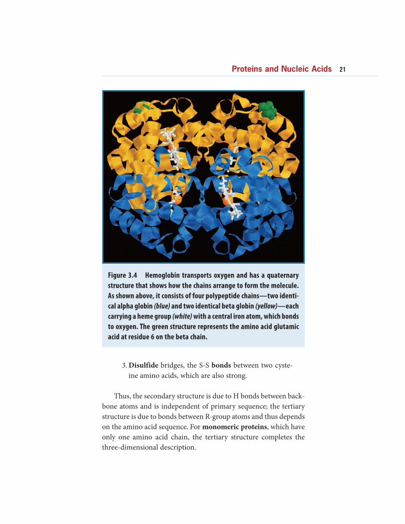
Figure 3.4 Hemoglobin transports oxygen and has a quaternary
structure that shows how the chains arrange to form the molecule.
As shown above, it consists of four polypeptide chains—two identi-
cal alpha globin (blue) and two identical beta globin (yellow)—each
carrying a heme group (white) with a central iron atom, which bonds
to oxygen. The green structure represents the amino acid glutamic
acid at residue 6 on the beta chain.
Proteins and Nucleic Acids 21
22 BIOCHEMISTRY
Quaternary StructureProteins that have more than one polypeptide chain require a
higher level of organization. In the quaternary structure, the
different chains are packed together to form the overall three-
dimensional structure of the protein. The individual polypeptide
chains can be arranged in a variety of shapes as part of the quater-
nary structure.
Globular or Fibrous StructuresThe final three-dimensional shape of a protein can be classified
as globular or fibrous. Globular means round, like a ball. Most
proteins are globular, including enzymes, membrane proteins, and
storage proteins. Fibrous proteins are elongated and look more
like ropes. Most have structural roles, such as collagen, the main
support of skin and bones. Fibrous proteins are usually composed
of many polypeptide chains. Some proteins have both fibrous and
globular components; for example, the muscle protein myosin has
a long fibrous tail and a globular head.
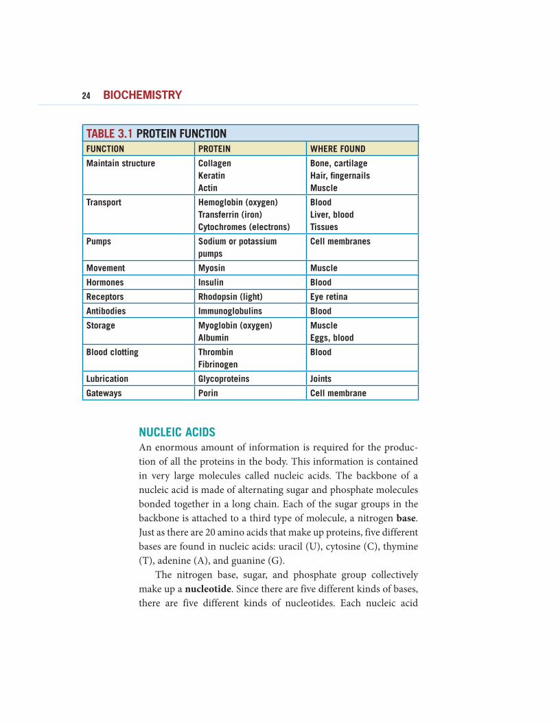
PROTEINS DO EVERYTHINGProteins are involved in all life processes in many different roles.
The human body makes about 50,000 different kinds of protein,
and each has a specific function.
The function of proteins depends on their structure. For
example, hemoglobin, the oxygen-carrying protein in red blood
cells, consists of four chains. Each chain contains one iron atom
and can bind one molecule of oxygen. Since the body needs dif-
ferent amounts of oxygen, the structure of hemoglobin makes
it easy to vary its capacity to bind oxygen and respond to what
the body needs. Immunoglobulins are proteins that function as
antibodies, which help the body fight disease and destroy foreign
invaders. Antibodies have four polypeptide chains arranged in a
Y-shape. This shape allows antibodies to link foreign substances
together, causing them to clump and lose their ability to harm the
body.
Actin is one of the proteins found in muscle. It consists of many
polypeptide chains arranged in a double helix to form long filaments
that are very strong. Tubulin is a protein that forms assemblies in the
form of hollow tubes called microtubules. The microtubules make
up cilia and flagella. Cilia are the short, hairlike structures that allow
some single-cell organisms to move and help transport materials in
larger organisms. In humans, cilia in the trachea, or windpipe, move
mucus out of the lungs. Flagella are the whiplike “tails” that propel
sperm cells, as well as some one-celled organisms.
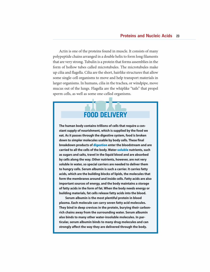
FOOD DELIVERY
The human body contains trillions of cells that require a con-
stant supply of nourishment, which is supplied by the food we
eat. As it passes through the digestive system, food is broken
down to simpler molecules usable by body cells. These final
breakdown products of digestion enter the bloodstream and are
carried to all the cells of the body. Water-soluble nutrients, such
as sugars and salts, travel in the liquid blood and are absorbed
by cells along the way. Other nutrients, however, are not very
soluble in water, so special carriers are needed to deliver them
to hungry cells. Serum albumin is such a carrier. It carries fatty
acids, which are the building blocks of lipids, the molecules that
form the membranes around and inside cells. Fatty acids are also
important sources of energy, and the body maintains a storage
of fatty acids in the form of fat. When the body needs energy or
building materials, fat cells release fatty acids into the blood.
Serum albumin is the most plentiful protein in blood
plasma. Each molecule can carry seven fatty acid molecules.
They bind in deep crevices in the protein, burying their carbon-
rich chains away from the surrounding water. Serum albumin
also binds to many other water-insoluble molecules. In par-
ticular, serum albumin binds to many drug molecules and can
strongly affect the way they are delivered through the body.
Proteins and Nucleic Acids 23
24 BIOCHEMISTRY
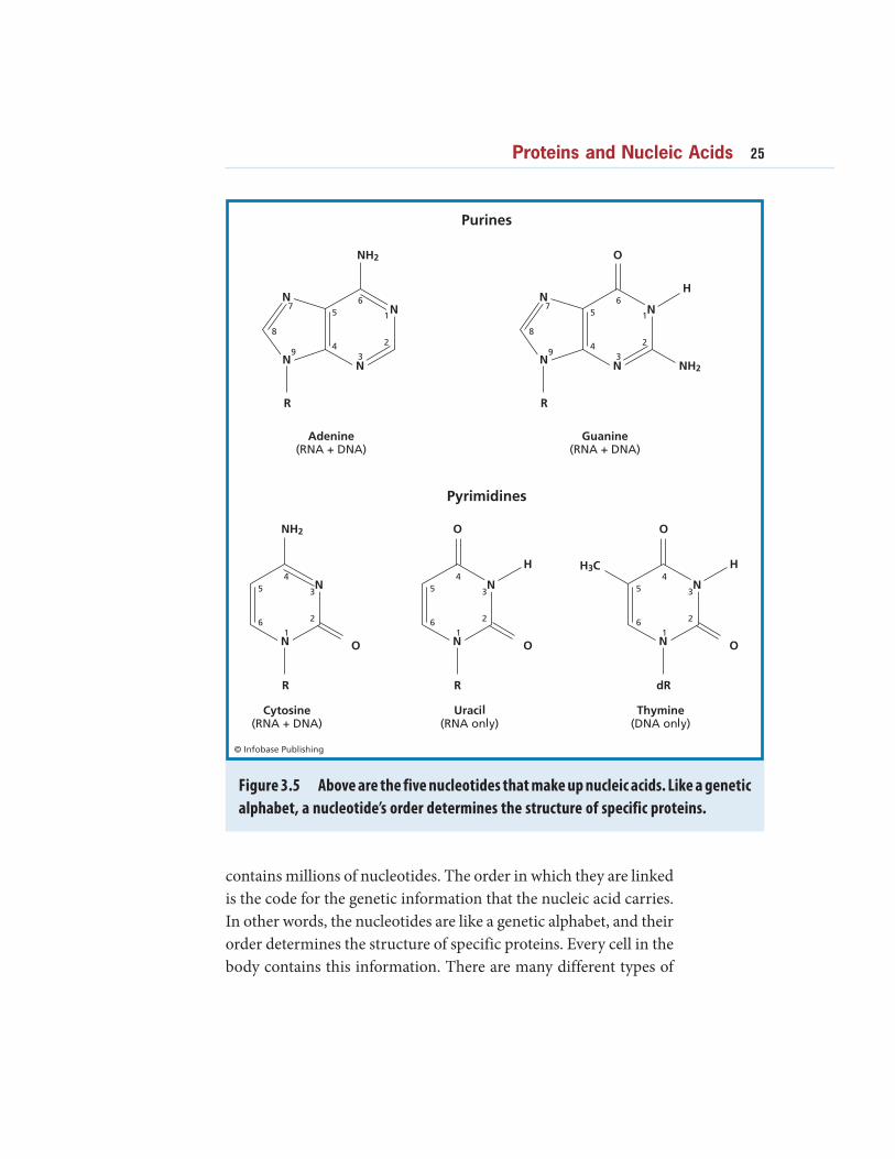
NUCLEIC ACIDSAn enormous amount of information is required for the produc-
tion of all the proteins in the body. This information is contained
in very large molecules called nucleic acids. The backbone of a
nucleic acid is made of alternating sugar and phosphate molecules
bonded together in a long chain. Each of the sugar groups in the
backbone is attached to a third type of molecule, a nitrogen base.
Just as there are 20 amino acids that make up proteins, five different
bases are found in nucleic acids: uracil (U), cytosine (C), thymine
(T), adenine (A), and guanine (G).
The nitrogen base, sugar, and phosphate group collectively
make up a nucleotide. Since there are five different kinds of bases,
there are five different kinds of nucleotides. Each nucleic acid
A Sampling of Isotopes
FUNCTION PROTEIN WHERE FOUND
Maintain structure Collagen
Keratin
Actin
Bone, cartilage
Hair, fingernails
Muscle
Transport Hemoglobin (oxygen)
Transferrin (iron)
Cytochromes (electrons)
Blood
Liver, blood
Tissues
Pumps Sodium or potassium
pumps
Cell membranes
Movement Myosin Muscle
Hormones Insulin Blood
Receptors Rhodopsin (light) Eye retina
Antibodies Immunoglobulins Blood
Storage Myoglobin (oxygen)
Albumin
Muscle
Eggs, blood
Blood clotting Thrombin
Fibrinogen
Blood
Lubrication Glycoproteins Joints
Gateways Porin Cell membrane
TABLE 3.1 PROTEIN FUNCTION
contains millions of nucleotides. The order in which they are linked
is the code for the genetic information that the nucleic acid carries.
In other words, the nucleotides are like a genetic alphabet, and their
order determines the structure of specific proteins. Every cell in the
body contains this information. There are many different types of
Figure 3.5 Above are the five nucleotides that make up nucleic acids. Like a genetic
alphabet, a nucleotide’s order determines the structure of specific proteins.
Proteins and Nucleic Acids 25
26 BIOCHEMISTRY
nucleic acids that help cells replicate and build proteins. The best
known are DNA and RNA.
DNA
In most living organisms, except viruses, genetic information is
stored in a molecule called deoxyribonucleic acid, or DNA. It gets
its name from the sugar group that it contains, deoxyribose. DNA
is made and found in the nucleus of living cells. The four nucleo-
tides found in DNA are: adenine (A), cytosine (C), guanine (G),
and thymine (T). These nucleotides form two long chains that twist
around each other in a spiral shape called a double helix.
The double helix has the ability to wind and unwind so that
the nucleic acid chain can duplicate itself. That duplication process
happens every time a cell divides.
The nucleotides in one strand of the double helix bond to
nucleotides in the other strand. This is called base-pairing. This
bonding is highly specific, because adenine nucleotides (A) always
bond to thymine (T), and guanine (G) always bonds to cytosine
(C). The double-stranded DNA molecule has a unique ability:
It can make exact copies of itself, in a process called replication.
When more DNA is needed, for example during reproduction or
growth, the H bonds between the nucleotides break, and the two
strands of the DNA molecule separate. New bases present in the
cell pair up with the bases on each of the two separate strands, thus
forming two new, double-stranded DNA molecules that are identi-
cal both to the original DNA molecule and to each other.
When a cell is not dividing, the DNA is not replicating and it
is in the form of loose white strings in the cell nucleus. The nucleic
acid strands are usually found uncoiled. To fit into the cell, the DNA
is cut into shorter lengths, and each length is tightly wrapped up
in a bundle called chromatin. During most of the life of a cell, the
chromatin is dispersed throughout the nucleus and cannot be seen
with a light microscope. However, when a cell starts to reproduce,
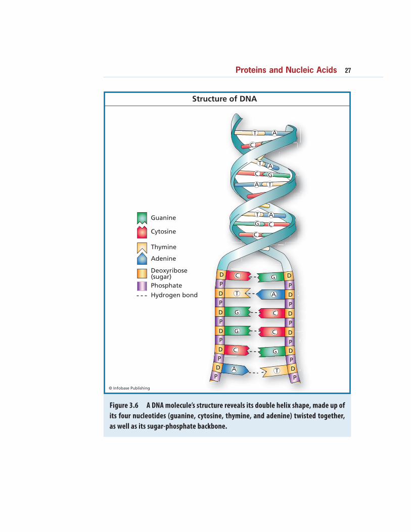
Figure 3.6 A DNA molecule’s structure reveals its double helix shape, made up of
its four nucleotides (guanine, cytosine, thymine, and adenine) twisted together,
as well as its sugar-phosphate backbone.
Proteins and Nucleic Acids 27
28 BIOCHEMISTRY
the chromatin unwinds so that the DNA can replicate. After DNA
replication, the chromatin coils up even tighter to form structures
called chromosomes. The chromosomes are about 100,000 times
shorter than fully stretched DNA, and therefore are 100,000 times
thicker, so they are big enough to be seen with a light microscope.
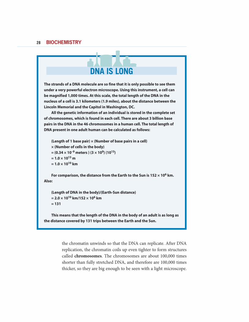
DNA IS LONG
The strands of a DNA molecule are so fine that it is only possible to see them
under a very powerful electron microscope. Using this instrument, a cell can
be magnified 1,000 times. At this scale, the total length of the DNA in the
nucleus of a cell is 3.1 kilometers (1.9 miles), about the distance between the
Lincoln Memorial and the Capitol in Washington, DC.
All the genetic information of an individual is stored in the complete set
of chromosomes, which is found in each cell. There are about 3 billion base
pairs in the DNA in the 46 chromosomes in a human cell. The total length of
DNA present in one adult human can be calculated as follows:
(Length of 1 base pair) × (Number of base pairs in a cell)
× (Number of cells in the body)
= (0.34 × 10⁻⁹ meters ) (3 × 10⁹) (1013)
= 1.0 × 10¹³ m
= 1.0 × 10¹⁰ km
For comparison, the distance from the Earth to the Sun is 152 × 106 km.
Also:
(Length of DNA in the body)/(Earth-Sun distance)
= 2.0 × 10¹⁰ km/152 × 106 km
= 131
This means that the length of the DNA in the body of an adult is as long as
the distance covered by 131 trips between the Earth and the Sun.
In a cell that is reproducing, the chromosomes are found in pairs,
with each chromosome of a pair containing one of the replicated
copies of the DNA.
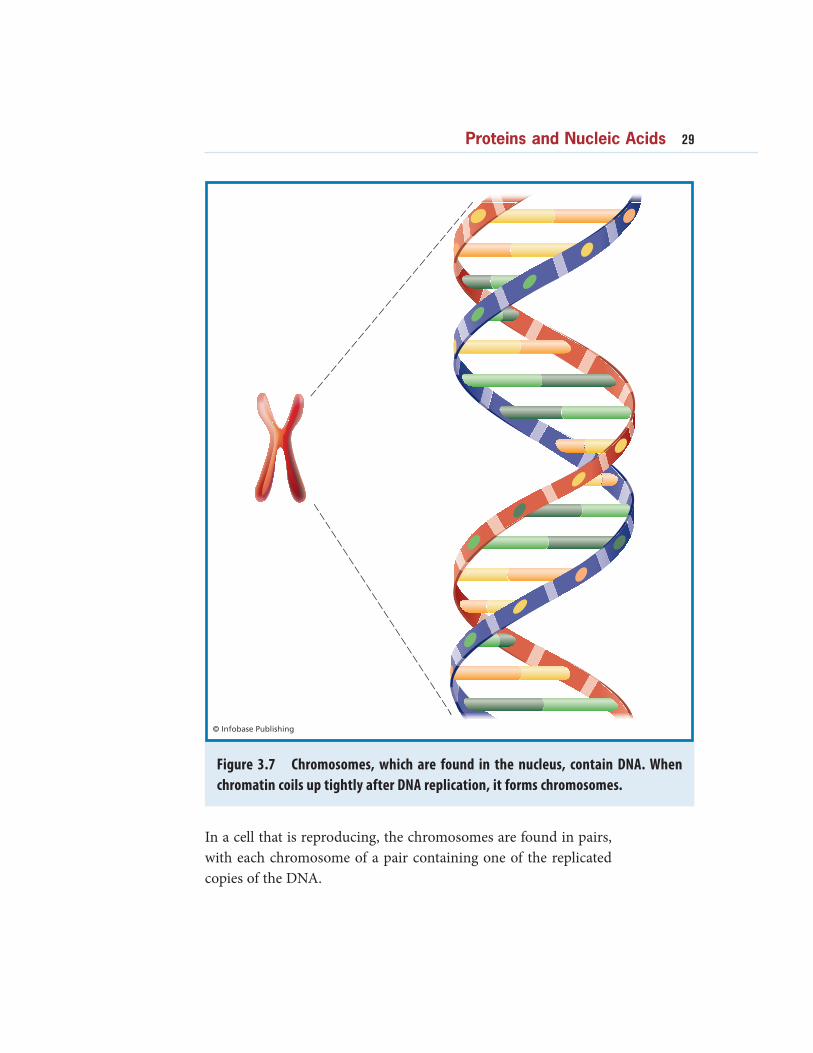
Figure 3.7 Chromosomes, which are found in the nucleus, contain DNA. When
chromatin coils up tightly after DNA replication, it forms chromosomes.
Proteins and Nucleic Acids 29
30 BIOCHEMISTRY
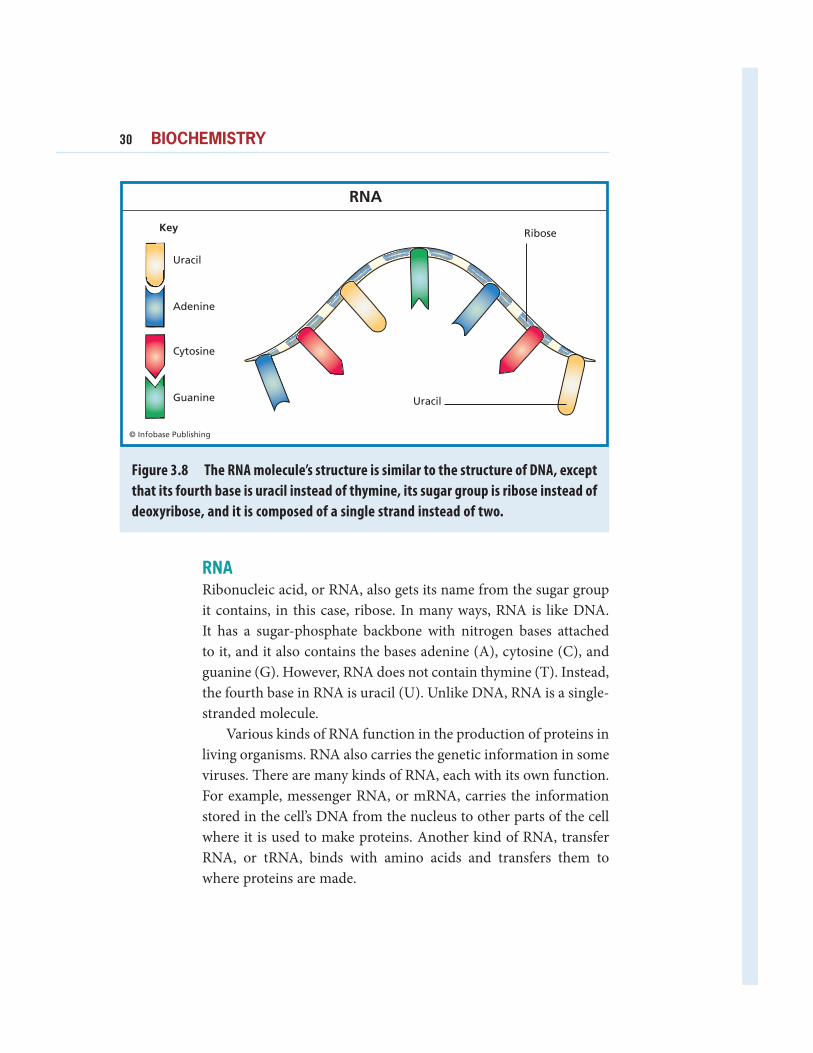
RNA
Ribonucleic acid, or RNA, also gets its name from the sugar group
it contains, in this case, ribose. In many ways, RNA is like DNA.
It has a sugar-phosphate backbone with nitrogen bases attached
to it, and it also contains the bases adenine (A), cytosine (C), and
guanine (G). However, RNA does not contain thymine (T). Instead,
the fourth base in RNA is uracil (U). Unlike DNA, RNA is a single-
stranded molecule.
Various kinds of RNA function in the production of proteins in
living organisms. RNA also carries the genetic information in some
viruses. There are many kinds of RNA, each with its own function.
For example, messenger RNA, or mRNA, carries the information
stored in the cell’s DNA from the nucleus to other parts of the cell
where it is used to make proteins. Another kind of RNA, transfer
RNA, or tRNA, binds with amino acids and transfers them to
where proteins are made.
Figure 3.8 The RNA molecule’s structure is similar to the structure of DNA, except
that its fourth base is uracil instead of thymine, its sugar group is ribose instead of
deoxyribose, and it is composed of a single strand instead of two.