Embed Size (px)
Citation preview

The Journal of Neuroscience, November 1993, 13(11): 4968-4978
Protein Tyrosine Phosphatases Expressed in the Developing Rat Brain
Mustafa Sahin and Susan Hockfield
Section of Neurobiology, Yale University School of Medicine, New Haven, Connecticut 06510
Previous studies of the developing nervous system have shown that cell-cell and cell-matrix interactions are involved in a variety of processes such as the proliferation, migration, and differentiation of neurons. While many cell-surface mol- ecules have been identified, the signal transduction mech- anisms through which they modify cellular responses are poorly understood. Recent studies have described a new and large family of enzymes, protein tyrosine phosphatases (PTPases), that may play a key role in transduction of cell surface events. Opposing the actions of protein tyrosine kinases (PTKs), PTPases can determine the state of tyrosine phosphorylation of a protein and regulate its function. Within the family of PTPases, two subgroups have been charac- terized: low-molecular-weight cytoplasmic (nonreceptor) PTPases and high-molecular-weight transmembrane (recep- tor) PTPases. Many receptor PTPases have fibronectin type Ill and/or lg-like domains in their extracellular domains, sug- gesting that they have dual functions: cell adhesion and signal transduction. Such molecules may play a role in cel- lular recognition events that mediate the accurate assembly of the nervous system.
Using polymerase chain reaction with degenerate primers and a neonatal rat cortex cDNA library, we have identified a number of putative PTPase domains expressed in brain. Three are characterized here. These three sequences are most abundantly expressed in the developing cortex and so are named cortex-enriched protein tyrosine phosphatases (CPTPs) 1,2, and 3. CPTPl and CPTP3 show sequence ho- mology to receptor PTPases and detect multiple high-mo- lecular-weight mRNAs that are expressed preferentially in the developing CNS. Analysis of a longer cDNA indicates that CPTPl and CPTP3 are the first and second phosphatase domains of a single receptor PTPase. CPTPS identifies a single, smaller mRNA species with sequence homology to nonreceptor PTPases. Within the CNS, mRNAs detected by all three CPTPs are expressed at highest levels during pre- natal and early postnatal days and are downregulated in the adult. In situ hybridization demonstrates that the CPTPs are expressed by progenitor cells and developing neurons. The
Received Apr. 6, 1993; accepted June 7, 1993.
We thank Suresh Karne for advice on PCR, James Dowling for assistance in DNA sequencing, and Joe Musco for help with photography. We also thank members of the Hockfield lab for critical reading of the manuscript. This work was supported by NIH Grant NS22807. M.S. is supported by a predoctoral fel- lowship from Merck Research Laboratories.
Correspondence should be addressed to Mustafa Sahin, Section ofNeurobiology, Yale University School of Medicine, Sterling Hall of Medicine C-405, New Haven, CT 06510.
Copyright 0 1993 Society for Neuroscience 0270-6474/93/134968-l 1$05.00/O
spatial and temporal regulation of CPTPs suggests that they may play a role in neuronal development.
[Key words: tyrosine phosphatase, polymerase chain re- action, neocortex, rat, embryo, in situ hybridization, neuro- genesis]
During the development of the mammalian CNS, a plate of morphologically undifferentiated progenitor cells undergoes a period of rapid cellular proliferation to give rise to all the cells, both neurons and glia, that will comprise the mature CNS. The cellular processes that mediate cellular proliferation, the migra- tion of newly born neurons to their final locations, the elabo- ration of characteristic dendritic arbors, and the precision of axon route and target specificity are beginning to be explored. A number of different studies indicate that many of the events in the generation and differentiation of cells in the developing nervous system require a complex series of cell-cell interactions. For example, the fate of cortical or retinal neurons is determined at least in part by interactions that occur at the time of a neuron’s final mitosis (Reh and Kljavin, 1989; McConnell and Kaz- nowski, 199 1). While the precise mechanisms that might govern these kinds of interactions are not currently well understood, proteins that mediate cell-cell recognition and that transduce signals from the cell surface to intracellular locations are likely to have important roles in such events (Elkins et al., 1990; Hynes and Lander, 1992).
One of the most ubiquitous intracellular signaling systems is phosphorylation of proteins on serine, threonine, and tyrosine residues. Regulation of protein function through tyrosine phos- phorylation is known to be critical in the control of many de- velopmental processes, including cellular proliferation and dif- ferentiation. Growing evidence suggests that tyrosine phosphorylation and dephosphorylation may also play key roles in neural development (Wagner et al., 199 1). The opposing ac- tions of protein tyrosine kinases (PTKs) and protein tyrosine phosphatases (PTPases) determine the state of protein tyrosine phosphorylation. A growing number of PTKs and PTPases have been identified in various species and tissues, including the mammalian CNS.
In the developing nervous system, studies of growth factor receptors and Drosophila mutants have significantly advanced our understanding of the functional significance of PTKs. The trk family of tyrosine kinases, first identified as genes with on- cogenic potential, have now been shown to function as high- affinity neurotrophin receptors (Chao, 1992). In addition, the receptors for other peptide growth factors, such as platelet-de- rived growth factor, basic fibroblast growth factor, epidermal growth factor, and insulin-like growth factor, are also trans- membrane tyrosine kinases that are expressed in neural tissues
The Journal of Neuroscience, November 1993, f3(11) 4969
(Chao, 1992; Schlessinger and Ullrich, 1992). The function of receptor tyrosine kinases has also been studied extensively with genetic techniques in Drosophila (reviewed in Shilo, 1992). Dro- sophila PTK mutants demonstrate that these molecules are cru- cial for the determination of neuroblast identity in the CNS @int little ball (Schejter and Shilo, 1989)], for establishing the number and spacing of photoreceptors in the eye imaginal disk [ellipse (Baker and Rubin, 1989)], for differentiation of the R7 photoreceptors [sevenless (Rubin, 199 l)], and for glial migration [breathless (Klambt et al., 1992)]. Several novel putative PTKs are expressed preferentially in the embryonic and early postnatal rodent CNS (Lai and Lemke, 1991), consistent with a role in mammalian neural development.
While there is ample evidence that PTKs play an important role in neural development, much less is known about the PTPases. Tyrosine dephosphorylation has been associated with cellular differentiation in a number of non-neural tissues. For instance, during granulocytic differentiation of leukemia cell lines, phosphotyrosine residues decrease while PTPase activity increases (Frank and Sartorelli, 1988). Since phosphorylation is a reversible process, one might predict that PTPases, like PTKs, play important roles in the development of the nervous system. Indeed, an increase in PTPase activity is associated with NGF- induced neuronal differentiation of PC 12 cells (Aparicio et al., 1992), and two recently identified Drosophila receptor PTPases are selectively expressed on subsets of developing axons (Tian et al., 199 1; Yang et al., 199 1). Perhaps of greatest interest in regard to possible roles in cell-cell signaling, many receptor PTPases (including those identified in the Drosophila nervous system) have fibronectin type III (FN-III) and/or immunoglob- ulin (Ig)-like domains in their extracellular domains, suggesting that they may have dual functions: cell adhesion and signal transduction.
While few PTPases have been reported in the developing brain, the level of tyrosine phosphorylation indicates that PTPases must be particularly active early in neurogenesis (Mah- er, 199 1). Furthermore, the increasingly large number of PTKs with demonstrated activity during neural development implies that there may also be a large group of PTPases involved in neuronal growth and differentiation. Given the possible function of PTPases in the determination of cellular phenotype and our interest in the generation of cellular diversity in the mammalian CNS (Hockfield and McKay, 1985; Geschwind and Hockfield, 1989; Hockfield and Sur, 1990; Martin et al., 1992), we have examined the expression of PTPases in the neonatal rat CNS. Using a polymerase chain reaction (PCR)-based approach, we have identified a number of DNA sequences that encode pu- tative PTPase domains and present here the characterization of three of these. All three are enriched in the nervous system during embryonic and early postnatal days. The temporal and spatial regulation of expression of the mRNAs identified by these sequences suggests that they encode proteins that may participate in neuronal development.
Materials and Methods RNA extraction and cDNA library synthesis. Total cellular RNA was extracted from cerebral neocortex of postnatal day 0 (PO) Sprague- Dawley rats using the guanidine thiocyanate/cesium chloride ultracen- trifugation method (Bothwell et al., 1990). PolyA+ RNA was isolated by one pass through an oligo-dT cellulose (type III, Collaborative Re- search) affinity column (Sambrook et al., 1989). Oligo-dT-primed cDNA synthesis was carried out with the Superscript Plasmid kit (GIBCO/ Bethesda Research Labs). Briefly, the kit uses a Not1 primer-adapter and RNaseH- M-MLV reverse transcriptase for the first-strand syn-
thesis and Escherichia coli RNase H, DNA pol I, and DNA ligase for the second-strand synthesis. Double-stranded cDNA was blunt-ended with T4 DNA pol, cut with NotI, and size fractionated by column chromatography. cDNAs larger than 500 base pairs (bp) were ligated directionally into a modified Bluescript vector, E6 1, gift of J. L. R. Rubenstein (Rubenstein et al., 1991). The cDNA library contained 3.2 x lo6 clones with an average insert size of 900 bp. For the Northern analysis, the same methods were utilized to extract total RNA from cortex without hippocampus at embryonic day 16 (El6), PO, P4, P14, P30, and adult; PO and P35 liver; P35 kidney; and P14 and adult spinal cord.
PCR amplijication. The cDNA library was used as a template for amplification using Taq polymerase (GeneAmp, Perkin Elmer) and de- generate primers (see Fig. 1 for position and sequence of primers). Twenty-five picomoles each of sense and antisense primers were used in 25 il reactions in Taq buffer (10 mM Tris-HCl pa 8.3, 50 mM KCl, 1.5 mM M&l,. 0.001% gelatin) with 0.6 units of Taa nolvmerase. 200 KM dNTPsT a&l 25 ng of cDNi library as template:@CR was c&ied out in a Perkin Elmer DNA Thermal Cycler for 35 cycles. Each cycle included a 30 set denaturation at 94°C and a 3 min extension at 72°C. In order to facilitate the annealing of the degenerate primers, the initial five cycles included a 1 min annealing step at 37°C and a slow ramp (1°C per 4 set) between annealing and extension. The subsequent 30 cycles utilized 1 min annealing step at 45°C with no ramp. One microliter aliquots of the reaction were used to ligate the amplified fragments into the TA vector (Invitrogen). Plasmids with inserts were chosen by blue/ white selection and examined by PCR with the original degenerate primers for the presence of PTPase domains. Inserts that gave a band of the appropriate size (350 bp) after PCR were sequenced by the di- deoxy-chain termination method (Sequenase, U.S. Biochemical) on both strands using M 13 (-40) forward and reverse primers. Sequence anal- yses were conducted using GCG software (Genetics Computer Group, 1991).
Northern hybridization. Northern analyses were performed using stan- dard methods (Bothwell et al., 1990). Total (25 pg) or polyA+ (l-2 pg) RNA was denatured in 2.2 M formaldehyde, 50% formamide, 1 x MOPS buffer at 65°C for 15 min. RNA was resolved by electrophoresis on a 1 .O% agarose gel containing 2.2 M formaldehyde and 1 x MOPS buffer, transferred to Zeta-probe (Bio-Rad) by capillary blotting, and then baked (80°C) under vacuum for 2 hr. Hybridization was carried out in 7% SDS, ‘1% BSA, 0.5 M phosphate buffer pH 6.8 (PB), 1 mM EDTA for at least 8 hr at 65°C (Church and Gilbert. 1984). Hvbridization solution contained l-3 x 10Qcpmlml of probe made by ranhom primed labeling of the PCR fragments for each PTPase clone (Boehringer Mannheim). For random priming, PCR fragments were either gel purified using GeneClean (American Bioanalytical) or isolated using Magic PCR Preps (Promega). After labeling with ‘*P-dCTP (Amersham), the specific ac- tivity of the probes was 2-8 x lo* cpm/hg. After hybridization, filters were washed twice in 5% SDS, 0.5% BSA, 40 mM PB, 1 mM EDTA and four times in 1% SDS, 40 mM PB, 1 mM EDTA at 65°C for 20 min. Cyclophilin, which is present at a constant relative abundance through- out development (Lenoir et al., 1986), was used as a control for equal loading of lanes. RNA molecular weight standards (GIBCO/Bethesda Research Labs) were included on the blots to estimate the sizes of the transcripts. Densitometry of the autoradiograms was performed on the LKB Ultrascan XL system.
In situ hybridization. In situ hybridization was performed as in Martin et al. (1992). Twelve-micron-thick frozen sections were thaw mounted onto gelatin-coated slides, postfixed, and dehydrated. Sections were pre- hybridized in 2 x saline-sodium citrate (SSC) 50% formamide at 50°C for 1 hr. Tissues were then hybridized in 50% formamide, 1 x Den- hardt’s, 0.75 M NaCI, 10% dextran sulfate, 15 mM dithiothreitol, 10 mM Tris-HCl pH 7.5,l mM EDTA, 0.5 mg/ml tRNA, 100 bg/ml salmon sperm DNA, and l-2 x lo6 cpm probe at 50°C for 8-12 hr. The 35S- CTP (New England Nuclear) labeled probes were generated using the T7 and SP6 promotors in the TA vector and the Riboprobe system (Promega). Neurofilament-middle (NF-M) antisense and CPTP3 sense probes were used as positive and negative controls, respectively (Martin et al., 1992). The negative control did not give a signal. Following hybridization, the slides were treated with 20 &ml RNase A at 37°C for 30 min. Final washes were done in 0.1 x SSC, 0.1% P-mercaptoe- than01 at 65°C for 30 min. Slides were then exposed to Kodak XAR film for 24-120 hr. Autoradiograms were used as negatives to print figures. To analyze the signal at higher resolution, the slides were dipped into emulsion, developed after 30-90 d, and counterstained with cresyl violet.

4970 Sahin and Hockfield * Tyrosine Phosphatases in Developing Brain
Figure I. PCR primers were con- structed from the most conserved regions in the catalytic domain. A, Three members of the transmembrane PTPase family are shown to illustrate the extent of similarity in their first phosphatase domains: human CD45 (Streuli et al., 1987), human LAR (Streuli et al., 1988), and mouse LRP (Matthews et al.. 1990). The alignments were produced using the BESTFIT function of the GCG program (Genetics Comuuter Groun. 199 1). Amino acids conserved among the three PTPases are indicated in boldface. Regions selected for primers are shown in boxes. B, Amino acid sequence used to generate the PCR primers. C, De- generate nucleic acid sequence of the primers.
A.
EQKATVIVMVTRCEEGNRNKCAEYWPSMEEGTRAFGiX'WKINQHKRCPDYIIQ 193
EQRTATVVMMTRLEEKSRVKCDQYWP..ARGTETCGLIQVTLLDTVELATYTVR 1481
EQNTATIVMVTNLKERKECKCAQYWP..DQGC!dTYGNVRVSVEDVTVLVDYTVR 408
CD45 794 KLNI...VNKKEKATGREVTHIQFTSWPDHGVPEDPHLLLKLRRRVNAFSNFFSGPIW
LAR 1482 TFALHKSGSSEKRE....LRQFQF'M?WPDHGVPEYPTPILAFLRRVKACNPLDA'3PMVV LRP 409 KFCIQQVGDVTNRKPQRLITQFHFTS~DF~FTPIGMLIW
B. V
DFWRMIW HCSAGVG
C. 5' GACTTCTGGAGAATGATATGG 3' 3' GTGACGTCACGACCACATCC 5'
CG DC A TT C
Results PCR fragments encoding putative PTPase domains were isolated from a rat cortex cDNA library To identify PTPases expressed in neonatal rat cortex, PCR am- plification was carried out with primers corresponding to the conserved catalytic domains of previously reported receptor PTPases. Oligonucleotide primers corresponding to amino acid sequences DFWRM(I/V)W (upstream) and HCSAGVG (down- stream) were synthesized using most common codon usage ta- bles to reduce degeneracy (Lathe, 1985) (Fig. 1). A PO rat neo- cortex cDNA library was used as the DNA template for the amplification. To identify a wide range of receptor PTPases, PCR was performed with low-stringency annealing conditions (see Materials and Methods). The PCR products were ligated directly into the TA vector without size selection. Of 5 1 isolates carrying inserts, 11 showed significant homology to the catalytic domain of previously reported PTPases.
The 11 sequences fall into five groups (Fig. 2). The first group contains six clones with identical sequences, all of which are highly homologous (97%) in nucleotide sequence and 99% iden- tical in amino acid sequence to phosphatase domain I of mouse leukocyte common antigen (LCA)-related phosphatase (LRP) (Matthews et al., 1990). The second group is made up of two
G
clones with sequences identical to phosphatase domain I of rat LCA-related molecule, LAR (Pot et al., 1991). Both LRP and LAR are ubiquitously expressed and have been detected in the brain in previous studies (Saito and Streuli, 1991). The re- maining clones (groups 3-5) contain sequences that are not iden- tical to any PTPases in the databases. They also lack significant identity to one another. We refer to these clones as cortex- enriched protein tyrosine phosphatases (CPTPs) 1, 2, and 3.
The deduced amino acid sequences of CPTPl and CPTP3 show high homology to human receptor PTPase LAR (Streuli et al., 1988) and HPTPG (Krueger et al., 1990). CPTPl encodes an amino acid sequence with 85% and 88% identity to the first phosphatase domains of human LAR and HPTPG, respectively. Nucleotide sequence identity is approximately 75% with many conservative base substitutions. CPTP3, on the other hand, is almost identical to the second catalytic domains of LAR and HPTPG (94% and 97% amino acid identity, respectively). Nu- cleotide sequence identity in this region is about 85%. The ho- mology of CPTPl and CPTP3 to rat LAR is slightly less than the homology to human LAR and HPTPG. These data indicate that CPTPl and CPTP3 belong to the LAR subfamily of re- ceptor PTPases, but neither is identical to rat LAR.
The sequence of CPTP2 (Fig. 2) includes regions that encode the consensus amino acids within the PTPase domains. The
(1) RAT LRP WMAIN-I 1 EQNTAT rVMVTNLKERKECXCAQYWP..DQGCWPYGNVRVSVEDVTVLWYT 50
(2) RAT LAR WMAIN-I 1 EQRTATWXMTRL EEKSRVKCEQYWP..ARGTEX'YGLIQVTLVM'VELATYT 50 (3) RAT CPTP-1 1 EQRSAmF.EKSRVKoQYWp.. NRGTEXYGFIQVTLL~LATFC 50 (4) RAT CFTP-2 1 E!fNWIIVMACREFEMGRJXCERYWPLYGEDPITFAPFKISCENEQARTDYF 52 (5) RAT CFTP-3 1 ENNSTIWHQYWP..AERSARYQYFWDPM?BYNMPQYI 50
(1) RAT LRP D-I 51 VRKFCIQQVGD~PQRLITaFHFTSWPDFGVPFTPI~~L~~~YA..GAIW 111
(2) RAT LAR D-I 51 MRTFALHKSGSSEKRE....LRQFQFMAWPDHGVPEY!?TPILAFLRRVKACNPLDA..GPMW 107 (3) RAT CPTP-1 51 VRTFSLHKNGSSEKRE....VRHFQFTAWPDHGVPEYPTPFLAFLRRVKTC!NPPDA..GPWV 106 (4) RAT CPTP-2 53 IRTLLL..EFQNESRR....LYQ FHYVNWPDHDVPSSFDSILDMISLMRKYQEH..FiDVPICI 107 (5) RAT CPTP-3 51 LREFKVTDARXQSRT....VRQFQFTBVPE~KSGFGFIDFIGQVHKTKEQFGQDZPISV 106
Figure 2. Deduced amino acid sequences of the PCR-amplified cDNAs from the PO rat neocortex library. PCR from the PO cortex library resulted in the identification of five groups of putative PTPase domains. Groups 1 and 2 correspond to phosphatase domain I of rat LRP and LAR, respectively. Novel sequences (groups 3-5) are called rat cortex-enriched protein tyrosine phosphatases (CPTPs). Amino acid sequences deduced from the nucleotide sequence of the PCR subclones flanked by the primers were aligned using the PILEUP function of the GCG program (Genetics Computer Group, 199 1). Amino acids encoded by the PCR primers are not shown. Amino acids shown in boldface are conserved among the five clones as well as among other PTPases.
The Journal of Neuroscience, November 1993, 73(11) 4971
A
B
C
D
E
12-
?.8- 8s-
n CPTPl7.6kb
q CPTPl 6.5 kb
= 300 q CPT!=-2
i q CPTP3 7.5 kb
6 0 CPTPI 6.5 kb 200
E 8 & P 100
n ”
El6 PO P4 PI4 P30 AD LIV
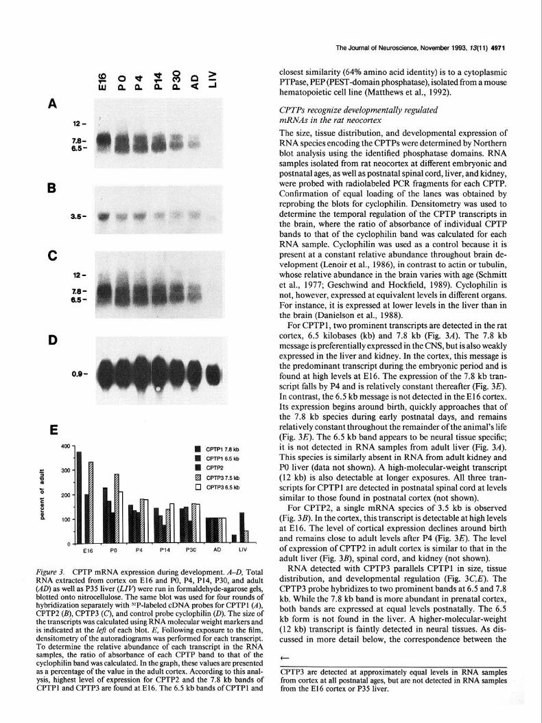
Figure 3. CPTP mRNA expression during development. A-D, Total RNA extracted from cortex on El6 and PO, P4, P14, P30, and adult (AD) as well as P35 liver (LIP) were run in formaldehyde-agarose gels, blotted onto nitrocellulose. The same blot was used for four rounds of hybridization separately with 32P-labeled cDNA probes for CPTPl (A), CPTPZ (B), CPTP3 (C), and control probe cyclophilin (0). The size of the transcripts was calculated using RNA molecular weight markers and is indicated at the left of each blot. E, Following exposure to the film, densitometry of the autoradiograms was performed for each transcript. To determine the relative abundance of each transcript in the RNA samples, the ratio of absorbance of each CPTP band to that of the cyclophilin band was calculated. In the graph, these values are presented as a percentage of the value in the adult cortex. According to this anal- ysis, highest level of expression for CPTPZ and the 7.8 kb bands of CPTPl and CPTP3 are found at E16. The 6.5 kb bands of CPTPl and
closest similarity (64% amino acid identity) is to a cytoplasmic PTPase, PEP (PEST-domain phosphatase), isolated from a mouse hematopoietic cell line (Matthews et al., 1992).
CPTPs recognize developmentally regulated mRNAs in the rat neocortex The size, tissue distribution, and developmental expression of RNA species encoding the CPTPs were determined by Northern blot analysis using the identified phosphatase domains. RNA samples isolated from rat neocortex at different embryonic and postnatal ages, as well as postnatal spinal cord, liver, and kidney, were probed with radiolabeled PCR fragments for each CPTP. Confirmation of equal loading of the lanes was obtained by reprobing the blots for cyclophilin. Densitometry was used to determine the temporal regulation of the CPTP transcripts in the brain, where the ratio of absorbance of individual CPTP bands to that of the cyclophilin band was calculated for each RNA sample. Cyclophilin was used as a control because it is present at a constant relative abundance throughout brain de- velopment (Lenoir et al., 1986), in contrast to actin or tubulin, whose relative abundance in the brain varies with age (Schmitt et al., 1977; Geschwind and Hockfield, 1989). Cyclophilin is not, however, expressed at equivalent levels in different organs. For instance, it is expressed at lower levels in the liver than in the brain (Danielson et al., 1988).
For CPTP 1, two prominent transcripts are detected in the rat cortex, 6.5 kilobases (kb) and 7.8 kb (Fig. 3A). The 7.8 kb message is preferentially expressed in the CNS, but is also weakly expressed in the liver and kidney. In the cortex, this message is the predominant transcript during the embryonic period and is found at high levels at E16. The expression of the 7.8 kb tran- script falls by P4 and is relatively constant thereafter (Fig. 3E). In contrast, the 6.5 kb message is not detected in the El6 cortex. Its expression begins around birth, quickly approaches that of the 7.8 kb species during early postnatal days, and remains relatively constant throughout the remainder of the animal’s life (Fig. 3E). The 6.5 kb band appears to be neural tissue specific; it is not detected in RNA samples from adult liver (Fig. 3A). This species is similarly absent in RNA from adult kidney and PO liver (data not shown). A high-molecular-weight transcript (12 kb) is also detectable at longer exposures. All three tran- scripts for CPTP 1 are detected in postnatal spinal cord at levels similar to those found in postnatal cortex (not shown).
For CPTP2, a single mRNA species of 3.5 kb is observed (Fig. 3B). In the cortex, this transcript is detectable at high levels at E16. The level of cortical expression declines around birth and remains close to adult levels after P4 (Fig. 3E). The level of expression of CPTP2 in adult cortex is similar to that in the adult liver (Fig. 3B), spinal cord, and kidney (not shown).
RNA detected with CPTP3 parallels CPTPl in size, tissue distribution, and developmental regulation (Fig. 3C,E). The CPTP3 probe hybridizes to two prominent bands at 6.5 and 7.8 kb. While the 7.8 kb band is more abundant in prenatal cortex, both bands are expressed at equal levels postnatally. The 6.5 kb form is not found in the liver. A higher-molecular-weight (12 kb) transcript is faintly detected in neural tissues. As dis- cussed in more detail below, the correspondence between the
t
CPTP3 are detected at approximately equal levels in RNA samples from cortex at all postnatal ages, but are not detected in RNA samples from the El6 cortex or P35 liver.

4972 Sahin and Hockfield l Tyrosine Phosphatases in Developing Brain
Figure 4. In situ hybridization demonstrates that CPTPs are expressed in the CNS during embryonic development. Parasagittal sections from El5 (A-D) and El7 (E-H) animals were hybridized to YS-labeled antisense RNA probes for CPTPl (A, E), CPTP2 (II, F), CPTP3 (C, G), and NF-M (D, H) as control probe. A, At El 5, a high CPTPl signal is detected in the cerebral cortex (c), midbrain (mb), and medulla (m). B, CPTPZ mRNA is present throughout the CNS including the spinal cord. C, CPTP3 mRNA is expressed at high levels in the cerebral cortex and the midbrain. There is less but significant labeling in the medulla and the spinal cord. D, NF-M signal is detected at the sacral end of the spinal cord, but only weakly in the medulla and at even lower levels in the telencephalon. E, At El 7, CPTPl mRNA is found at highest levels within the cortex (c) surrounding the lateral ventricle (v) and in the midbrain (mb). Lower signal is detected in the medulla (m), spinal cord (s), and the DRG (d). F, In a near-adjacent section, CPTP2 message is detected throughout the CNS and in the DRG, with highest expression in the cortex. G, At El 7, CPTP3 signal is seen at high levels in the cortex and at somewhat lower levels in more caudal regions of the CNS. H, NF-M signal is expressed at higher levels in the DRG than the CNS; a weak signal is detectable in the spinal cord and medulla and an even weaker signal in the cortex. Scale bar, 1 mm.
transcripts detected by CPTPl and CPTP3 suggests that they may represent the two phosphatase domains of a single PTPase gene.
CPTPs are expressed in the CNS during embryonic and postnatal development To determine the spatial distribution of CPTP mRNAs during development, we performed in situ hybridization using radio- labeled antisense RNA. Both embryonic and postnatal CNS tissues were analyzed by this method. Near adjacent sections were probed with NF-M as a positive control for neuronal RNA expression.
CPTPI. During embryogenesis, CPTP 1 expression is higher in the nervous system than in non-neural tissues. CPTPl mRNA is detected in El 5 embryos at high levels in the CNS (Fig. 4A). At this stage, CPTPl mRNA is widely distributed in the CNS including the cortex, midbrain, medulla, and spinal cord and peripherally in the dorsal root ganglia (DRG). At E15, many parts of the CNS do not exhibit a strong hybridization signal to
NF-M probe (Fig. 40). Thus, CPTPl appears to be expressed before neurons start expressing neurofilament. By El 7, CNS expression of CPTPl has increased, with highest transcript lev- els detected in the developing dorsal telencephalon and mid- brain (Fig. 4E). Around the lateral ventricles, the level ofCPTP1 expression is higher in the developing cortex than in the gan- glionic eminence. More caudal regions of the nervous system such as the spinal cord and DRG express CPTPl , but at some- what lower levels than in the cortex and midbrain.
During postnatal development, CPTPl transcripts are de- tected nonuniformly in the brain. At P4, the highest levels of CPTPl are found in the neocortex, hippocampus, and cerebel- lum (Fig. 54). Within the neocortex, CPTPl mRNA appears most abundant in the superficial layers and the subplate (Figs. 54, 6B). This hybridization pattern is markedly different from the pattern of NF-M expression, where hybridization is most intense in the middle layers of the neocortex (Fig. 5D), corre- lating with a higher neuronal density. CPTPl and NF-M also show different patterns of expression in the developing cere-

The Journal of Neuroscience. November 1993, f3(11) 4973
Figure 5. CPTP expression in postnatal brain. Parasagittal sections from P4 (A-D) and adult (E-H) brains were hybridized to YS-labeled antisense RNA probes for CPTPl (A, E), CPTP2 (B, F), CPTP3 (C, G), and NF-M (0, H) as control. A, At P4, a high level of CPTPl signal is detected in the neocortex (c), cerebellum (cb), and hippocampus (h). Within the neocortex, signal is especially high in the superficial layers and the subplate (arrow). In the cerebellar cortex, two bands of CPTPl signal are detected, in the external and internal granular layers (see also Fig. 60). In the hippocampus (h), all CA fields and the dentate gyrus exhibit high levels of CPTPl hybridization. B, CPTPZ signal is seen most prominently in the cerebellum, while the rest of the brain displays diffuse hybridization. C, CPTP3 mRNA is detected in a pattern very similar to CPTPl (in A). Signal is most intense in the neocortex, hippocampus, dentate gyrus, and cerebellar cortex. D, NF-M is abundant in neocortex, hippocampus, thalamus, and cerebellum. Within the neocortex, the highest signal is in the middle layers. In the cerebellum, the most superficial layer (external granular layer) shows less signal than the deeper layers. E, In adults, the highest levels of CPTPl mRNA is detected in the CA fields of hippocampus, dentate gyrus, and entorhinal cortex. The neocortex displays only a diffuse signal. F, CPTP2 message is detected at very low levels throughout the brain. G, CPTP3 mRNA is expressed diffusely in the neocortex and hippocampal formation. H, NF-M message in the adult is most prominent in the hippocampus and neocortex, but is detectable throughout the brain. Scale bar, 1 mm.
bellum. At P4, CPTPl is expressed in both the external and internal granular layers (Figs. 5A, 60) while NF-M appears to be excluded from the external granular layer (Fig. SD). CPTPl expression in the adult neocortex is lower than at P4 while adult hippocampus and entorhinal cortex continue to express this message at significant levels (Fig. 5E).
The expression of CPTPl in the spinal cord follows a devel- opmental regulation similar to that in the forebrain. CPTPl is expressed throughout the rostrocaudal extent of the spinal cord at embryonic ages (Fig. 4E). At P4, CPTPl is expressed in the spinal gray matter, with somewhat higher levels of expression in the dorsal horn (Fig. 7A). NF-M is expressed at higher levels in the ventral than in the dorsal horn (Fig. 70). At higher mag- nification, CPTPI signal is seen associated with neurons (Fig. 6F). In the adult spinal cord, CPTPl expression is diminished relative to early postnatal levels, but remains higher in gray matter than in white matter (Fig. 7E).
CPTPZ. CPTP2 mRNA is widely distributed in the embryo, but the highest in situ signals are found in the CNS, DRG, and liver. Within the CNS at El 5 and E17, the telencephalon ex- hibits the strongest hybridization (Fig. 4&F). Postnatally, neo- cortical CPTP2 expression is markedly downregulated. At P4, the CPTP2 message is still expressed in the cerebellar cortex, but there is only a weak and diffuse signal present in the neo- cortical gray matter (Fig. 5B). This low level of expression per- sists in the adult brain (Fig. 50. Within the spinal cord, CPTP2 is found at high levels in the gray matter during early postnatal periods. Hybridization is seen in both ventral and dorsal horns at P4 (Fig. 7B). In the adult spinal cord, the signal is less intense although still restricted to the gray matter (Fig. 70.
CPTP3. The hybridization pattern of CPTP3 in the CNS generally follows the pattern of CPTPl. In the embryonic CNS, CPTP3 mRNA is detected at highest levels in the cortex and midbrain at both El 5 and El 7 (Fig. 4C,G). Neocortical ex- pression of CPTP3 persists into the early postnatal period. At P4, the hippocampus and cerebellum also show strong hybrid-
ization to CPTP3. The CPTP3 expression in the cerebellum appears to be highest in the external and internal granular layers, similar to that seen with CPTPI (Fig. 5C). In the adult, a diffuse signal is seen in the neocortex and hippocampus (Fig. 5G). In the spinal cord, CPTP3 is found throughout the gray matter at P4 (Fig. 7C). The distribution of the signal remains the same, but the intensity declines in the adult spinal cord (Fig. 7G).
CPTPI and CPTP3 represent the two phosphatase domains of a single PTPase gene Virtually all transmembrane PTPases have two conserved in- tracellular phosphatase domains. The similarity between the first phosphatase domains of different PTPases is higher than that between first and second domains within any single phos- phatase. The same is true for the second phosphatase domains. Interestingly, CPTPl is highly homologous to the first, and CPTP3 to the second, catalytic domains of LAR and HPTPG. Moreover, CPTPl and CPTP3 have almost identical develop- mental expression profiles on northern blots and very similar patterns of expression by in situ hybridization. These obser- vations suggest that CPTPl and CPTP3 may represent the first and second catalytic domains of a new receptor PTPase. In order to verify this possibility, PCR was used to screen for cDNAs that contain both CPTPl and CPTP3. A nondegenerate oligo- nucleotide primer internal to CPTPl (corresponding to amino acids LATFCVR) and the degenerate downstream PTPase primer (see Fig. 1 C) were used to amplify cDNAs from the PO library. This amplification resulted in two PCR products with approx- imate molecular weights of 100 and 1100 bp (data not shown). Subcloning and sequencing of the large PCR product revealed that it contained CPTPl sequences on the 5’ end and CPTP3 sequences on the 3’ end. The region in between CPTPl and CPTP3 also showed high homology to rat and human LAR and to HPTPG (82%, 84%, and 88% amino acid identity, respec- tively). For simplicity, in the remainder of this article, we con- sider CPTPl and CPTP3 together and refer to them as CPTPl.

4974 Sahin and Hockfield l Tyrosine Phosphatases in Developing Brain
Figure 6. CPTPl mRNA is detected in neurons. Emulsion-dipped slides were counterstained with cresyl violet and photographed under bright-field optics for cell localization (A, C, E) or under dark-field optics for silver grain visu- alization (B, D, F). A and B, Horizontal section through the neocortex on P6 demonstrates CPTPl mRNA in all lay- ers of the cortex, with highest levels in layer 2-3 and the subnlate (s). Much less label is detected in the underlying white matter (wm). C and D, CPTPl mRNA is detected at high level in the external (single arrow) and internal (double arrow) granular layers of the cerebellum at P4. Between the two granular layers, the Purkinje cell layer is relatively devoid of hybridization. E and F, In the ventral horn of P4 spinal cord, CPTPl signal is very high over neurons in the gray matter (arrows). Note that hybridization in the neigh- boring white matter (wm) is much less intense. Scale bars: 200 Nrn for A-D, 100 Nrn for E and F.
Discussion We have identified five sequences encoding putative PTPase domains that are expressed in the developing rat neocortex. By in situ hybridization we demonstrate that three of these se-
quences, CPTPl, CPTPZ, and CPTP3, are expressed in the CNS by progenitor cells and by developing neurons. Although we have information for only a relatively small stretch of the cDNAs, each contains the consensus sequences found in the catalytic domains of previously characterized PTPases, strongly sug-

The Journal of Neuroscience, November 1993. 13(11) 4975
Figure 7. CPTP expression is restricted to gray matter in the spinal cord. Transverse sections from P4 (A-D) and adult (E-H) spinal cord were hybridized to ?S-labeled antisense RNA probes for CPTPl (A, E), CPTP2 (B, F), CPTP3 (C, G), and NF-M (0, H) as control probe. At both ages, the sections for CPTPl and NF-M are taken from cervical segments while those for CPTPZ and CPTP3 are from thoracic segments. A, CPTPl is expressed throughout the spinal gray matter, with somewhat higher levels of expression in the spinal dorsal horn. B-D, Expression of CPTPZ (B), CPTP3 (C), and NF-M (D) is similarly restricted to the gray matter. NF-M (0) is expressed at higher levels in the ventral horn. E-G, In the adult, CPTP- 1 (E), CPTPZ Q, and CPTP3 (G) expression is diminished, but still greater in gray than in white matter. H, At this stage, NF-M is highly expressed throughout the spinal gray matter. Scale bar, 1 mm.
gesting that they encode phosphatases. Analysis of a longer cDNA clone indicates that CPTPl and CPTP3 represent the two phos- phatase domains of a single PTPase gene.
In our PCR screen, each of the three CPTP sequences was obtained only once. Therefore, we cannot exclude the possibility that the nucleotide sequences of these clones contain rare in- corporation errors due to the infidelity of Taq polymerase. How- ever, when multiple copies of previously identified sequences were obtained (six clones of LRP and two of LAR), the sequences we obtained were identical to one another. Furthermore, in the case of rat LAR, the sequence of our PCR product was identical to the cDNA sequence in the GenBank. Therefore, if PCR errors occurred, they were probably infrequent. It is unlikely that in- corporation errors could account for the three new putative phosphatase domains reported here.
CPTPI may encode a novel receptor PTPase Recent PCR and low-stringency hybridization studies have led to the identification of many novel PTPases (reviewed in Fischer et al., 1991; Saito and Streuli, 1991; Charbonneau and Tonks, 1992; Pot and Dixon, 1992). Within the family of PTPases, two subfamilies have been described: low-molecular-weight cyto- plasmic (nonreceptor) PTPases and high-molecular-weight transmembrane (receptor) PTPases. With the exception of two (HPTPP and DPTPIOD), all transmembrane phosphatases have two conserved intracellular phosphatase domains. The catalytic activity is associated with the domain proximal to the mem- brane while the second, more distal domain appears to have regulatory functions.
Both putative phosphatase domains of CPTPl detect multiple high-molecular-weight transcripts, similar in size to receptor PTPases. In addition, they both show high sequence similarity to LAR and HPTPG, two receptor PTPases. Thus, the sequence, as well as the size, of the RNA transcripts detected by CPTPl strongly suggests that CPTPl encodes a transmembrane PTPase. The in situ hybridization results presented here show that both domains of CPTPl are expressed at very high levels in the developing brain. In particular, the ventricular zone of the cor- tical analage at E 15 and El 7, where progenitor cells are giving rise to postmitotic neurons, is perhaps the highest region of CPTPl expression. Expression is markedly downregulated in
the adult, suggesting that these putative PTPases may have a role in the proliferation and early differentiation of neurons.
The sequence of CPTPl is similar to two recently reported phosphatase domains isolated by PCR from human pm-B-cell cDNA (Adachi et al., 1992). Like CPTPl, these two PCR prod- ucts (240 bp each) show high similarity to human LAR and HPTPG. One of the pm-B-cell clones is 93% identical in peptide sequence and 83% identical in nucleotide sequence to the first domain of CPTPl. The other clone differs in only two of its 80 amino acids from the second domain (96% peptide, 88O/o nu- cleotide identity). The limited amount of sequence information reported for the pre-B-cell cDNAs prevents us from determining whether CPTPl represents the rat homolog of this human PTPase.
The putative phosphatase domains ofCPTP1 are most similar to LAR and HPTPd, two receptor-like PTPases that are highly similar to one another (63% in the extracellular region, 88% in the intracellular region). The extracellular domains of LAR and HPTPG contain three Ig-like repeats and eight FN-III-like re- peats (Saito and Streuli, 199 1). The combination of Ig- and FN- III-like repeats is also found in cell adhesion molecules like NCAM (Rutishauser, 1983) fasciclin II, neuroglian (Grenning- loh et al., 1990) and TAG- 1 (Furley et al., 1990), as well as in a tumor suppressor gene product, DCC (deleted in colorectal carcinomas) (Fearon et al., 1990). The extracellular domains of NCAM, fasciclin II, and neuroglian mediate homophilic binding (Rutishauser, 1983; Grenningloh et al., 1990). Homophilic binding has not been reported for any of the receptor PTPases, nor have other ligands for the extracellular domains of almost any receptor PTPases (except for CD45) been identified. The high degree of similarity among the phosphatase domains of CPTPl, LAR, and HPTPG leads to the prediction that the ex- tracellular region of CPTPl will also contain Ig- and FN-II-like motifs. Such a structure might allow CPTPl to play a dual role, cell-cell (or cell-matrix) adhesion and signal transduction, in neuronal differentiation.
CPTPI may give rise to more than a single gene product
On Northern blots both putative phosphatase domains of CPTPl recognize multiple transcripts, with two prominent bands at 7.8 and 6.5 kb. The presence of multiple bands may be due to

4976 Sahin and Hockfield * Tyrosine Phosphatases in Developing Brain
differential processing of mRNA from a single gene, to the ex- istence of multiple genes encoding the CPTPs, or to cross-hy- bridization to other mRNAs. Several lines of evidence support the first of these possibilities, that is, the presence of differential splicing and/or polyadenylation sites. First, on genomic Southern blots, carried out under high-stringency conditions, CPTPl hy- bridizes to a single gene. Second, it is not likely that the two bands represent cross-hybridization to other gene products, such as rat LAR, because the calculated melting temperature of CPTPl-LAR hybrids is lower than the stringency conditions used for the Northern blots. In fact, all bands remain present when the stringency of the Northern is increased by raising the wash temperature to 70°C. Furthermore, Northems using the rat LAR domain I as a probe detect a single band around 8 kb and never reveal the 6.5 kb band. Taken together, these data indicate that the 6.5 and 7.8 kb CPTPl transcripts represent two different mRNAs derived from a single gene, distinct from rat LAR.
Many receptor PTPases, including CD45 (Streuli et al., 1987), LAR (Zhang and Longo, 1992) LRP (Matthews et al., 1990; Sap et al., 1990) DPTPlOD and DPTP99A ( Tian et al., 1991; Yang et al., 1991), have variants with different splicing and polyadenylation sites. Alternative splicing of the CD45 gene is particularly interesting because it occurs in the region encoding the extracellular domain of the molecule. Eight isoforms of CD45 are produced using all possible combinations of the three exons encoding the extracellular domain (Trowbridge, 199 1). These isoforms are expressed by different cell types and may have different ligands (Thomas, 1989). Therefore, receptor PTPases have the potential to generate a large number of cell type-specific isoforms by alternative splicing. This raises the possibility that different isoforms of CPTPl may be expressed by different groups of neural cells. Until we obtain additional sequence of these transcripts, we will not be able to resolve possible differential distributions of the mRNAs. However, the differential temporal regulation of the two major CPTP 1 transcripts is consistent with this hypothesis. The expression of 7.8 kb and 6.5 kb transcripts is regulated differently during neural development. On Northern blots, the 7.8 kb band is expressed at very high levels in the embryonic cortex while the 6.5 kb is not detected in the cortex until after birth. The in situ signal detected in the CNS of em- bryos therefore reflects the expression of the 7.8 kb message, while the signal detected in postnatal brain most likely reflects expression of both 7.8 and 6.5 kb messages. The differential regulation of the two CPTPl mRNAs suggests that the two transcripts may encode gene products with different functions during pre- and postnatal brain development. We are currently pursuing this possibility by screening a cDNA library as well as by raising antibodies to analyze protein expression.
CPTP2 may encode a nonreceptor PTPase CPTP2 recognizes a 3.5 kb message with more sequence ho- mology to nonreceptor than to receptor PTPases. Two groups have recently reported PCR-generated clones with very high homology to CPTP2. The deduced amino acid sequence of CPTP2 is identical to a sequence isolated from mouse myeloid leukemia cells (Yi et al., 199 1) and is highly similar (one out of 107 amino acids different) to a sequence from rat kidney cDNA (Moriyama et al., 1992). Although the nucleic acid sequence was not reported, the molecular weight and tissue distribution of the mouse myeloid mRNA are similar to CPTP2. These sequence data strongly suggest that CPTP2, the mouse myeloid
PTPase, and the rat kidney PTPase all represent a single, ubiq- uitously expressed, nonreceptor PTPase.
One major difference between the mouse myeloid PTPase and CPTP2 is that the level of the myeloid PTPase expression was reported to be equal in all tissues examined, including fetal and adult brain. This is in contrast to the downregulation of CPTP2 we report here. The difference may be explained by the use of @actin as a control for equal loading of RNAs probed with the myeloid PTPase probe. In the brain, actin isoforms are expressed at much higher levels during development than in the adult (Schmitt et al., 1977). An actin control may, then, have resulted in an underestimation of the mouse myeloid PTPase mRNA in the fetal brain.
Nontransmembrane PTPases have been isolated from the brain by other groups. T-cell PTPase (Cool et al., 1989) rat PTPase- 1 (Guan et al., 1990) and STEP (striatum-enriched phosphatase; Lombroso et al., 199 1) are expressed in the brain. Rat PTPase- 1 is found at high levels in adult hippocampus, while STEP is primarily expressed in the adult striatum. The developmental expression profiles of these genes have not been reported. At present, CPTP2 is the only putative nonreceptor PTPase that shows preferentially high levels of expression in embryonic brain. Our in situ hybridization results show that the CPTP2 is ex- pressed at highest level in the developing cortex during the embryonic period, the time of rapid cellular proliferation. As development continues, the level of CPTP2 expression in the CNS decreases such that only a low level of expression can be detected in the adult cortex or spinal cord. CPTP2 expression in the adult liver is about the same as in the adult brain, much less than the embryonic brain.
During the preparation of this report, a full cDNA sequence of a phosphatase from murine P 19EC cells (named P 19-PTP) appeared in the GenBank. At the nucleic acid level CPTP2 shows 93% homology to P19-PTP. The deduced peptide se- quence of the phosphatase domain of P19-PTP is identical to the CPTP2 peptide sequence except for one amino acid (Den Herzog et al., 1992). The tissue distribution and developmental regulation of P 19-PTP has not been reported, but based on size and sequence information, P19-PTP is likely to be the murine homolog of CPTP2.
Potential roles for PTPases in the developing brain
CPTP 1 and CPTP2 both encode putative PTPases that are wide- ly distributed in the developing CNS. Within the cortex, both CPTP2 and the 7.8 kb transcript of CPTPl show very high levels of expression during the period of corticogenesis. Both are also expressed at high levels in the external granule cell layer of the developing cerebellum and are downregulated throughout the nervous system in adult animals. Such temporal regulation of expression suggests that these PTPases may be involved in proliferation, migration, and early differentiation of neurons. The 6.5 kb CPTPl message is expressed at the same level throughout the postnatal period, implying that it has a consti- tutive role in mature neural functions.
The role of intercellular interactions in normal physiological processes and during development is well established. With the identification of increasing numbers of cell-cell and cell-matrix adhesion molecules, greater insights into the molecular mech- anisms underlying cellular interactions have been gained. For example, it has become increasingly clear that cell adhesion is not merely a matter of extracellular stickiness, but is a complex
The Journal of Neuroscience, November 1993, 13(11) 4977
phenomenon involving transmembrane signaling and cytoplas- mic responses. This has been demonstrated most clearly in the immune system. For instance, in the interaction between leu- kocytes and endothelium, initial adhesion leads to “activation” of the leukocytes that is mediated by second messengers (Spring- er, 1990; Butcher, 199 1). Similarly, the binding of helper T-cells to antigen-presenting cells is followed by cytoskeletal reorga- nization and is regulated by phosphorylation (Kupfer and Sing- er, 1989). Both events involve cell adhesion molecules of the Ig and integrin families. Similar signaling mediated by integrins is involved in adhesive functions of platelets (reviewed in Shattil and Brugge, 199 1).
In the developing nervous system, the molecular mechanisms that underlie processes such as neuronal migration and axonal guidance are being studied in detail (Bixby and Harris, 1991; Hynes and Lander, 1992). While many cell adhesion molecules have been shown to play a role in these processes, the signal transduction mechanism by which cell surface events are trans- lated into cellular responses is poorly understood. Many cellular processes are regulated by protein phosphorylation, balanced by the competing activities of kinases and phosphatases. Cell- cell contacts and protein phosphorylation can reflect two com- ponents of a single process, as illustrated by genetic studies in Drosophila. Null mutations of either the cell adhesion molecule (fasciclin I) or a cytoplasmic PTK (abl) have little observable effect on the nervous system. However, embryos double mutant for fasciclin Z and abl show a pronounced disorganization of the nervous system, presumably due to a developmental misrouting of growth cones (Elkins et al., 1990). The identification of two receptor PTPases expressed on subsets of growing axons in Dro- sophila further suggests that a PTPase might perform both cell- cell recognition and intracellular signaling functions during de- velopment (Tian et al., 1991; Yang et al., 1991); however, the test of such a hypothesis has not yet been reported.
In vitro studies of vertebrate neurons also suggest that tyrosine phosphorylation regulated by extracellular signals is involved in neurite outgrowth. Inhibition of PTKs by genistein facilitates substrate-induced neurite outgrowth (Bixby and Jhabvala, 1992). Furthermore, binding of soluble Ll and NCAM to growth cone membranes reduces tyrosine phosphorylation of tubulin (Atashi et al., 1992). Although the mechanism of the decreased phos- phorylation is not known, PTPases may be involved either by directly dephosphorylating tubulin or by regulating the activity of the PTKs. PTPases such as CD45 and LRP have already been shown to regulate src family PTKs (Sefton and Campbell, 199 1; Zheng et al., 1992).
The distribution of the PTPases described here suggests a role in cell-cell communication early in neural development. In the developing cortex and brainstem, CPTPl and CPTP2 are ex- pressed in many areas, but both are most abundant in the regions immediately surrounding the ventricles, which are the site of cellular proliferation. While the cells that occupy the prolifer- ative zones of the embryonic brain are largely homogeneous in appearance, they give rise to all of the phenotypically diverse types of neurons and glia in the mature brain (for discussion, see Geschwind and Hockfield, 1989). The mechanisms that con- trol cellular differentiation have been the object of intensive study over the last several years. Reports from two laboratories indicate that some aspects of the cell fate are determined as a consequence of signals transmitted and received during the ter- minal mitosis (Reh and Kljavin, 1989; McConnell and Kaz- nowski, 199 1). The potential dual roles of receptor PTPases as
cell adhesion and transmembrane signaling proteins would be well suited to mediate this kind of signal transduction.
Receptor PTPases and membrane associated nonreceptor PTPases, including LAR, LRP, and the CPTPs identified in this study, are new candidates that may play adhesion and/or sig- naling roles mediating the accurate assembly of the nervous system. It might be safely predicted that the full repertoire of PTPases expressed in the brain has not yet been explored. While the sequences we described here are expressed quite abundantly in the neonatal cortex and throughout the developing nervous system, members of the PTPase family expressed in more spe- cific temporal and spatial patterns probably remain to be dis- covered.
Note added in proof The sequence of CPTPl reported here corresponds to a portion of two sequences that recently appeared in GenBank (accension nos. L11587 and L12329).
References Adachi M, Sekiya M, Arimura Y, Takekawa M, Itoh F, Hinoda Y, Imai
K, Yachi A (1992) Protein-tyrosine phosphatase expression in pre-B cell NALM-6. Cancer Res 52:737-740.
Aparicio LF, Ocrant I, Boylan JM, Gruppuso PA (1992) Protein ty- rosine phosphatase activation during nerve growth factor-induced neuronal differentiation of PC12 cells. Cell Growth Differ 3:363-367.
Atashi JR, Klinz SG, Ingraham CA, Matten WT, Schachner M, Maness PF (1992) Neural cell adhesion molecules modulate tyrosine phos- phorylation of tubulin in nerve growth cone membranes. Neuron 8:83 l-842.
Baker NE, Rubin GM (1989) Effect on eye development of dominant mutations in Drosophila homologue of EGF receptor. Nature 340: 150-153.
Bixby JL, Harris WA (1991) Molecular mechanisms of axon growth and guidance. Annu Rev Cell Biol 7: 117-l 59.
Bixby JL, Jhabvala P (1992) Inhibition of tyrosine phosphorylation potentiates substrate-induced neurite growth. J Neurobiol 23:468- 480.
Bothwell A, Yancopoulos GD, Alt FW (1990) Methods for cloning and analysis of eukaryotic genes. Boston: Jones and Bartlett.
Butcher EC (199 1) Leukocyte-endothelial cell recognition: three (or more) steps to specificity and diversity. Cell 67: 1033-1036.
Chao MV (1992) Neurotrophin receptors: a window into neuronal differentiation. Neuron 9:583-593.
Charbonneau H, Tonks NK (1992) 1002 protein phosphatases? Annu Rev Cell Biol 8:463-493.
Church GM, Gilbert W (1984) Genomic sequencing. Proc Nat1 Acad Sci USA 81:1991-1995.
Cool DE, Tonks NK, Charbonneau H, Walsh KA, Fischer EH, Krebs EG (1989) cDNA isolated from a human T-cell library encodes a member of the protein-tyrosine-phosphatase family. Proc Nat1 Acad Sci USA 86:5257-5261.
Danielson PE, Forss-Petter S, Brow MA, Calavetta L, Douglass J, Milner RJ. Sutcliffe JG (1988) ~1 B15: a cDNA clone of the rat mRNA encoding cyclophilin. DNA 7:261-267.
Den Herzog J, Pals CEGM, Jonk LJC, Kruijer W (1992) Differential expression of a novel murine non-receptor protein tyrosine phos- phatase during differentiation of P 19 embryonal carcinoma cells. Bio- them Biophys Res Commun 184:1241-1249.
Elkins T, Zinn K, McAllister L, Hoffman FM, Goodman CS (1990) Genetic analysis of a Drosophila neural cell adhesion molecule: in- teraction of fusciclin Z and abelson tyrosine kinase mutations. Cell 60~565-575.
Fearon ER, Cho KR, Nigro JM, Simons JW, Ruppert JM, Hamilton SR. Preisineer AC. Thomas G. Kinzler KW. Voaelstein B (1990) Identificatio> of a chromosome’ 18q gene that is altered in colorectal cancers. Science 247149-56.
Fischer EH. Charbonneau H. Tonks NK (199 1) Protein tvrosine phos- phatases:’ a diverse family of intracellular and transmembrane en- zymes. Science 253:401-406.
Frank DA, Sartorelli AC (1988) Alterations in tyrosine phosphory-

4979 Sahin and Hockfield * Tyrosine Phosphatases in Developing Brain
lation during the granulocytic maturation of HL-60 leukemia cells. Cancer Res 4852-58.
Furley AJ, Morton SB, Manalo D, Karagogeos D, Dodd J, Jesse11 TM (1990) The axonal glycoprotein TAG-l is an immunoglobulin su- perfamily member with neurite outgrowth-promoting activity. Cell 61:157-170.
Genetics Computer Group (199 1) Program manual for the GCG pack- age, version 7. Madison, WI: Genetics Computer Group.
Geschwind DH, Hockfield S (1989) Identification of proteins that are developmentally regulated during early cerebral corticogenesis in the rat. J Neurosci 9:430343 17.
Grenningloh G, Bieber AJ, Rehm EJ, Snow PM, Traquina ZR, Hortsch M. Pate1 NH. Goodman CS (1990) Molecular genetics of neuronal recognition in Drosophila: evolution and function-of immunoglobulin superfamily cell adhesion molecules. Cold Spring Harbor Symp Quant Biol .55:327-340.
Guan K, Haun RS, Watson SJ, Geahlen RL, Dixon JE (1990) Cloning and expression of a protein-tyrosine-phosphatase. Proc Nat1 Acad Sci USA 87:1501-1505.
Hockfield S, McKay RDG (1985) Identification of major cell classes in the developing mammalian nervous system. J Neurosci 5:3310- 3328.
Hockfield S, Sur M (1990) Monoclonal antibody Cat-301 identifies Y-cells in the dorsal lateral geniculate nucleus of the cat. J Comp Neurol 300:320-330.
Hynes RO, Lander AD (1992) Contact and adhesive specificities in the associations, migrations and targeting of cells and axons. Cell 68: 303-322.
Klambt C, Glazer L, Shilo B-Z (1992) breathless, a Drosophila FGF receptor homolog, is essential for migration of tracheal and specific midline glial cells. Genes Dev 6: 1668-1678.
Krueger NX. Streuli M. Saito H (1990) Structural diversitv and evo- lution of human receptor-like protein tyrosine phosphatasks. EMBO J 9:3241-3252.
Kupfer A, Singer SJ (1989) The specific interaction of helper T cells and antigen-presenting B cells. IV. Membrane and cytoskeletal re- organizations in the bound T cell as a function of antigen dose. J Exp Med 170:1697-1713.
Lai C, Lemke G (199 1) An extended family of protein-tyrosine kinase genes differentially expressed in the vertebrate nervous system. Neu- ron 6:691-704.
Lathe R (1985) Synthetic oligonucleotide probes deduced from amino acid sequence data: theoretical and practical considerations. J Mol Biol 183:1-12.
Lenoir D, Battenberg E, Bloom FE, Milner RJ (1986) The brain- specific gene lB236 is expressed postnatally in the developing brain. J Neurosci 6:522-530.
Lombroso PJ, Murdoch G, Lemer M (199 1) Molecular characteriza- tion of a protein-tyrosine-phosphatase enriched in striatum. Proc Nat1 Acad Sci USA 88~7242-7246.
Maher PA (199 1) Tissue-dependent regulation of protein tyrosine ki- nase activity during embryonic development. J Cell Biol 112:955- 963.
Martin KA, Grant SGN, Hockfield S (1992) The mas proto-oncogene is developmentally regulated in the rat central nervous system. Dev Brain Res 68:75-82.
Matthews RJ, Cahir ED, Thomas ML (1990) Identification of an additional member of the protein-tyrosine-phosphatase family: evi- dence for alternative splicing in the tyrosine phosphatase domain. Proc Nat1 Acad Sci USA 87:4444-4448. ̂ ̂
Matthews RJ. Browne DB. Flores E. Thomas ML (1992) Character- ization of hematopoietic’intracellular protein tyrosine phosphatases: description of a phosphatase containing an SH2 domain and another enriched in proline-, glutamic acid-, serine-, and threonine-rich se- quences. Mol Cell Biol 12:2396-2405.
McConnell SK, Kaznowski CE (199 1) Cell cycle dependence of lam- inar determination in developing neocortex.. Science 254:282-285.
Morivama T. Fuiiwara Y. Imai E. Takenaka M. Kawanishi S. Inoue T. Noguchi T, Tanaka T,‘Kamada T, Ueda N ‘( 1992) cDNA cloning of rat LRP, a receptor like protein tyrosine phosphatase, and evidence for its gene regulation in cultured rat mesangial cells. Biochem Bio- phys Res Commun 188:34-39.
Pot DA, Dixon JE (1992) A thousand and two protein tyrosine phos- phatases. Biochim Biophys Acta 1136:35-43.
Pot DA, Woodford TA, Remboutsika E, Haun RS, Dixon JE (1991) Cloning, bacterial expression, purification, and characterization of the cytoplasmic domain of rat LAR, a receptor-like protein tyrosine phos- phatase. J Biol Chem 266:19688-19696. -
Reh TA. Kliavin IJ (1989) Aee ofdifferentiation determines rat retinal germinalcell phenotype! induction of differentiation by dissociation. J Neurosci 9:4179-4189.
Rubenstein JLR, Brice AEJ, Ciaranello RD, Denney D, Porteus MH, Usdin TB (1991) Subtractive hybridization system using single- stranded phagemids with directional inserts. Nucleic Acids Res 18: 4833-4842.
Rubin GM (199 1) Signal transduction and fate of the R7 photoreceptor in Drosophila. Trends Genet 7~372-377.
Rutishauser U (1983) Molecular and biological properties of a neural cell adhesion molecule. Cold Spring Harbor Symp Quant Biol 48: 501-526.
Saito H, Streuli M (1991) Molecular characterization of protein ty- rosine phosphatases. Cell Growth Differ 2:59-65.
Sambrook J, Fritsch EF, Maniatis T (1989) Molecular cloning: a lab- oratory manual, 2d ed. Cold Spring Harbor, NY: Cold Spring Harbor Laboratory.
Sap J, D’Eustachio P, Givol D, Schlessinger J (1990) Cloning and expression of a widely expressed receptor tyrosine phosphatase. Proc NatlAcadSciUSA87:6112-6116.
Schejter ED, Shilo B-Z (1989) The Drosophila EGF receptor homolog (DER) gene is allelic tofaint little ball, a locus essential for embryonic development. Cell 56: 1093-l 104.
Schlessinger J, Ullrich A (1992) Growth factor signaling by receptor tyrosine kinases. Neuron 9:383-39 1.
Schmitt H, Gozes I, Littauer UZ (1977) Decrease in levels and rates of synthesis of tubulin and actin in developing rat brain. Brain Res 121:327-342.
Sefton BM, Campbell M-A (199 1) The role of tyrosine protein phos- phorylation in lymphocyte activation. Annu Rev Cell Biol 7:257- 214.
Shattil SJ, Brugge JS( 199 1) Protein tyrosine phosphorylation and the adhesive functions of platelets. Curr Opin Cell Biol 3:869-879.
Shilo B-Z (1992) Roles of receptor tyrosine kinases in Drosophila development. FASEB J 6:2915-2922.
Springer TA (1990) Adhesion receptors of the immune system. Science 346:425433.
Streuli M, Hall LR, Saga Y, Schlossman SF, Saito H (1987) Differential usage of three exons generates at least five different mRNAs encoding human leukocyte common antigens. J Exp Med 166: 1548-l 566.
Streuli M, Krueger NX, Hall LR, Schlossman SF, Saito H (1988) A new member of the immunoglobulin superfamily that has a cyto- plasmic region homologous to the leukocyte common antigen. J Exp Med 168:1553-1562.
Thomas ML (1989) The leukocyte common antigen family. Annu Rev Immunol7:339-369.
Tian S-S, Tsoulfas P, Zinn K (199 1) Three receptor-like protein-ty- rosine phosphatases are selectively expressed on central nervous sys- tem axons in the Drosophila embryo. Cell 67:675-685.
Trowbridge IS (199 1) CD45: a prototype for transmembrane protein tyrosine phosphatases. J Biol Chem 266:235 17-23520.
Wagner KR, Me L, Huganir RL (199 1) Protein tyrosine kinases and phosphatases in the nervous system. Curr Opin Neurobiol 1:65-73.
Yang X, Seow KT, Bahri SM, Oon SH, Chia W (199 1) Two Drosophila receptor-like tyrosine phosphatase genes are expressed in a subset of developing axons and pioneer neurons in the embryonic CNS. Cell 67:661-673.
Yi T, Cleveland JL, Ihle JN (1991) Identification of novel protein tyrosine phosphatases of hematopoietic cells by polymerase chain reaction amplification. Blood 78:2222-2228.
Zhang JS, Longo FM (1992) LAR tyrosine phosphatase receptor: a novel GC-rich transcript expressed in rat CNS. Sot Neurosci Abstr 18:949.
Zheng XM, Wang Y, Pallen CJ (1992) Cell transformation and acti- vation of pp60 (c-src) by overexpression of a protein tyrosine phos- phatase. Nature 359:336-339.


![The emerging roles of phosphatases in Hedgehog pathway...ture, stability, activity, protein-protein interaction [6]. In contrast to protein kinases, protein phosphatases have been](https://img.pdfslide.us/doc/110x75/60ee63efe2bdd8639d7712a6/the-emerging-roles-of-phosphatases-in-hedgehog-pathway-ture-stability-activity.jpg)


![orion.towson.eduorion.towson.edu/~karne/teaching/c657sl/embsql.pdf · / * Filename: pro 'ect . sc / * Instru Dr . karne ogram used for company database dname [15] # include](https://img.pdfslide.us/doc/110x75/5c9a9bfc09d3f2a06c8c26d7/orion-karneteachingc657slembsqlpdf-filename-pro-ect-sc-instru.jpg)