Embed Size (px)
Citation preview
Protein Translocation Across the Endoplasmic Reticulum.
I. Detection in the Microsomal Membrane of a Receptor
for the Signal Recognition Particle
REID GILMORE, GONTER BLOBEL, and PETER WALTER Laboratory of Cell Biology, The Rockefeller University, New York, New York 10021
ABSTRACT Salt-extracted microsomal membranes (K-RM) contain an activity that is capable of releasing the signal recognition particle (SRP)-mediated elongation arrest of the synthesis of secretory polypeptides (Walter, P., and G. Blobel, 1981, J. Cell Biol., 91:557-561). This arrest- releasing activity was shown to be a function of an integral microsomal membrane protein, termed the SRP receptor (Gilmore, R., P. Walter, and G. Blobel, 1982, J. Cell BioL, 95:470-477). We attempted to solubilize the arrest-releasing activity of the SRP receptor by mild protease digestion of K-RM using either trypsin or elastase. We found, however, that neither a trypsin, nor an elastase "solubilized" supernatant fraction exhibited the arrest-releasing activity. Only when either the trypsin- or elastase-derived supernatant fraction was combined with the trypsinized membrane fraction, which by itself was also inactive, was the arrest-releasing activity restored. Release of the elongation arrest was followed by the translocation of the secretory protein across the microsomal membrane and the removal of the signal peptide. Thus, although we have been unable to proteolytically sever the arrest-releasing activity from K-RM and thereby to uncouple the release of the elongation arrest from the process of chain translocation, we have been able to proteolytically dissect and reconstitute the arrest-releasing activity. Furthermore, we found that the arrest-releasing activity of the SRP receptor can be inactivated by alkylation of K-RM with N-ethylmaleimide.
Recently, the function of signal recognition protein (SRP) in the translocation of secretory proteins a c r o s s (1-3) or integra- tion of certain integral membrane proteins in to (4) the endo- plasmic reticulum membrane has been elucidated.
The investigation of SRP began with the observation (5) that dog pancreas rough microsomes (RM) lose their ability to translocate nascent secretory proteins upon extraction by salt. Translocation activity, however, could be restored to the salt- extracted membranes (K-RM) by the readdition of the salt extract (5, 6). The active component in the salt extract was subsequently purified to homogeneity and shown to be an 11S complex of six nonidentical polypeptide chains (6). Based on its mechanism of action (1-3), the purified protein was termed signal recognition protein (SRP). Recently, it was shown that SRP contains, in addition to the six polypeptides, a 7S RNA molecule (identified as the small cytoplasmic 7SL RNA [7, 8]), and therefore the nomenclature Was changed to signal recognition particle (SRP).
SRP has been shown to cause a signal sequence-induced, site-specific arrest of chain elongation. Thus, translation of a mRNA for a secretory protein (bovine pituitary preprolactin)
"I-HE JOURNAL OF C [ t t BIOLOGY • VOLUME 95 NOVEMBER 1982 463-469 © The Rockefel ler Univers i ty Press • 0021-9525/82/11/0463/07 $1.00
in the wheat germ cell-free system, in the presence of SRP, did not yield synthesis of completed preprolactin molecules, but instead yielded synthesis of a discrete NH2 terminal segment of preprolactin comprising 60-70 amino acid residues (3). When salt-extracted microsomal membranes (K-RM) were added to an SRP arrested cell-free translation system, the elongation arrest was released, followed by the translocation of nascent preprolactin across the microsomal membrane and the removal of the signal peptide by signal peptidase, thereby yielding segregated prolactin molecules (3).
Our present work (here and in the following paper [9]) was directed towards identifying, purifying, and characterizing the arrest-releasing activity contained in the K-RM fraction. Using immobilized SRP as an affinity adsorbent we isolated an integral membrane protein that exhibited the arrest-releasing activity. Because of the direct interaction of this protein with SRP we refer to it as the SRP receptor.
The existence of an SRP receptor had been postulated (3) based upon the following lines of evidence: (a) K-RM release the SRP-mediated elongation arrest of secretory protein syn- thesis, but phospholipid vesicles prepared from K-RM do not
463
(3), (b) a salt-dependent interaction between SRP and RM is observed that is not disrupted by extraction of the ribosomes from the membrane with EDTA (10), and (c) a membrane- bound component of the translocation system was previously detected using pretense digestions of K-RM (11, 12). The SRP receptor was envisioned to fimction during the binding of the SRP-arrested ribosome to the membrane, thereby releasing the elongation arrest and initiating the translocation of the nascent chain (3).
In this paper we describe a quantitative assay for the SRP receptor based upon its arrest-releasing activity. Using this assay we show (a) that the SRP receptor can be proteolytically dissected into a soluble cytoplasmic domain and a membrane- bound domain, (b) that neither the cytoplasmic domain nor the membrane-bound domain can release the translation arrest when assayed separately, but when the two fractions are com- bined the arrest-releasing activity is restored, (c) that the arrest- releasing activity is inhibited by treatment of K-RM with N- ethylmaleimide (NEM), and (d) that the translocation of nas- cent secretory proteins occurs whenever the SRP-induced elon- gation arrest is released by the SRP receptor.
MATERIALS AND METHODS
Materials laSS]Met (1,000 Ci/mmol) was obtained from New England Nuclear, Boston,
MA; trypsin from Boehringer Mannheim, Federal Republic of Germany, and elastase from Merck and Co., Inc., Federal Republic of Germany. Trasylol (10,000 kallikrein inhibitor U/ml) was from FBA Pharmaceuticals, New York, NY. Other pretense inhibitors were from Sigma Chemical Co., St. Louis, Me. The nonionic detergent Nikkol (oetaethyleneglycol-mono-N-dodecyl ether) was from Nikko Chemicals Co., Ltd., Tokyo, Japan.
Preparation of Microsomal Membranes, Signal Recognition Particle, and Salt-Extracted Microsomal Membranes
Rough microsomal membranes (RM), signal recognition particle (SRP), and salt-extracted rough microsomal membranes (K-RM) were prepared as described previously (1) except that adsorbed ribosomes were removed from RM by extraction with 25 mM EDTA (I 1) before the extraction of SRP. The gel filtration step using a Sepharose CL-2B colunm was omitted. The triethanolamine buffer used for all preparative procedures was prepared as a 1 M stock solution adjusted to pH 7.5 at room temperature with acetic acid and, as such, is referred to as TEA.
Protease Digestions of K-RM K-RM were subjected to limited pretense digestions after suspending the
membranes in l mI of 50 mM TEA, 1 mM dithiothreitol (DTT) at a membrane concentration of 2 eq/#l. Aliquots of a stock solution of trypsin were added to obtain the desired pretense concentrations (0-50 #g/ml of trypsin). After 60 min of incubation at 0°C, the samples were adjusted to 0.5 mM diisopropylfluoro- phosphate and incubated for an additional 15 rain at 0°C. Trasylol (100 #1) was added, and the concentration of KOAc was adjusted to 500 mM by the addition of the appropriate volume of 4 M KOAc. A trypsin-solubilized supernatant fraction was separated from the trypsinized membranes by centrifugation for 30 min at 100,000 g,v. The membranes were washed once by resuspension in 5 ml of 50 mM TEA, 0.25 M sucrose, 1 mM DTT, followed by centrifugation for 30 min at 100,000 g,v. The membrane pellet (Tx-K-RM, where x denotes the concentration of trypsin used in gtg/ml) was resuspended in 50 mM TEA, 0.25 M sucrose, 1 mM DTT, frozen in liquid N2, and stored at -80°C. The trypsin- derived supernatant fraction was concentrated by ultrafiltration using an Amicon PM-30 membrane to a final volume of 200 #1. This supernatant fraction was applied to a 1.0 ml Sephadex G-25 column equilibrated in 50 mM TEA, 150 mM KOAc, 1 mM DTT. The excluded volume (300/~1) containing the protease- derived supernatant fraction (T:Sup) was collected, frozen in liquid N2 and stored at -80°C.
The elastase-derived supernatant fraction (El-Sup) was prepared in an iden- tical manner except that the 10 mg/ml elastas¢ stock solution was pretreated with an equal volume of trasylol before use to inhibit nonelastolytic activity (12). Trasylol was not added after the pretense digestion.
464 THE JOURNAL OF CELL B,OLOGV. VOLUME 95, 1982
NEM Alkylation of K-RM Dithiothreitol was removed from K-RM before treatment with NEM by
ccntrift~gation and resuspension of the membranes in 50 mM TEA, 0.25 M sucrose. The samples were incubated with NEM at the specified concentration for 30 min at 25°C. The NEM was quenched with a 10-fold molar excess of DTT- with respect to sulfhydryl groups before use in the in vitro translations.
Assay for the Arrest-releasing Activity A premixed aliquot of bovine pituitary RNA (0.08 A~o U/25 ~1 translation)
and rabbit reticulocyte RNA (0.01 A~,o U/25/zl translation) was translated in a staphylococcal nuclease-treated (13) wheat germ system at 25°C for 1 h. The 25- /~1 translations contained 7.5/tl of wheat germ $23, 25 ~Ci of [35S]Met, and were supplemented with human placental RNase inhibitor (14) and a mixture of protease inhibitors described previously (1). All translations were adjusted to a final ion concentration of 140 mM KOAc, 2.85 mM Mg (OAc)~. The translations contained 0.002% Nikkol to stabilize the SRP activity (6). The 25-#1 translations were supplemented with 10 U of SRP to arrest the elongation of ~90% of the nascent preprolactin chains (1). Control translations, which were not supple- mented with SRP, were conducted for all experiments although in most cases the autoradiograph of the translation products of a control translation is not shown. The translations were further supplemented with K-RM or fractions derived from K-RM as described in the text and the figure legends.
PAGE in SDS The procedures for the preparation of samples for PAGE (1, 15), the subse-
quent autoradiography of dried slab gels (15), and the quantitation of radioactivity in specific polypeptides have been described (1, 11).
Definitions and Calculations 1 equivalent (eq) is the amount of a fraction (supernatant fluid or membrane)
that is derived from l t~l of a RM suspension at a concentration of 50 A ~ U/ml. 1 eq is derived from ~ 1 nag of tissue.
One unit of translocation activity (U) (a) for a membrane is the amount of membranes that gives the same amount of processing (i.e., translocation) as 1 cq ofRM, (b) for SRP is the amount that has to be added back to 1 eq of K-RM to restore activity to that of I eq of RM.
Bovine preprolactin contains eight methionyl residues (15, 16), whereas pro- lactin contains seven (16). The incorporation of [a~S]Met into prolactin was corrected for the loss of one methionyl residue contained within the signal peptide (15) using the ratio of 8/7. The incorporation of [~S]Met into both preprolactin (pPL) and prolactin (PL) was normalized with respect to the incorporation of laSS]Met into globin (GLO), which served as an internal standard in the arrest- releasing activity assay. For example, the globin-normalized incorporation of [25S]Met into prolactin (PLo) was calculated for an assay translation containing SRP and K-RM using the following formula:
GLO PL~ = (PL X 8/7) X GLO-
where PL and GLO are the uncorrected cpm incorporated into prolactin and globin, respectively, in the assay translation, and GLO- is the uncorrected cpm incorporated into globin in the control translation that contained neither SRP nor K-RM. The percent SRP inhibition of preprolactin plus prolactin synthesis was calculated as below for the assay translation containing SRP and K-RM:
100 x (pPLo + PLo) % Inhibition = 100
(pPL- + PL-)
where pPLo and PLo are the globin-normalized incorporation of laSS]Met into preprolactin and prolactin in the assay translation, and pPL- and PL are the incorporation of laSS]Met into preprolactin and prolactin in the control transla- tion. No incorporation of [~S]Met into prolactin (PL-) occurs in the control translation, so this term can be deleted from the denominator.
RESULTS
Assay for the Arrest-releasing Activity of the SRP Receptor
As a prerequisite for the characterization of the SRP receptor it was essential to develop a quantitative assay based on its arrest-releasing activity (3). The paradigm for this assay was to
add a sufficient quantity of SRP to a cell-free translation system to induce an elongation arrest for >90% of the nascent preprolactin molecules. The arrest-releasing activity was then measured upon the addition of K-RM or K-RM-derived frac- tions. To distinguish between SRP-induced arrest and any nonspecific inhibition of the translation system by added frac- tions, we monitored the translation of globin mRNA, which is not affected by SRP (1). Thus, globin mRNA, when cotran- slated with preprolactin mRNA (under conditions in which neither RNA competes for translation) serves as an internal standard: the amount of newly synthesized globin can be used to normalize for nonspecific translation inhibitory effects caused by K-RM or fractions derived from K-RM.
The data from the arrest-releasing activity assays including necessary controls are generally displayed in several panels. The upper panel usually shows an autoradiograph of an SDS gel with the preprolactin (pPL), prolactin (PL), and globin (GLO) bands indicated. The lower panels usually show quan- tification of the autoradiographic data as (a) the incorporation of [asS]Met into preprolactin (pPL) and/or prolactin (PL) that is normalized with respect to the incorporation of [a~S]Met into globin and (b) the determination of the % inhibition of syn- thesis by SRP. A decrease in the percent inhibition corresponds to an increase in the arrest-releasing activity and as such represents the assay for the SRP receptor.
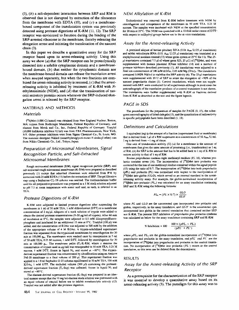
The data in Fig. 1 show that SRP can be added in a quantity such as to cause a >90% translation arrest of preprolactin synthesis (panel A, compare lane h (minus SRP) with lane a (plus SRP); for quantification see the data point corresponding to 0 eq of K-RM in panel C). Increasing the amounts of K- RM in the translation led to a release of the elongation arrest (note the increasing amounts of prolactin synthesized in lanes b--g of panel A; for quantification see the corresponding points in panels B and C). The release by K-RM of the SRP-induced translation arrest yields only prolactin, and not preprolactin (see panel B). This indicates that the translocation of secretory proteins is directly coupled to the release of the elongation arrest by the SRP receptor. A complete release (i.e., 0% inhi- bition) of the SRP-mediated translation arrest was not ob- served, as a significant generalized inhibition of translation occurred (for example, see GLO in lane g) when saturating quantities of K-RM were added to the assays. This inhibition prevented the accurate quantification of secretory protein syn- thesis at higher concentrations of K-RM.
Tryptic Dissection of the Arrest- releasing Activity
Trypsin digestion of K-RM has previously been used to demonstrate that K-RM contain integral membrane protein(s) required for translocation (11). Limited trypsin digestions were used to dissect this activity into a fraction containing trypsin "solubilized" fragment(s) that could be separated by centrifu- gation from a fraction containing the trypsinized membranes. The trypsin-digested membrane fraction was not translocation competent, but translocation could be restored by the addition of the solubilized fragment fraction to this membrane fraction (1 I). These experiments were confirmed (12) and extended: a 60,000-dalton protein fragment was purified from elastase di- gests of K-RM (17) and subsequently shown by immunopre- cipitation to be derived from a 72,000-dalton integral mem- brane protein (18).
We attempted to dissect the arrest-releasing activity of K- RM using an analogous limited protease digestion based upon
FIGURE I K-RM release SRP-mediated arrest of prolactin synthesis. (A) A mixture of bovine pituitary and rabbit reticulocyte RNA was translated in a 25-#I wheat germ system in the presence (lanes a -g ) or absence (lane h) of 10 U of SRP. The translations were supple- mented with the fol lowing quantities of K-RM: (a and h) 0.0 eq, (b) 0.25 eq, (c) 0.5 eq, (d ) 0.75 eq, (e) 1.0 eq, ( f) 1.5 eq, (g) 2.0 eq. The translation products were analyzed by SDS PAGE and visualized by autoradiography. (B) Bands corresponding to preprolactin (pPL), prolactin (P/.), and globin ( GLO} were excised from the gel and the globin-normalized incorporation (see Methods) of [35S]Met into preprolactin (&) and prolactin (0) was determined. (C) The percent SRP-induced inhibit ion of preprolactin plus prolactin synthesis (II) was calculated as described in Materials and Methods. The promi- nent band in panel A between preprolactin and prolactin is pre- growth hormone.
the premise that the activity was associated with a protein domain located on the cytoplasmic side of the microsomal membrane and that the activity might be converted into a soluble form by mild proteolysis.
We therefore treated K-RM with various concentrations of trypsin. The incubation mixtures were separated by centrifu- gation into a soluble fraction (termed Tx-Sup, where x denotes
GILmore et AL. Detection of the SRP Receptor 465
the concentration of trypsin) and into a trypsinized membrane fraction (termed Tx-K-RM, with x again denoting the trypsin concentration used), and both fractions were assayed, either alone or in combination, in the SRP receptor assay.
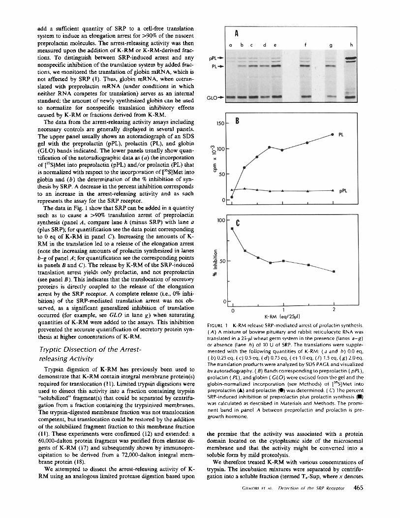
The data in Fig. 2 (displayed in a manner similar to that of Fig. 1) demonstrated that the arrest-releasing activity present in K-RM was inactivated in T~-K-RM depending on the concentration of trypsin used in their preparation. To substan- tiate this conclusion we carried out a titration experiment in which we varied the concentrations of Tx-K-RM (resulting from incubation of K-RM with 0.0, 0.3, 1.0, 5.0, or 50 #g/ml
of trypsin) in the SRP receptor assay. The data in Fig. 3 show that at low concentrations of trypsin (up to 0.3 #g/ml) there is little inactivation, whereas at high concentrations of trypsin (50 ptg/ml) there is an almost complete inactivation of arrest-re- leasing activity.
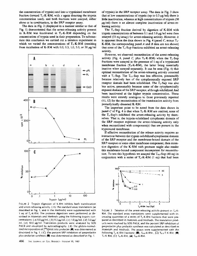
The Tx-Sup fraction derived by digestion of K-RM with trypsin concentrations of between 0.1 and 5.0 tzg/ml were then assayed (33 eq/assay) for arrest-releasing activity. However, it is apparent from the data shown in Fig. 4 (panel C, minus Ts- K-RM, the corresponding panel A and B data are not shown) that none of the T,-Sup fractions exhibited an arrest-releasing activity.
However, we observed reconstitution of the arrest-releasing activity (Fig. 4, panel C, plus Ts-K-RM) when the Tx-Sup fractions were assayed in the presence of 1 cq of a trypsinized membrane fraction (Ts-K-RM), the latter being essentially inactive when assayed separately. It can be seen (Fig. 4) that optimal reconstitution of the arrest-releasing activity occurred with a TL-SUp. The To.l-SUp was less effective, presumably because relatively less of the cytoplasmically exposed SRP receptor domain had been sohibilized. The Ts-Sup was also less active, presumably because some of the cytoplasmically exposed domain of the SRP receptor, although solubilized, had been inactivated at the higher trypsin concentration. These results were entirely analogous to those previously reported (11, 12) for the reconstitution of the translocation activity from protcolytically dissected K-RM.
The important point to be noted from the data shown in panel C of Fig. 4 is that when Ts-K-RM are omitted, none of the T,-Sup's exhibited the arrest-releasing activity by them- selves. That is, the trypsin-solubilized cytoplasmic domain of the SRP receptor expresses the arrest-releasing activity only when reconstituted with component(s) that are present in the trypsinized membrane.
If effective rcconstitution of the release activity requires an interaction between the trypsin-solubilized cytoplasmic domain of the SRP receptor and the membrane-bound domain of the SRP receptor or some other membrane component, then exten- sive digestion of the K-RM with protease might also render this membrane-bound component incompetent for reconstitu- tion. To test this hypothesis we assayed the To.a-Sup (40 eq) in conjunction with a series of T~-K-RM (1 eq) that had been
FIGURE 2 Trypsin digestion of K-RM inhibits both translocation and arrest-releasing activity. (A) The standard assay translations (as described in Fig. 1, and in the Methods) were supplemented with 1 eq of Tx-K-RM. The protease digestions were performed as de- scribed in Materials and Methods using the fo l lowing trypsin con- centrations: (a) 0.0/~g/ml, (b ) 0.3/~g/ml, (c) 1.0 p,g/ml, (d ) 5.0/~g/ ml, (e) 50.0 /sg/ml. Translation products were analyzed by SDS PAGE and visualized by autoradiography. (B) The globin-normal- ized incorporation of [3sS]Met into prolactin (O) was determined as described in Fig. 1. (C) The percent SRP inhibi t ion of preprolactin plus prolactin synthesis (11) was determined as described in Fig. 1.
466 THE JOURNAL OF CELL BIOLOGY • VOLUME 95, 1982
lOO
- ~ ~ i = 505
50 _c ~0.3
o I * I I I I I
0 1 2 3 4 T x - K - R M ( e q / 2 5 p t )
FIGURE ,3 Titration of the arrest-releasing activity present in Tx-K- RM. The standard assay translations were supplemented with in- creasing quantities of a series of Tx-K-RM fractions that were pre- pared as described in Materials and Methods. The translation prod- ucts were resolved by SDS PAGE, and the percent SRP inhibi t ion of preprolactin plus prolactin synthesis was calculated as described in Materials and Methods. The assays were supplemented with the fol lowing T~-K-RM fractions: ( l l L To.o-K-RM , ([Z]) To.3-T-K-RM, (0) T1-K-RM, (O) Ts-K-RM, (A) Tso-K-RM.
EIGURE 5 Extensive trypsin digestion of K-RM prevents functional reconstitution. (A) The standard assay translations were conducted either in the absence (lanes a - f ) or presence (lanes g - / ) of 40 eq of a To 3-Sup fraction. The translations were further supplemented with 1 eq of the fol lowing Tx-K-RM fractions: (a and g) To-K-RM, (b and h) TrK-RM, (cand i) Ts-K-RM, ( d a n d j ) T10-K-RM, (eand k) T~6- K-RM, ( f and I) Tso-K-RM. Translation products were resolved by SDS PAGE and visualized by autoradiography. (B) The bands cor- responding to prolactin (PL) and globin (GLO) were excised from the gel and the globin-normalized incorporation of [35S]Met into prolactin was determined for the assays conducted in the presence of (0, lanes g-I) or absence (0, a-f), of the To.3-Sup fraction. Calculations were performed as described in Materials and Methods.
FtGure 4 Reconstitution of both translocation and arrest-releasing activity. (A) The standard assay translations were supplemented with 1 eq of Ts-K-RM and 33 eq of the fol lowing series of Tx-Sup fractions that were prepared as described in Materials and Methods: (a) To.0-Sup, (b ) To.~-Sup, (c) To.a-Sup, (d ) T~-Sup and (e) Ts-Sup. No Tx-Sup was added to the translation in lane f. The translation products were resolved by SDS PAGE and visualized by autoradiog- raphy. (B) The globin-normalized incorporation of laSS]Met into preprolactin (A) and prolactin (Q) was determined for the assays shown in lanes a - e of panel A as described in Materials and Methods. (C) The percent SRP inhibi t ion of preprolactin plus pro- lactin synthesis was determined as described in Materials and Meth- ods for the assays that were supplemented with both Ts-K-RM and a Tx-Sup fraction (II, corresponding to lanes a - e in panel A) and for the assays that were supplemented with 33 eq of the various Tx-Sup fractions (0, the corresponding translation products are not shown).
prepared from K-RM by digestion with trypsin concentrations of between 1.0 and 50.0 btg/ml.
When the Tx-K-RM's were assayed without the added T0.3- Sup fraction no arrest-releasing activity was observed for mem- branes that were digested with >1 #g/ml of trypsin (see Fig. 5,
panel A, lanes a-e and panel B, open circles). When assayed in the presence of the T0.3-Sup (see Fig. 5, panel A, lanes g-l, and panel B, closed circles) the various Tx-K-RM showed a com- parably retarded loss of the arrest-releasing activity, but even- tually, at the highest trypsin concentration (50.0 ~tg/ml), the reconstitutable arrest-releasing activity was virtually inacti- vated. We conclude that, at high trypsin concentrations, the membrane-bound domain of the SRP receptor or some other component required for reconstitution can be rendered incom- petent for interaction with the cytoplasmic domain of the SRP receptor.
Dissect ion o f SRP Receptor by Elastase
Our results so far show a striking analogy between the reconstitution of the translocation activity (11, 12) and the arrest-releasing activity using trypsin-derived fractions of K- RM, suggesting that a single component in the trypsin-derived supernatant fraction might be responsible for restoring these two activities. The active component responsible for restoring the translocation activity has been purified from elastase-sol- ubilized supernatant fractions (17), and was shown to be de- rived by proteolysis from an integral membrane protein (18).
GILMORE ET AL. Detection of the SRP Receptor 467
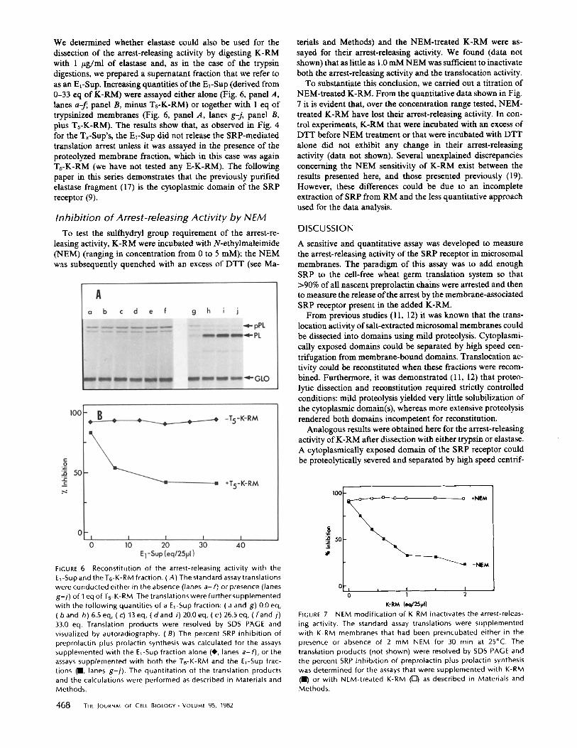
We determined whether elastase could also be used for the dissection of the arrest-releasing activity by digesting K-RM with 1 /~g/ml of elastase and, as in the case of the trypsin digestions, we prepared a supernatant fraction that we refer to as an EE-SUp. Increasing quantities of the El-SUp (derived from 0-33 eq of K-RM) were assayed either alone (Fig. 6, panel A, lanes a-~ panel B, minus Ts-K-RM) or together with 1 eq of trypsinized membranes (Fig. 6, panel A, lanes g-j, panel B, plus Ts-K-RM). The results show that, as observed in Fig. 4 for the Tx-Sup's, the Ez-Sup did not release the SRP-mediated translation arrest unless it was assayed in the presence of the proteolyzed membrane fraction, which in this case was again Ts-K-RM (we have not tested any E-K-RM). The following paper in this series demonstrates that the previously purified elastase fragment (17) is the cytoplasmic domain of the SRP receptor (9).
Inhibition of Arrest-releasing Activity by NEM To test the sulfhydryl group requirement of the arrest-re-
leasing activity, K-RM were incubated with N-ethylmaleimide (NEM) (ranging in concentration from 0 to 5 mM); the NEM was subsequently quenched with an excess of DTT (see Ma-
terials and Methods) and the NEM-treated K-RM were as- sayed for their arrest-releasing activity. We found (data not shown) that as little as 1.0 mM NEM was sufficient to inactivate both the arrest-releasing activity and the translocation activity.
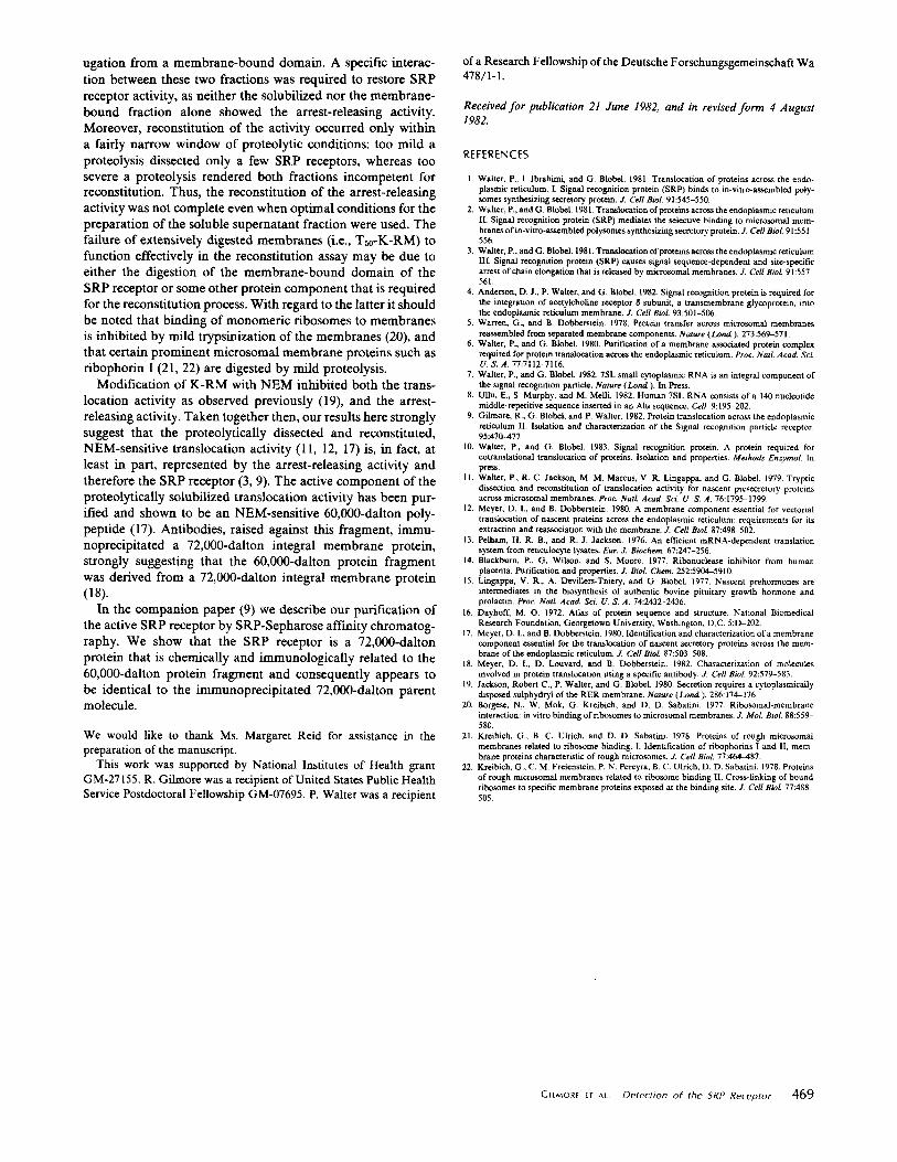
To substantiate this conclusion, we carried out a titration of NEM-treated K-RM. From the quantitative data shown in Fig. 7 it is evident that, over the concentration range tested, NEM- treated K-RM have lost their arrest-releasing activity. In con- trol experiments, K-RM that were incubated with an excess of DTT before NEM treatment or that were incubated with DTT alone did not exhibit any change in their arrest-releasing activity (data not shown). Several unexplained discrepancies concerning the NEM sensitivity of K-RM exist between the results presented here, and those presented previously (19). However, these differences could be due to an incomplete extraction of SRP from RM and the less quantitative approach used for the data analysis.
D I S C U S S I O N
A sensitive and quantitative assay was developed to measure the arrest-releasing activity of the SRP receptor in microsomal membranes. The paradigm of this assay was to add enough SRP to the cell-free wheat germ translation system so that >90% of all nascent preprolactin chains were arrested and then to measure the release of the arrest by the membrane-associated SRP receptor present in the added K-RM.
From previous studies (11, 12) it was known that the trans- location activity of salt-extracted microsomal membranes could be dissected into domains using mild proteolysis. Cytoplasmi- cally exposed domains could be separated by high speed cen- trifugation from membrane-bound domains. Translocation ac- tivity could be reconstituted when these fractions were recom- bined. Furthermore, it was demonstrated (11, 12) that proteo- lyric dissection and reconstitution required strictly controlled conditions: mild proteolysis yielded very little solubilization of the cytoplasmic domain(s), whereas more extensive proteolysis rendered both domains incompetent for reconstitution.
Analogous results were obtained here for the arrest-releasing activity of K-RM after dissection with either trypsin or elastase. A cytoplasmically exposed domain of the SRP receptor could be proteolytically severed and separated by high speed centrif-
FIGURE 6 Reconstitution of the arrest-releasing activity with the El-Sup and the Ts-K-RM fraction. (A) The standard assay translations were conducted either in the absence (lanes a- f) or presence (lanes g-j) of I eq of Ts-K-RM. The translations were further supplemented with the following quantities of a El-Sup fraction: ( a and g) 0.0 eq, ( band h) 6.5 eq, (c) 13eq, ( d a n d i) 20.0 eq, (e) 26.5 eq, ( f and j) 33.0 eq. Translation products were resolved by SDS PAGE and visualized by autoradiography. (B) The percent SRP inhibition of preprolactin plus prolactin synthesis was calculated for the assays supplemented with the El-Sup fraction alone (41,, lanes a-f), or the assays supplemented with both the Ts-K-RM and the EI-Sup frac- tions (R, lanes g-i). The quantitation of the translation products and the calculations were performed as described in Materials and Methods.
468 ]-HE JOURNAL OF CELL BIOLOGY-VOLUME 95, 1982
50
0 [ - | i ! I I 0 1 2
K-RM (eq/25VI )
FIGURE 7 NEM modification of K-RM inactivates the arrest-releas- ing activity. The standard assay translations were supplemented with K-RM membranes that had been preincubated either in the presence or absence of 2 mM NEM for 30 min at 25°C. The translation products (not shown) were resolved by SDS PAGE and the percent SRP inhibition of preprolactin plus prolactin synthesis was determined for the assays that were supplemented with K-RM (ll) or with NEM-treated K-RM ([Z]) as described in Materials and Methods.
ugation from a membrane-bound domain. A specific interac- tion between these two fractions was required to restore SRP receptor activity, as neither the solubilized nor the membrane- bound fraction alone showed the arrest-releasing activity. Moreover, reconstitution of the activity occurred only within a fairly narrow window of proteolytic conditions: too mild a proteolysis dissected only a few SRP receptors, whereas too severe a proteolysis rendered both fractions incompetent for reconstitution. Thus, the reconstitution of the arrest-releasing activity was not complete even when optimal conditions for the preparation of the soluble supernatant fraction were used. The failure of extensively digested membranes (i.e., T~o-K-RM) to function effectively in the reconstitution assay may be due to either the digestion of the membrane-bound domain of the SRP receptor or some other protein component that is required for the reconstitution process. With regard to the latter it should be noted that binding of monomeric ribosomes to membranes is inhibited by mild trypsinization of the membranes (20), and that certain prominent microsomal membrane proteins such as ribophorin I (21, 22) are digested by mild proteolysis.
Modification of K-RM with NEM inhibited both the trans- location activity as observed previously (19), and the arrest- releasing activity. Taken together then, our results here strongly suggest that the proteolytically dissected and reconstituted, NEM-sensitive translocation activity (1 l, 12, 17) is, in fact, at least in part, represented by the arrest-releasing activity and therefore the SRP receptor (3, 9). The active component of the proteolytically solubilized translocation activity has been pur- ified and shown to be an NEM-sensitive 60,000-dalton poly- peptide 07). Antibodies, raised against this fragment, immu- noprecipitated a 72,000-dalton integral membrane protein, strongly suggesting that the 60,000-dalton protein fragment was derived from a 72,000-dalton integral membrane protein (18).
In the companion paper (9) we describe our purification of the active SRP receptor by SRP-Sepharose affinity chromatog- raphy. We show that the SRP receptor is a 72,000-dalton protein that is chemically and immunologically related to the 60,000-dalton protein fragment and consequently appears to be identical to the immunoprecipitated 72,000-dalton parent molecule.
We would like to thank Ms. Margaret Reid for assistance in the preparation of the manuscript.
This work was supported by National Institutes of Health grant GM-27155. R. Gilmore was a recipient of United States Public Health Service Postdoctoral Fellowship GM-07695. P. Walter was a recipient
of a Research Fellowship of the Deutsche F orschungsgemeinschaft Wa 478/1 - 1.
Rece ived f o r publ ica t ion 21 J u n e 1982, a n d in revised f o r m 4 A u g u s t
1982.
REFERENCES
1. Walter, P., I. Ibrahimi, and G. BlobeL. L981. Translocatinn of proteins across the endo- plasmic reticulum. I. Signal recognition protein (SRP) binds to in-vitro-assembled poly- somes synthesizing secretory protein. J. Cell Biol. 91:545-550.
2. Walter, P., and G. BlobeL 1981. Translocation of proteins across the endoplasmic reticulum II. Signal recognition protein (SRP) mediates the selective binding to microsomal mem- branes of in-vitro-assembled polysomes synthesizing secretory protein. J. Cell Biol. 91:551- 556.
3. Walter, P.,andG. Blobel. 1981.Translocationofproteinsacrosstheendoplasmic reticulum III. Signal recognition protein (SRP) causes signal sequence-dependent and site-specific arrest of chain elongation that is released by microsomal membranes. J. Cell BioL 91:557 561.
4. Anderson, D. J., P. Walter, and G. Blobel. 1982. Signal recognition protein is required for the integration of acetylcholine receptor ~ subunit, a transmembrane glycoprotein, into the endoplasmic reticuinm membrane. J. Cell BioL 93:501-506.
5. Warren, G., and B. Dobberstein. 1978. Protein transfer across microsomal membranes reassembled from separated membrane components. Nature (Lond). 273:569-571.
6. Walter, P., and G. Blobel. 1980. Purification of a membrane associated protein complex required for protein translocation across the endoplasmic reticulum. Proc. Natt Acad. ScL U. S. A. 77:7112-7116.
7. Walter, P., and G. BlobeL 1982. 7SL small cytoplasmic RNA is an integral component of the signal recognition particle. Nature (Lond). In Press.
8. Ullu, E., S. Murphy, and M. Melli. 1982. Human 7SL RNA consists of a 140 nucleotide middle-repetitive sequence inserted in an AIu sequence. Cell. 9:195 202.
9. Gilmore, R, G. Blobel, and P. Walter. 1982. Protein translocation across the endoplasmic reticulum II. Isolation and characterization of the Signal recognition particle receptor. 95:470-477
10. Walter, P., and G. Blobel. 1983. Signal recognition protein. A protein required for cotranslational translocation of proteins. Isolation and properties. Methods EnzymoL In press.
II. Waller, P., R. C. Jackson, M. M. Marcus, V. R. Lingappa, and G. Blobet. 1979. Tryptic dissection and reconstitution of translocation activity for nascent presecretory proteins across microsomal membranes. Proc. NatL d cad. ScL U. S. ,4.76:1795-1799.
12. Meyer, D. I., and B. Dobberstein. 1980. A membrane component essential for vectorial transtocation of nascent proteins across the endoplasmic reticulum: requirements for its extraction and reassoclation with the membrane. J. Cell Biol. 87:498 502.
13. Pelham, H. R. B., and R. J. Jackson. 1976. Art efficient mRNA-dependent translation system from reticulocyte lysates. Eur. J. Biochem. 67:247-256.
14. Blackburn, P., G. Wilson, and S. Moore. 1977. Ribonuclease inhibitor from human placenta. Purification and properties..L Biol. Chem. 252:5904-5910.
15. Lingappa, V. R., A. Devillers-Thiery, and G. Biobel. 1977. Nascent prehormones are intermediates in the biosynthesis of authentic bovine pituitary growth hormone and prolactin. Proc. Natt Acad. Sct U. S. A. 74:2432-2436.
16. Dayhoff, M. O. 1972. Atlas of protein sequence and structure. National Biomedical Research Foundation, Georgetown University, Washington, D.C. 5:D-202.
17. Meyer~ D. I., and B. Dobberstein. 1980. Identification and characterization of a membrane component essential for the translocatinn of nascent secretory proteins across the mem- brane of the endoplasmic reticuinm. J. Cell Biol. 87:503-508.
18. Meyer, D. I., D. Louvard, and B. Dobberstein. 1982. Characterization of molecotes involved in protein translocation using a specific antibody. 3. Cell Biol. 92:579-583.
19. Jackson, Robert C., P. Walter, and G. Blobel. 1980. Secretion requires a cytoplasmically disposed sulphydryl of the RER membrane. Nature (Lond). 286:174-176.
20. Borgcse, N:, W. Mok, G. Kreibich, and D. D Sabatini. 1977. Ribosomal-membrane interaction: in vitro binding of ribosomes to microsomal membranes. J. MoL Biol. 88:559- 580.
21. Kreibich, G., B. C. Ulrich, and D. D. Sabatini. 1978. Proteins of rough microsomal membranes related to ribosome binding. I. Identification of ribophorins I and 1I, mem- brane proteins characteristic of rough microsomes, k Cell Blot 77:464~87.
22. Kreibich, G., C. M. Freienstein, P. N. Pereyra, B. C. Ulrich, D. D. Sabatini. 1978. Proteins of rough microsomal membranes related to ribosome binding II. Cross-linking of bound ribosomes to specific membrane proteins exposed at the binding site. J. Cell BioL 77:488- 505.
GltMOR[ er AL Detection of the SRP Receptor 469


![Endoplasmic reticulum[1]](https://img.pdfslide.us/doc/110x75/58ed5fc71a28aba1678b4611/endoplasmic-reticulum1.jpg)