Embed Size (px)
Citation preview

Proteinstructureprediction
CS/CME/BioE/Biophys/BMI279Oct.10and12,2017
RonDror
1

Outline
• Why predict protein structure? • Can we use (pure) physics-based methods? • Knowledge-based methods • Two major approaches to protein structure
prediction – Template-based (“homology”) modeling (e.g., Phyre2) – Ab initio modeling (e.g., Rosetta)
• What’s the best structure prediction method? • Structure prediction games • Comparing protein structures
2

Why predict protein structure?
3
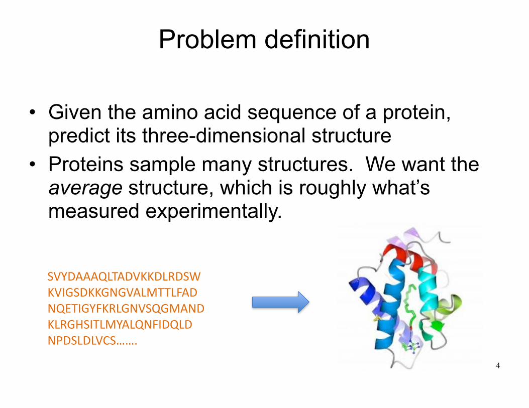
Problem definition
• Given the amino acid sequence of a protein, predict its three-dimensional structure
• Proteins sample many structures. We want the average structure, which is roughly what’s measured experimentally.
4
SVYDAAAQLTADVKKDLRDSWKVIGSDKKGNGVALMTTLFADNQETIGYFKRLGNVSQGMANDKLRGHSITLMYALQNFIDQLDNPDSLDLVCS…….

How are predicted structures used?
• Drug development – Computational screening of candidate drug compounds – Figuring out how to optimize a promising candidate
compound – Figuring out which binding site to target
• Predicting the function of a protein • Identifying the mechanism by which a protein
functions, and how one might alter that protein’s function (e.g., with a drug)
• Interpreting experimental data – For example, a computationally predicted approximate
structure can help in determining an accurate structure experimentally, as we’ll see later in this course 5

Why not just solve the structures experimentally?
• Some structures are very difficult to solve experimentally – Sometimes many labs work for decades to solve the structure of one protein
• Sequence determination far outpaces experimental structure determination – We already have far more sequences than experimental structures, and this
gap will likely grow
6http://www.dnastar.com/blog/wp-content/uploads/2015/08/ProteinDBGrowthBar3.png

Can we use (pure) physics-based methods?
7

Simulation vs. experiment for 12 fast-folding proteins, up to 80 residues each
Lindorff-Larsen et al., Science, 2011
Why not just simulate the folding process by molecular dynamics?
8
Chignolin BBA
NTL9WW domain
VillinTrp-cage
Protein GHomeodomain
Protein BBBL
λ-repressorα3D

For most proteins, this doesn’t work
1. Folding timescales are usually much longer than simulation timescales.
2. Current molecular mechanics force fields aren’t sufficiently accurate.
3. Disulfide bonds form during the real folding process, but this is hard to mimic in simulation.
9

Can we find simpler physics-based rules that predict protein structure?
• For example, look at patterns of hydrophobic, hydrophilic, or charged amino acids?
• People have tried for a long time without much success
10

Knowledge-based methods
11

Basic idea behind knowledge-based (data-driven) methods
• We have experimental structures for over 100,000 proteins.
• Can we use that information to help us predict new structures?
• Yes!
12http://www.duncanmalcolm.com/blog/startup-data-analytics-metric-
Wecanalsousethe>50millionproteinsequencesintheUniProtdatabase

Proteins with similar sequences tend to have similar structures
• Proteins with similar sequences tend to be homologs, meaning that they evolved from a common ancestor
• The fold of the protein (i.e., its overall structure) tends to be conserved during evolution
• This tendency is very strong. Even proteins with 15% sequence identity usually have similar structures. – During evolution, sequence changes more quickly
than structure • Also, there only appear to be 1,000–10,000
naturally occurring protein folds13

For most human protein sequences, we can find a homolog with known structure
14
Schwede, Structure 2013
The plot shows the fraction of amino acids in human proteins that can be mapped to similar sequences in PDB structures. Different colors indicate % sequence identity.
Unstructured(disordered)aminoacids

What if we can’t identify a homolog in the PDB?
• We can still use information based on known structures – We can construct databases of observed structures of
small fragments of a protein – We can use the PDB to build empirical, “knowledge-
based” energy functions
15

Two major approaches to protein structure prediction
16

Two main approaches to protein structure prediction
• Template-based modeling (homology modeling) – Used when one can identify one or more likely
homologs of known structure • Ab initio structure prediction
– Used when one cannot identify any likely homologs of known structure
– Even ab initio approaches usually take advantage of available structural data, but in more subtle ways
17

18
Template-based (“homology”) modeling (e.g., Phyre2)
Two major approaches to protein structure prediction

Template-based structure prediction: basic workflow
• User provides a query sequence with unknown structure
• Search the PDB for proteins with similar sequence and known structure. Pick the best match (the template).
• Build a model based on that template – One can also build a model based on multiple
templates, where different templates are used for different parts of the protein.
19

What does it mean for two sequences to be similar?
• Basic measure: count minimum number of amino acid residues one needs to change, add, or delete to get from one sequence to another – Sequence identity: amino acids that match exactly
between the two sequences – Not trivial to compute for long sequences, but there
are efficient dynamic programming algorithms to do so
20
What does it mean for two sequences to be similar?
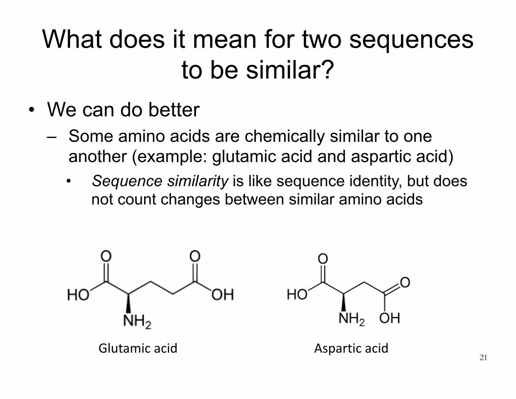
• We can do better – Some amino acids are chemically similar to one
another (example: glutamic acid and aspartic acid) • Sequence similarity is like sequence identity, but does
not count changes between similar amino acids
21Glutamicacid Asparticacid

What does it mean for two sequences to be similar?
• We can do even better – Once we’ve identified some homologs to a query sequence
(i.e., similar sequences in the sequence database), we can create a profile describing the probability of mutation to each amino acid at each position
– We can then use this profile to search for more homologs – Iterate between identification of homologs and profile
construction – Measure similarity of two sequences by comparing their profiles – Often implemented using hidden Markov models (HMMs)
(but you are not responsible for knowing about HMMs)
22

We’ll use the Phyre2 template-based modeling server as an example
• Try it out: http://www.sbg.bio.ic.ac.uk/phyre2/ • Why use Phyre2 as an example of template-
based modeling? – Among the better automated structure
prediction servers – Among the most widely used, and arguably
the easiest to use – Approach is similar to that of other template-
based modeling methods – Great name!
23
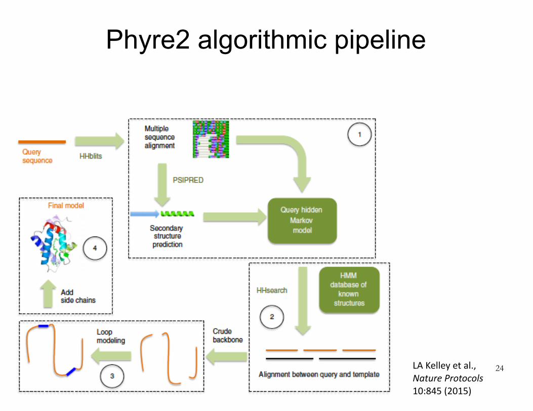
Phyre2 algorithmic pipeline
24LAKelleyetal.,NatureProtocols10:845(2015)

Phyre2 algorithmic pipeline
25
Identifysimilarsequencesinproteinsequencedatabase

Phyre2 algorithmic pipeline
26
Chooseatemplatestructureby:(1)comparingsequenceprofilesand(2)predictingsecondarystructureforeachresidueinthequerysequenceandcomparingtocandidatetemplatestructures.Secondarystructure(alphahelix,betasheet,orneither)ispredictedforsegmentsofquerysequenceusinganeuralnetworktrainedonknownstructures.

Phyre2 algorithmic pipeline
27
Computeoptimalalignmentofquerysequencetotemplatestructure

Buildacrudebackbonemodel(nosidechains)bysimplysuperimposingcorrespondingaminoacids.Someofthequeryresidueswillnotbemodeled,becausetheydon’thavecorrespondingresiduesinthetemplate(insertions).Therewillbesomephysicalgapsinthemodeledbackbone,becausesometemplateresiduesdon’thavecorrespondingqueryresidues(deletions).
Phyre2 algorithmic pipeline

Phyre2 algorithmic pipeline
29
Useloopmodelingtopatchupdefectsinthecrudemodelduetoinsertionsanddeletions.Foreachinsertionordeletion,searchalargelibraryoffragments(2-15residues)ofPDBstructuresforonesthatmatchlocalsequenceandfitthegeometrybest.Tweakbackbonedihedralswithinthesefragmentstomakethemfitbetter.

Phyre2 algorithmic pipeline
30
Addsidechains.Useadatabaseofcommonlyobservedstructuresforeachsidechain(thesestructuresarecalledrotamers).Searchforcombinationsofrotamersthatwillavoidstericclashes(i.e.,atomsendingupontopofoneanother).

Modeling based on multiple templates• In “intensive mode,” Phyre
2 will use multiple templates that cover (i.e., match well to) different parts of the query sequence. – Build a crude backbone
model for each template – Extract distances between
residues for “reliable” parts of each model
– Perform a simplified protein folding simulation in which these distances are used as constraints. Additional constraints enforce predicted secondary structure
– Fill in the side chains, as for single-template models
31
LAKelleyetal.,NatureProtocols10:845(2015)You’renotresponsibleforthis

32
Ab initio modeling (e.g., Rosetta)
Two major approaches to protein structure prediction

Two main approaches to protein structure prediction
• Template-based modeling (homology modeling) – Used when one can identify one or more likely
homologs of known structure • Ab initio structure prediction
– Used when one cannot identify any likely homologs of known structure
– Even ab initio approaches usually take advantage of available structural data, but in more subtle ways
33

Ab initio structure prediction
• Also known as “de novo structure prediction” • Many approaches proposed over time • Probably the most successful is fragment
assembly, as exemplified by the Rosetta software package
34

We’ll use Rosetta as an example of ab-initio structure prediction
• Software developed over the last 15–20 years by David Baker (U. Washington) and collaborators
• Software at: https://www.rosettacommons.org/software • Structure prediction server: http://robetta.bakerlab.org/ • Why use Rosetta as an example?
– Among the better ab initio modeling packages (for some years it was the best)
– Approach is similar to that of many ab initio modeling packages
– Rosetta provides a common framework that has become very popular for a wide range of molecular prediction and design tasks, especially protein design35
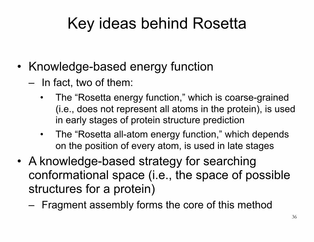
Key ideas behind Rosetta
• Knowledge-based energy function – In fact, two of them:
• The “Rosetta energy function,” which is coarse-grained (i.e., does not represent all atoms in the protein), is used in early stages of protein structure prediction
• The “Rosetta all-atom energy function,” which depends on the position of every atom, is used in late stages
• A knowledge-based strategy for searching conformational space (i.e., the space of possible structures for a protein) – Fragment assembly forms the core of this method
36

Rosetta energy function• At first this was the only energy function used by
Rosetta (hence the name) • Based on a simplified representation of protein
structure: – Do not explicitly represent solvent (e.g., water) – Assume all bond lengths and bond angles are fixed – Represent the protein backbone using torsion angles
(three per amino acid: Φ, Ψ, ω) – Represent side chain position using a single “centroid,”
located at the side chain’s center of mass • Centroid position determined by averaging over all
structures of that side chain in the PDB37

Rosetta energy function
38
FromRohletal.,MethodsinEnzymology2004 You’renotresponsibleforthedetails!

Rosetta energy function
39
FromRohletal.,MethodsinEnzymology2004 You’renotresponsibleforthedetails!

Rosetta energy function: take-aways
• The (coarse-grained) Rosetta energy function is essentially entirely knowledge-based – Based on statistics computed from the PDB
• Many of the terms are of the form –loge[P(A)], where P(A) is the probability of some event A – This is essentially the free energy of event A. Recall
definition of free energy:
40
P(A) = exp −GAkBT( )GA = −kBT loge P(A)( )

Rosetta all-atom energy function
• Still makes simplifying assumptions: – Do not explicitly represent solvent (e.g., water) – Assume all bond lengths and bond angles are fixed
• Functional forms are a hybrid between molecular mechanics force fields and the (coarse-grained) Rosetta energy function – Partly physics-based, partly knowledge-based
41

Are these potential energy functions or free energy functions?
• The energy functions of previous lectures were potential energy functions
• One can also attempt to construct a free energy function, where the energy associated with a conformation is the free energy of the set of “similar” conformations (for some definition of “similar”)
• The Rosetta energy functions are sometimes described as potential energy functions, but they are closer to approximate free energy functions – This means that searching for the “minimum” energy is more valid – Nevertheless, typical protocol is to repeat the search process
many times, cluster the results, and report the largest cluster as the solution. This rewards wider and deeper wells.
42
How does Rosetta search the conformational space?
• Two steps: – Coarse search: fragment assembly – Refinement
• Perform coarse search many times, and then perform refinement on each result
43

Coarse search: fragment assembly
• Uses a large database of 3-residue and 9-residue fragments, taken from structures in the PDB
• Monte Carlo sampling algorithm proceeds as follows: 1. Start with the protein in an extended conformation 2. Randomly select a 3-residue or 9-residue section 3. Find a fragment in the library whose sequence resembles it 4. Consider a move in which the backbone dihedrals of the
selected section are replaced by those of the fragment. Calculate the effect on the entire protein structure.
5. Evaluate the Rosetta energy function before and after the move.
6. Use the Metropolis criterion to accept or reject the move. 7. Return to step 2
• The real search algorithm adds some bells and whistles44

Refinement
• Refinement is performed using the Rosetta all-atom energy function, after building in side chains
• Refinement involves a combination of Monte Carlo moves and energy minimization
• The Monte Carlo moves are designed to perturb the structure much more gently than those used in the coarse search – Many still involve the use of fragments
45

What’s the best structure prediction method?
46

What’s the best protein structure prediction method?
• Currently, it’s probably I-TASSER – http://zhanglab.ccmb.med.umich.edu/I-TASSER/
• I-TASSER is template-based, but it uses threading, meaning that when selecting a template it maps the query sequence onto the template structure and evaluates the quality of the fit – This allows detection of very remote homology
• I-TASSER combines many algorithms – It incorporates a surprisingly large number of different components
and strategies, including an ab initio prediction module – It runs many algorithms in parallel and then looks for a consensus
between the results • Example: at least seven different threading algorithms
– Inelegant but effective47
You’renotresponsibleforthis

Structure prediction games
48
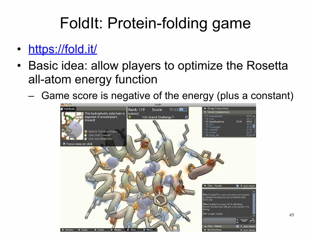
FoldIt: Protein-folding game• https://fold.it/ • Basic idea: allow players to optimize the Rosetta
all-atom energy function – Game score is negative of the energy (plus a constant)
49

50

EteRNA: RNA design game • Similar idea, but:
– For RNA rather than protein. – Goal is RNA design. Users collective design RNA sequences, which are tested
experimentally. • From Rhiju Das (Stanford) and Adrien Treuille (CMU)
51

Comparing protein structures
52

Comparing structures of a protein
• The most common measure of similarity between two structures for a given protein is root mean squared distance/deviation (RMSD), defined as where x gives the coordinates for one structure and w the coordinates for the other
• We generally want to align the structures, which can be done by finding the rigid-body rotation and translation of one structure that will minimize its RMSD from the other – The relevant measure of similarity is RMSD after alignment.
53
1n
xi −wi( )2i=1
n
∑