Embed Size (px)
Citation preview
3320 March 28, 2014|Volume 20|Issue 12|WJG|www.wjgnet.com
Online Submissions: http://www.wjgnet.com/esps/[email protected]:10.3748/wjg.v20.i12.3320
World J Gastroenterol 2014 March 28; 20(12): 3320-3326 ISSN 1007-9327 (print) ISSN 2219-2840 (online)
© 2014 Baishideng Publishing Group Co., Limited. All rights reserved.
BRIEF ARTICLE
Protective effects of intravenous anesthetics on kidney tissue in obstructive jaundice
Sinan Hatipoglu, Huseyin Yildiz, Ertan Bulbuloglu, Ismail Coskuner, Ergul Belge Kurutas, Filiz Hatipoglu, Harun Ciralik, Mehmet Sait Berhuni
Sinan Hatipoglu, Department of General Surgery Unit, School of Medicine, Adiyaman University, 02040 Adiyaman, TurkeyHuseyin Yildiz, Ismail Coskuner, Department of Anesthesiol-ogy and Reanimation Unit, School of Medicine, Kahramanmaras Sutcuimam University, 46100 Kahramanmaras, TurkeyErtan Bulbuloglu, Mehmet Sait Berhuni, Department of Gen-eral Surgery Unit, School of Medicine, Kahramanmaras Sutcui-mam University, 46100 Kahramanmaras, TurkeyErgul Belge Kurutas, Department of Biochemistry Unit, School of Medicine, Kahramanmaras Sutcuimam University, 46100 Kahramanmaras, TurkeyFiliz Hatipoglu, Department of Obstetrics and Gynecology Unit, School of Medicine, Adiyaman University, 02040 Adiyaman, TurkeyHarun Ciralik, Department of Pathology Unit, School of Medi-cine, Kahramanmaras Sutcuimam University, 46100 Kahraman-maras, TurkeyAuthor contributions: Hatipoglu S and Yildiz H contributed equally to this work; Hatipoglu S, Yildiz H and Bulbuloglu E designed the research; Hatipoglu S, Yildiz H and Bulbuloglu E performed the research; Hatipoglu S, Yildiz H, Bulbuloglu E, Coskuner I, Kurutas EB, Ciralik H, Hatipoglu F and Berhuni MS contributed new reagents/analytic tools; Hatipoglu S, Yildiz H and Bulbuloglu E analyzed the data; Hatipoglu S wrote the paper.Correspondence to: Sinan Hatipoglu, MD, Assistant Profes-sor, Department of General Surgery Unit, School of Medicine, Adiyaman University, Altınsehir Street, 02040 Adiyaman, Turkey. [email protected]: +90-505-4509402 Fax: +90-416-2231693Received: November 11, 2013 Revised: January 14, 2014 Accepted: January 19, 2014Published online: March 28, 2014
AbstractAIM: To evaluate the protective effects on kidney tis-sue of frequently used intravenous anesthetics (ket-amine, propofol, thiopental, and fentanyl) in rats with obstructive jaundice.
METHODS: There is an increased incidence of postop-erative acute renal failure in patients with obstructive
jaundice. Thirty-two Wistar-albino rats were randomly divided into four equal groups. Laparatomy was per-formed on each animal in the four groups and common bile ducts were ligated and severed on day 0. After 7 d, laparotomy was again performed using ketamine, pro-pofol, thiopental, or fentanyl anesthesia whose antioxi-dative properties are well known in oxidative stress in a rat liver model of obstructive jaundice. After 2 h, the rats were sacrificed. Renal tissue specimens were ana-lyzed for catalase, superoxide dismutase and malondi-aldehyde enzymes activities. All values are expressed as the mean ± SD. P values less than 0.05 were con-sidered statistically significant.
RESULTS: All animals survived without complications until the end of the study. Enlargement in the bile duct and obstructive jaundice were observed in all rats. Catalase was found to be significantly lower in the fen-tanyl group than in the ketamine (P = 0.039), propofol (P = 0.012), and thiopental (P = 0.001) groups. Super-oxide dismutase activities were similar in all groups (P > 0.05). Malondialdehyde was found to be significantly lower in the ketamine group than in the propofol (P = 0.028), thiopental (P = 0.002) and fentanyl (P = 0.005) groups. Malondialdehyde was also lower in the fentanyl group than in the thiopental group (P = 0.001). The re-sults showed that obstructive jaundice sensitizes renal tissue to damage under the different anesthetics.
CONCLUSION: Among the agents tested, ketamine and propofol generated the least amount of oxidative stres on renal tissues in this rat model of obstructive jaundice created by common bile duct ligation. The im-portance of free radical injury in renal tissue in obstruc-tive jaundice under different intravenous anesthetics during hepatobiliary and liver transplant surgery should be considered for prevention of postoperative acute re-nal failure.
© 2014 Baishideng Publishing Group Co., Limited. All rights reserved.
BRIEF ARTICLE
Key words: Obstructive jaundice; Postoperative acute renal failure; Oxidative stress; Intravenous anesthetics; Renal tissue damage
Core tip: There is an increased incidence of postop-erative acute renal failure in patients with obstructive jaundice. Recent studies suggested that the free oxy-gen radicals produced in obstructive jaundice may play a major role in the etiopathogenesis of acute renal fail-ure. We evaluated the protective effects on kidney tis-sue of frequently used intravenous anesthetics, whose antioxidative properties are well known, in a rat model of obstructive jaundice. Among the agents tested, ketamine and propofol generated the least amount of oxidative stres on renal tissues in this rat model of obstructive jaundice created by common bile duct liga-tion.
Hatipoglu S, Yildiz H, Bulbuloglu E, Coskuner I, Kurutas EB, Hatipoglu F, Ciralik H, Berhuni MS. Protective effects of intrave-nous anesthetics on kidney tissue in obstructive jaundice. World J Gastroenterol 2014; 20(12): 3320-3326 Available from: URL: http://www.wjgnet.com/1007-9327/full/v20/i12/3320.htm DOI: http://dx.doi.org/10.3748/wjg.v20.i12.3320
INTRODUCTIONToday, palliative and curative operations are performed on many patients with obstructive jaundice (OJ) under anesthesia. As a result of improvements in liver trans-plant surgery in the last 50 years, more complicated and prolonged operations are being conducted. Patients with severe OJ usually have a number of metabolic disorders and one or multiple organ function failure. Renal dys-function is one of the serious complications in patients with OJ[1-3].
An association between OJ and acute renal failure (ARF) has been recognized for well over a century. The renal damage is due to biliary disorders either present on admission to hospital or which develop postoperatively. One third of patients with OJ have deterioration of re-nal function before surgical intervention[4]. On the other hand, surgery on patients with OJ is known to be associ-ated with increased risk of postoperative renal failure[5-8]. Early diagnosis and prevention of spontaneous evolution of the disease can improve prognosis.
Patients with intra- or extra-hepatic bile duct ob-struction are susceptible to ARF especially after major surgery[9]. Surgical treatment for the relief of OJ is still complicated by postoperative ARF in almost 10%of patients[3]. Patients with OJ are often subjected to either general or sedation anesthesia, usually using drugs which are metabolized by the liver and/or are eliminated by the kidney and the liver. Some intravenous anesthetic agents have been shown to increase production of reactive oxygen species and cause tissue damage[9-13]. In contrast, some intravenous anesthetic drugs are capable of reduc-
ing oxidative stress[13,14].To date, there have been no reports of the effects of
intravenous anesthetic agents on oxidative stress in renal tissues in rats with OJ. Biliary obstruction is associated with an intense state of oxidative stress. Antioxidant de-fenses [as demonstrated by superoxide dismutase (SOD) and catalase (CAT) activities] are decreased and lipid per-oxidation [as demonstrated by malondialdehyde (MDA) levels] are increased in rat models with OJ[13,15]. In this study, we therefore investigated the effects on renal tis-sues of frequently used intravenous anesthetics (ketamine, propofol, thiopental, and fentanyl), in a rat model of oxidative stress caused by OJ through common bile duct ligation. We used these intravenous anesthetics as their antioxidative properties are well known.
MATERIALS AND METHODSAnimalsThe experimental protocol was approved by the Animal Ethics Review Committee of the Faculty of Medicine, University of Kahramanmaras and adhered to the Na-tional Institutes of Health Guidelines for the Use of Ex-perimental Animals. Thirty-two male Wistar rats (300-375 g) were subjected to controlled conditions of tempera-ture (about 22 ℃) and illumination (12 h light:12 h dark cycle), and were provided with food and water ad libitum. They were fed a commercial diet. Rats were placed in in-dividual metabolic cages and acclimatized for 1 wk before the study commenced.
Experimental designIn this prospective experimental study, rats were divided randomly into four groups, each group containing eight animals. Food was withdrawn for 12 h before the opera-tion, with water available ad libitum during this period. Each rat was weighed during each anesthetic and anesthe-tized with ketamine (50 mg/kg) intramuscularly. As de-scribed by Lee et al[16], experimental jaundice was created by ligation of the common bile duct[13,15]. The abdominal cavity was opened with a midline incision after disinfec-tion of the skin. The common bile duct was located and OJ induced by double ligation with 5/0 silk and transsec-tion of the common bile duct in the supraduodenal part between the lowermost tributary of the bile duct and the uppermost tributary of the pancreatic duct. The abdomi-nal wall was then closed with 3-0 silk in two layers. Cages were examined daily.
After 7 d, Group Ⅰ rats received intramuscular single-dose ketamine (50 mg/kg), Group Ⅱ received intra-muscular single-dose propofol (10 mg/kg), Group Ⅲ received intramuscular single-dose thiopental (20 mg/kg), and Group IV received intramuscular single-dose fentan-yl (50 mcg/kg). Two hours later, the rats were sacrificed.
Sample collectionThe animals were anesthetized and a second laparotomy was performed through a similar incision. The left and
3321 March 28, 2014|Volume 20|Issue 12|WJG|www.wjgnet.com
Hatipoglu S et al . Anesthetic protection of kidney in jaundice
right kidneys of each rat were carefully removed in all groups and stored in iced 0.9% NaCl solution for a short time. A 0.5 cm × 0.5 cm sample of kidneys (left or right) which contain both renal cortical and medullar tissue were washed with physiological saline to remove hematoma and blotted on filter paper. The renal tissue was immediately frozen in liquid nitrogen and stored at -80 ℃ for later measurement of MDA, SOD and CAT activities.
Antioxidant studyIn order to determine tissue antioxidant levels, the renal tissue samples were removed from the freezer, brought to room temperature, then homogenized with three vol-umes of ice-cold 1.15% KCl. Activities of antioxidant enzymes and levels of lipid peroxidation were measured in the supernatant after centrifugation at 14000 rpm. SOD activity was measured by the method described by Fridovich[17]. CAT activity was determined by measuring the decrease in hydrogen peroxide concentration at 230 nm by the method of Beutler[18]. Lipid peroxidation was reflected by MDA levels, which were measured by the method described by Ohkawa et al[19]. All enzyme activi-ties are expressed as units per milligram protein (U/mg protein).
Statistical analysisAll values are expressed as mean ± SD. The Kolmogo-rov-Smirnov statistic was used to test the normality of the distribution. Differences between SOD groups were evaluated by Kruskal-Wallis variance analysis followed by a post hoc (Bonferroni correction) Mann-Whitney U test. Differences between MDA and CAT groups were evaluated by one way analysis of variance (ANOVA) for continuous variables with post hoc procedures (Bonferroni correction). P-values < 0.05 were considered statistically significant. Data were analyzed using SPSS 9.05 for Win-dows® statistical package (Chicago, IL, United States).
RESULTSAll animals survived without complications until the end of the study. Enlargement of the bile duct and OJ were observed in all rats. The mean values of the parameters studied are presented in Table 1. The results showed that the presence of OJ sensitized the renal tissue to damage under the different anesthetics.
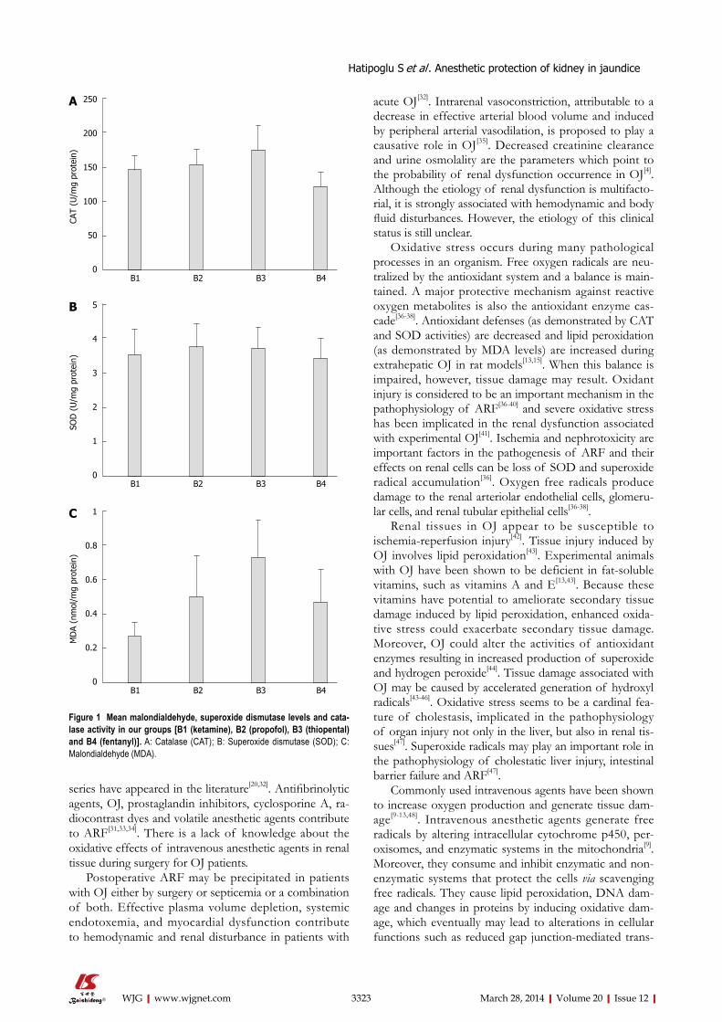
CAT was found to be significantly lower in the fen-tanyl group than in the ketamine (P = 0.039), propofol (P = 0.012), and thiopental (P = 0.001) groups. Although CAT was higher in the thiopental group than in the ket-amine and propofol groups, this difference was not sta-tistically significant (Table 1, Figure 1A).
SOD activity was similar between all groups and in-tergroup differences were not found (P > 0.05) (Table 1, Figure 1B).
MDA was found to be significantly lower in the ket-amine group than in the propofol (P = 0.028), thiopental (P = 0.002) and fentanyl (P = 0.005) groups. MDA was also lower in the fentanyl group than in the thiopental group (P = 0.001). MDA was similar between the propo-fol and thiopental groups and no other significant inter-group difference was found (Table 1, Figure 1C).
DISCUSSIONMany clinical observations and experimental studies point to the frequent occurrence of different organ complica-tions in patients with OJ. One of the main consequences of biliary obstruction is its effect on renal function, which markedly increases patient morbidity and mortal-ity. Acute renal failure is a life-threatening complication of OJ which continues to be a significant challenge, in-volving 6%-18% of patients, and is associated with high mortality (20%-100%)[20-23]. Patients with intra- or extra-hepatic bile duct obstruction are susceptible to ARF especially when undergoing major surgery, and postoper-ative ARF in patients with OJ remains a clinically signifi-cant complication[9,24]. ARF occurs in approximately 9% of patients requires surgery for relief of OJ, and contrib-utes to eventual mortality in 76% of those who develop it. Postoperative mortality has been directly attributed to ARF in approximately 5%-16% of patients after surgery for OJ[25,26].
When mechanical biliary obstruction is diagnosed, surgical, endoscopic or radiologic intervention is usually recommended. On the other hand, despite advances in preoperative evaluation and postoperative care, surgical intervention for relief of obstructive jaundice still carries high morbidity and mortality rates, mainly due to sepsis and renal dysfunction[25,27-30]. The presence of OJ (total bilirubin < 8 mg/dL) is an independent risk factor for the development of postoperative renal dysfunction[31].
Although the association between OJ and ARF has been recognized since 1910 when Clairmont and Von Haberer[22] first postulated that jaundice might predispose to postoperative renal failure, surprisingly few reports or
3322 March 28, 2014|Volume 20|Issue 12|WJG|www.wjgnet.com
Table 1 Mean malondialdehyde, superoxide dismutase levels and catalase levels in renal tissue of rats
Groups Catalase Superoxide dismutase
Malondialdehyde
B1 (ketamine) 146.11 ± 20.911 3.52 ± 0.73 0.27 ± 0.0804,5,6
B2 (propofol) 154.11 ± 21.462 3.74 ± 0.67 0.50 ± 0.244
B3 (thiopental) 174.8 ± 36.63 3.70 ± 0.61 0.73 ± 0.225,7
B4 (fentanyl) 122.48 ± 20.541,2,3 3.41 ± 0.59 0.47 ± 0.196,7
Data are expressed as mean ± SD. Mean malondialdehyde (MDA), superoxide dismutase (SOD) levels and catalase (CAT) levels in renal tissue of rats (8 rats in each group). CAT and SOD activities are expressed as U/mg protein. MDA enzyme activities are expressed as nmol/mg protein. P-values < 0.05 were considered statistically significant. In the CAT group: 1P = 0.039 for ketamine vs fentanyl comparison; 2P = 0.012 for propofol vs fentanyl comparison; 3P = 0.001 for thiopental vs fentanyl comparison. In the MDA group: 4P = 0.028 for ketamine vs propofol comparison; 5P = 0.002 for ketamine vs thiopental comparison; 6P = 0.005 for ketamine vs fentanyl comparison; 7P = 0.001 for thiopental vs fentanyl comparison.
Hatipoglu S et al . Anesthetic protection of kidney in jaundice
3323 March 28, 2014|Volume 20|Issue 12|WJG|www.wjgnet.com
acute OJ[32]. Intrarenal vasoconstriction, attributable to a decrease in effective arterial blood volume and induced by peripheral arterial vasodilation, is proposed to play a causative role in OJ[35]. Decreased creatinine clearance and urine osmolality are the parameters which point to the probability of renal dysfunction occurrence in OJ[4]. Although the etiology of renal dysfunction is multifacto-rial, it is strongly associated with hemodynamic and body fluid disturbances. However, the etiology of this clinical status is still unclear.
Oxidative stress occurs during many pathological processes in an organism. Free oxygen radicals are neu-tralized by the antioxidant system and a balance is main-tained. A major protective mechanism against reactive oxygen metabolites is also the antioxidant enzyme cas-cade[36-38]. Antioxidant defenses (as demonstrated by CAT and SOD activities) are decreased and lipid peroxidation (as demonstrated by MDA levels) are increased during extrahepatic OJ in rat models[13,15]. When this balance is impaired, however, tissue damage may result. Oxidant injury is considered to be an important mechanism in the pathophysiology of ARF[36-40] and severe oxidative stress has been implicated in the renal dysfunction associated with experimental OJ[41]. Ischemia and nephrotoxicity are important factors in the pathogenesis of ARF and their effects on renal cells can be loss of SOD and superoxide radical accumulation[36]. Oxygen free radicals produce damage to the renal arteriolar endothelial cells, glomeru-lar cells, and renal tubular epithelial cells[36-38].
Renal tissues in OJ appear to be susceptible to ischemia-reperfusion injury[42]. Tissue injury induced by OJ involves lipid peroxidation[43]. Experimental animals with OJ have been shown to be deficient in fat-soluble vitamins, such as vitamins A and E[13,43]. Because these vitamins have potential to ameliorate secondary tissue damage induced by lipid peroxidation, enhanced oxida-tive stress could exacerbate secondary tissue damage. Moreover, OJ could alter the activities of antioxidant enzymes resulting in increased production of superoxide and hydrogen peroxide[44]. Tissue damage associated with OJ may be caused by accelerated generation of hydroxyl radicals[43-46]. Oxidative stress seems to be a cardinal fea-ture of cholestasis, implicated in the pathophysiology of organ injury not only in the liver, but also in renal tis-sues[47]. Superoxide radicals may play an important role in the pathophysiology of cholestatic liver injury, intestinal barrier failure and ARF[47].
Commonly used intravenous agents have been shown to increase oxygen production and generate tissue dam-age[9-13,48]. Intravenous anesthetic agents generate free radicals by altering intracellular cytochrome p450, per-oxisomes, and enzymatic systems in the mitochondria[9]. Moreover, they consume and inhibit enzymatic and non-enzymatic systems that protect the cells via scavenging free radicals. They cause lipid peroxidation, DNA dam-age and changes in proteins by inducing oxidative dam-age, which eventually may lead to alterations in cellular functions such as reduced gap junction-mediated trans-
series have appeared in the literature[20,32]. Antifibrinolytic agents, OJ, prostaglandin inhibitors, cyclosporine A, ra-diocontrast dyes and volatile anesthetic agents contribute to ARF[31,33,34]. There is a lack of knowledge about the oxidative effects of intravenous anesthetic agents in renal tissue during surgery for OJ patients.
Postoperative ARF may be precipitated in patients with OJ either by surgery or septicemia or a combination of both. Effective plasma volume depletion, systemic endotoxemia, and myocardial dysfunction contribute to hemodynamic and renal disturbance in patients with
250
200
150
100
50
0
CAT
(U/m
g pr
otei
n)
B1 B2 B3 B4
5
4
3
2
1
0
SOD
(U
/mg
prot
ein)
B1 B2 B3 B4
1
0.8
0.6
0.4
0.2
0
MD
A (n
mol
/mg
prot
ein)
B1 B2 B3 B4
C
B
A
Figure 1 Mean malondialdehyde, superoxide dismutase levels and cata-lase activity in our groups [B1 (ketamine), B2 (propofol), B3 (thiopental) and B4 (fentanyl)]. A: Catalase (CAT); B: Superoxide dismutase (SOD); C: Malondialdehyde (MDA).
Hatipoglu S et al . Anesthetic protection of kidney in jaundice
3324 March 28, 2014|Volume 20|Issue 12|WJG|www.wjgnet.com
mission, activation of transcription factors, intracellular calcium and pH changes, and/or cell death[9-13,49].
Transient functional impairment of renal cation and water transport in the proximal convoluted tubule oc-curred 3 to 4 d following bile duct ligation in rats[6]. Maxi-mum plasma concentrations and renal clearance of bile acids occurred between the 3rd or 4th postoperative day following common bile duct ligation. This peak coincided with maximal disruption of the proximal convoluted tu-bule architecture and postoperative changes in renal func-tion such as increased urine flow rate and decreased urine osmolality and sodium excretion[6]. Because of these re-sults, we chose to sacrifice our rats on the 7th postopera-tive day and specimens of renal tissues were resected.
Ketamine has been extensively studied as a safe and reliable dissociative sedative/anesthetic agent in various clinical situations. Ketamine’s properties as a protective agent against oxidative stress and ischemia/reperfusion injury of the brain, kidney, skeletal muscle, heart, and in-testine have been reported[50-54]. In our experiment, MDA levels were lower in the ketamine group compared with the other groups, confirming ketamine as an agent which protects against oxidative stress. MDA is one of the fairly reactive metabolic products resulting from the effect of free oxygen radicals on tissues and from a series of reac-tions during lipid peroxidation. The tissue MDA level is a sensitive indicator of lipid peroxidation and thus of oxi-dative stress[55]. Since ketamine lowered MDA levels more than the other agents used, we can conclude that it has an influence over the antioxidant defense system, while reducing lipid peroxidation.
Propofol and thiopental are another type of highly lipid-soluble anesthetic which have demonstrated anti-oxidant properties by inhibiting lipid peroxidation[56-58]. Both are often used to reduce cerebral edema during liver transplantation in fulminant hepatic failure patients[13]. Propofol is widely used for the induction and mainte-nance of general anesthesia, as well as for sedation of in-tubated postoperative patients on mechanical ventilation. Propofol has been proven to ameliorate ischemic/re-perfusion injury in several organs, including the heart[59], lungs[60], brain[61], and kidney[62]. Propofol has been found to limit oxidative injury in the liver and other tissues[63]. According to our literature searches, the oxidative ef-fects of propofol and thiopental on renal tissue injury due to OJ have not been studied before now. Regarding the markers of oxidative stress, MDA was highest in the thiopental group, and was significantly higher than in the ketamine or fentanyl groups. Although CAT was higher in the thiopental group than in the ketamine and propo-fol groups, this difference was not statistically significant. SOD catalyzes the produced superoxide radicals into H2O2, whereas CAT prevents oxidative damage by dis-sociating H2O2 and inhibiting lipid peroxidation[64]. In our experiment, SOD activity was similar amongst all groups and no significant intergroup difference was found.
Fentanyl is one of many opioid receptor agonists and has effects on the brain, heart, and liver[65]. Regarding its
oxidative effects on renal tissue in OJ however, little is known. In our experiment, CAT was found to be signifi-cantly lower in the fentanyl group than in the ketamine, propofol, and thiopental groups.
The association between ARF and OJ is well es-tablished. However, despite the substantial number of clinical reviews, animal studies, and various pathogenic mechanisms and therapeutic strategies proposed, the main pathophysiological mechanisms are still obscure. Therefore, postoperative ARF remains a major challenge in hepatobiliary and liver transplant surgery. It is impor-tant to recognize ARF early and take adequate measures to prevent its occurrence. As free oxygen radicals appear to play a significant role in ARF etiopathogenesis, one option is preoperative and postoperative antioxidant treatment to prevent ARF in OJ. According to our exper-iment, ketamine and propofol generated the least amount of oxidative stress in renal tissues in this rat model of OJ created by common bile duct ligation. In addition, close collaboration of clinicians, especially hepatobiliary and liver transplant surgeons and anesthesiologists, is very important during the preoperative, perioperative, and postoperative process to prevent ARF.
COMMENTSBackgroundThe association between acute renal failure and obstructive jaundice is well established and there is an increased incidence of postoperative acute renal failure in patients with obstructive jaundice. Recent studies suggest that the free oxygen radicals produced in obstructive jaundice may play a major role in the etiopathogenesis of acute renal failure. The authors evaluated the protec-tive effects on kidney tissue of frequently used intravenous anesthetics whose antioxidative properties are well known in oxidative stress in a rat liver model of obstructive jaundice. Research frontiersThe importance of free radical injury on renal tissue in obstructive jaundice un-der difference intravenous anesthetics should be considered during hepatobili-ary surgery for prevention of postoperative acute renal failure.Innovations and breakthroughsTo date, no one has reported the effects on renal tissues of intravenous anes-thetic agents on oxidative stress in a rat model of obstructive jaundice. Biliary obstruction is associated with an intense state of oxidative stress. Antioxidant defenses (as demonstrated by superoxide dismutase and catalase activities) are decreased and lipid peroxidation (as demonstrated by malondialdehyde levels) are increased in rat models with obstructive jaundice. Ketamine and propofol generated the least amount of oxidative stress in renal tissues in this rat model of obstructive jaundice created by common bile duct ligation.Peer reviewThe paper describes how different anesthetics could potentially reduce the risk of acute renal failure in patients with obstructive jaundice by reducing the oxida-tive stress inflicted by jaundice in combination with acute surgery. So that, it is of interest and should be of interest to the readers.
REFERENCES1 Lazzara S, Pergolizzi FP, Melita G, Cavaleri A, Tigano D,
Riso F. [Alpha-glucosidase and alanine-amino-peptidase in the early diagnosis of renal failure in obstructive jaundice]. Chir Ital 1997; 49: 51-52 [PMID: 10392185]
2 Sural S, Sharma RK, Gupta A, Sharma AP, Gulati S. Acute renal failure associated with liver disease in India: etiology and outcome. Ren Fail 2000; 22: 623-634 [PMID: 11041294 DOI: 10.1081/JDI-100100903]
COMMENTS
Hatipoglu S et al . Anesthetic protection of kidney in jaundice
3325 March 28, 2014|Volume 20|Issue 12|WJG|www.wjgnet.com
3 Cahill CJ, Pain JA, Bailey ME. Bile salts, endotoxin and renal function in obstructive jaundice. Surg Gynecol Obstet 1987; 165: 519-522 [PMID: 3120329]
4 Raicević Sibinović S, Nagorni A, Brzacki V, Radisavljević M. [Prediction of renal dysfunction in patients with obstructive icterus]. Med Pregl 2011; 64: 503-506 [PMID: 22097119]
5 Govil D, Anand AC, Mishra MC, Kapur BM, Tandon RK. Renal functions in obstructive jaundice: a pre and post op-erative assessment. J Assoc Physicians India 1993; 41: 151-153 [PMID: 8226598]
6 Kaler B, Karram T, Morgan WA, Bach PH, Yousef IM, Bom-zon A. Are bile acids involved in the renal dysfunction of obstructive jaundice? An experimental study in bile duct ligated rats. Ren Fail 2004; 26: 507-516 [PMID: 15526908 DOI: 10.1081/JDI-200031753]
7 Bouillot JL, Ledorner G, Alexandre JH. [Risk factors in sur-gery of obstructive jaundice. Retrospective studies apropos of 176 patients]. Gastroenterol Clin Biol 1985; 9: 238-243 [PMID: 4007379]
8 Acalovschi I, Chirileanu T. Acute renal failure in obstructive diseases of the extrahepatic biliary ducts. Med Interne 1984; 22: 203-208 [PMID: 6494768]
9 Kramer HJ. Impaired renal function in obstructive jaundice: roles of the thromboxane and endothelin systems. Nephron 1997; 77: 1-12 [PMID: 9380222 DOI: 10.1159/000190241]
10 Fassoulaki A, Andreopoulou K, Williams G, Pateras C. The effect of single and repeated doses of thiopentone and fen-tanyl on liver function in the rat. Anaesth Intensive Care 1986; 14: 145-147 [PMID: 3740388]
11 Okutomi T, Nomoto K, Nakamura K, Goto F. Autogenous production of hydroxyl radicals from thiopental. Acta Anaes-thesiol Scand 1995; 39: 338-342 [PMID: 7793212 DOI: 10.1111/j.1399-6576.1995.tb04073.x]
12 Abidova SS. [Effect of propofol and ketamine on lipid me-tabolism and lipid peroxidation in rats]. Eksp Klin Farmakol 2002; 65: 46-48 [PMID: 12596533]
13 Yildiz H, Coskuner I, Bulbuloglu E, Silay E, Kurutas EB, Dogan Z, Kantarceken B, Oksuz H, Senoglu N, Yuzbasioglu MF, Cetinkaya A, Ciralik H. The protective effects of ket-amine and propofol in obstructive jaundice: an experimental study. Bratisl Lek Listy 2012; 113: 139-144 [PMID: 22428761 DOI: 10.4149/BLL_2012_034]
14 Kevin LG, Novalija E, Stowe DF. Reactive oxygen species as mediators of cardiac injury and protection: the relevance to anesthesia practice. Anesth Analg 2005; 101: 1275-1287 [PMID: 16243980 DOI: 10.1213/01.ANE.0000180999.81013.D0]
15 Singh S, Shackleton G, Ah-Sing E, Chakraborty J, Bailey ME. Antioxidant defenses in the bile duct-ligated rat. Gastroenter-ology 1992; 103: 1625-1629 [PMID: 1426883]
16 Lee E. The effect of obstructive jaundice on the migration of reticulo-endothelial cells and fibroblasts into early ex-perimental granulomata. Br J Surg 1972; 59: 875-877 [PMID: 4637088]
17 Fridovich I. Superoxide radical: an endogenous toxicant. Annu Rev Pharmacol Toxicol 1983; 23: 239-257 [PMID: 6307121 DOI: 10.1146/annurev.pa.23.040183.001323]
18 Beutler E. Red Cell Metabolism: A Manual of Biochemical Methods. 2nd ed. New York: Grune & Stratton, Inc., 1975: 66-69
19 Ohkawa H, Ohishi N, Yagi K. Assay for lipid peroxides in animal tissues by thiobarbituric acid reaction. Anal Biochem 1979; 95: 351-358 [PMID: 36810 DOI: 10.1016/0003-2697(79)90738-3]
20 Fogarty BJ, Parks RW, Rowlands BJ, Diamond T. Renal dys-function in obstructive jaundice. Br J Surg 1995; 82: 877-884 [PMID: 7648096 DOI: 10.1002/bjs.1800820707]
21 Clarke DL, Pillay Y, Anderson F, Thomson SR. The current standard of care in the periprocedural management of the patient with obstructive jaundice. Ann R Coll Surg Engl 2006; 88: 610-616 [PMID: 17132306 DOI: 10.1308/003588406X14932
7]22 Coratelli P, Passavanti G. Pathophysiology of renal failure in
obstructive jaundice. Miner Electrolyte Metab 1990; 16: 61-65 [PMID: 2182995]
23 Allison MEM. The kidney and the liver. Pr6- and postopera-tive factors. In: Blumgart LG, editor. Surgery of Liver and Biliary Tract. 1st ed. London: Churchill Livingstone, 1990: 405-421
24 Yamamoto T, Hishida A. [Renal damage in liver cirrhosis: pathophysiology and management]. Nihon Rinsho 1994; 52: 159-164 [PMID: 8114286]
25 Wait RB, Kahng KU. Renal failure complicating obstructive jaundice. Am J Surg 1989; 157: 256-263 [PMID: 2644864 DOI: 10.1016/0002-9610(89)90540-0]
26 Thompson JN, Edwards WH, Winearls CG, Blenkharn JI, Benjamin IS, Blumgart LH. Renal impairment following bili-ary tract surgery. Br J Surg 1987; 74: 843-847 [PMID: 3664254 DOI: 10.1002/bjs.1800740932]
27 Nanji AA, Scudamore CH, Filipenko JD, Owen DA. Hepa-torenal syndrome associated with obstructive jaundice. J Clin Gastroenterol 1985; 7: 431-433 [PMID: 4067231 DOI: 10.1097/00004836-198510000-00013]
28 Assimakopoulos SF, Scopa CD, Vagianos CE. Pathophysi-ology of increased intestinal permeability in obstructive jaundice. World J Gastroenterol 2007; 13: 6458-6464 [PMID: 18161914 DOI: 10.3748/wjg.13.6458]
29 Pain JA, Cahill CJ, Bailey ME. Perioperative complica-tions in obstructive jaundice: therapeutic considerations. Br J Surg 1985; 72: 942-945 [PMID: 3936565 DOI: 10.1002/bjs.1800721203]
30 Greig JD, Krukowski ZH, Matheson NA. Surgical morbid-ity and mortality in one hundred and twenty-nine patients with obstructive jaundice. Br J Surg 1988; 75: 216-219 [PMID: 3349328 DOI: 10.1002/bjs.1800750309]
31 Byers J, Sladen RN. Renal function and dysfunction. Curr Opin Anaesthesiol 2001; 14: 699-706 [PMID: 17019168 DOI: 10.1097/00001503-200112000-00017]
32 Naranjo A, Cruz A, López P, Chicano M, Martín-Malo A, Sitges-Serra A, Muntané J, Padillo J. Renal function after do-pamine and fluid administration in patients with malignant obstructive jaundice. A prospective randomized study. J Gastrointestin Liver Dis 2011; 20: 161-167 [PMID: 21725513]
33 Kramer HJ, Schwarting K, Bäcker A, Meyer-Lehnert H. Re-nal endothelin system in obstructive jaundice: its role in im-paired renal function of bile-duct ligated rats. Clin Sci (Lond) 1997; 92: 579-585 [PMID: 9205418]
34 Kucuk C, Sozuer E, Ikizceli I, Avsarogullari L, Keceli M, Akgun H, Muhtaroglu S. Role of oxygen free radical scav-engers in acute renal failure complicating obstructive jaun-dice. Eur Surg Res 2003; 35: 143-147 [PMID: 12740534 DOI: 10.1159/000070043]
35 O’Neill PA, Wait RB, Kahng KU. Role of renal sympathetic nerve activity in renal failure associated with obstructive jaundice in the rat. Am J Surg 1991; 161: 662-667 [PMID: 1862825 DOI: 10.1016/0002-9610(91)91251-D]
36 Paller MS, Hoidal JR, Ferris TF. Oxygen free radicals in ischemic acute renal failure in the rat. J Clin Invest 1984; 74: 1156-1164 [PMID: 6434591 DOI: 10.1172/JCI111524]
37 Burke TJ, Schrier RW. Pathophysiology of Cell Ischemia. In: Schrier RW, Gottschalk CW. Diseases of the Kidney. 5 ed. Boston: Little Brown and Co., 1993: 1257-1286
38 Yoshioka T, Ichikawa I. Cellular defence mechanisms against ischaemic and toxic injury. Nephrol Dial Transplant 1994; 9 Suppl 4: 34-36 [PMID: 7800265]
39 Schrier RW, Burke TJ. New aspects in pathogenesis of acute renal failure. Nephrol Dial Transplant 1994; 9 Suppl 4: 9-14 [PMID: 7800274]
40 Yoshioka T, Fogo A, Beckman JK. Reduced activity of an-tioxidant enzymes underlies contrast media-induced renal injury in volume depletion. Kidney Int 1992; 41: 1008-1015
Hatipoglu S et al . Anesthetic protection of kidney in jaundice
3326 March 28, 2014|Volume 20|Issue 12|WJG|www.wjgnet.com
[PMID: 1513081 DOI: 10.1038/ki.1992.153]41 Cruz A, Padillo FJ, Túnez I, Muñoz C, Granados J, Pera-
Madrazo C, Montilla P. Melatonin protects against renal oxidative stress after obstructive jaundice in rats. Eur J Pharmacol 2001; 425: 135-139 [PMID: 11502279 DOI: 10.1016/S0014-2999(01)01173-6]
42 Tajiri K, Miyakawa H, Liu J, Kamiyama T, Marumo F, Sato C. Enhanced renal susceptibility to ischemia-reperfusion injury in the rat with obstructive jaundice. Hepatogastroenterology 1997; 44: 789-795 [PMID: 9222691]
43 Tsai LY, Lee KT, Tsai SM, Lee SC, Yu HS. Changes of lipid peroxide levels in blood and liver tissue of patients with obstructive jaundice. Clin Chim Acta 1993; 215: 41-50 [PMID: 8513567 DOI: 10.1016/0009-8981(93)90247-2]
44 Tsai LY, Lee KT, Lu FJ. Biochemical events associated with ligation of the common bile duct in Wistar rats. J Formos Med Assoc 1997; 96: 17-22 [PMID: 9033177]
45 Sikuler E, Buchs AE, Yaari A, Keynan A. Hemodynamic characterization of conscious and ketamine-anesthetized bile duct-ligated rats. Am J Physiol 1991; 260: G161-G166 [PMID: 1987805]
46 Halliwell B, Gutteridge JM. Oxygen toxicity, oxygen radi-cals, transition metals and disease. Biochem J 1984; 219: 1-14 [PMID: 6326753]
47 Assimakopoulos SF, Mavrakis AG, Grintzalis K, Papapos-tolou I, Zervoudakis G, Konstantinou D, Chroni E, Vagianos CE, Georgiou C. Superoxide radical formation in diverse organs of rats with experimentally induced obstructive jaundice. Redox Rep 2008; 13: 179-184 [PMID: 18647488 DOI: 10.1179/135100008X308902]
48 Murphy PG, Bennett JR, Myers DS, Davies MJ, Jones JG. The effect of propofol anaesthesia on free radical-induced lipid peroxidation in rat liver microsomes. Eur J Anaesthesiol 1993; 10: 261-266 [PMID: 8330595]
49 Bachowski S, Kolaja KL, Xu Y, Ketcham CA, Stevenson DE, Walborg EF, Klaunig JE. Role of oxidative stress in the mech-anism of dieldrin’s hepatotoxicity. Ann Clin Lab Sci 1997; 27: 196-209 [PMID: 9142372]
50 Reeker W, Werner C, Möllenberg O, Mielke L, Kochs E. High-dose S(+)-ketamine improves neurological outcome following incomplete cerebral ischemia in rats. Can J Anaesth 2000; 47: 572-578 [PMID: 10875722 DOI: 10.1007/BF03018950]
51 Lee HT, Ota-Setlik A, Fu Y, Nasr SH, Emala CW. Differen-tial protective effects of volatile anesthetics against renal ischemia-reperfusion injury in vivo. Anesthesiology 2004; 101: 1313-1324 [PMID: 15564938 DOI: 10.1097/00000542-200412000-00011]
52 Salman AE, Dal D, Salman MA, Iskit AB, Aypar U. The ef-fect of ketamine on acute muscular ischaemia reperfusion in rats. Eur J Anaesthesiol 2005; 22: 712-716 [PMID: 16163919 DOI: 10.1017/S0265021505001171]
53 Kato R, Foëx P. Myocardial protection by anesthetic agents against ischemia-reperfusion injury: an update for anesthe-
siologists. Can J Anaesth 2002; 49: 777-791 [PMID: 12374705 DOI: 10.1007/BF03017409]
54 Cámara CR, Guzmán FJ, Barrera EA, Cabello AJ, Garcia A, Fernández NE, Caballero E, Ancer J. Ketamine anesthesia reduces intestinal ischemia/reperfusion injury in rats. World J Gastroenterol 2008; 14: 5192-5196 [PMID: 18777596 DOI: 10.3748/wjg.14.5192]
55 Nielsen F, Mikkelsen BB, Nielsen JB, Andersen HR, Grand-jean P. Plasma malondialdehyde as biomarker for oxidative stress: reference interval and effects of life-style factors. Clin Chem 1997; 43: 1209-1214 [PMID: 9216458]
56 Almaas R, Saugstad OD, Pleasure D, Rootwelt T. Effect of barbiturates on hydroxyl radicals, lipid peroxidation, and hypoxic cell death in human NT2-N neurons. Anesthesiology 2000; 92: 764-774 [PMID: 10719955 DOI: 10.1097/00000542-200003000-00020]
57 Demopoulos HB, Flamm ES, Seligman ML, Jorgensen E, Ransohoff J. Antioxidant effects of barbiturates in model membranes undergoing free radical damage. Acta Neurol Scand Suppl 1977; 64: 152-153 [PMID: 268766]
58 Smith DS, Rehncrona S, Siesjö BK. Inhibitory effects of dif-ferent barbiturates on lipid peroxidation in brain tissue in vi-tro: comparison with the effects of promethazine and chlor-promazine. Anesthesiology 1980; 53: 186-194 [PMID: 7425331 DOI: 10.1097/00000542-198009000-00002]
59 Lim KH, Halestrap AP, Angelini GD, Suleiman MS. Propo-fol is cardioprotective in a clinically relevant model of nor-mothermic blood cardioplegic arrest and cardiopulmonary bypass. Exp Biol Med (Maywood) 2005; 230: 413-420 [PMID: 15956771]
60 Balyasnikova IV, Visintine DJ, Gunnerson HB, Paisansathan C, Baughman VL, Minshall RD, Danilov SM. Propofol at-tenuates lung endothelial injury induced by ischemia-reper-fusion and oxidative stress. Anesth Analg 2005; 100: 929-936 [PMID: 15781500 DOI: 10.1213/01.ANE.0000147707.49192.88]
61 Ergün R , Akdemir G, Sen S, Taşçi A, Ergüngör F. Neuroprotective effects of propofol following global cerebral ischemia in rats. Neurosurg Rev 2002; 25: 95-98 [PMID: 11954772]
62 Wang HH, Zhou HY, Chen CC, Zhang XL, Cheng G. Propo-fol attenuation of renal ischemia/reperfusion injury involves heme oxygenase-1. Acta Pharmacol Sin 2007; 28: 1175-1180 [PMID: 17640480 DOI: 10.1111/j.1745-7254.2007.00566.x]
63 Lin LN, Wang WT, Wu JZ, Hu ZY, Xie KJ. [Protective effect of propofol on liver during ischemia-reperfusion injury in patients undergoing liver surgery]. Zhongguo Wei Zhong Bing Ji Jiu Yi Xue 2004; 16: 42-44 [PMID: 14706203]
64 Kono Y, Fridovich I. Superoxide radical inhibits catalase. J Biol Chem 1982; 257: 5751-5754 [PMID: 6279612]
65 Chinev S, Bakalova R, Peneva V, Uzunova P, Galabova T, Sokolova Z, Ribarov S. Nitrous oxide with fentanyl and droperidol minimizes lipid peroxidation in the liver. Eur J Anaesthesiol 1995; 12: 155-162 [PMID: 7781635]
P- Reviewers: Boyuk A, Coelho AMM, Sandblom G S- Editor: Gou SX L- Editor: Cant MR E- Editor: Zhang DN
Hatipoglu S et al . Anesthetic protection of kidney in jaundice
© 2014 Baishideng Publishing Group Co., Limited. All rights reserved.
Published by Baishideng Publishing Group Co., LimitedFlat C, 23/F., Lucky Plaza,
315-321 Lockhart Road, Wan Chai, Hong Kong, ChinaFax: +852-65557188
Telephone: +852-31779906E-mail: [email protected]
http://www.wjgnet.com
I S S N 1 0 0 7 - 9 3 2 7
9 7 7 1 0 07 9 3 2 0 45
1 2