Embed Size (px)
Citation preview
Protective Effects of Chrysin Against Oxidative Stressand Inflammation Induced by Lead Acetate in Rat Kidneys:a Biochemical and Histopathological Approach
Sefa Kucukler1 & Fulya Benzer2 & Serkan Yildirim3& Cihan Gur1 & Fatih Mehmet Kandemir1 &
Aydin Sukru Bengu4& Adnan Ayna5 & Cuneyt Caglayan6
& Muhammet Bahaeddin Dortbudak3
Received: 17 April 2020 /Accepted: 23 June 2020# Springer Science+Business Media, LLC, part of Springer Nature 2020
AbstractIn this study, the protective effects of chrysin (CR) on lead acetate (PbAc)-induced renal toxicity in Sprague-Dawley rats wereinvestigated with biochemical, histopathological, and immunohistochemical methods. In the study, rats were given orally at 30mg/kg/body weight (BW) PbAc after CR of 25 and 50 mg/kg/BW was administered to them orally (a total of 7 administrationsfor 7 days). The results showed that CR reduced urea and creatinine levels by alleviating PbAc-induced kidney damage. It wasdetermined that CR decreases PbAc-induced lipid peroxidation due to its antioxidant properties and increases catalase (CAT),superoxide dismutase (SOD), glutathione peroxidase (GPx) activities, and glutathione (GSH) levels. It was also detected that CRprotects DNA from the toxic effects of PbAc and reduces 8-hydroxy-2′-deoxyguanosine (8-OHdG) levels. Biochemical andimmunohistochemical findings demonstrated that CR had anti-inflammatory and antiapoptotic effects and reduced nuclear factorkappa-B (NF-κB), interleukin-33 (IL-33), prostaglandin-E2 (PGE-2), tumor necrosis factor-α (TNF-α), p53 levels, and theactivities of cyclooxygenase-2 (COX-2) and inducible nitric oxide synthase (iNOS), which were increased with PbAc adminis-tration. Moreover, CR was found to increase the levels of aquaporin-1 (AQP-1) and nephrine in PbAc-induced kidney tissue. CRdecreased the contents of lead (Pb), zinc (Zn), iron (Fe), sodium (Na), and copper (Cu) and increased those of potassium (K)calcium (Ca) in renal tissue. These results indicated that CR considerably alleviates kidney toxicity caused by PbAc.
Keywords Apoptosis . Chrysin . Inflammation . Lead acetate . Nephrotoxicity . Oxidative stress
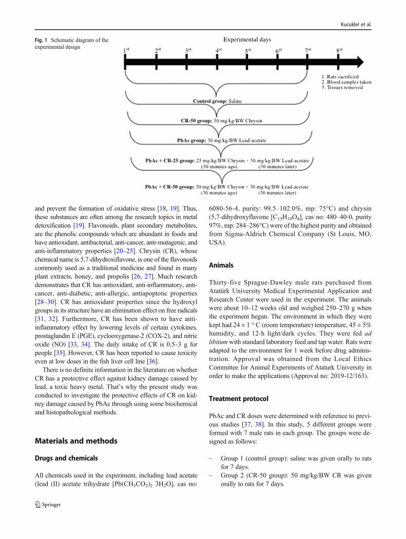
Introduction
Lead (Pb), an environmental pollutant and toxic agent, is amongthe heavy metals [1, 2]. Contaminated food, water, and air
pollution are major sources of Pb toxicity [3, 4]. According tothe World Health Organization, Pb is among the 10 most dan-gerous substances for public health [5]. Pb excretion is verydifficult and can be stored in soft tissues, bones, and other im-portant organs for a long time [6, 7]. In addition, it has toxiceffects onmany tissues, especially kidney [6, 8, 9].Although themechanism of Pb toxicity in the kidneys cannot be understoodprecisely, studies suggest that oxidative stress has an importantcontribution to Pb toxicity [10, 11]. Moreover, studies havereported that Pb can induce apoptosis by causing mitochondrialdegradation and DNA damage [12, 13] (Fig. 1).
In the treatment of toxicity caused by heavy metals, includingPb, chelators are used to excrete heavy metal from the body [7,14]. It has been reported that chelators used in lead treatment,alongwith some undesirable side effects [15], did not havemucheffect on the lead accumulated in tissue [16, 17]. Therefore,research has focused on various alternative approaches to thetreatment of Pb toxicity, particularly plant-based drugs [7].
Herbal products and their active components protect the tis-sues and organs from the attacks reactive oxygen species (ROS)
* Fatih Mehmet [email protected]
1 Department of Biochemistry, Faculty of Veterinary Medicine,Ataturk University, 25240 Erzurum, Turkey
2 Department of Midwifery, Faculty of Health Science, MunzurUniversity, 62000 Tunceli, Turkey
3 Department of Pathology, Faculty of Veterinary Medicine, AtaturkUniversity, 25240 Erzurum, Turkey
4 Department of Medical Services and Tecniques, Program of MedicalLaboratory Tecniques, Bingol University, 12000 Bingöl, Turkey
5 Department of Chemistry, Faculty of Sciences and Arts, BingolUniversity, 12000 Bingöl, Turkey
6 Department of Biochemistry, Faculty of VeterinaryMedicine, BingolUniversity, 12000 Bingöl, Turkey
Biological Trace Element Researchhttps://doi.org/10.1007/s12011-020-02268-8
and prevent the formation of oxidative stress [18, 19]. Thus,these substances are often among the research topics in metaldetoxification [19]. Flavonoids, plant secondary metabolites,are the phenolic compounds which are abundant in foods andhave antioxidant, antibacterial, anti-cancer, anti-mutagenic, andanti-inflammatory properties [20–25]. Chrysin (CR), whosechemical name is 5,7-dihydroxiflavone, is one of the flavonoidscommonly used as a traditional medicine and found in manyplant extracts, honey, and propolis [26, 27]. Much researchdemonstrates that CR has antioxidant, anti-inflammatory, anti-cancer, anti-diabetic, anti-allergic, antiapoptotic properties[28–30]. CR has antioxidant properties since the hydroxylgroups in its structure have an elimination effect on free radicals[31, 32]. Furthermore, CR has been shown to have anti-inflammatory effect by lowering levels of certain cytokines,prostaglandin E (PGE), cyclooxygenase-2 (COX-2), and nitricoxide (NO) [33, 34]. The daily intake of CR is 0.5–3 g forpeople [35]. However, CR has been reported to cause toxicityeven at low doses in the fish liver cell line [36].
There is no definite information in the literature on whetherCR has a protective effect against kidney damage caused bylead, a toxic heavy metal. That’s why the present study wasconducted to investigate the protective effects of CR on kid-ney damage caused by PbAc through using some biochemicaland histopathological methods.
Materials and methods
Drugs and chemicals
All chemicals used in the experiment, including lead acetate(lead (II) acetate trihydrate [Pb(CH3CO2)2 3H2O], cas no:
6080-56-4, purity: 99.5–102.0%, mp: 75°C) and chrysin(5,7-dihydroxyflavone [C15H10O4], cas no: 480–40-0, purity97%,mp: 284–286°C) were of the highest purity and obtainedfrom Sigma-Aldrich Chemical Company (St Louis, MO,USA).
Animals
Thirty-five Sprague-Dawley male rats purchased fromAtatürk University Medical Experimental Application andResearch Center were used in the experiment. The animalswere about 10–12 weeks old and weighed 250–270 g whenthe experiment began. The environment in which they werekept had 24 ± 1 ° C (room temperature) temperature, 45 ± 5%humidity, and 12-h light/dark cycles. They were fed adlibitumwith standard laboratory feed and tap water. Rats wereadapted to the environment for 1 week before drug adminis-tration. Approval was obtained from the Local EthicsCommittee for Animal Experiments of Ataturk University inorder to make the applications (Approval no: 2019-12/163).
Treatment protocol
PbAc and CR doses were determined with reference to previ-ous studies [37, 38]. In this study, 5 different groups wereformed with 7 male rats in each group. The groups were de-signed as follows:
– Group 1 (control group): saline was given orally to ratsfor 7 days.
– Group 2 (CR-50 group): 50 mg/kg/BW CR was givenorally to rats for 7 days.
Fig. 1 Schematic diagram of theexperimental design
Kucukler et al.
– Group 3 (PbAc group): 30 mg/kg/BW PbAc was givenorally to rats for 7 days.
– Group 4 (PbAc + CR-25 group): 30 mg/kg/BW PbAcwas given orally to rats 30 min after 25 mg/kg/BW CRthe administration for 7 days.
– Group 5 (PbAc + CR-50 group): 30 mg/kg/BW PbAcwas given orally to rats 30 min after the administrationof 50 mg/kg/BW CR for 7 days.
Collection of samples
Twenty-four hours after the last drug administration, the ani-mals’ body weights were measured and then they decapitatedunder mild isoflurane (IsoFlo; Abbott, Queenborough, UK)anesthesia. As soon as they were decapitated, blood sampleswere collected from their Vena jugularis. Blood samples werecollected in anticoagulant free tubes and centrifuged at1200×g for 15 min. Blood serum was used to determine renalfunction. After the kidneys from rats were washed with ice-cold physiologic saline (0.85%NaCl), one of them was storedat − 80 ° C for biochemical analysis and the other in 10%buffered formalin solution for histological examination untilused.
Determination of serum urea and creatinine levels
Serum urea [39] and creatinine [40] levels were analyzed witha commercial kit (Diasis Diagnostic Systems, Istanbul,Turkey) according to the manufacturer’s instructions.
Preparation of tissue homogenates
To obtain homogenate from the kidneys, the tissues were di-luted 1:20 v/w with phosphate-buffered saline (PBS; pH 7.4).The resulting mixture was rapidly homogenized with a tissuelysate device (TissueLyser II, Qiagen). The homogenate wasthen centrifuged at + 4°C and 3000 rpm for 30 min. Thesupernatant was used for biochemical analysis.
Determination of lipid peroxidation and antioxidantenzyme activities in kidney tissue
The level of lipid peroxidation was determined by analyzingthe amount of malondialdehyde (MDA) at 532 nm accordingto the method developed by Placer et al. [41]. The amount ofMDA was expressed as nmol/g tissue. Superoxide dismutaseactivity was measured by the method designed by Sun et al.[42]. The results were expressed as U/g protein. The measure-ment of catalase activity was performed according to themethod of Aebi [43]. The results were expressed as catal/gprotein. Glutathione peroxidase (GPx) activity was measuredaccording to the method developed by Lawrence, Burk [44].
Results were expressed as U/g protein. The method developedby Sedlak, Lindsay [45] was used to determine the level ofglutathione. Results were expressed as nmol/g of tissue. Totalprotein analysis was performed according to the method de-veloped by Lowry et al. [46] using bovine serum albumin(BSA) as standard.
Determination of AQP-1 levels in renal tissue
Aquaporin-1 (AQP-1) levels was performed by using ratenzyme-linked immunosorbent assay (ELISA) kit (cat. no:201-11-0566; assay range: 0.15–40 ng/ml) obtained fromSunred Biological Technology Company.
Determination of p53 levels in renal tissue
p53 levels were analyzed with rat ELISA kit according to themanufacturer’s instructions (Sunred, Shanghai, China) (cat.no: 201-11-0072; assay range: 0.05–10 ng/ml). The color in-tensity at the end of the procedures was read at 450 nm withthe ELISA microplate reader (Bio-Tek, Winooski, VT, USA).
Determination of inflammatory response levels inkidney tissue
Interleukin-33 (IL-33) (cat. no: 201-11-3102; assay range: 1.5- 400 ng/L), COX-2 (cat. no: 201-11-0297; assay range: 0.5–150 ng/ml), PGE-2 (cat. no: 201-11-0505; assay range: 0.05–15 ng/ml), nuclear factor kappa-B (NF-κB) (cat. no: 201-11-0288; assay range: 0.08–20 ng/ml), and inducible nitric oxidesynthase (iNOS) (cat. no: 201-11-0741; assay range: 0.8–200ng/ml) in renal tissue were measured by ELISA to determinethe degree of inflammatory response levels in accordance withthe manufacturer’s instructions (Sunred, Shanghai, China).
Determination of DNA damage level in kidney tissue
The degree of DNA damage in kidney tissue was determinedby measuring the level of 8-hydroxy-2′-deoxyguanosine (8-OHdG) using a commercial kit (Sunred, Shangai, China) (cat.no: 201-11-0032; assay range: 0.05–20 ng/ml).
Histopathological examination of kidney tissue
Necropsy was performed for histopathological evaluation ofthe renal tissues and tissue samples were fixed in 10% forma-lin solution for 48 h. Tissues were embedded in paraffinblocks with routine-tracking procedures. From each blockwere taken 4-μm-thick cross-sections. All preparations werestained with hematoxylin-eosin (HE) and examined with alight microscope (Leica DM 1000, Germany).
Protective Effects of Chrysin Against Oxidative Stress and Inflammation Induced by Lead Acetate in Rat...
Immunohistochemical examination of kidney tissue
Cross-sections obtained from kidney tissues were trans-f e r r e d t o a d h e s i v e ( p o l y - L -Ly s i n ) s l i d e s f o rimmunperoxidase examination and passed through xyloland alcohol series. After washing with phosphate-bufferedsaline (PBS), endogenous peroxidase was inactivated for1 0 m in a t 3% H2O2 (Me r c k , K50505100 8301.08600.1000). In the microwave oven set at 500 watts,the antigen in the tissues was released by exposure to re-trieval solution (abcam, ab93678) for 2 × 5 min. Tissueswere incubated with nephrin and tumor necrosis factor-α(TNF-α) antibodies (catalog no. Ab-216692, Abcam, UK,sc-52B83, Santa Cruz, USA) for 30 min,in an incubator setat 37 °C. Immunohistochemistry procedures were followedaccording to the kit instructions (AbcamHRP/DABDetection IHC kit). 3-3 ’Diaminobenzidine (DAB) wasused as chromogen. Background staining was performedwith hematoxylin. Immuno-positivity of the samples wasexpressed as none (-), mild (+), moderate (++), and severe(+++).
Determination of elemental content of kidney tissue
In this study, ICP-MS NexION® 2000 (PerkinElmer®Inc., USA) device with quartz nebulizer gasifier, cyclonicspray chember, and integrated auto-sampler was used forthe element analysis of samples. The washing solutioncontaining 1% hydrochloric acid ultra-pure water was pre-pared using 18.3 MΩ ultra-pure water and the ICP-MSmethod was performed. In the preparation of the sample,0.2 g were weighed, and transferred to the microwaveoven teflon cups and added 10 mL nitric acid. ICP-MScalibration solutions were prepared by dilution with com-mercially available multi-element standards of 1% (nitricacid, ultra-pure water). In addition, ICP-MS calibrationwas performed before each measurement. With a peristal-tic pump, the samples were sent to the cyclonic spraychamber with argon gas flow. ICP-MS NexION instru-ment software was used to control the instrument, includ-ing calibration, interferences, data collection, and dataanalysis. In addition to argon gas, helium gas was usedto prevent interference.
Statistical analysis
Statistical analysis of biochemical data was done withone-way ANOVA test in IBM SPSS program (version20.0; IBM Co, North Castle, NY). Tukey’s multiplecomparison test was used for comparisons betweenthe groups. All values were expressed as mean ± stan-dard error (SEM), and p < 0.05 was considered signif-icant. Kruskal-Wallis test, which is one of the nonpara-metric tests, was applied to the data obtained fromhistopathological examinations in order to determinethe differences between the groups. The comparisonof binary groups was done using Mann-Whitney Utest. SPSS 13.0 package program was used for thesestatistical analyzes.
Results
Analysis results of serum urea and creatinine levels
The results indicated that PbAc increases serum urea and cre-atinine levels by disrupting kidney function (P < 0.05). CRwas found to reduce serum urea and creatinine levels by de-creasing kidney toxicity caused by PbAc in a dose-dependentmanner (25 and 50 mg/kg/BW). It was determined that therewas no significant difference between the control and CRgroups. Furthermore, it was observed that the rats who lostweight with PbAc administration reached the weight of theanimals in the control group with CR treatment. Serum ureaand creatinine levels and body weights of all groups are pre-sented in Table 1.
Analysis of lipid peroxidation and antioxidantmarkers in kidney tissue
It was determined that PbAc administration increased MDAlevels by causing lipid peroxidation in kidney tissue and de-creased the activities of antioxidant enzymes (SOD, CAT andGPx) and GSH levels. On the other hand, it was found that CRadministration decreased lipid peroxidation; thus, MDA levelsreduced. It was also detected that CR treatment significantlyincreased SOD, CAT, and GPx activities and GSH levels
Table 1 Effects of CR on kidneyfunction parameters and bodyweights in PbAc-inducednephrotoxicity in rats
Parameters Control CR-50 PbAc PbAc + CR-25 PbAc + CR-50
Urea (mg/dL) 3.68 ± 0.14d 3.49 ± 0.06d 9.23 ± 0.25a 6.58 ± 0.24b 5.05 ± 0.11c
Creatinine(mg/dL)
0.45 ± 0.01d 0.43 ± 0.01d 2.16 ± 0.05a 1.31 ± 0.04b 0.87 ± 0.03c
Body weight (g) 278.29 ± 3.26ab 283.86 ± 2.43a 240.57 ± 1.95d 261.00 ± 4.90c 274.29 ± 3.40b
Data of rats in each group were expressed as mean ± SEM (n = 7). The different letters (a–d ) on the same lineindicate a statistically significant difference (p < 0.05) between the groups
Kucukler et al.
compared to PbAc group (P < 0.05). Lipid peroxidation andthe levels of antioxidant markers in kidney tissue are given inTable 2.
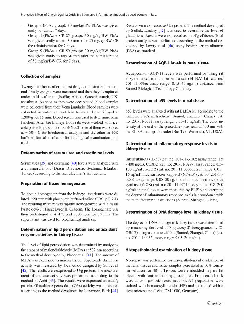
Analysis results of AQP-1 levels in kidney tissue
AQP-1 levels are given in Fig. 2. The results demonstratedthat PbAc decreased AQP-1 levels due to the damage to thekidneys. Furthermore, CR administration was observed to sig-nificantly increase AQP-1 levels compared to PbAc group byalleviating kidney damage P < 0.05.
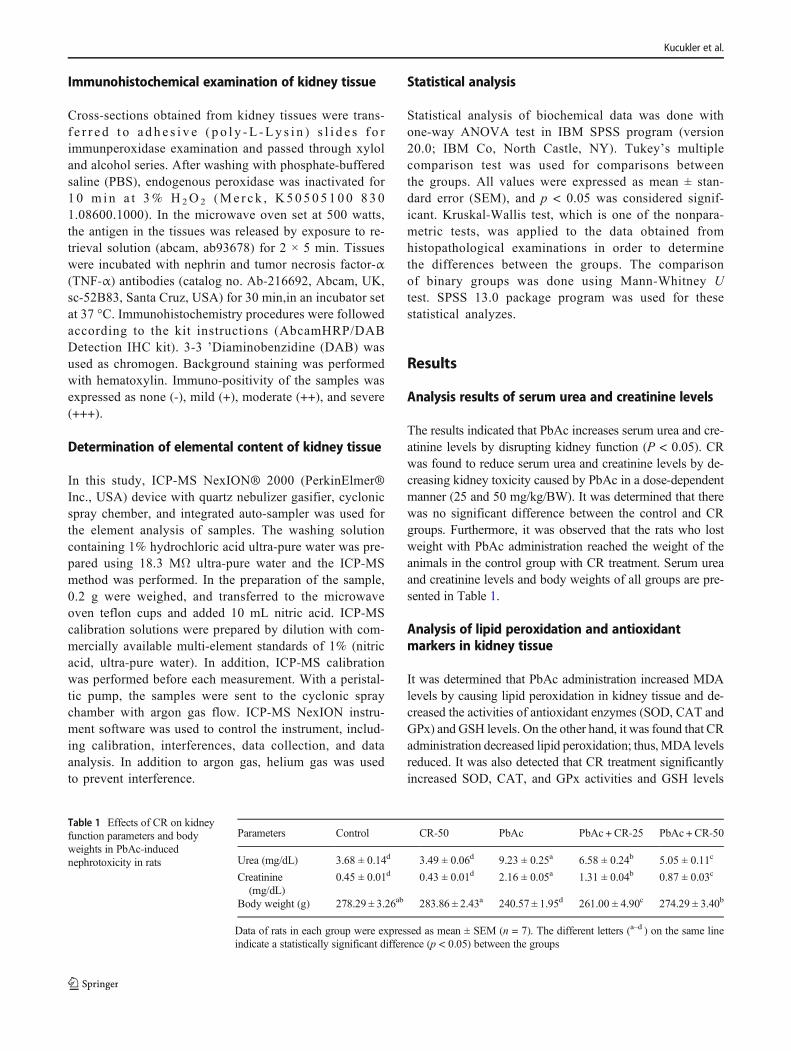
Analysis results of p53 levels in kidney tissue
p53 levels analyzed by ELISAmethod are given in Fig. 3. Theresults demonstrated that PbAc made cells undergo apoptosisby increasing the p53 levels. CR was found to protect the cellsagainst apoptosis by suppressing p53 expression.
Analysis of inflammatory response levels in kidneytissue
Analysis results of inflammatory markers of kidney tissue arepresented in Table 3. The results showed that PbAc led to
inflammation by significantly increasing IL-33, PGE-2,COX-2, NF-κB, and iNOS levels compared to the controlgroup. Nevertheless, CR was found to reduce PGE-2, COX-2, NF-κB, and iNOS levels in a dose-dependent manner,thereby alleviating inflammation in the kidney. It was foundthat IL-33 levels did not make a significant difference betweenPbAc + CR-25 and PbAc + CR-50 groups.
DNA damage level
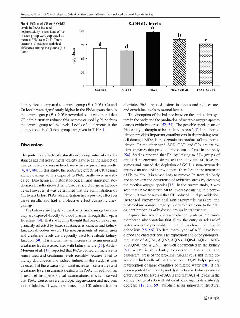
As stated in Fig. 4, it was observed that there were increases in8-OHdG levels since PbAc damaged DNA. CR was observedto protect DNA from damage caused by PbAc and to limit therise of 8-OHdG levels.
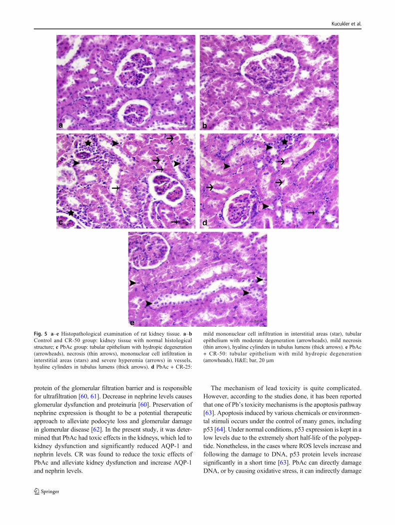
Histopathological findings
Histopathological examination showed that the kidney tissuesof the rats in the control and CR-50 groups had normal ap-pearance (Fig. 5a–b). Mononuclear cell infiltration in the in-terstitium of the PbAc group, severe hydropic degenerationand necrosis in the tubules, severe hyperemia in the vessels,and hyaline cylinders in some tubulus lumens were observed(Fig. 5c). Hydropic degeneration and mild coagulation
Table 2 Effects of CR onoxidative stress parameters inPbAc-induced nephrotoxicity inrats
Parameters Control CR-50 PbAc PbAc + CR-25 PbAc + CR-50
MDA (nmol/g tissue) 37.99 ± 0.65d 36.82 ± 0.66d 60.24 ± 0.81a 50.38 ± 0.57b 44.77 ± 0.56c
GSH (nmol/g tissue) 2.71 ± 0.03a 2.76 ± 0.03a 1.54 ± 0.03d 1.86 ± 0.02c 2.06 ± 0.04b
SOD (U/g protein) 23.42 ± 0.59a 23.96 ± 0.50a 12.85 ± 0.30d 15.02 ± 0.37c 17.46 ± 0.63b
CAT (katal/g protein) 34.42 ± 0.63a 34.37 ± 0.73a 22.36 ± 0.56c 26.08 ± 0.27b 27.55 ± 0.49b
GPx (U/g protein) 26.68 ± 0.48a 26.96 ± 0.43a 15.82 ± 0.22d 17.91 ± 0.36c 21.56 ± 0.44b
Data of rats in each group were expressed as mean ± SEM (n = 7). The different letters (a–d ) on the same lineindicate a statistically significant difference (p < 0.05) between the groups. (MDA, malondialdehyde; GSH,glutathione; SOD, superoxide dismutase; CAT, catalase; GPx, glutathione peroxidase)
Fig. 2 Effects of CR on AQP-1levels in PbAc-inducednephrotoxicity in rats. Data of ratsin each group were expressed asmean ± SEM (n = 7). Differentletters (a–d) indicate statisticaldifference among the groups (p <0.05)
Protective Effects of Chrysin Against Oxidative Stress and Inflammation Induced by Lead Acetate in Rat...
necrosis, mild mononuclear cell (MNH) infiltration, and hy-peremia in interstitial areas were detected in moderate tubulusepithelium in the PbAc + CR-25 group (Fig. 5d). In the PbAc+ CR-50 group, the lesions were very mild and statisticallysignificant P ˂ 0.05 differences were found when compared tothe PbAc group (Fig. 5e). Histopathological findings of allgroups are summarized in Table 4.
Immunohistochemical findings
As a result of immunohistochemical examination of the renaltissues, severe nephrin expression was observed in the tubulusepithelium in the control and CR-50 groups, but TNF-α ex-pression was not observed (Fig. 6, 7a–b). Negative nephrinexpression in tubular epithelium in PbAc group and severeTNF-α expression in interstitial tissues, perivascular, and glo-meruli were determined (Fig. 6, 7c). Nephrine was slightlyexpressed in renal tubule epithelium of the PbAc + CR-25group (Fig. 6d) and TNF-α expression was moderate in theinterstitial area (Fig. 7d). In the PbAc + CR-50 group,nephrine expression was severe in the tubulus epithelium(Fig. 6e). Mild TNF-α expression was detected in the intersti-tial area (Fig. 7e). In this group, expression levels of immu-nohistochemical markers were statistically significant (P ˂
0.05) differences when compared with PbAc group.Immunohistochemical findings are summarized in Table 4.
Levels of elements in kidney tissue
According to the data obtained by ICP-MS method, the PbAcand PbAc + CR-25 groups were found to have the highest Pbaccumulation in kidney tissue compared to the control group(P < 0.05). It was determined that 50 mg/kg/BW administra-tion of CR reduced Pb accumulation caused by PbAc. Thegroups with the highest K level were found to have CR ad-ministration with PbAc. In the PbAc and CR groups, K levelswere significantly lower than the control group (P < 0.05), butthere was no significant difference between them (P > 0.05).The PbAc group had the highest Na levels in the kidney tissuecompared to the control group. In the CR-50 group, Na levelsdecreased significantly (P < 0.05), but there was no significantdifference between CR administration with PbAc and controlgroups (P > 0.05). PbAc + CR-25 and PbAc + CR-50 were thegroups with the highest Ca levels compared to the controlgroup. While there was no significant difference betweenCR-50 and PbAc groups (P > 0.05), it was found that Calevels decreased compared to the control group (P < 0.05). Itwas observed that CR decreased significantly Fe levels in
Fig. 3 Effects of CR on p53levels in PbAc-inducednephrotoxicity in rats. Data of ratsin each group were expressed asmean ± SEM (n = 7). Differentletters (a–d) indicate statisticaldifference among the groups (p <0.05)
Table 3 Effects of CR oninflammation parameters inPbAc-induced nephrotoxicity inrats
Parameters Control CR-50 PbAc PbAc +CR-25 PbAc +CR-50
NF-κB (ng/g tissue) 37.70 ± 0.76d 36.04 ± 0.59d 69.75 ± 0.73a 58.89 ± 0.99b 49.35 ± 0.80c
IL-33 (ng/g tissue) 1.49 ± 0.03c 1.40 ± 0.03c 2.25 ± 0.04a 1.76 ± 0.05b 1.71 ± 0.04b
PGE-2 (ng/g tissue) 9.71 ± 0.31d 9.01 ± 0.25d 19.42 ± 0.30a 16.43 ± 0.27b 13.64 ± 0.34c
COX-2 (ng/gtissue)
397.38 ± 3.91d 390.31 ± 4.59d 552.02 ± 4.07a 500.40 ± 4.79b 449.61 ± 6.51c
iNOS (ng/g tissue) 181.68 ± 2.78d 176.50 ± 2.17d 267.20 ± 3.32a 240.86 ± 2.87b 216.74 ± 4.35c
Data of rats in each group were expressed as mean ± SEM (n = 7). The different letters (a–d ) on the same lineindicate a statistically significant difference (p < 0.05) between the groups. (NF-kB, nuclear factor kappa-B; IL-33,interleukin-33; PGE-2, prostaglandin E2; COX-2, cyclooxygenase-2; iNOS, inducible nitric oxide synthase)
Kucukler et al.
kidney tissue compared to control group (P < 0.05). Cu andZn levels were significantly higher in the PbAc group than inthe control group (P < 0.05); nevertheless, it was found thatCR administration reduced this increase caused by PbAc fromthe control group to low levels. Levels of all elements in thekidney tissue in different groups are given in Table 5.
Discussion
The protective effects of naturally occurring antioxidant sub-stances against heavy metal toxicity have been the subject ofmany studies, and researchers have achieved promising results[4, 47, 48]. In this study, the protective effects of CR againstkidney damage of rats exposed to PbAc orally were investi-gated. Biochemical, histopathological, and immunohisto-chemical results showed that PbAc caused damage in the kid-neys. However, it was determined that the administration ofCR to rats before PbAc administration had a positive effect onthese results and had a protective effect against kidneydamage.
The kidneys are highly vulnerable to toxic damage becausethey are exposed directly to blood plasma through their openfenestrae [49]. That’s why, it is thought that one of the organsprimarily affected by toxic substances is kidneys and kidneyfunction disorders occur. The measurements of serum ureaand creatinine levels are frequently used to evaluate kidneyfunction [50]. It is known that an increase in serum urea andcreatinine levels is associated with kidney failure [51]. Abdel-Moneim et al. [49] reported that PbAc caused an increase inserum urea and creatinine levels possibly because it led tokidney dysfunction and kidney failure. In this study, it wasdetected that there was a significant increase in serum urea andcreatinine levels in animals treated with PbAc. In addition, asa result of histopathological examinations, it was observedthat PbAc caused severe hydropic degeneration and necrosisin the tubules. It was determined that CR administration
alleviates PbAc-induced lesions in tissues and reduces ureaand creatinine levels to normal levels.
The disruption of the balance between the antioxidant sys-tem in the body and the production of reactive oxygen speciescauses oxidative stress [52, 53]. The possible mechanism ofPb toxicity is thought to be oxidative stress [15]. Lipid perox-idation provides important contributions in determining renalcell damage. MDA is the degradation product of lipid perox-idation. On the other hand, SOD, CAT, and GPx are antiox-idant enzymes that provide antioxidant defense in the body[54]. Studies reported that Pb, by linking to SH- groups ofantioxidant enzymes, decreased the activities of these en-zymes and caused the depletion of GSH, a non-enzymaticantioxidant and lipid peroxidation. Therefore, in the treatmentof Pb toxicity, it is aimed both to remove Pb from the bodyand to prevent the occurrence of oxidative stress by cleaningthe reactive oxygen species [15]. In the current study, it wasseen that PbAc increased MDA levels by causing lipid perox-idation. It was observed that CH reduced lipid peroxidation,increased enzymatic and non-enzymatic markers andprotected membrane integrity in kidney tissue due to the anti-oxidant properties of hydroxyl groups in its structure.
Aquaporins, which are water channel proteins, are trans-membrane glycoproteins that allow the entry or release ofwater across the permeable epithelium, such as renal tubularepithelium [55, 56]. To date, many types of AQP have beencloned and characterized. The expression and/or physiologicalregulation of AQP-1, AQP-2, AQP-3, AQP-4, AQP-6, AQP-7, AQP-8, and AQP-11 are well documented in the kidney[57] AQP1 is abundantly expressed in the apical andbasolateral areas of the proximal tubular cells and in the de-scending limb cells of the Henle loop. AQP1 helps quicklyreabsorption of large quantities of filtered water [58]. It hasbeen reported that toxicity and dysfunction in kidneys consid-erably affect the levels of AQPs and that AQP-1 levels in thekidney tissues of rats with different toxic agents dramaticallydecrease [19, 55, 59]. Nephrin is an important structural
Fig. 4 Effects of CR on 8-OHdGlevels in PbAc-inducednephrotoxicity in rats. Data of ratsin each group were expressed asmean ± SEM (n = 7). Differentletters (a–d) indicate statisticaldifference among the groups (p <0.05)
Protective Effects of Chrysin Against Oxidative Stress and Inflammation Induced by Lead Acetate in Rat...
protein of the glomerular filtration barrier and is responsiblefor ultrafiltration [60, 61]. Decrease in nephrine levels causesglomerular dysfunction and proteinuria [60]. Preservation ofnephrine expression is thought to be a potential therapeuticapproach to alleviate podocyte loss and glomerular damagein glomerular disease [62]. In the present study, it was deter-mined that PbAc had toxic effects in the kidneys, which led tokidney dysfunction and significantly reduced AQP-1 andnephrin levels. CR was found to reduce the toxic effects ofPbAc and alleviate kidney dysfunction and increase AQP-1and nephrin levels.
The mechanism of lead toxicity is quite complicated.However, according to the studies done, it has been reportedthat one of Pb’s toxicity mechanisms is the apoptosis pathway[63]. Apoptosis induced by various chemicals or environmen-tal stimuli occurs under the control of many genes, includingp53 [64]. Under normal conditions, p53 expression is kept in alow levels due to the extremely short half-life of the polypep-tide. Nonetheless, in the cases where ROS levels increase andfollowing the damage to DNA, p53 protein levels increasesignificantly in a short time [63]. PbAc can directly damageDNA, or by causing oxidative stress, it can indirectly damage
Fig. 5 a–e Histopathological examination of rat kidney tissue. a–bControl and CR-50 group: kidney tissue with normal histologicalstructure; c PbAc group: tubular epithelium with hydropic degeneration(arrowheads), necrosis (thin arrows), mononuclear cell infiltration ininterstitial areas (stars) and severe hyperemia (arrows) in vessels,hyaline cylinders in tubulus lumens (thick arrows). d PbAc + CR-25:
mild mononuclear cell infiltration in interstitial areas (star), tubularepithelium with moderate degeneration (arrowheads), mild necrosis(thin arrow), hyaline cylinders in tubulus lumens (thick arrows). e PbAc+ CR-50: tubular epithelium with mild hydropic degeneration(arrowheads), H&E; bar, 20 μm
Kucukler et al.
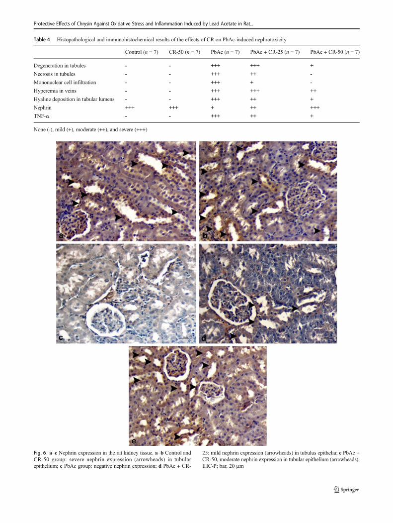
Table 4 Histopathological and immunohistochemical results of the effects of CR on PbAc-induced nephrotoxicity
Control (n = 7) CR-50 (n = 7) PbAc (n = 7) PbAc + CR-25 (n = 7) PbAc + CR-50 (n = 7)
Degeneration in tubules - - +++ +++ +
Necrosis in tubules - - +++ ++ -
Mononuclear cell infiltration - - +++ + -
Hyperemia in veins - - +++ +++ ++
Hyaline deposition in tubular lumens - - +++ ++ +
Nephrin +++ +++ + ++ +++
TNF-α - - +++ ++ +
None (-), mild (+), moderate (++), and severe (+++)
Fig. 6 a–e Nephrin expression in the rat kidney tissue. a–b Control andCR-50 group: severe nephrin expression (arrowheads) in tubularepithelium; c PbAc group: negative nephrin expression; d PbAc + CR-
25: mild nephrin expression (arrowheads) in tubulus epithelia; e PbAc +CR-50, moderate nephrin expression in tubular epithelium (arrowheads),IHC-P; bar, 20 μm
Protective Effects of Chrysin Against Oxidative Stress and Inflammation Induced by Lead Acetate in Rat...
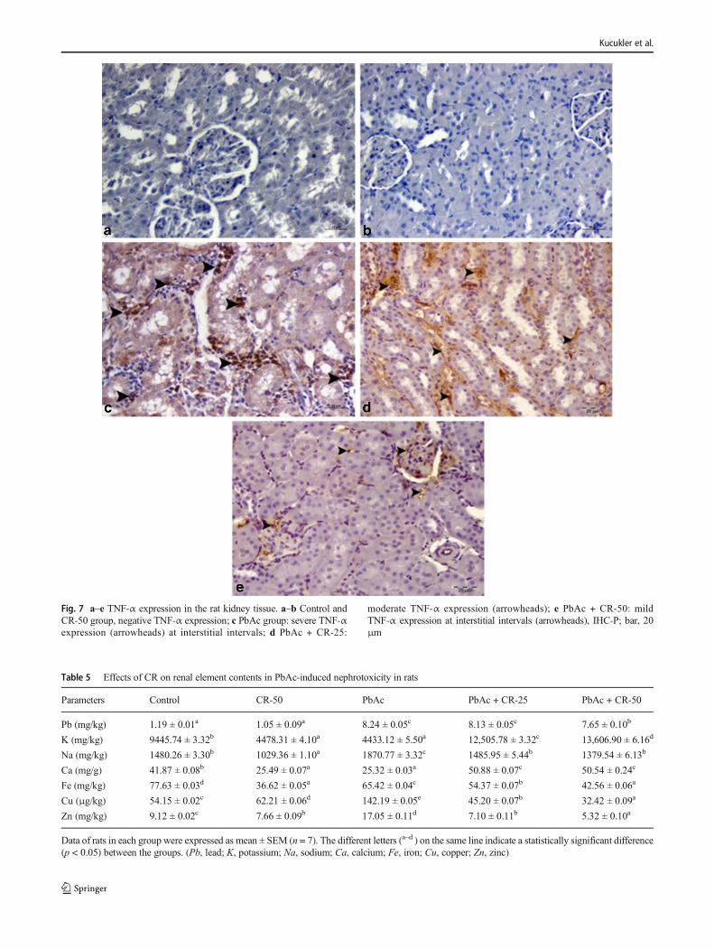
Fig. 7 a–e TNF-α expression in the rat kidney tissue. a–b Control andCR-50 group, negative TNF-α expression; c PbAc group: severe TNF-αexpression (arrowheads) at interstitial intervals; d PbAc + CR-25:
moderate TNF-α expression (arrowheads); e PbAc + CR-50: mildTNF-α expression at interstitial intervals (arrowheads), IHC-P; bar, 20μm
Table 5 Effects of CR on renal element contents in PbAc-induced nephrotoxicity in rats
Parameters Control CR-50 PbAc PbAc + CR-25 PbAc + CR-50
Pb (mg/kg) 1.19 ± 0.01a 1.05 ± 0.09a 8.24 ± 0.05c 8.13 ± 0.05c 7.65 ± 0.10b
K (mg/kg) 9445.74 ± 3.32b 4478.31 ± 4.10a 4433.12 ± 5.50a 12,505.78 ± 3.32c 13,606.90 ± 6.16d
Na (mg/kg) 1480.26 ± 3.30b 1029.36 ± 1.10a 1870.77 ± 3.32c 1485.95 ± 5.44b 1379.54 ± 6.13b
Ca (mg/g) 41.87 ± 0.08b 25.49 ± 0.07a 25.32 ± 0.03a 50.88 ± 0.07c 50.54 ± 0.24c
Fe (mg/kg) 77.63 ± 0.03d 36.62 ± 0.05a 65.42 ± 0.04c 54.37 ± 0.07b 42.56 ± 0.06a
Cu (μg/kg) 54.15 ± 0.02c 62.21 ± 0.06d 142.19 ± 0.05e 45.20 ± 0.07b 32.42 ± 0.09a
Zn (mg/kg) 9.12 ± 0.02c 7.66 ± 0.09b 17.05 ± 0.11d 7.10 ± 0.11b 5.32 ± 0.10a
Data of rats in each group were expressed as mean ± SEM (n = 7). The different letters (a–d ) on the same line indicate a statistically significant difference(p < 0.05) between the groups. (Pb, lead; K, potassium; Na, sodium; Ca, calcium; Fe, iron; Cu, copper; Zn, zinc)
Kucukler et al.
cells and DNA [65]. This explains the situation in the currentstudy that PbAc causes severe damage to the kidneys by in-ducing an increase in p53 levels. However, it has been deter-mined that CR reduces p53 levels by alleviating oxidativestress and DNA damage due to its antioxidant effect, thusprotecting kidneys from PbAc toxicity.
There is growing evidence of the link between oxidativestress and inflammatory response. Oxidative stress providesimportant contributions to the inflammation process. It hasbeen reported that oxidant molecules affect all phases of theinflammatory process, such as the release of endogenous dan-ger signal molecules, their perception by natural immune cellsfrom the Toll-like receptors (TLRs) and NOD-like receptor(NLRs) families, and the activation of signal pathways thatinitiate an adaptive cellular reaction to these signals [66].The responses initiated by TLRs are transmitted by activationof NF-κB [67]. Therefore, oxidative stress activates NF-κBand initiates inflammation mechanism. This is one of thestrongest evidence supporting the link between oxidativestress and inflammation in disease progression [68]. NF-κBstimulates the release of pro-inflammatory cytokines, particu-larly TNF-α. In addition, expression of iNOS and COX-2proteins is regulated by NF-κB. Therefore, suppression ofNF-κB is of great therapeutic importance [19, 69]. Liu et al.[70] reported that Pb affects kidney tissue and causes NF-κBactivation and inflammation. Flavonoids play an importantrole in the regulation of cellular functions such as cell cyclesignals and modulation of inflammatory pathways [71].Rehman et al. [72] showed that CR effectively inhibited theincrease in ferric nitrilotriacetate-mediated TNF-α, COX-2,iNOS, and PGE2 expressions. Kandemir et al. [73] reportedthat paracetamol-induced inflammation in kidney tissue im-proves CR and decreases IL-33 levels. Similar to the literature,in the present study, PbAc increased NF-κB, IL-33, PGE-2,COX-2, and iNOS levels in renal tissue due to oxidativestress. As a result, inflammation in the kidney tissue occurred.CR reduced the inflammation caused by PbAc and decreasedNF-κB, IL-33, PGE-2, COX-2, and iNOS levels significantlycompared to PbAc group. Immunohistochemical examinationrevealed that TNF-α was strongly expressed in the PbAc-treated group, while CR decreased TNF-α expression.
DNA is a highly sensitive macromolecule to oxidativedamage [74]. 8-OHdG is a widely used biomarker fordetermining oxidative damage in DNA [75]. ROS isthought to play an active role in the formation of 8-OHdG [19, 75]. According to the data obtained from thisstudy, PbAc increased the formation of 8-OHdG by caus-ing oxidative damage in DNA. Also, CR improved PbAc-induced oxidative DNA damage with antioxidant proper-ties, approximating the formation of 8-OHdG to that ofthe control group. Similarly, Rani et al. [76] reported thatCR significantly reduced the 8-OHdG level dose-dependently.
In the studies evaluating the effectiveness of antioxidantsas chelating agents, although antioxidants are reported to benot as effective as traditional chelators [77, 78], there are thestudies showing that flavonoids have chelating properties inaddition to their antioxidant properties [79]. It has been report-ed that CR is also capable of metal chelation [80]. In our study,we found that administration of 25 mg/kg/BW of CR did notmake a significant difference in the amount of Pb in renaltissue compared to the PbAc group, but that of 50 mg/kg/BW reduced chelation of Pb significantly.
There is sufficient information that heavy metals, in-cluding Pb, may have adverse effects on the concentra-tions of essential metals. However, the information aboutthe effect of electrolytes in the body is insufficient [81].Xia et al. [82] reported that Pb has no effect on theamount of Cu and Zn in kidney tissue. Aksu et al. [83]stated that PbAc administration increases zinc accumula-tion in the kidney and has no effect on Cu and Fe levelsand also increases Zn level with the use of phenolic com-pounds. In our study, it was found that PbAc significantlyincreased Cu, Zn, and Na amounts in kidney tissue anddecreased K, Ca, and Fe amounts in comparison to thecontrol group. However, it was seen that K and Ca in-creased; Fe, Cu, and Zn decreased in the kidneys of therats given CR with PbAc, and Na did not make a signif-icant difference compared to the control group. Given thatZn homeostasis is provided by the kidneys, the informa-tion that PbAc accumulates in the kidneys along withdamage to the kidneys confirms our data [83]. Also, Pbcauses Fe absorption to reduce by linking to similar areaswith Fe [84]. This explains why PbAc reduces the amountof Fe in the kidney in the current study.
Conclusion
Our findings confirmed that PbAc led to toxicity in the kid-neys because of inflammation and apoptosis associated withoxidative stress. It was also detected that toxicity decreasedAQP-1 levels. However, it was concluded that the antioxidant,anti-inflammatory and antiapoptotic properties of CR also ap-ply to PbAc-induced nephrotoxicity, and that CR is a promis-ing compound in the treatment of renal toxicity. Still, themechanism of this effect of CR needs to be supported byfurther studies.
Funding Information This study was funded by the Unit of ScientificResearch Projects in Munzur University. (Project No: YLMUB017-24)
Compliance with Ethical Standards
Conflict of Interest The authors declare that they have no conflict ofinterest.
Protective Effects of Chrysin Against Oxidative Stress and Inflammation Induced by Lead Acetate in Rat...
References
1. Phyu MP, Tangpong J (2014) Neuroprotective effects of xanthonederivative ofGarcinia mangostana against lead-induced acetylcho-linesterase dysfunction and cognitive impairment. Food ChemToxicol 70:151–156. https://doi.org/10.1016/j.fct.2014.04.035
2. Khalil SR, Khalifa HA, Abdel-Motal SM, Mohammed HH, ElewaYHA, Mahmoud HA (2018) Spirulina platensis attenuates the as-sociated neurobehavioral and inflammatory response impairmentsin rats exposed to lead acetate. Ecotoxicol Environ Saf 157:255–265. https://doi.org/10.1016/j.ecoenv.2018.03.068
3. Soleimani E, Goudarzi I, Abrari K, Lashkarbolouki T (2016) Thecombined effects of developmental lead and ethanol exposure onhippocampus dependent spatial learning andmemory in rats: role ofoxidative stress. Food Chem Toxicol 96:263–272. https://doi.org/10.1016/j.fct.2016.07.009
4. Khalil SR, Elhady WM, Elewa YHA, Abd El-Hameed NE, Ali SA(2018) Possible role of Arthrospira platensis in reversing oxidativestress-mediated liver damage in rats exposed to lead. BiomedPharmacother 97:1259–1268. https://doi.org/10.1016/j.biopha.2017.11.045
5. Shojaeepour S, Fazeli M, Oghabian Z, Pourgholi L, Mandegary A(2018) Oxidative stress in opium users after using lead-adulteratedopium: the role of genetic polymorphism. Food Chem Toxicol 120:571–577. https://doi.org/10.1016/j.fct.2018.07.061
6. Song XB, Liu G, Liu F, Yan ZG, Wang ZY, Liu ZP, Wang L(2017) Autophagy blockade and lysosomal membrane perme-abilization contribute to lead-induced nephrotoxicity in primaryrat proximal tubular cells. Cell Death Dis 8(6):e2863. https://doi.org/10.1038/cddis.2017.262
7. Abd El-Hack ME, Abdelnour SA, Abd El-Moneim AEE, Arif M,Khafaga A, Shaheen H, Samak D, Swelum AA (2019) Putativeimpacts of phytogenic additives to ameliorate lead toxicity in ani-mal feed. Environ Sci Pollut Res Int 26(23):23209–23218. https://doi.org/10.1007/s11356-019-05805-8
8. Oyagbemi AA, Omobowale TO, Akinrinde AS, Saba AB,Ogunpolu BS, Daramola O (2015) Lack of reversal of oxidativedamage in renal tissues of lead acetate-treated rats. Environ Toxicol30(11):1235–1243. https://doi.org/10.1002/tox.21994
9. Patrick L (2006) Lead toxicity, a review of the literature. Part 1:exposure, evaluation, and treatment. Altern Med Rev 11(1):2–22
10. Matovic V, Buha A, Ethukic-Cosic D, Bulat Z (2015) Insight intothe oxidative stress induced by lead and/or cadmium in blood, liverand kidneys. Food Chem Toxicol 78:130–140. https://doi.org/10.1016/j.fct.2015.02.011
11. Farmand F, Ehdaie A, Roberts CK, Sindhu RK (2005) Lead-induced dysregulation of superoxide dismutases, catalase, glutathi-one peroxidase, and guanylate cyclase. Environ Res 98(1):33–39.https://doi.org/10.1016/j.envres.2004.05.016
12. Liu J, Jia DY, Cai SZ, Li CP, Zhang MS, Zhang YY, Yan CH,WangYP (2015)Mitochondria defects are involved in lead-acetate-induced adult hematopoietic stem cell decline. Toxicol Lett 235(1):37–44. https://doi.org/10.1016/j.toxlet.2015.03.007
13. Dobrakowski M, Pawlas N, Kasperczyk A, Kozlowska A,Olewinska E, Machon-Grecka A, Kasperczyk S (2017) OxidativeDNA damage and oxidative stress in lead-exposed workers. HumExp Toxicol 36(7):744–754. https: / /doi.org/10.1177/0960327116665674
14. Caglayan C, Kandemir FM, Darendelioglu E, Yildirim S, KucuklerS, Dortbudak MB (2019) Rutin ameliorates mercuric chloride-induced hepatotoxicity in rats via interfering with oxidative stress,inflammation and apoptosis. J Trace Elem Med Biol 56:60–68.https://doi.org/10.1016/j.jtemb.2019.07.011
15. Alcaraz-Contreras Y, Mendoza-Lozano RP, Martinez-Alcaraz ER,Martinez-Alfaro M, Gallegos-Corona MA, Ramirez-Morales MA,
Vazquez-Guevara MA (2016) Silymarin and dimercaptosuccinicacid ameliorate lead-induced nephrotoxicity and genotoxicity inrats. Hum Exp Toxicol 35(4):398–403. https://doi.org/10.1177/0960327115591373
16. Andersen O, Aaseth J (2016) A review of pitfalls and progress inchelation treatment of metal poisonings. J Trace ElemMed Biol 38:74–80. https://doi.org/10.1016/j.jtemb.2016.03.013
17. BaSalamah MA, Abdelghany AH, El-Boshy M, Ahmad J, Idris S,Refaat B (2018) Vitamin D alleviates lead induced renal and testic-ular injuries by immunomodulatory and antioxidant mechanisms inrats. Sci Rep 8(1):4853. https://doi.org/10.1038/s41598-018-23258-w
18. Benzer F, Kandemir FM, Kucukler S, Comakli S, Caglayan C(2018) Chemoprotective effects of curcumin on doxorubicin-induced nephrotoxicity in wistar rats: by modulating inflammatorycytokines, apoptosis, oxidative stress and oxidative DNA damage.Arch Physiol Biochem 124(5):448–457. https://doi.org/10.1080/13813455.2017.1422766
19. Caglayan C, Kandemir FM, Yildirim S, Kucukler S, Eser G (2019)Rutin protects mercuric chloride-induced nephrotoxicity viatargeting of aquaporin 1 level, oxidative stress, apoptosis and in-flammation in rats. J Trace Elem Med Biol 54:69–78. https://doi.org/10.1016/j.jtemb.2019.04.007
20. Kuzu M, Kandemir FM, Yildirim S, Kucukler S, Caglayan C, TurkE (2018) Morin attenuates doxorubicin-induced heart and braindamage by reducing oxidative stress, inflammation and apoptosis.Biomed Pharmacother 106:443–453. https://doi.org/10.1016/j.biopha.2018.06.161
21. Kandemir FM, Kucukler S, Caglayan C, Gur C, Batil AA, Gülçin İ(2017) Therapeutic effects of silymarin and naringin onmethotrexate-induced nephrotoxicity in rats: biochemical evalua-tion of anti-inflammatory, antiapoptotic, and antiautophagic prop-erties. J Food Biochem 41(5):e12398
22. Benzer F, Kandemir FM, Ozkaraca M, Kucukler S, Caglayan C(2018) Curcumin ameliorates doxorubicin-induced cardiotoxicityby abrogation of inflammation, apoptosis, oxidative DNA damage,and protein oxidation in rats. J BiochemMol Toxicol 32(2). https://doi.org/10.1002/jbt.22030
23. Kandemir FM, Yildirim S, Kucukler S, Caglayan C, Mahamadu A,Dortbudak MB (2018) Therapeutic efficacy of zingerone againstvancomycin-induced oxidative stress, inflammation, apoptosisand aquaporin 1 permeability in rat kidney. Biomed Pharmacother105:981–991. https://doi.org/10.1016/j.biopha.2018.06.048
24. Kandemir FM, Ozkaraca M, Küçükler S, Caglayan C, Hanedan B(2018) Preventive effects of hesperidin on diabetic nephropathyinduced by streptozotocin via modulating TGF-β1 and oxidativeDNA damage. Toxin Rev 37(4):287–293
25. Celik H, Kucukler S, Comakli S, Ozdemir S, Caglayan C, YardimA, Kandemir FM (2019) Morin attenuates ifosfamide-induced neu-rotoxicity in rats via suppression of oxidative stress, neuroinflam-mation and neuronal apoptosis. Neurotoxicology 76:126–137.https://doi.org/10.1016/j.neuro.2019.11.004
26. Hanedan B,OzkaracaM, Kirbas A, Kandemir FM,AktasMS, KilicK, Comakli S, Kucukler S, Bilgili A (2018) Investigation of theeffects of hesperidin and chrysin on renal injury induced by colistinin rats. Biomed Pharmacother 108:1607–1616. https://doi.org/10.1016/j.biopha.2018.10.001
27. Samarghandian S, Farkhondeh T, Azimi-Nezhad M (2017)Protective effects of chrysin against drugs and toxic agents. Dose-Response 15(2):1559325817711782. https://doi.org/10.1177/1559325817711782
28. Aksu EH, Ozkaraca M, Kandemir FM, Omur AD, Eldutar E,Kucukler S, Comakli S (2016) Mitigation of paracetamol-inducedreproductive damage by chrysin in male rats via reducing oxidativestress. Andrologia 48(10):1145–1154. https://doi.org/10.1111/and.12553
Kucukler et al.
29. Tahir M, Sultana S (2011) Chrysin modulates ethanol metabolismin Wistar rats: a promising role against organ toxicities. AlcoholAlcohol 46(4):383–392. https://doi.org/10.1093/alcalc/agr038
30. Eldutar E, Kandemir FM, Kucukler S, Caglayan C (2017)Restorative effects of chrysin pretreatment on oxidant-antioxidantstatus, inflammatory cytokine production, and apoptotic and au-tophagic markers in acute paracetamol-induced hepatotoxicity inrats: an experimental and biochemical study. J Biochem MolToxicol 31(11). https://doi.org/10.1002/jbt.21960
31. Mantawy EM, El-BaklyWM, Esmat A, BadrAM, El-Demerdash E(2014) Chrysin alleviates acute doxorubicin cardiotoxicity in ratsvia suppression of oxidative stress, inflammation and apoptosis. EurJ Pharmacol 728:107–118. https://doi.org/10.1016/j.ejphar.2014.01.065
32. Aksu EH, Kandemir FM, Kucukler S, Mahamadu A (2018)Improvement in colistin-induced reproductive damage, apoptosis,and autophagy in testes via reducing oxidative stress by chrysin. JBiochem Mol Toxicol 32(11):e22201. https://doi.org/10.1002/jbt.22201
33. Zheng X, Meng WD, Xu YY, Cao JG, Qing FL (2003) Synthesisand anticancer effect of chrysin derivatives. Bioorg Med Chem Lett13(5):881–884. https://doi.org/10.1016/s0960-894x(02)01081-8
34. Ha SK, Moon E, Kim SY (2010) Chrysin suppresses LPS-stimulated proinflammatory responses by blocking NF-kappaBand JNK activations in microglia cells. Neurosci Lett 485(3):143–147. https://doi.org/10.1016/j.neulet.2010.08.064
35. Mani R, Natesan V (2018) Chrysin: sources, beneficial pharmaco-logical activities, and molecular mechanism of action.Phytochemistry 145:187–196. https://doi.org/10.1016/j.phytochem.2017.09.016
36. Tsuji PA, Walle T (2008) Cytotoxic effects of the dietary flavoneschrysin and apigenin in a normal trout liver cell line. Chem BiolInteract 171(1):37–44. https://doi.org/10.1016/j.cbi.2007.08.007
37. Asad A, Hamid S, Qama K (2018) Effect of Lead acetate on base-ment membrane of seminiferous tubules of adult rat testis and pro-tective effects of Ficus carica: a histological study. J CollPhysicians Surg Pak 28(10):731–734 3010
38. Temel Y, Kucukler S, Yildirim S, Caglayan C, Kandemir FM(2020) Protective effect of chrysin on cyclophosphamide-inducedhepatotoxicity and nephrotoxicity via the inhibition of oxidativestress, inflammation, and apoptosis. Naunyn Schmiedeberg's ArchPharmacol 393(3):325–337. https://doi.org/10.1007/s00210-019-01741-z
39. Talke H, Schubert GE (1965) Enzymatic urea determination in theblood and serum in the Warburg optical test. Klin Wochenschr 43:174–175. https://doi.org/10.1007/BF01484513
40. Newman D (1999) Renal function and nitrogen metabolites. Tietztextbook of clinical chemistry:1204-1270
41. Placer ZA, Cushman LL, Johnson BC (1966) Estimation of productof lipid peroxidation (malonyl dialdehyde) in biochemical systems.Anal Biochem 16(2):359–364. https://doi.org/10.1016/0003-2697(66)90167-9
42. Sun Y, Oberley LW, Li Y (1988) A simple method for clinicalassay of superoxide dismutase. Clin Chem 34(3):497–500
43. Aebi H (1984) Catalase in vitro. Methods Enzymol 105:121–126.https://doi.org/10.1016/s0076-6879(84)05016-3
44. Lawrence RA, Burk RF (1976) Glutathione peroxidase activity inselenium-deficient rat liver. Biochem Biophys Res Commun 71(4):952–958. https://doi.org/10.1016/0006-291x(76)90747-6
45. Sedlak J, Lindsay RH (1968) Estimation of total, protein-bound,and nonprotein sulfhydryl groups in tissue with Ellman’s reagent.Anal Biochem 25(1):192–205. https://doi.org/10.1016/0003-2697(68)90092-4
46. Lowry OH, Rosebrough NJ, Farr AL, Randall RJ (1951) Proteinmeasurement with the folin phenol reagent. J Biol Chem 193(1):265–275
47. Moustafa GG, Khalil S, Hussein MM, Labib M (2012) The cyto-toxic and ultrastrctural perturbations of aluminum exposed nile cat-fish with special reference to the mitigating effect of vitamin C
48. Khalil SR, Hussein MM (2015) Neurotransmitters and neuronalapoptotic cell death of chronically aluminum intoxicated Nile cat-fish (Clarias gariepinus) in response to ascorbic acid supplementa-tion. Neurotoxicology 51:184–191. https://doi.org/10.1016/j.neuro.2015.09.008
49. Abdel-Moneim AM, El-Toweissy MY, Ali AM, Allah AAMA,Darwish HS, Sadek IA (2015) Curcumin ameliorates lead (Pb2+)-induced hemato-biochemical alterations and renal oxidative dam-age in a rat model. Biol Trace Elem Res 168(1):206–220
50. Qi SS, Zheng HX, Jiang H, Yuan LP, Dong LC (2020) Protectiveeffects of chromium picolinate against diabetic-induced renal dys-function and renal fibrosis in streptozotocin-induced diabetic rats.Biomolecules 10(3). https://doi.org/10.3390/biom10030398
51. Soussi A, Gargouri M, Akrouti A, El Feki A (2018) Antioxidantand nephro-protective effect of Juglans regia vegetable oil againstlead-induced nephrotoxicity in rats and its characterization by GC-MS. EXCLI J 17:492–504. https://doi.org/10.17179/excli2018-1235
52. Köksal E, Bursal E, Gülçin İ, Korkmaz M, Çağlayan C, Gören AC,Alwasel SH (2017) Antioxidant activity and polyphenol content ofTurkish thyme (Thymus vulgaris) monitored by liquid chromatog-raphy and tandem mass spectrometry. Int J Food Prop 20(3):514–525
53. Taslimi P, Kandemir FM, Demir Y, IleriturkM, Temel Y, CaglayanC, Gulcin I (2019) The antidiabetic and anticholinergic effects ofchrysin on cyclophosphamide-induced multiple organ toxicity inrats: pharmacological evaluation of some metabolic enzyme activ-ities. J Biochem Mol Toxicol:e22313. doi:https://doi.org/10.1002/jbt.22313
54. Zheng HX, Qi SS, He J, Hu CY, Han H, Jiang H, Li XS (2020)Cyanidin-3-glucoside from black rice ameliorates diabetic nephrop-athy via reducing blood glucose, suppressing oxidative stress andinflammation, and regulating transforming growth factor beta1/Smad expression. J Agric Food Chem 68(15):4399–4410. https://doi.org/10.1021/acs.jafc.0c00680
55. Gao J, Wang X, Chang Y, Zhang J, Song Q, Yu H, Li X (2006)Acetazolamide inhibits osmotic water permeability by interactionwith aquaporin-1. Anal Biochem 350(2):165–170. https://doi.org/10.1016/j.ab.2006.01.003
56. Kishore BK, Krane CM, Di Iulio D, Menon AG, Cacini W (2000)Expression of renal aquaporins 1, 2, and 3 in a rat model ofcisplatin-induced polyuria. Kidney Int 58(2):701–711. https://doi.org/10.1046/j.1523-1755.2000.00216.x
57. Matsuzaki T, Yaguchi T, Shimizu K, Kita A, Ishibashi K, Takata K(2017) The distribution and function of aquaporins in the kidney:resolved and unresolved questions. Anat Sci Int 92(2):187–199.https://doi.org/10.1007/s12565-016-0325-2
58. Pallone TL, Kishore BK, Nielsen S, Agre P, Knepper MA (1997)Evidence that aquaporin-1 mediates NaCl-induced water fluxacross descending vasa recta. Am J Phys 272(5 Pt 2):F587–F596.https://doi.org/10.1152/ajprenal.1997.272.5.F587
59. Kandemir FM, Yildirim S, Caglayan C, Kucukler S, Eser G (2019)Protective effects of zingerone on cisplatin-induced nephrotoxicityin female rats. Environ Sci Pollut Res Int 26(22):22562–22574.https://doi.org/10.1007/s11356-019-05505-3
60. Zhang X, Williams MC, Rentsendorj O, D’Agnillo F (2018)Reversible renal glomerular dysfunction in guinea pigs exposedto glutaraldehyde-polymerized cell-free hemoglobin. Toxicology402:37–49
61. Moraes A, Magalhães V (2018) Renal tubular damage caused bycylindrospermopsin (cyanotoxin) in mice. Toxicol Lett 286:89–95
62. Li X, Chuang PY, D’Agati VD, Dai Y, Yacoub R, Fu J, Xu J, TakuO, Premsrirut PK, Holzman LB (2015) Nephrin preserves podocyte
Protective Effects of Chrysin Against Oxidative Stress and Inflammation Induced by Lead Acetate in Rat...
viability and glomerular structure and function in adult kidneys. JAm Soc Nephrol 26(10):2361–2377
63. Xu J, Lian LJ, Wu C, Wang XF, Fu WY, Xu LH (2008) Leadinduces oxidative stress, DNA damage and alteration of p53, Baxand Bcl-2 expressions in mice. Food Chem Toxicol 46(5):1488–1494. https://doi.org/10.1016/j.fct.2007.12.016
64. Caglayan C, Kandemir FM, Yıldırım S, Kucukler S, Kılınc MA,SaglamYS (2018) Zingerone ameliorates cisplatin-induced ovarianand uterine toxicity via suppression of sex hormone imbalances,oxidative stress, inflammation and apoptosis in female wistar rats.Biomed Pharmacother 102:517–530
65. Paulis MG, Hassan OA, Abbass MF, Mohammad MAH (2018)Structural and lipid peroxidation effects of lead on rat hippocampusand its attenuation by hydrogen rich water. J Chem Neuroanat 91:55–62. https://doi.org/10.1016/j.jchemneu.2018.04.004
66. Lugrin J, Rosenblatt-Velin N, Parapanov R, Liaudet L (2014) Therole of oxidative stress during inflammatory processes. Biol Chem395(2):203–230. https://doi.org/10.1515/hsz-2013-0241
67. Khalil SR,MohammedWA, Zaglool AW, ElhadyWM, FaragMR,El Sayed SAM (2019) Inflammatory and oxidative injury is in-duced in cardiac and pulmonary tissue following fipronil exposurein Japanese quail: mRNA expression of the genes encoding inter-leukin 6, nuclear factor kappa B, and tumor necrosis factor-alpha.Environ Pollut 251:564–572. https://doi.org/10.1016/j.envpol.2019.05.012
68. Turillazzi E, Neri M, Cerretani D, Cantatore S, Frati P, Moltoni L,Busardo FP, Pomara C, Riezzo I, Fineschi V (2016) Lipid peroxi-dation and apoptotic response in rat brain areas induced by long-term administration of nandrolone: the mutual crosstalk betweenROS and NF-kB. J Cell Mol Med 20(4):601–612
69. Kaulmann A, Legay S, Schneider YJ, Hoffmann L, Bohn T (2016)Inflammation related responses of intestinal cells to plum and cab-bage digesta with differential carotenoid and polyphenol profilesfollowing simulated gastrointestinal digestion. Mol Nutr Food Res60(5):992–1005
70. Liu B, Zhang H, Tan X, Yang D, Lv Z, Jiang H, Lu J, Baiyun R,Zhang Z (2017) GSPE reduces lead-induced oxidative stress byactivating the Nrf2 pathway and suppressing miR153 and GSK-3β in rat kidney. Oncotarget 8(26):42226–42237
71. Gargouri M, Magné C, Dauvergne X, Ksouri R, El Feki A, MetgesM-AG, Talarmin H (2013) Cytoprotective and antioxidant effectsof the edible halophyte Sarcocornia perennis L.(swampfire) againstlead-induced toxicity in renal cells. Ecotoxicol Environ Saf 95:44–51
72. RehmanMU, TahirM, Khan AQ, Khan R, Lateef A, QamarW, AliF, Sultana S (2013) Chrysin suppresses renal carcinogenesis viaamelioration of hyperproliferation, oxidative stress and inflamma-tion: plausible role of NF-κB. Toxicol Lett 216(2-3):146–158
73. Kandemir F, Kucukler S, Eldutar E, Caglayan C, Gülçin I (2017)Chrysin protects rat kidney from paracetamol-induced oxidative
stress, inflammation, apoptosis, and autophagy: a multi-biomarkerapproach. Sci Pharm 85(1):4
74. Esplugas R, LLovet MI, Bellés M, Serra N, Vallvé JC, DomingoJL, Linares V (2018) Renal and hepatic effects following neonatalexposure to low doses of Bisphenol-A and 137Cs. Food ChemToxicol 114:270–277
75. Caglayan C, Temel Y, Kandemir FM, Yildirim S, Kucukler S(2018) Naringin protects against cyclophosphamide-induced hepa-totoxicity and nephrotoxicity through modulation of oxidativestress, inflammation, apoptosis, autophagy, and DNA damage.Environ Sci Pollut Res 25(21):20968–20984
76. Rani N, Bharti S, Bhatia J, Tomar A, Nag T, Ray R, Arya DS(2015) Inhibition of TGF-β by a novel PPAR-γ agonist, chrysin,salvages β-receptor stimulated myocardial injury in rats throughMAPKs-dependent mechanism. Nutr Metab 12(1):11
77. Gurer H, Ozgunes H, Neal R, Spitz DR, Ercal N (1998) Antioxidanteffects of N-acetylcysteine and succimer in red blood cells fromlead-exposed rats. Toxicology 128(3):181–189. https://doi.org/10.1016/s0300-483x(98)00074-2
78. Flora SJ, Pande M, Mehta A (2003) Beneficial effect of combinedadministration of some naturally occurring antioxidants (vitamins)and thiol chelators in the treatment of chronic lead intoxication.Chem Biol Interact 145(3):267–280. https://doi.org/10.1016/s0009-2797(03)00025-5
79. Gautam P, Flora SJ (2010) Oral supplementation of gossypin dur-ing lead exposure protects alteration in heme synthesis pathway andbrain oxidative stress in rats. Nutrition 26(5):563–570. https://doi.org/10.1016/j.nut.2009.06.008
80. Flora SJ, Pachauri V (2010) Chelation in metal intoxication. Int JEnviron Res Public Health 7(7):2745–2788. https://doi.org/10.3390/ijerph7072745
81. Fiati Kenston SS, Su H, Li Z, Kong L, Wang Y, Song X, Gu Y,Barber T, Aldinger J, Hua Q, Li Z, Ding M, Zhao J, Lin X (2018)The systemic toxicity of heavy metal mixtures in rats. Toxicol Res(Camb) 7(3):396–407. https://doi.org/10.1039/c7tx00260b
82. Xia D, Yu X, Liao S, Shao Q, Mou H, Ma W (2010) Protectiveeffect of Smilax glabra extract against lead-induced oxidative stressin rats. J Ethnopharmacol 130(2):414–420
83. Aksu D, Sağlam Y, Yildirim S, Aksu T (2017) Effect of pomegran-ate (Punica granatum L.) juice on kidney, liver, heart and testishistopathological changes, and the tissues lipid peroxidation andantioxidant status in lead acetate-treated rats. Cell Mol Biol(Noisy le Grand) 63 (10)
84. KwongWT, Friello P, Semba RD (2004) Interactions between irondeficiency and lead poisoning: epidemiology and pathogenesis. SciTotal Environ 330(1-3):21–37
Publisher’s Note Springer Nature remains neutral with regard to jurisdic-tional claims in published maps and institutional affiliations.
Kucukler et al.


![Regulatory effect of chrysin on expression of lenticular ... · subclass. Chrysin occurs naturally in the leaves of the Indian trumpet tree, Oroxylum indicum [29], passion flower](https://img.pdfslide.us/doc/110x75/5f3c802c317027416448b31d/regulatory-effect-of-chrysin-on-expression-of-lenticular-subclass-chrysin-occurs.jpg)