Embed Size (px)
Citation preview
Proresolving and cartilage-protective actions of resolvinD1 in inflammatory arthritis
The Harvard community has made thisarticle openly available. Please share howthis access benefits you. Your story matters
Citation Norling, Lucy V., Sarah E. Headland, Jesmond Dalli, Hildur H.Arnardottir, Oliver Haworth, Hefin R. Jones, Daniel Irimia, CharlesN. Serhan, and Mauro Perretti. 2016. “Proresolving and cartilage-protective actions of resolvin D1 in inflammatory arthritis.”JCI insight 1 (5): e85922. doi:10.1172/jci.insight.85922. http://dx.doi.org/10.1172/jci.insight.85922.
Published Version doi:10.1172/jci.insight.85922
Citable link http://nrs.harvard.edu/urn-3:HUL.InstRepos:27320251
Terms of Use This article was downloaded from Harvard University’s DASHrepository, and is made available under the terms and conditionsapplicable to Other Posted Material, as set forth at http://nrs.harvard.edu/urn-3:HUL.InstRepos:dash.current.terms-of-use#LAA
Proresolving and cartilage-protective actions of resolvin D1 in inflammatory arthritis
Lucy V. Norling1, Sarah E. Headland1, Jesmond Dalli2, Hildur H. Arnardottir2, Oliver Haworth1, Hefin R. Jones1, Daniel Irimia3, Charles N. Serhan2, and Mauro Perretti1
1William Harvey Research Institute, Barts and the London School of Medicine, Queen Mary University of London, London, United Kingdom
2Center for Experimental Therapeutics and Reperfusion Injury, Harvard Institutes of Medicine, Department of Anesthesiology, Perioperative and Pain Medicine, Brigham and Women’s Hospital (BWH) and Harvard Medical School, Boston, Massachusetts, USA
3Center for Engineering in Medicine, Massachusetts General Hospital, Harvard Medical School, Shriners Hospital for Children, Boston, Massachusetts, USA
Abstract
Rheumatoid arthritis (RA) is a debilitating disease characterized by persistent accumulation of
leukocytes within the articular cavity and synovial tissue. Metabololipidomic profiling of arthritic
joints from omega-3 supplemented mice identified elevated levels of specialized proresolving lipid
mediators (SPM) including resolvin D1 (RvD1). Profiling of human RA synovial fluid revealed
physiological levels of RvD1, which — once applied to human neutrophils — attenuated
chemotaxis. These results prompted analyses of the antiarthritic properties of RvD1 in a model of
murine inflammatory arthritis. The stable epimer 17R-RvD1 (100 ng/day) significantly attenuated
arthritis severity, cachexia, hind-paw edema, and paw leukocyte infiltration and shortened the
remission interval. Metabololipidomic profiling in arthritic joints revealed 17R-RvD1 significantly
reduced PGE2 biosynthesis, while increasing levels of protective SPM. Molecular analyses
indicated that 17R-RvD1 enhanced expression of genes associated with cartilage matrix synthesis,
and direct intraarticular treatment induced chondroprotection. Joint protective actions of 17R-
RvD1 were abolished in RvD1 receptor–deficient mice termed ALX/fpr2/3−/−. These
License: This work is licensed under the Creative Commons Attribution 4.0 International License. To view a copy of this license, visit http://creativecommons.org/licenses/by/4.0/.
Address correspondence to: Mauro Perretti, Queen Mary University of London, William Harvey Research Institute, Charterhouse Square, London, UNK EC1M 6BQ, United Kingdom. Phone: 44.207.8828782; [email protected]. Serhan and M. Perretti are co–senior authors.
Conflict of interest: C.N. Serhan is an inventor on patents (resolvins) assigned to BWH and licensed to Resolvyx Pharmaceuticals. C.N. Serhan is a scientific founder of Resolvyx Pharmaceuticals and owns equity in the company of undetermined value. C.N. Serhan’s interests were reviewed and are managed by the BWH and Partners HealthCare in accordance with their conflict-of-interest policies.
Author contributionsLVN designed and performed experiments, analyzed data, and wrote the manuscript. SEH performed experiments and analyzed data. JD and HHA analyzed metabololipidomics data. HRJ analyzed data and performed statistical analyses. OH assisted with flow cytometry analyses. DI provided unique tools for leukocyte chemotaxis. CNS and MP provided conceptual expertise and contributed to manuscript preparation.
HHS Public AccessAuthor manuscriptJCI Insight. Author manuscript; available in PMC 2016 May 21.
Published in final edited form as:JCI Insight. 2016 April 21; 1(5): e85922–. doi:10.1172/jci.insight.85922.
Author M
anuscriptA
uthor Manuscript
Author M
anuscriptA
uthor Manuscript

investigations open new therapeutic avenues for inflammatory joint diseases, providing
mechanistic substance for the benefits of omega-3 supplementation in RA.
Introduction
Rheumatoid arthritis (RA) is a chronic debilitating disease characterized by persistent
synovitis, which leads to joint damage, increased disability, and accelerated cardiovascular
disease, leading to higher mortality (1). It is now appreciated that chronic inflammatory
diseases such as arthritis may persist due to a failure of resolution responses (2). Current
treatment strategies for RA aim to intervene early to consistently limit inflammation in order
to maintain joint integrity (3). Accordingly, patient prognosis has dramatically improved
since earlier diagnosis, along with the revolutionary advent of biologics and treatment with
disease-modifying antirheumatics and methotrexate (4). Despite this, current expectations
are that only 50% of treated patients will exhibit a reduction in disease activity score from
high to a sustained level of low or, more rarely, remission (4). In addition, treatment controls
yet seldom repairs joint damage (5), and patients on therapies with biologics develop an
increased risk of opportunistic infections (4, 6). Thus, new therapeutic approaches that can
counter disease chronicity and maintain joint integrity are required.
In order to circumvent the progress of acute self-resolving to persistent-chronic
inflammation, the inflammatory response must be actively resolved. It is now appreciated
that this process is not passive as once believed, but it is governed by the spatiotemporal
production of endogenous proresolving mediators (2). Uncovering these mediators and
elucidating their mechanisms of action has shed light on the promising nature of these
compounds as potential therapeutics (7). Omega-3 polyunsaturated fatty acids (PUFA),
which are abundant in marine oils, are precursors to a new genus of bioactive lipid mediators
that comprise the lipoxins, resolvins (Rvs), protectins, and maresins (MaRs) (collectively
termed specialized proresolving lipid mediators [SPM]; refs. 2, 7), which exhibit potent and
stereoselective protective properties such as limiting further neutrophil recruitment,
enhancing containment and clearance of infections, and reducing inflammatory pain (2, 7,
8). Dietary supplementation with fish oils has proved efficacious in reducing joint pain,
swollen joint count and NSAID usage in RA patients, yet there is an unmet need to
determine the mechanism of action(s) of these supplements (9, 10). Thus, SPM may offer a
molecular mechanism and basis for the beneficial effects of omega-3 consumption in
specific RA clinical endpoints (9, 10).
In the present study, we performed lipid mediator metabololipidomics on arthritic joints
from mice fed standard versus omega-3–enriched chow to directly compare the local
bioactive lipid mediator metabolome. Among the mediators quantified, we identified
elevated levels of RvD1, which was also identified in human RA synovial fluids. When its
17R epimer (selected for its increased metabolic stability, ref. 11) was administered to mice
during inflammatory arthritis, it significantly reduced joint inflammation. Herein, we report
mechanisms of action of RvD1 and tested its antiarthritic potential in an inflammatory
polyarthritis model evoked by transfer of arthritogenic serum that mimics the presentation
and histopathology of human RA.
Norling et al. Page 2
JCI Insight. Author manuscript; available in PMC 2016 May 21.
Author M
anuscriptA
uthor Manuscript
Author M
anuscriptA
uthor Manuscript

Results
Dietary supplementation with omega-3 PUFA reduces arthritis and modulates the local lipid mediator profile
We tested whether a fish oil–enriched diet could modulate the local bioactive lipid mediator
metabolome and assessed the functional consequences of regulating tissue levels of these
potent mediators in a neutrophillic model of arthritis triggered by transfer of arthritogenic
serum (12). This model of arthritis is dependent upon IgG, FcγRs, complement, leukotriene
B4 (LTB4), and IL-1β and is not dependent upon the adaptive immune system, yet requires a
variety of innate immune cells — including mast cells, macrophages, and neutrophils —
allowing the study of the innate immune system in arthritis (12). Mice fed omega-3–
enriched chow exhibited a reduced arthritic score as compared with those on a standard
chow diet (Figure 1A). We next performed lipid mediator metabololipidomics on arthritic
paws. Mediators from the arachidonic acid (AA), eicosapentaenoic acid (EPA), and
docosahexaenoic acid (DHA) bioactive metabolomes were identified and quantified in
murine paws in accordance with published criteria, including matching retention times (RTs)
on liquid chromatography (LC) and tandem mass spectrometry (MS/MS) fragmentation
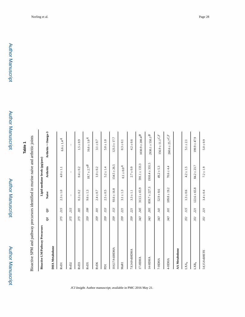
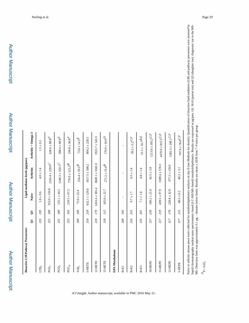
spectra (ref. 13 and Table 1).
Distinct profiles of lipid mediators were identified in naive, arthritic, and omega-3–
supplemented mice undergoing arthritis; lipid mediators could be separated utilizing linear
discriminant analysis (Figure 1B). Thus, correlations between individual lipid mediators and
the 3 treatment groups were plotted (Figure 1C). Higher levels of MaR1 were found in naive
paws as compared with arthritic paws. Comparatively, arthritic joints were associated with
higher levels of proinflammatory prostanoids including thromboxane (TX) B2, prostaglandin
(PG) D2, PGE2, and PGF2α, as well as leukotriene (LT) B4. Whereas the paws of arthritic
mice supplemented with an omega-3–enriched diet were characterized by elevated levels of
SPM, including EPA-derived RvE2 and RvE3 and DHA derived Rvs, of which RvD1 and
RvD5 were significantly increased (Table 1 and Figure 1C). These findings suggest that
local production of SPM within the joints of omega-3–supplemented mice may contribute to
the reduction in arthritis severity.
Identification of SPM in human RA synovial fluids
We next profiled synovial fluid from human RA patients to determine the bioactive lipid
mediator metabolome found within human joints (refer to Table 2 for patient demographics/
treatment regimes). Using LC-MS/MS–based lipid mediator metabololipidomics, we
identified mediators from all 3 major bioactive metabolomes in human synovial fluids
including DHA-derived Rvs and AA-derived eicosanoids (Figure 2A). Similarly to murine
arthritic joints, AA-derived autacoids classically associated with joint pain and inflammation
were detected within the synovial effusate, including PGE2, PGD2, PGF2α, TXB2, and
LTB4. In these fluids, we also identified the pathway marker for the protective lipoxin family
5,15-diHETE, as well as the D-series Rvs, RvD1 and RvD3. All of these mediators were
identified in accordance with published criteria including matching RTs in LC (Figure 2A)
and characteristic/diagnostic MS/MS fragmentation patterns (Figure 2B). Of note, the levels
of these proresolving mediators in these inflammatory exudates were detected within
Norling et al. Page 3
JCI Insight. Author manuscript; available in PMC 2016 May 21.
Author M
anuscriptA
uthor Manuscript
Author M
anuscriptA
uthor Manuscript

physiologically relevant concentrations (RvD1, ~31 pM, bioactive concentrations: 10 pM–
100 nM; RvD3, ~23 pM, bioactive concentrations: 1 pM–10 nM) (refs. 8, 14, 15, and Table
3).
Proresolving lipid mediators directly impact neutrophil migration
As arthritic synovial exudates contain abundant neutrophils, we utilized microfluidic
chambers that provide defined spatiotemporal concentration gradients to study the role of
SPM in regulating neutrophil chemotaxis in real time and at a single cell level. We directly
compared the actions of lipoxin A4 (LXA4), LXB4, RvD1, or RvD2 on neutrophil migration
toward IL-8, a classical neutrophil chemotaxis signal. Initially, neutrophils were exposed to
IL-8 (10 nM) alone, then exposed to a set concentration (1 nM) of SPM concomitantly with
the IL-8 gradient over the subsequent 15 minutes. In the absence of IL-8 superfusion, any
captured PMN remained rounded and rapidly detached during the first minute of flow
(Supplemental Figure 1A, still images); this was also the case following exposure to SPM
alone (data not shown).
Displacement and directionality were determined for each individual cell by tracking the cell
centroid utilizing ImageJ software analysis. Following exposure to IL-8, cells migrated in
the direction of the chemotactic gradient as seen by vertical cell trajectory paths (Figure 3A;
veh), with general direction depicted by the rose plot (Figure 3B; veh). The inset in Figure
3B shows the polarized morphology of neutrophils after exposure to IL-8 plus vehicle (0.1%
ethanol). In the presence of LXA4, RvD1, or RvD2, neutrophils no longer responded to the
chemotactic signal and instead migrated in the direction of fluid flow (Figure 3A and B).
With 1 nM LXB4, cells still responded to IL-8 and retained their polarized morphology.
Rose plots for each individual donor were plotted separately (Supplemental Figure 1B–F).
The chemotaxis index was calculated for each SPM, as graphically illustrated in Figure 3C.
LXA4, RvD1, and RvD2 all significantly blunted PMN chemotaxis by approximately 75%.
The directionality of neutrophil migration was also calculated for each SPM (Figure 3D);
exposure to LXA4 or RvD2 significantly reduced direct travel (i.e., increased random
movement). These results demonstrate that, at low nanomolar concentrations, LXA4, RvD1,
and RvD2 each selectively impact and reduce directional migration toward IL-8. We
subsequently selected RvD1, which significantly attenuated PMN chemotaxis yet had little
effect on random migration, to test in a neutrophilic model of arthritis.
Treatment of mice with 17R-RvD1 confers protection from inflammatory arthritis
RA is a progressive inflammatory disease characterized by extensive infiltration of
leukocytes into the synovial cavity and articular tissues. Given that RvD1 significantly
regulated human neutrophil responses and its 17R-epimer retains the bioactions of RvD1
while displaying higher metabolic stability (11), we next investigated the therapeutic
potential of 17R-RvD1 (7S,8R,17R-trihydroxy-4Z,9E,11E,13Z,15E,19Z-docosahexaenoic
acid) in protecting against leukocyte-mediated join damage. Mice were injected with
arthritogenic K/BxN serum and monitored over 8 days. Administration of K/BxN serum
triggered robust arthritis, which rapidly developed over the first few days (Figure 4A). 17R-
RvD1–treated mice exhibited a lower clinical score and reduced hind paw edema compared
with vehicle-treated mice (Figure 4A and B). 17R-RvD1 also protected mice from the
Norling et al. Page 4
JCI Insight. Author manuscript; available in PMC 2016 May 21.
Author M
anuscriptA
uthor Manuscript
Author M
anuscriptA
uthor Manuscript
transient weight loss associated with arthritis (Figure 4C). Disease penetrance, defined as the
proportion of mice developing a clinical score greater than 1 out of a maximum 12, was
100% regardless of treatment group (Figure 4D), yet the clinical data indicates 17R-RvD1
significantly reduced disease severity.
In addition to the macroscopic assessment of arthritis, histological analyses were performed
on knee joints to assess inflammatory infiltrate, cartilage degradation, and evidence of bone
erosion. Microscopic analyses of the naive, nonarthritic articular joint presented little
evidence for leukocyte infiltration and demonstrated intact cartilage and bone. Arthritic
serum provoked a marked accumulation of leukocytes within the joint, as well as evident
pannus formation and synovial hypertrophy as compared with naive animals, whereas 17R-
RvD1 treatment limited leukocyte recruitment, synovitis, and pannus intrusion (Figure 4E–
G, and Supplemental Figure 2A and B). Blinded analyses of histopathological sections
revealed a significant tissue protection by 17R-RvD1 (Figure 4H). Levels of KC (CXCL1), a
key chemokine known to drive inflammatory arthritis and promote leukocyte recruitment,
were assessed (16). Plasma levels were elevated during arthritis and were significantly
dampened following 17R-RvD1 treatment (Figure 4I). Further studies were performed to
decipher which leukocytes infiltrated arthritic joints. Flow cytometric analyses identified
neutrophil (Ly6GhiCD11bhi), monocyte (Ly6ChiLy6Glo), and macrophage (F4/80hiLy6Glo)
accumulation within arthritic paws. Each of these leukocyte subtypes was significantly
reduced in paws from 17R-RvD1–treated mice (Figure 4J–L).
Current arthritis treatment strategies aim to intervene early to reduce synovitis and prevent
cartilage degradation and bone erosion (3). Hence, our interest was to determine whether
17R-RvD1 would be effective at reducing the clinical signs of early experimental arthritis.
Therapeutic administration of Rvs at the peak of an acute inflammatory response is known
to accelerate the resolution process (17). Therefore, the time taken to remit (remission
interval) based on a clinical score was calculated analogously to the resolution interval
widely used to calculate the loss of neutrophils from an inflammatory site by 50% (17, 18).
17R-RvD1 was tested as a therapeutic agent after the second dosing of K/BxN serum when
mice exhibited overt signs of arthritis (100 ng, i.p. daily from day 4–10). Rv treatment
limited the arthritis severity (Tmax; veh day 8, max. score 9.5 vs. 17R-RvD1 day 6, max.
score 7) and shortened the remission interval (Ri; veh 15 days, 17R-RvD1 9 days) (Figure
4M). This experiment demonstrates the potential of 17R-RvD1 as a therapeutic for limiting
tissue damage in patients with early RA.
Administration of 17R-RvD1 selectively modulates the local bioactive lipid metabolome within arthritic paws
Because RvD1 regulates proinflammatory eicosanoid biosynthesis during acute peritonitis
(15), we next performed lipid mediator metabololipidomics on arthritic paws obtained 8
days after K/BxN serum administration to elucidate the potential mechanism(s) underlying
17R-RvD1 actions. Identification was conducted using established criteria (13), including
LC RT and at least 6 fragment diagnostic ions in the MS/MS as illustrated for LTB4 and
PGE2 (Figure 5A and B). In these paws, mediators from each of the 3 major bioactive
metabolomes were identified, including the following eicosanoids: LTB4, LXA4, and LXB4
Norling et al. Page 5
JCI Insight. Author manuscript; available in PMC 2016 May 21.
Author M
anuscriptA
uthor Manuscript
Author M
anuscriptA
uthor Manuscript
from the lipoxygenase pathway and PGE2, PGD2, PGF2α, and TXB2 from the
cyclooxygenase pathways (Figure 5C). Multiple reaction monitoring (MRM) was utilized to
quantify the individual mediator amounts. We found that 17R-RvD1 downregulated
production of proinflammatory LTB4, an essential mediator required for the development of
experimental arthritis (19), along with TXB2 and PGE2, PGD2, and PGF2α while activating
protective LXA4 and LXB4 biosynthesis (Figure 5C). We also found that 17R-RvD1
stimulated the biosynthesis of EPA-derived SPM including the E-series Rvs RvE2 and RvE3
by approximately 50%. From the DHA bioactive metabolome, protectin Dx (PDx) was
significantly elevated in mice treated with 17R-RvD1. Thus, this regulation of lipid
mediators may contribute to the joint protective actions following pharmacological delivery
of 17R-RvD1.
Gene expression analysis was performed on paw tissue from arthritic mice treated with
vehicle and 17R-RvD1 observing selective modulatory functions. A trend for
downmodulation of Il1b, the key cytokine driving K/BxN arthritis; Ly6g, a marker of
neutrophil influx; and Ptsg2 (cyclooxygenase-2), essential for PG biosynthesis, was detected
in mice given 17R-RvD1. On the other hand, higher expression of Alox15 (15-lipoxygenase)
transcript, a key gene in SPM biosynthesis, was detected in mice treated with 17R-RvD1
(Supplemental Table 1). Most notably, a significant upregulation of key genes involved in
cartilage matrix synthesis was observed, namely type II collagen and aggrecan (Figure 6A).
This unpredicted finding brought us to investigate the impact of 17R-RvD1 treatments on
cartilage erosion during K/BxN arthritis. After the onset of arthritis (day 3), mice were
administered 17R-RvD1 (100 ng) directly into the knee joint, along with PBS (+0.1%
ethanol) into contralateral knees as control, with histological analyses being performed on
day 5. Cartilage integrity was first assessed in naive mice, whereby the cartilage remained
intact regardless of PBS or 17R-RvD1 treatment, as measured by toluidine blue staining
(Figure 6B, left panels, and Figure 6C analyses). However, a marked reduction in toluidine
blue staining on the cartilage surface was observed in PBS-injected knees of arthritic mice,
indicating loss of glycosaminoglycans (Figure 6B, upper right panel, white arrowheads).
Remarkably, a significant protection from cartilage damage was evident in 17R-RvD1–
treated joints as compared with paired contralateral control knees (Figure 6B, lower right
panel, and Figure 6C, analyses).
To determine whether this was a direct effect on the chondrocytes rather than an indirect
reduction of leukocyte/stromal cell–induced cartilage degradation, we took advantage of the
human chondrocyte (C28/I2) micromass system to evaluate extracellular matrix (ECM)
accumulation (20). Treatment of micromasses with 17R-RvD1 (0.1–100 nM) resulted in a
slight but nonsignificant increase in ECM accumulation (Figure 6D). When chondrocytes
were stimulated with IL-1β, a characteristic loss in ECM was quantified. Of note, 17R-RvD1
treatments significantly protected against the catabolic effects of concomitant IL-1β
stimulation, leading to increased ECM deposition (Figure 6D and representative images of
Alcian blue stained micromasses). As chondrocytes express the 17R-RvD1 receptor ALX/
FPR2 (21) and 17R-RvD1 demonstrated direct actions on chondrocytes, we sought to
determine whether this was via receptor ligation of ALX/FPR2. Addition of the specific
antagonist WRW4 did not have any effect on resting or IL-1β–treated micromasses alone.
However, the accumulative effects of 17R-RvD1 were reversed by the addition of WRW4,
Norling et al. Page 6
JCI Insight. Author manuscript; available in PMC 2016 May 21.
Author M
anuscriptA
uthor Manuscript
Author M
anuscriptA
uthor Manuscript
suggesting this protective effect was mediated via ALX/FPR2 (Figure 6E). Further support
of ALX/FPR2 receptor dependency for the protective actions of 17R-RvD1 was attained in
vivo by utilizing mice nullified with the orthologue receptor Fpr2/3 (22). Accordingly, we
subjected these mice to arthritis and treated them daily with either vehicle (PBS + 0.1%
ethanol [EtOH], i.p.) or 17R-RvD1 (100 ng, i.p.). In the absence of Fpr2/3, 17R-RvD1 no
longer attenuated the clinical score (Figure 6F), the swelling of hind paws, or the arthritis-
related manifestation of cachexia, and disease penetrance was almost identical between the 2
genotypes (data not shown). Histopathology of murine hind paws indicated substantial
leukocyte infiltration (Supplemental Figure 3A), with near maximal score (3/3) for both
genotypes (Supplemental Figure 3B). In summary, joint protection afforded by 17R-RvD1
was abolished in the absence of ALX/Fpr2/3.
Discussion
RA is a chronic progressive autoimmune disease characterized by inflammation of the joints
that manifests as swelling, pain, and functional impairment. This condition can also lead to
muscle wasting and osteoporosis and is associated with systemic inflammatory
comorbidities such as periodontitis and cardiovascular disease (1, 23). Although current
therapeutics have revolutionized the management of RA, existing treatments aimed at
limiting joint inflammation are associated with unwanted side effects including
gastrointestinal (GI) disturbances and increased risk of infections. In addition, although
current therapeutics may lead to significant improvement in patient prognosis by slowing or
stopping disease progression, treatment rarely repairs joint damage (5).
We report here an elevated local production of SPM in arthritic joints from mice
supplemented with omega-3–enriched chow, a biochemical response that correlates with a
reduction in arthritis severity. Using targeted lipid mediator metabololipidomics, we
identified RvD1 as an SPM that was both elevated in arthritic paws from omega-3–
supplemented mice and present in human RA synovial fluids, detected within the
concentration range for physiological activities. Thus, when we tested the stable epimer
17R-RvD1 (11), a selective modulation of local lipid mediator biosynthesis and reduction in
joint leukocyte infiltration was observed paralleled by macroscopic amelioration of arthritis
severity, cachexia, and hind-paw edema. We also uncovered tissue-protective functions of
17R-RvD1, such as stimulation of chondrocyte matrix production and protection from
cartilage degradation. Together, these findings identify 17R-RvD1 as a prototype for new
therapeutic approaches for the treatment of arthritis.
Timely resolution of an inflammatory response is pertinent to maintain tissue homeostasis.
To date, we and others have identified a number of endogenous mediators that activate
resolution pathways to actively switch off inflammation, including annexin-A1 (24), gaseous
mediators (e.g., hydrogen sulphide, ref. 25; and carbon monoxide, refs. 26, 27) as well as
SPM (lipoxins, Rvs, protectins, and MaRs; refs. 2, 7). Dysregulated inflammation is a
common determinant of many pathologies, yet it was only recently appreciated that diseases
such as RA may persist, at least in part, due to a failure of resolution (28). Accordingly,
essential protective roles for endogenous SPM and their receptors are indicated in
experimental arthritis. Null mice lacking either 12/15-LOX (a key enzyme involved in SPM
Norling et al. Page 7
JCI Insight. Author manuscript; available in PMC 2016 May 21.
Author M
anuscriptA
uthor Manuscript
Author M
anuscriptA
uthor Manuscript

biosynthesis) or ALX/FPR2 (a high-affinity receptor for RvD1, LXA4, and annexin-A1),
display exacerbated disease severity and tissue damage in inflammatory arthritis (29, 30).
Additionally, mice nullified for 5-LOX (another key enzyme involved in SPM biosynthesis)
exhibit intensified and prolonged duration of infectious arthritis (31). Furthermore,
inhibiting COX-2 activity delays the resolution of collagen-induced arthritis, which can be
restored with PGE2-mediated LXA4 production (32). Together, results from these studies
emphasize the importance of endogenous SPM for the control of arthritis. Early studies
demonstrated the protective effects of supplementing mice with fish oil–enriched diet, with
reduced susceptibility and lower severity scores when subjected to collagen-induced arthritis
(33, 34). Congruently, we found that arthritic mice fed a fish oil– enriched diet displayed
attenuated arthritis that was associated with a reduction in proinflammatory lipid mediator
levels and an enhanced biosynthesis of omega-3–derived SPM.
Enzymes of the SPM biosynthetic pathway, as well as the RvD1 receptor, are expressed in
the rheumatoid synovium. Compared with osteoarthritis (OA) synovia, elevated levels of 5-
and 15-LOX, as well as ALX/FPR2, are detected in synovial tissue of RA patients (35, 36).
In the present study, we identified both RvD1 and RvD3, along with the lipoxin pathway
marker 5,15-diHETE, in synovial fluids from RA patients. These results add to the recent
identification of RvD5, MaR1, and LXA4 within synovial fluid (37). From these findings,
we propose that metabololipidomic profiling can assist in RA patient stratification with the
definition of lipid mediator profiles associated with specific cohorts and in the assignment of
precision medical treatments and personalized RA patient care.
Aberrant infiltration of immune cells into the joint and the subsequent destruction of bone
and cartilage drive loss of function. During active phases of RA, neutrophils present within
synovial fluids contribute to disease pathogenesis via release of cytotoxic enzymes and
reactive oxygen species (38). We previously reported that RvD1 (0.1–100 nM) significantly
dampened neutrophil recruitment to TNF-α stimulated endothelium (39), a pivotal cytokine
that drives RA (6). Using microfluidics chambers to study bioactions at the single cell level
under flow of continuous chemokine gradients, we observed potent actions of RvD1 at low
nanomolar concentrations, blunting directed neutrophil locomotion. Thus, we utilized the
K/BxN serum transfer model of arthritis to test the efficacy of the stable epimer 17R-RvD1.
In this model of symmetric polyarthritis, daily treatment with 17R-RvD1 preserved joint
integrity and lessened the histopathology of arthritis, with reduced synovitis and pannus
intrusion and overall leukocyte infiltration. We previously demonstrated that 17R-RvD1–
enriched nanoparticles limit leukocyte recruitment into the inflamed temporomandibular
joint (40). Our current results emphasize the protective actions of 17R-RvD1 in limiting
joint inflammation.
It is known that a lipid-cytokine-chemokine cascade drives neutrophil recruitment during
experimental arthritis (16). Mechanistically, we found that administration of 17R-RvD1
downregulated Ptsg2 and elevated Alox15 gene expression, resulting in the selective
modulation of the local lipid mediator metabolome. We measured reduced LTB4 and PG
levels that contribute to the pain and perpetuation of joint inflammation and identified select
regulation of SPM levels that may contribute to the protective effects afforded by 17R-RvD1
administration. We also determined that 17R-RvD1 reduced systemic KC (CXCL1) levels, a
Norling et al. Page 8
JCI Insight. Author manuscript; available in PMC 2016 May 21.
Author M
anuscriptA
uthor Manuscript
Author M
anuscriptA
uthor Manuscript
principal chemokine that recruits neutrophils, and downregulated Il1b transcripts, a key
cytokine that amplifies arthritis. The main cellular source of this cytokine in this
experimental model of arthritis is neutrophils (16), and correspondingly, we observed a
reduction in neutrophil Ly6g transcripts. The antiarthritic actions of 17R-RvD1 were lost in
Fpr2/3-null mice, reiterating this essential signaling pathway for RvD1 in murine
inflammation (15, 41).
Once damaged, articular cartilage has limited intrinsic capability to self-repair. Importantly,
we uncovered unique tissue protective functions of 17R-RvD1 in maintaining cartilage
integrity during inflammatory arthritis. Chondrocytes were identified as target cells for the
protective actions of RvD1, stimulating ECM deposition. We provide evidence that 17R-
RvD1 protects chondrocytes from IL-1β–induced degradation via direct ALX/FPR2 receptor
ligation. This finding complements the array of known functions that proresolving mediators
exert to repair injured tissue in the context of wound healing (42), tissue regeneration (43,
44), and restitution of gut epithelial barrier function (45). Our results indicate that mediators
of resolution could be modeled to repair cartilage if, and when, their receptor target is
presented on the chondrocyte. Recent work indicates that ALX/FPR2 is expressed by human
chondrocytes and surface expression is upregulated in catabolic settings, like those evoked
with IL-1β (21).
The results we present here using synthetic RvD1 as a prototype therapeutic tool from the
SPM portfolio, a choice dictated by detection of this bioactive mediator in mouse and human
joints, opens the opportunity to exploit this line of research for innovative therapeutic
strategies in RA that combine potent antiinflammatory with tissue-protective properties.
Modeling RvD1 analogues or agonists at their receptor provide another avenue for
therapeutic development (24). It is important to point out the prediction that, compared with
current therapeutic options, RvD1 and — in general — SPM-based therapies will be
beneficial in multiple respects. Firstly, SPM do not provoke immune suppression (a major
side effect associated with biologics, e.g., anti-TNF therapy; ref. 6) and rather stimulate host
phagocyte functions to enhance phagocytosis and killing of microorganisms (8, 46, 47).
Secondly, SPM exert bone-sparing properties (a major secondary effect associated with
long-term glucocorticoid therapy is bone loss). In the context of experimental periodontitis,
RvE1 prevents alveolar bone loss (48), accelerates bone regeneration (43), and inhibits
osteoclast maturation and bone resorption (49). Additionally, parathyroid hormone
stimulates RvD1 and RvD2 to enhance macrophage efferocytosis of apoptotic osteoblasts,
highlighting a role for SPM in bone homeostasis (50). Thirdly, SPM are potent analgesics
(51), and relevantly, 17R-RvD1 is antihyperalgesic in a model of arthritic pain (52). Finally,
with the present study, we demonstrate chondroprotective functions of SPM, combating
catabolic stimulation and promoting direct anabolic responses enabling protection from
cartilage degradation during inflammatory arthritis.
In summary, we provide new mechanisms underlying the antiarthritic properties evoked by
omega-3 consumption in arthritis (10) and focus on a specific omega-3–derived SPM,
namely RvD1 and its stable epimer 17R-RvD1, to establish proof-of-concept for the validity
of this approach. We defined bioactions for this proresolving endogenous lipid mediator in
the context of joint inflammation and chondroprotection. Together, these results give the
Norling et al. Page 9
JCI Insight. Author manuscript; available in PMC 2016 May 21.
Author M
anuscriptA
uthor Manuscript
Author M
anuscriptA
uthor Manuscript
foundation for the development of innovative therapeutic strategies to avert joint destruction
during RA with limited side effects, ultimately for the benefit of the patients.
Methods
Animals
Male 12-week-old, approximately 30 g, C57Bl/6 (Charles River Laboratories) or Fpr2/3-null
(ALX-null) mice (generated on a C57Bl/6 background as described, and back-backcrossed
10 times; ref. 22) were maintained on a standard chow pellet diet and had access to water ad
libitum, with a 12-hour light-dark cycle. For omega-3 diet-enrichment studies, mice were
maintained on a CRM (P) plus 10% salmon oil or standard CRM (P) chow (Special diet
services, LBS Biotech) 3 weeks prior to and throughout the duration of experiments.
Real-time neutrophil chemotaxis using microfluidics chambers
Microfluidics chambers were engineered for testing the actions of bioactive lipid mediators
on the chemotactic behavior of human neutrophils as described previously (53). The main
channel for assessing neutrophil chemotactic behavior was modified by physical adsorption
of P-selectin (50 µg/ml, R&D Systems) and ICAM-1 (10 µg/ml, R&D Systems) for 30
minutes, followed by blocking with 2% human serum albumin (HSA, Sigma-Aldrich) in
HBSS (Sigma-Aldrich) immediately before use. Steady-state gradients of IL-8 (0–10 nM,
R&D Systems) were formed in the 2 gradient generators connected to the main channel, one
containing vehicle (0.1% ethanol) and the other containing an overlaying uniform
concentration (1 nM) of the specialized proresolving lipid mediator for testing (LXA4,
LXB4, RvD1, or RvD2; Cayman Chemicals). Approximately 10 µl of capillary blood was
then collected from healthy volunteers using a BD genie lancet (BD Biosciences), and
diluted 1:10 with HBSS, 0.2% HSA. Neutrophils were captured from flowing blood over
approximately 3 minutes, and the first valve opened for infusion of the IL-8 gradient (fluid
flow removed the majority of RBC and other cells that were not tethered to the coated
chamber), allowing direct monitoring of neutrophils captured on the chamber surface. After
15 minutes, the gradient was switched to the second gradient generator containing a uniform
concentration of the SPM combined with 10 nM IL-8 gradient. Neutrophil migration in the
chemokine gradient and their response(s) to addition of different SPM were recorded every 6
seconds over a 30-minute period with a digital camera and Image Pro Plus software. Cell
migration was analyzed using the cell-tracking function in ImageJ software, and tracks were
analyzed utilizing Ibidi Chemotaxis and Migration Tool. Only cells that started and
remained within the field of view over the entire course of video capture were analyzed. At
least 20 cells were tracked per donor, with 3–4 donors per test compound.
K/BxN serum transfer model of inflammatory arthritis
K/BxN serum was generated in house by breeding NOD/Shiltj mice (Charles River
Laboratories) with KRN transgenic mice (gift from Mohini Gray, Edinburgh University,
Edinburgh, United Kingdom); the subsequent offspring (K/BxN mice) express both the T
cell receptor (TCR) transgene KRN and the MHC class II molecule Ag7 and spontaneously
develop a severe inflammatory arthritis. Serum was collected from K/BxN mice at 10 weeks
of age, which causes a similar arthritis when transferred to recipient mice due to
Norling et al. Page 10
JCI Insight. Author manuscript; available in PMC 2016 May 21.
Author M
anuscriptA
uthor Manuscript
Author M
anuscriptA
uthor Manuscript
autoantibodies recognizing glucose-6-phosphate isomerase (GPI) depositing on the cartilage
surface. Serum transfer–induced arthritis was performed by i.p. injection of 100 µl of
arthritogenic serum on days 0 and 2 in WT or ALX/Fpr2/3-null mice. Disease was
monitored by assessing the clinical score (maximum 12 points per animal, 3 per limb with
the following scoring system: 0, no evidence of inflammation; 1, inflammation in one of the
following aspects: individual phalanges joints, localized wrist/ankle, or swelling on surface
of paw; 2, inflammation on two aspects of paw; 3, major swelling on all aspects of paw).
Paw edema was assessed by water displacement plethysmometry (Ugo Basile). Mice were
weighed daily for signs of cachexia. For prophylactic treatment regime, WT mice were
administered vehicle (0.1% EtOH) or 17R-RvD1 (100 ng) in 100 µl of saline i.p. daily (from
day 0, 5 minutes prior to arthritogenic serum administration). For therapeutic treatment
protocol, WT mice were administered vehicle (0.1% EtOH) or 17R-RvD1 (100 ng) in 100 µl
of saline i.p. daily following overt signs of arthritis (day 4) over a 7-day period. The arthritis
remission interval was calculated based on arthritic score and calculated by analogy to the
resolution indices widely used to calculate the loss of neutrophils from an inflammatory site
(17, 18). At the end of experiments, blood was collected via cardiac puncture into
heparinized syringes and centrifuged to obtain platelet-free plasma. For intraarticular
treatments, WT mice were administered K/BxN serum on days 0 and 2 (100 µl, i.p.) and
were treated locally on day 3 with vehicle (left knee; 5 µl PBS containing 0.1% EtOH) or
17R-RvD1 (right knee; 5 µl, 100 ng 17R-RvD1), and joints were harvested for histological
analysis on day 5.
Molecular analyses
Arthritic ankle joints were collected on day 8 and stored in RNAlater at −80°C prior to
homogenising using Precellys ceramic beads (Bertin Technologies). RNA was extracted
using RNeasy Plus mini kit (Qiagen), and genomic DNA contamination eliminated with
Turbo DNA-free kit (Applied Biosystems). Complementary DNA was synthesized using
SuperScriptIII reverse transcriptase and OligoDt primers (Invitrogen). Quantitative PCR
(qPCR) was performed using QuantiTect primers (Qiagen) and ABI Prism 7900 sequence
detector system (Applied Biosystems). Relative expression values were calculated following
normalization to endogenous housekeeping gene Rpl32 and using the 2−(ΔΔCt method
normalized to a naive mouse (calibrator sample).
Flow cytometry
Leukocytes were isolated from arthritic paws following tissue digestion. Briefly, paws were
collected after 8 days of arthritis, skin was carefully removed, and digestion buffer was
added (collagenase D [Roche; 0.5 µg/ml] and DNAse [Sigma-Aldrich; 40 µg/ml] in serum-
free RPMI). Paws were incubated at 37°C with gentle agitation, and liberated cells were
collected via a 70-µm cell-strainer and kept on ice. Another 15 ml of digestion buffer was
added to the paws and incubated for a further 30 minutes. Liberated cells were centriinsight.
fuged at 400 g for 10 minutes and resuspended in PBS for counting. Leukocytes were first
stained with Zombie NIR (BioLegend; 1:500, 20 min, 4°C) to identify live cells, and distinct
leukocyte subtypes were identified using the following antibodies from eBioscience: CD45
(25 µg/ml, clone 30-F11), CD11b (0.2 µg/ml, clone M1/70), Ly6G (25 µg/ml, clone
Norling et al. Page 11
JCI Insight. Author manuscript; available in PMC 2016 May 21.
Author M
anuscriptA
uthor Manuscript
Author M
anuscriptA
uthor Manuscript

RB6-8CS), Ly6C (0.4 µg/ml, clone HK1.4), and F4/80 (10 µg/ml, clone BM8). Samples
were analyzed using an LSR Fortessa flow cytometer and FlowJo software (Tree Star Inc.).
Histology
Joints were decalcified and paraffin embedded. Sections (8 µm) were stained with H&E or
1% aqueous toluidine blue, and standard light microscopy was used to determine the degree
of leukocyte infiltration/synovitis/pannus formation, bone erosion, and cartilage damage,
which were each graded from 0 (no disease) to 3 (severe); a maximum score of 9 was
possible, as averaged by 2 independent examiners, who were blinded to the experiment.
Cartilage integrity was calculated from percentage area of cartilage stained with toluidine
blue using thresholds applied with ImageJ software.
Determination of plasma KC (CXCL1) levels
Mice were sacrificed on day 8 following induction of arthritis, and blood was collected via
cardiac puncture into heparinized syringes and centrifuged to obtain platelet-free plasma.
KC (CXCL1) values were determined by ELISA.
Targeted LC-MS/MS–based lipidomics of arthritic paws and synovial fluids
Paws were collected and immediately transferred to liquid nitrogen prior to homogenization
in 1 ml ice cold MeOH containing deuterated internal standards (d4-LTB4, d8-5S-HETE, d4-
PGE2, d5-LXA4, and d5-LTC4, 500 pg each) and homogenized using a glass dounce.
Synovial fluid (0.5 ml) from RA patients (Dx Biosamples) were placed in 1 ml ice cold
MeOH containing deuterated internal standards (d4-LTB4, d8-5S-HETE, d4-PGE2, d5-LXA4,
and d5-RvD2, 500 pg each). All samples were kept at −20°C for 45 minutes to allow for
protein precipitation and subjected to solid phase extraction as in ref. 13. Methyl formate
fractions were then brought to dryness using a TurboVap LP (Biotage), and products were
suspended in water-methanol (50:50 vol:vol) for LC-MS-MS. A Shimadzu LC-20AD HPLC
and a Shimadzu SIL-20AC autoinjector (Shimadzu), paired with a QTrap 6500 (AB Sciex)
was utilized and operated as described (13). To monitor each LM and respective pathways,
an MRM method was developed with diagnostic ion fragments and identification using
recently published criteria (13), including matching RT to synthetic and authentic materials
and at least 6 diagnostic ions for each LM. Calibration curves were obtained for each using
authentic compound mixtures and deuterium labeled LM at 3.12, 6.25, 12.5, 25, 50, 100, and
200 pg. Linear calibration curves were obtained for each LM, which gave r2 values of 0.98–
0.99.
Chondrocyte micromass assay
The human chondrocyte cell line C28/I2 (gift from Mary Goldring, Cornell Medical
College, New York, New York, USA; ref. 54) was utilized for micromass assays as described
previously (20). Micromasses were stimulated with or without IL-1β (30 ng/ml) alone or in
combination with 17R-RvD1 (0.1–100 nM) for 24 hours, and ECM accumulation was
calculated following staining with Alcian blue as reported (21). Briefly, micromasses were
fixed (4% glutaraldehyde, 15 min) acidified with HCl, and stained with Alcian blue 8GS
overnight before dye extraction with guanidine hydrochloride (200 µl, 6M). Absorbance of
Norling et al. Page 12
JCI Insight. Author manuscript; available in PMC 2016 May 21.
Author M
anuscriptA
uthor Manuscript
Author M
anuscriptA
uthor Manuscript
extracted dye was measured at 620 nm using a Multiskan Bichromatic 348
spectrophotometer and normalized to DNA content using SYBRgreen dye (excitation 485
nm and emission 535 nm) with a TECAN M200 spectrophotometer. These values were used
to generate ECM accumulation concentrations and are expressed as percentage change from
control micromasses. In some experiments, the FPR2/ALX receptor antagonist WRW4 (10
µM) was added to micromasses 10 minutes prior to 17R-RvD1 addition, and ECM
accumulation was evaluated after 24 hours.
Statistics
Statistical analyses were performed with mean ± SEM, where n is the biological replicate
(individual mice or human donors). Data were analyzed using either 2-tailed paired or
unpaired Student’s t test, 1-way ANOVA with appropriate post hoc analysis, Mann-Whitney
t test, or 2-way ANOVA with repeated measures where appropriate. Analyses were
performed using GraphPad Prism; in all cases, P < 0.05 was considered significant.
Multivariate analysis of arthritis metabololipidomics was performed using linear
discriminant analysis, where bioactive lipid mediator predictors were entered simultaneously
to the model (carried out in SPSS 23; IBM).
Study approval
Human peripheral blood was collected according to a protocol approved by Barts and the
London Research Ethics Committee (London, United Kingdom [QMREC 2014:61]).
Written informed consent was received from participants prior to inclusion in the study
according to the declaration of Helsinki. All animal experiments were approved and
performed under the guidelines of the Ethical Committee for the Use of Animals, Barts and
The London School of Medicine, and in accordance with the UK Home Office regulations
(Guidance on the Operation of Animals, Scientific Procedures Act, 1986).
Supplementary Material
Refer to Web version on PubMed Central for supplementary material.
Acknowledgments
This work was funded by the Arthritis Research UK (Career Development Fellowship 19909 to L.V. Norling) and Wellcome Trust Programme grant 086867/Z/08/Z to M. Perretti. S.E. Headland is supported by an Arthritis Research UK foundation Fellowship 20842. J. Dalli was funded by a Sir Henry Dale Fellowship jointly funded by the Wellcome Trust and the Royal Society (grant 107613/Z/15/Z). This work forms part of the research themes contributing to the translational research portfolio of Barts and the London Cardiovascular Biomedical Research Unit, which is supported and funded by the NIH Research. C.N. Serhan and work carried out in the Center for Experimental Therapeutics and Reperfusion Injury laboratories at BWH are supported by the NIH (grant P01GM095467). H.H. Arnardottir was funded by an Arthritis Foundation Postdoctoral Fellowship (6095).
References
1. Bacon PA, Townend JN. Nails in the coffin: increasing evidence for the role of rheumatic disease in the cardiovascular mortality of rheumatoid arthritis. Arthritis Rheum. 2001; 44(12):2707–2710. [PubMed: 11762929]
2. Buckley CD, Gilroy DW, Serhan CN. Proresolving lipid mediators and mechanisms in the resolution of acute inflammation. Immunity. 2014; 40(3):315–327. [PubMed: 24656045]
Norling et al. Page 13
JCI Insight. Author manuscript; available in PMC 2016 May 21.
Author M
anuscriptA
uthor Manuscript
Author M
anuscriptA
uthor Manuscript
3. Raza K, Buckley CE, Salmon M, Buckley CD. Treating very early rheumatoid arthritis. Best Pract Res Clin Rheumatol. 2006; 20(5):849–863. [PubMed: 16980210]
4. Feldmann M, Maini RN. Perspectives from masters in rheumatology and autoimmunity: can we get closer to a cure for rheumatoid arthritis? Arthritis Rheumatol. 2015; 67(9):2283–2291. [PubMed: 26138641]
5. Klarenbeek NB, et al. The impact of four dynamic, goal-steered treatment strategies on the 5-year outcomes of rheumatoid arthritis patients in the BeSt study. Ann Rheum Dis. 2011; 70(6):1039–1046. [PubMed: 21415052]
6. Siebert S, Tsoukas A, Robertson J, McInnes I. Cytokines as therapeutic targets in rheumatoid arthritis and other inflammatory diseases. Pharmacol Rev. 2015; 67(2):280–309. [PubMed: 25697599]
7. Serhan C. Pro-resolving lipid mediators are leads for resolution physiology. Nature. 2014; 510(7503):92–101. [PubMed: 24899309]
8. Chiang N, et al. Infection regulates pro-resolving mediators that lower antibiotic requirements. Nature. 2012; 484(7395):524–528. [PubMed: 22538616]
9. Goldberg RJ, Katz J. A meta-analysis of the analgesic effects of omega-3 polyunsaturated fatty acid supplementation for inflammatory joint pain. Pain. 2007; 129(1–2):210–223. [PubMed: 17335973]
10. Calder P. Omega-3 polyunsaturated fatty acids and inflammatory processes: nutrition or pharmacology? Br J Clin Pharmacol. 2013; 75(3):645–662. [PubMed: 22765297]
11. Sun YP, et al. Resolvin D1 and its aspirin-triggered 17R epimer. Stereochemical assignments, anti-inflammatory properties, and enzymatic inactivation. J Biol Chem. 2007; 282(13):9323–9334. [PubMed: 17244615]
12. Monach P, et al. The K/BxN mouse model of inflammatory arthritis: theory and practice. Methods Mol Med. 2007; 136:269–282. [PubMed: 17983155]
13. Colas RA, Shinohara M, Dalli J, Chiang N, Serhan CN. Identification and signature profiles for pro-resolving and inflammatory lipid mediators in human tissue. Am J Physiol Cell Physiol. 2014; 307(1):C39–C54. [PubMed: 24696140]
14. Dalli J, et al. Resolvin D3 and aspirin-triggered resolvin D3 are potent immunoresolvents. Chem Biol. 2013; 20(2):188–201. [PubMed: 23438748]
15. Norling LV, Dalli J, Flower RJ, Serhan CN, Perretti M. Resolvin D1 limits polymorphonuclear leukocyte recruitment to inflammatory loci: receptor-dependent actions. Arterioscler Thromb Vasc Biol. 2012; 32(8):1970–1978. [PubMed: 22499990]
16. Chou RC, et al. Lipid-cytokine-chemokine cascade drives neutrophil recruitment in a murine model of inflammatory arthritis. Immunity. 2010; 33(2):266–278. [PubMed: 20727790]
17. Schwab JM, Chiang N, Arita M, Serhan CN. Resolvin E1 and protectin D1 activate inflammation-resolution programmes. Nature. 2007; 447(7146):869–874. [PubMed: 17568749]
18. Bannenberg GL, et al. Molecular circuits of resolution: formation and actions of resolvins and protectins. J Immunol. 2005; 174(7):4345–4355. [PubMed: 15778399]
19. Chen M, et al. Neutrophil-derived leukotriene B4 is required for inflammatory arthritis. J Exp Med. 2006; 203(4):837–842. [PubMed: 16567388]
20. Greco KV, et al. High density micromass cultures of a human chondrocyte cell line: a reliable assay system to reveal the modulatory functions of pharmacological agents. Biochem Pharmacol. 2011; 82(12):1919–1929. [PubMed: 21946086]
21. Headland SE, et al. Neutrophil-derived microvesicles enter cartilage and protect the joint in inflammatory arthritis. Sci Transl Med. 2015; 7(315):315ra190.
22. Dufton N, et al. Anti-inflammatory role of the murine formyl-peptide receptor 2: ligand-specific effects on leukocyte responses and experimental inflammation. J Immunol. 2010; 184(5):2611–2619. [PubMed: 20107188]
23. Scher JU, et al. Periodontal disease and the oral microbiota in new-onset rheumatoid arthritis. Arthritis Rheum. 2012; 64(10):3083–3094. [PubMed: 22576262]
24. Perretti M, Leroy X, Bland EJ, Montero-Melendez T. Resolution pharmacology: opportunities for therapeutic innovation in inflammation. Trends Pharmacol Sci. 2015; 36(11):737–755. [PubMed: 26478210]
Norling et al. Page 14
JCI Insight. Author manuscript; available in PMC 2016 May 21.
Author M
anuscriptA
uthor Manuscript
Author M
anuscriptA
uthor Manuscript
25. Caliendo G, Cirino G, Santagada V, Wallace JL. Synthesis and biological effects of hydrogen sulfide (H2S): development of H2S-releasing drugs as pharmaceuticals. J Med Chem. 2010; 53(17):6275–6286. [PubMed: 20462257]
26. Dalli J, et al. Proresolving lipid mediator profiles in baboon pneumonia are regulated by inhaled carbon monoxide. Am J Respir Cell Mol Biol. 2015; 53(3):314–325. [PubMed: 25568926]
27. Chiang N, et al. Inhaled carbon monoxide accelerates resolution of inflammation via unique proresolving mediator-heme oxygenase-1 circuits. J Immunol. 2013; 190(12):6378–6388. [PubMed: 23650615]
28. Yacoubian S, Serhan CN. New endogenous anti-inflammatory and proresolving lipid mediators: implications for rheumatic diseases. Nat Clin Pract Rheumatol. 2007; 3(10):570–590. [PubMed: 17906612]
29. Kronke G, et al. 12/15-lipoxygenase counteracts inflammation and tissue damage in arthritis. J Immunol. 2009; 183(5):3383–3389. [PubMed: 19675173]
30. Dufton N, et al. Anti-inflammatory role of the murine formyl-peptide receptor 2: ligand-specific effects on leukocyte responses and experimental inflammation. J Immunol. 2010; 184(5):2611–2619. [PubMed: 20107188]
31. Blaho VA, Zhang Y, Hughes-Hanks JM, Brown CR. 5-Lipoxygenase-deficient mice infected with Borrelia burgdorferi develop persistent arthritis. J Immunol. 2011; 186(5):3076–3084. [PubMed: 21270404]
32. Chan MM, Moore AR. Resolution of inflammation in murine autoimmune arthritis is disrupted by cyclooxygenase-2 inhibition and restored by prostaglandin E2-mediated lipoxin A4 production. J Immunol. 2010; 184(11):6418–6426. [PubMed: 20435922]
33. Leslie CA, Gonnerman WA, Ullman MD, Hayes KC, Franzblau C, Cathcart ES. Dietary fish oil modulates macrophage fatty acids and decreases arthritis susceptibility in mice. J Exp Med. 1985; 162(4):1336–1349. [PubMed: 3930652]
34. Leslie CA, Conte JM, Hayes KC, Cathcart ES. A fish oil diet reduces the severity of collagen induced arthritis after onset of the disease. Clin Exp Immunol. 1988; 73(2):328–332. [PubMed: 3180514]
35. Gheorghe KR, et al. Expression of 5-lipoxygenase and 15-lipoxygenase in rheumatoid arthritis synovium and effects of intraarticular glucocorticoids. Arthritis Res Ther. 2009; 11(3):R83. [PubMed: 19497113]
36. Hashimoto A, et al. Antiinflammatory mediator lipoxin A4 and its receptor in synovitis of patients with rheumatoid arthritis. J Rheumatol. 2007; 34(11):2144–2153. [PubMed: 17918787]
37. Giera M, et al. Lipid and lipid mediator profiling of human synovial fluid in rheumatoid arthritis patients by means of LC-MS/MS. Biochim Biophys Acta. 2012; 1821(11):1415–1424. [PubMed: 22841830]
38. Wright HL, Moots RJ, Edwards SW. The multifactorial role of neutrophils in rheumatoid arthritis. Nat Rev Rheumatol. 2014; 10(10):593–601. [PubMed: 24914698]
39. Norling LV, Dalli J, Flower RJ, Serhan CN, Perretti M. Resolvin D1 limits polymorphonuclear leukocyte recruitment to inflammatory loci: receptor-dependent actions. Arterioscler Thromb Vasc Biol. 2012; 32(8):1970–1978. [PubMed: 22499990]
40. Norling LV1, Spite M, Yang R, Flower RJ, Perretti M, Serhan CN. Cutting edge: Humanized nano-proresolving medicines mimic inflammation-resolution and enhance wound healing. J Immunol. 2011; 186(10):5543–5547. [PubMed: 21460209]
41. Krishnamoorthy S, Recchiuti A, Chiang N, Fredman G, Serhan CN. Resolvin D1 receptor stereoselectivity and regulation of inflammation and proresolving microRNAs. Am J Pathol. 2012; 180(5):2018–2027. [PubMed: 22449948]
42. Tang Y, Zhang MJ, Hellmann J, Kosuri M, Bhatnagar A, Spite M. Proresolution therapy for the treatment of delayed healing of diabetic wounds. Diabetes. 2013; 62(2):618–627. [PubMed: 23043160]
43. Hasturk H, et al. Resolvin E1 regulates inflammation at the cellular and tissue level and restores tissue homeostasis in vivo. J Immunol. 2007; 179(10):7021–7029. [PubMed: 17982093]
Norling et al. Page 15
JCI Insight. Author manuscript; available in PMC 2016 May 21.
Author M
anuscriptA
uthor Manuscript
Author M
anuscriptA
uthor Manuscript

44. Dalli J, et al. The novel 13S,14S-epoxy-maresin is converted by human macrophages to maresin 1 (MaR1), inhibits leukotriene A4 hydrolase (LTA4H), and shifts macrophage phenotype. FASEB J. 2013; 27(7):2573–2583. [PubMed: 23504711]
45. Leoni G, et al. Annexin A1-containing extracellular vesicles and polymeric nanoparticles promote epithelial wound repair. J Clin Invest. 2015; 125(3):1215–1227. [PubMed: 25664854]
46. Spite M, et al. Resolvin D2 is a potent regulator of leukocytes and controls microbial sepsis. Nature. 2009; 461(7268):1287–1291. [PubMed: 19865173]
47. Chiang N, Dalli J, Colas RA, Serhan CN. Identification of resolvin D2 receptor mediating resolution of infections and organ protection. J Exp Med. 2015; 212(8):1203–1217. [PubMed: 26195725]
48. Hasturk H, et al. RvE1 protects from local inflammation and osteoclast- mediated bone destruction in periodontitis. FASEB J. 2006; 20(2):401–403. [PubMed: 16373400]
49. Herrera BS, et al. An endogenous regulator of inflammation, resolvin E1, modulates osteoclast differentiation and bone resorption. Br J Pharmacol. 2008; 155(8):1214–1223. [PubMed: 18806821]
50. McCauley LK, Dalli J, Koh AJ, Chiang N, Serhan CN. Cutting edge: parathyroid hormone facilitates macrophage efferocytosis in bone marrow via proresolving mediators resolvin D1 and resolvin D2. J Immunol. 2014; 193(1):26–29. [PubMed: 24890726]
51. Ji RR, Xu ZZ, Strichartz G, Serhan CN. Emerging roles of resolvins in the resolution of inflammation and pain. Trends Neurosci. 2011; 34(11):599–609. [PubMed: 21963090]
52. Lima-Garcia JF, Dutra RC, da Silva K, Motta EM, Campos MM, Calixto JB. The precursor of resolvin D series and aspirin-triggered resolvin D1 display anti-hyperalgesic properties in adjuvant-induced arthritis in rats. Br J Pharmacol. 2011; 164(2):278–293. [PubMed: 21418187]
53. Kasuga K, et al. Rapid appearance of resolvin precursors in inflammatory exudates: novel mechanisms in resolution. J Immunol. 2008; 181(12):8677–8687. [PubMed: 19050288]
54. Goldring MB, et al. Interleukin-1 β-modulated gene expression in immortalized human chondrocytes. J Clin Invest. 1994; 94(6):2307–2316. [PubMed: 7989586]
Norling et al. Page 16
JCI Insight. Author manuscript; available in PMC 2016 May 21.
Author M
anuscriptA
uthor Manuscript
Author M
anuscriptA
uthor Manuscript
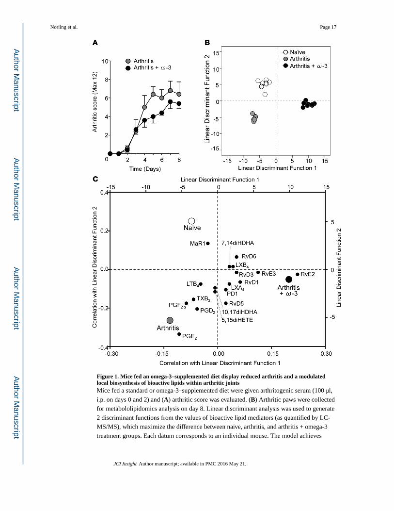
Figure 1. Mice fed an omega-3–supplemented diet display reduced arthritis and a modulated local biosynthesis of bioactive lipids within arthritic jointsMice fed a standard or omega-3–supplemented diet were given arthritogenic serum (100 µl,
i.p. on days 0 and 2) and (A) arthritic score was evaluated. (B) Arthritic paws were collected
for metabololipidomics analysis on day 8. Linear discriminant analysis was used to generate
2 discriminant functions from the values of bioactive lipid mediators (as quantified by LC-
MS/MS), which maximize the difference between naive, arthritis, and arthritis + omega-3
treatment groups. Each datum corresponds to an individual mouse. The model achieves
Norling et al. Page 17
JCI Insight. Author manuscript; available in PMC 2016 May 21.
Author M
anuscriptA
uthor Manuscript
Author M
anuscriptA
uthor Manuscript

complete discrimination of the 3 groups. (C) Correlations of each lipid mediator with the 2
linear discriminant functions are shown on the bottom x and left y axes. Group centroids for
the 2 discriminant functions are plotted on the top x and right y axes to show the
directionality and association of each lipid with the 3 treatment groups. n = 7–9 mice per
group.
Norling et al. Page 18
JCI Insight. Author manuscript; available in PMC 2016 May 21.
Author M
anuscriptA
uthor Manuscript
Author M
anuscriptA
uthor Manuscript
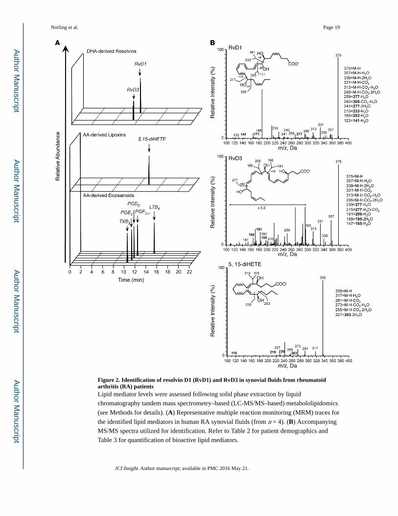
Figure 2. Identification of resolvin D1 (RvD1) and RvD3 in synovial fluids from rheumatoid arthritis (RA) patientsLipid mediator levels were assessed following solid phase extraction by liquid
chromatography tandem mass spectrometry–based (LC-MS/MS–based) metabololipidomics
(see Methods for details). (A) Representative multiple reaction monitoring (MRM) traces for
the identified lipid mediators in human RA synovial fluids (from n = 4). (B) Accompanying
MS/MS spectra utilized for identification. Refer to Table 2 for patient demographics and
Table 3 for quantification of bioactive lipid mediators.
Norling et al. Page 19
JCI Insight. Author manuscript; available in PMC 2016 May 21.
Author M
anuscriptA
uthor Manuscript
Author M
anuscriptA
uthor Manuscript
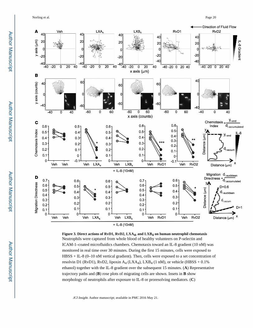
Figure 3. Direct actions of RvD1, RvD2, LXA4, and LXB4 on human neutrophil chemotaxisNeutrophils were captured from whole blood of healthy volunteers on P-selectin and
ICAM-1–coated microfluidics chambers. Chemotaxis toward an IL-8 gradient (10 nM) was
monitored in real time over 30 minutes. During the first 15 minutes, cells were exposed to
HBSS + IL-8 (0–10 nM vertical gradient). Then, cells were exposed to a set concentration of
resolvin D1 (RvD1), RvD2, lipoxin A4 (LXA4), LXB4 (1 nM), or vehicle (HBSS + 0.1%
ethanol) together with the IL-8 gradient over the subsequent 15 minutes. (A) Representative
trajectory paths and (B) rose plots of migrating cells are shown. Insets in B show
morphology of neutrophils after exposure to IL-8 or proresolving mediators. (C)
Norling et al. Page 20
JCI Insight. Author manuscript; available in PMC 2016 May 21.
Author M
anuscriptA
uthor Manuscript
Author M
anuscriptA
uthor Manuscript

Quantification of neutrophil chemotaxis index and (D) directness of neutrophil migration as
depicted by the graphs; n = 3–4 donors per compound. *P < 0.05, **P < 0.01, and ***P <
0.001 with 2-tailed paired Student’s t tests.
Norling et al. Page 21
JCI Insight. Author manuscript; available in PMC 2016 May 21.
Author M
anuscriptA
uthor Manuscript
Author M
anuscriptA
uthor Manuscript

Figure 4. 17R-RvD1 suppresses inflammatory arthritis by limiting leukocyte infiltration to arthritic jointsArthritis was induced with arthritogenic serum (100 µl, i.p. on days 0 and 2). Mice were
treated daily with vehicle (0.1% ethanol) or 17R-RvD1 (100 ng), and (A) arthritic score, (B)
paw edema, (C) weight loss, and (D) disease incidence were evaluated. Insets in A are
representative photographs of hind paw arthritis with and without daily 17R-RvD1
treatment; n = 14–17 mice per group. ***P < 0.001 or *P < 0.05 2-way ANOVA with
repeated measures. (E–G) Representative H&E-stained sections of knees from naive and
Norling et al. Page 22
JCI Insight. Author manuscript; available in PMC 2016 May 21.
Author M
anuscriptA
uthor Manuscript
Author M
anuscriptA
uthor Manuscript

arthritic mice on day 8 after arthritis induction (×4 magnification); n = 5–7 mice per group.
Scale bars: 200 µm. F, femur; T, tibia; m, meniscus; IFP, infrapatellar fat pad; PF, pannus
formation. (H) Histological score calculated by degree of leukocyte infiltration, cartilage,
and bone erosion; n = 5–7 mice per group, *P < 0.05 vs. naive, #P < 0.05 vs. arthritis, 1-way
ANOVA with Bonferroni post-hoc test. (I) Plasma KC levels; n = 8–10 mice per group, *P <
0.05 (vs. naive), #P < 0.05 (vs. arthritis) 1-way ANOVA with Bonferroni post-hoc test. (J–L)
Arthritic paws were digested to liberate leukocytes; cells were counted by light microscopy
and leukocyte subsets defined using specific antibodies by flow cytometry; n = 6 mice per
group, *P < 0.05 unpaired Student’s t test. (M) Mice were treated daily following overt signs
of arthritis (day 4) for 1 week with vehicle (0.1% ethanol) or 17R-RvD1 (100 ng), and
arthritic score was evaluated over 32 days. Arthritis remission indices were calculated; n =
11–12 mice per group.
Norling et al. Page 23
JCI Insight. Author manuscript; available in PMC 2016 May 21.
Author M
anuscriptA
uthor Manuscript
Author M
anuscriptA
uthor Manuscript
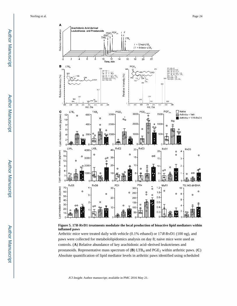
Figure 5. 17R-RvD1 treatments modulate the local production of bioactive lipid mediators within inflamed pawsArthritic mice were treated daily with vehicle (0.1% ethanol) or 17R-RvD1 (100 ng), and
paws were collected for metabololipidomics analysis on day 8; naive mice were used as
controls. (A) Relative abundance of key arachidonic acid–derived leukotrienes and
prostanoids. Representative mass spectrum of (B) LTB4 and PGE2 within arthritic paws. (C)
Absolute quantification of lipid mediator levels in arthritic paws identified using scheduled
Norling et al. Page 24
JCI Insight. Author manuscript; available in PMC 2016 May 21.
Author M
anuscriptA
uthor Manuscript
Author M
anuscriptA
uthor Manuscript

multiple reaction monitoring (MRM). n = 5–7 per group. *P < 0.05 and ***P < 0.001 vs.
naive and #P < 0.05 vs. arthritis with 1-way ANOVA and Bonferroni post-hoc test.
Norling et al. Page 25
JCI Insight. Author manuscript; available in PMC 2016 May 21.
Author M
anuscriptA
uthor Manuscript
Author M
anuscriptA
uthor Manuscript
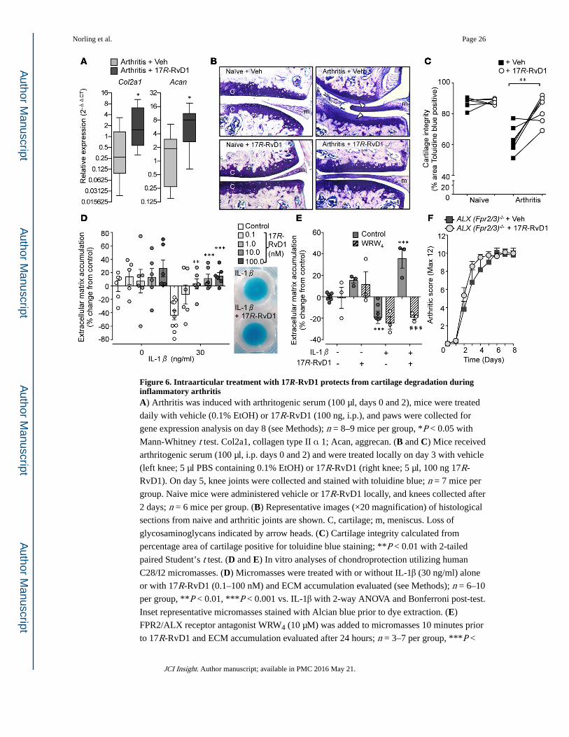
Figure 6. Intraarticular treatment with 17R-RvD1 protects from cartilage degradation during inflammatory arthritisA) Arthritis was induced with arthritogenic serum (100 µl, days 0 and 2), mice were treated
daily with vehicle (0.1% EtOH) or 17R-RvD1 (100 ng, i.p.), and paws were collected for
gene expression analysis on day 8 (see Methods); n = 8–9 mice per group, *P < 0.05 with
Mann-Whitney t test. Col2a1, collagen type II α 1; Acan, aggrecan. (B and C) Mice received
arthritogenic serum (100 µl, i.p. days 0 and 2) and were treated locally on day 3 with vehicle
(left knee; 5 µl PBS containing 0.1% EtOH) or 17R-RvD1 (right knee; 5 µl, 100 ng 17R-
RvD1). On day 5, knee joints were collected and stained with toluidine blue; n = 7 mice per
group. Naive mice were administered vehicle or 17R-RvD1 locally, and knees collected after
2 days; n = 6 mice per group. (B) Representative images (×20 magnification) of histological
sections from naive and arthritic joints are shown. C, cartilage; m, meniscus. Loss of
glycosaminoglycans indicated by arrow heads. (C) Cartilage integrity calculated from
percentage area of cartilage positive for toluidine blue staining; **P < 0.01 with 2-tailed
paired Student’s t test. (D and E) In vitro analyses of chondroprotection utilizing human
C28/I2 micromasses. (D) Micromasses were treated with or without IL-1β (30 ng/ml) alone
or with 17R-RvD1 (0.1–100 nM) and ECM accumulation evaluated (see Methods); n = 6–10
per group, **P < 0.01, ***P < 0.001 vs. IL-1β with 2-way ANOVA and Bonferroni post-test.
Inset representative micromasses stained with Alcian blue prior to dye extraction. (E)
FPR2/ALX receptor antagonist WRW4 (10 µM) was added to micromasses 10 minutes prior
to 17R-RvD1 and ECM accumulation evaluated after 24 hours; n = 3–7 per group, ***P <
Norling et al. Page 26
JCI Insight. Author manuscript; available in PMC 2016 May 21.
Author M
anuscriptA
uthor Manuscript
Author M
anuscriptA
uthor Manuscript
0.001 vs. vehicle, ###P < 0.001 vs. respective control with 2-way ANOVA and Bonferroni
post-test. (F) Dependency of Fpr2/3 (ALX) receptor for 17R-RvD1 protection from
inflammatory arthritis. Arthritis was induced in Fpr2/3-null (ALX-null) mice (see Methods),
mice were treated daily with vehicle (PBS with 0.1% EtOH) or 17R-RvD1 (100 ng, i.p.),
and arthritic score was assessed; n = 9–10 mice per group.
Norling et al. Page 27
JCI Insight. Author manuscript; available in PMC 2016 May 21.
Author M
anuscriptA
uthor Manuscript
Author M
anuscriptA
uthor Manuscript
Author M
anuscriptA
uthor Manuscript
Author M
anuscriptA
uthor Manuscript
Norling et al. Page 28
Tab
le 1
Bio
activ
e SP
M a
nd p
athw
ay p
recu
rsor
s id
entif
ied
in m
urin
e na
ive
and
arth
ritic
join
ts
Bio
acti
ve L
M/P
athw
ay P
recu
rsor
sL
ipid
med
iato
r le
vels
(pg
/paw
)
Q1
Q3
Nai
veA
rthr
itis
Art
hrit
is +
Om
ega-
3
DH
A M
etab
olom
e
RvD
137
521
52.
3 ±
1.0
4.0
± 1
.16.
6 ±
1.4
A
RvD
237
521
5–
––
RvD
337
518
10.
3 ±
0.2
0.4
± 0
.21.
5 ±
0.9
RvD
535
919
99.
6 ±
1.3
18.7
± 2
.7B
16.6
± 1
.6A
RvD
635
910
12.
4 ±
0.7
1.0
± 0
.23.
1 ±
0.7
PD1
359
153
2.5
± 0
.55.
2 ±
1.4
5.0
± 1
.0
10S,
17S-
diH
DH
A35
915
393
.6 ±
16.
815
4.5
± 2
6.5
123.
3 ±
17.
7
MaR
135
922
13.
1 ±
1.3
0.1
± 0
.0A
0.3
± 0
.1
7S,1
4S-d
iHD
HA
359
221
3.3
± 1
.12.
7 ±
0.9
4.2
± 0
.6
17-H
DH
A34
324
531
3.5
± 6
5.9
591.
1 ±
133
.310
30.9
± 2
00.4
B
14-H
DH
A34
320
585
9.7
± 2
27.3
1910
.4 ±
331
.529
30.1
± 5
50.1
B
7-H
DH
A34
314
152
.9 ±
8.6
49.2
± 5
.315
6.9
± 1
1.1C
,F
4-H
DH
A34
310
110
9.8
± 1
9.2
70.6
± 4
.426
0.6
± 2
5.7C
,F
AA
Met
abol
ome
LX
A4
351
115
1.3
± 0
.64.
2 ±
1.5
5.1
± 2
.1
LX
B4
351
221
122.
6 ±
65.
884
.2 ±
23.
719
9.9
± 4
7.9
5S,1
5S-d
iHE
TE
351
221
3.4
± 0
.47.
2 ±
1.9
5.0
± 0
.9
JCI Insight. Author manuscript; available in PMC 2016 May 21.
Author M
anuscriptA
uthor Manuscript
Author M
anuscriptA
uthor Manuscript
Norling et al. Page 29
Bio
acti
ve L
M/P
athw
ay P
recu
rsor
sL
ipid
med
iato
r le
vels
(pg
/paw
)
Q1
Q3
Nai
veA
rthr
itis
Art
hrit
is +
Om
ega-
3
LTB
433
519
51.
8 ±
0.6
4.0
± 1
.41.
5 ±
0.2
PGE
235
118
935
3.0
± 1
56.8
2103
.4 ±
229
.0C
628.
0 ±
89.
8F
PGD
235
118
915
5.1
± 4
4.5
1246
.3 ±
320
.1C
398.
4 ±
68.
9E
PGF 2
α35
319
321
9.5
± 9
7.5
770.
9 ±
123
.2B
194.
8 ±
36.
8F
TxB
236
916
973
.0 ±
25.
425
4.4
± 5
9.2B
73.6
± 1
4.3E
5-H
ET
E31
921
963
2.1
± 1
29.6
1617
.5 ±
508
.280
4.2
± 2
28.5
12-H
ET
E31
917
914
53.4
± 3
81.4
3849
.1 ±
104
1.0
1923
.7 ±
543
.3
15-H
ET
E31
911
516
5.8
± 2
2.7
271.
2 ±
19.
4B17
6.9
± 1
6.6D
EPA
Met
abol
ome
RvE
134
919
5–
––
RvE
233
325
39.
7 ±
1.7
6.5
± 1
.439
.2 ±
5.2
C,F
RvE
333
320
17.
1 ±
1.8
4.8
± 1
.451
.5 ±
16.
7B,E
18-H
EPE
317
259
100.
2 ±
21.
942
.5 ±
3.8
1213
.6 ±
183
.2C
,F
15-H
EPE
317
219
439.
5 ±
97.
915
89.4
± 5
78.3
4470
.9 ±
815
.5C
,E
12-H
EPE
317
179
228.
8 ±
32.
937
7.2
± 1
39.0
1583
.3 ±
298
.1C
,F
5-H
EPE
317
115
48.5
± 6
.540
.2 ±
3.1
707.
6 ±
78.
8C,F
Nai
ve o
r ar
thri
tic m
ouse
paw
s w
ere
colle
cted
for
met
abol
olip
idom
ics
anal
ysis
on
day
8 (s
ee M
etho
ds f
or d
etai
ls).
Qua
ntif
icat
ion
of b
ioac
tive
lipid
med
iato
rs (
LM
) an
d pa
thw
ay p
recu
rsor
s w
ere
asse
ssed
by
liqui
d ch
rom
atog
raph
y ta
ndem
mas
s sp
ectr
omet
ry–b
ased
(L
C-M
S/M
S–ba
sed)
met
abol
olip
idom
ics.
Res
ults
are
exp
ress
ed a
s pg
/paw
. Q1:
M-H
(pa
rent
ion)
and
Q3
(dau
ghte
r io
n): d
iagn
ostic
ion
in th
e M
S-M
S. D
etec
tion
limit
was
app
roxi
mat
ely
0.1
pg; –
den
otes
bel
ow li
mits
. Res
ults
are
mea
n ±
SE
M f
rom
7–9
mic
e pe
r gr
oup.
AP
< 0
.05,
JCI Insight. Author manuscript; available in PMC 2016 May 21.

Author M
anuscriptA
uthor Manuscript
Author M
anuscriptA
uthor Manuscript
Norling et al. Page 30B
P <
0.0
1, a
nd
CP
< 0
.001
com
pare
d w
ith n
aive
and
DP
< 0
.05,
EP
< 0
.01,
and
F P <
0.0
01 c
ompa
red
with
art
hriti
c w
ith 1
-way
AN
OV
A a
nd T
ukey
’s m
ultip
le c
ompa
riso
n te
st.
JCI Insight. Author manuscript; available in PMC 2016 May 21.
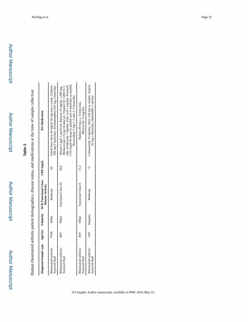
Author M
anuscriptA
uthor Manuscript
Author M
anuscriptA
uthor Manuscript
Norling et al. Page 31
Tab
le 2
Hum
an r
heum
atoi
d ar
thri
tis p
atie
nt d
emog
raph
ics,
dis
ease
sta
tus,
and
med
icat
ions
at t
he ti
me
of s
ampl
e co
llect
ion
Dia
gnos
is/S
ampl
e ty
peA
ge/S
exE
thni
city
AC
R F
unct
iona
l Cla
ss/
Dis
ease
Act
ivit
yC
RP
(m
g/l)
RA
Med
icat
ion
Rhe
umat
oid
arth
ritis
/Sy
novi
al f
luid
70/M
Whi
teM
oder
ate
20E
nbre
l Sur
e St
ick
50 m
g/m
l 50
mg
once
a w
eek,
Cel
ebre
x20
0 m
g 2
times
/day
, Ace
tam
inop
hen
650
mg
3 tim
es/d
ay
Rhe
umat
oid
arth
ritis
/Sy
novi
al f
luid
48/F
Whi
teFu
nctio
nal C
lass
III
29.6
But
ran
5 µg
/h 1
pat
ch/w
k, R
ituxa
n 10
mg/
ml,
1,00
0 m
g,M
etho
trex
ate
2.5
mg
10 ta
bs/w
k, V
itam
in D
3 1,
000
IU 5
tabs
, Pre
dnis
one
7 m
g/da
y, F
olic
aci
d 1
mg/
day,
Pen
nsai
d1.
5% li
quid
40
drop
s to
pai
nful
are
a 4
times
/day
as
need
ed,
Rox
icod
one
5 m
g 1–
2 ta
bs 2
–3 ti
mes
/day
Rhe
umat
oid
arth
ritis
/Sy
novi
al f
luid
40/F
Whi
teFu
nctio
nal C
lass
II
17.2
Hum
ira
40 m
g s.
c. 4
tim
es/d
ay,
Mel
oxic
am 1
5 m
g/da
y
Rhe
umat
oid
arth
ritis
/Sy
novi
al f
luid
74/F
His
pani
cM
oder
ate
<3
Lef
luno
mid
e 10
mg/
day,
Ale
ve 2
20 m
g as
nee
ded,
Asp
irin
81 m
g 2
times
/day
, Ost
eobi
flex
1 ta
b/da
y
JCI Insight. Author manuscript; available in PMC 2016 May 21.
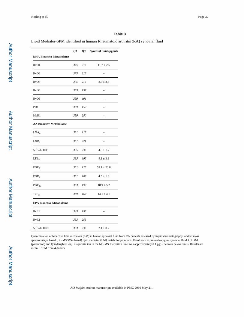
Author M
anuscriptA
uthor Manuscript
Author M
anuscriptA
uthor Manuscript
Norling et al. Page 32
Table 3
Lipid Mediator-SPM identified in human Rheumatoid arthritis (RA) synovial fluid
Q1 Q3 Synovial fluid (pg/ml)
DHA Bioactive Metabolome
RvD1 375 215 11.7 ± 2.6
RvD2 375 215 –
RvD3 375 215 8.7 ± 3.3
RvD5 359 199 –
RvD6 359 101 –
PD1 359 153 –
MaR1 359 250 –
AA Bioactive Metabolome
LXA4 351 115 –
LXB4 351 221 –
5,15-diHETE 335 235 4.3 ± 1.7
LTB4 335 195 9.1 ± 3.9
PGE2 351 175 53.1 ± 23.8
PGD2 351 189 4.5 ± 1.3
PGF2α 353 193 18.9 ± 5.2
TxB2 369 169 14.1 ± 4.1
EPA Bioactive Metabolome
RvE1 349 195 –
RvE2 333 253 –
5,15-diHEPE 333 235 2.1 ± 0.7
Quantification of bioactive lipid mediators (LM) in human synovial fluid from RA patients assessed by liquid chromatography tandem mass spectrometry– based (LC-MS/MS– based) lipid mediator (LM) metabololipidomics. Results are expressed as pg/ml synovial fluid. Q1: M-H (parent ion) and Q3 (daughter ion): diagnostic ion in the MS-MS. Detection limit was approximately 0.1 pg; – denotes below limits. Results are mean ± SEM from 4 donors.
JCI Insight. Author manuscript; available in PMC 2016 May 21.