Embed Size (px)
Citation preview
Jf. Exp.Biol. (1970), S3, 299-316 2 9 9With 4 plates and 9 text-figures
Printed in Great Britain
PROPERTIES OF ACTION POTENTIALS FROMINSECT MOTOR NERVE FIBRES
BY K. G. PEARSON, R. B. STEIN AND S. K. MALHOTRA
Department of Physiology and Biological SciencesElectron Microscopy Laboratory, University of Alberta, Edmonton, Canada
{Received 2 April 1970)
INTRODUCTION
Extracellular recording from small nerve branches provides a method for recordingfrom several nerve fibres simultaneously. This is extremely desirable in studying theorganization of a neuronal system. Furthermore, extracellular records are insensitiveto small movements, are stable over long periods, and can easily be repeated withsubsequent experimental animals. They also involve relatively little dissection, andhence reduce the risk of injury to nerve fibres. We have derived various theoreticalpredictions for records taken from small nerve trunks using different electrodeconfigurations (see Pearson, 1969). The present paper illustrates some of thesetheoretical results and their application to experimental work.
The preparations we have used are a levator motor nerve from the metathoracicsegment of the cockroach and the tergal nerve to the third abdominal segment of thelocust. The cockroach nerve is not ideal for this particular study, but is of interest inour work on the functional organization of motor units (Pearson & Bergman, 1969;Pearson & lies, 1970) and on the patterns of neural activity during behaviouralsequences. Use of a 'typical', rather than a specially selected nerve, illustrates theadvantages and disadvantages of the various recording methods. The locust nerve, onthe other hand, was selected because of its long length. The following properties ofaction potentials from different nerve fibres were measured in these preparations:monophasic and triphasic amplitudes, conduction velocity and duration. Triphasic ormonophasic amplitudes were used to predict fibre diameter, as checked from histo-logical sections. Conduction velocity was less useful since there were systematicdeviations from the expected square root relation between velocity and diameter. Thesystematic effects of fibre diameter on the properties of nerve action potentials shouldbe of general physiological interest.
METHODS
Cockroach preparation. The metathoracic nerve 6B ramus 4 (6Br4 in the notation ofPipa & Cook, 1959) of Periplaneta americana contains motor fibres which innervatethe main and posterior coxal levator muscles (181 and 182 in the notation of Carbonell,1947). This nerve trunk is easily seen after pinning back the coxa to expose its dorsalsurface and removing the tergal remotor muscles (174, 175 and 176) from the dorsalcoxal rim (see Pearson & Bergman, 1969). Further dissection of the part of the coxalrim connected to the posterior coxal levator muscles reveals the branch point of
300 K. G. PEARSON, R. B. STEIN AND S. K. MALHOTRA
6Br4; one branch going to the main levator muscles and the other to the posteriorlevator muscles. Neglecting the fine sensory branch 6Br3 (which was cut from themain nerve), the unbranched length of the nerve 6B from ramus 2 to the branch pointof 6Br4 is about 2 mm.
Locust preparation. The tergal nerve to the third abdominal segment of Locustamigratoria has a long unbranched length (6 mm) and contains about twenty motornerve fibres. The origin of this nerve is the third abdominal ganglion, which, togetherwith the first two abdominal ganglia, is fused to the metathoracic ganglion. The tergalnerve was exposed by removing the ventral cuticle from the thorax and first threeabdominal segments. The smaller fibres within this nerve are spontaneously activeand the larger fibres are readily activated by light touch to the ventral abdominalsegments.
Electrodes and recording equipment. All electrodes consisted of 75 /i silver wires. Forrecording a hook-shaped electrode was manipulated under the nerve and the nervewas carefully lifted until between 1 and 1-5 mm was clear of the haemolymph. Thissection was coated with petroleum jelly (Vaseline) to prevent drying. A second electrodewas then placed in the haemolymph close to this section of nerve and the two electrodeswere connected to a preamplifier. This gave a stable triphasic record. Extreme carewas needed when lifting the nerve clear of the haemolymph to prevent damage to theindividual fibres. A syringe was usually used to suck the haemolymph away from thenerve rather than attempting to draw the nerve out of the haemolymph. Signs ofdamage in a fibre were inflexions in the first positive or negative peaks and/or theabsence of a third phase.
Several methods of monophasic recording were tried. The most satisfactory was tolift the nerve out of saline, coat it as described above, and then cut the nerve justdistal to the point of recording. Immediately after cutting the nerve, the potentialwas diphasic, but within a few minutes a good and fairly stable monophasic potentialwas recorded.
To study the full time course of the action potentials, a second, more proximallysituated electrode pair was used. Records from the proximal electrode pair could bepulse height analysed (Stein, 1968) so that a given fibre could be selected to trigger theoscilloscope sweep. The second pair of electrodes was also needed to measure con-duction velocity. Because of the short length of the cockroach nerve, smaller lengthsof nerve were lifted clear of the haemolymph when two sets of electrodes were used.The preamplifiers, a Tektronix Type 122 and an Isleworth Type A101, were con-nected to a Tektronix 502A oscilloscope, and the traces were photographed.
Source resistance. The source resistance of the action potentials was measured byplacing a variable resistor across the input to the amplifier. The resistance value whichreduced the amplitude to about half gave an approximate measure of the sourceresistance. This was less than 200 k£i for monophasic recording and less than 100 kfifor triphasic recording. These values are sufficiently low compared to the inputresistance of the preamplifiers (10 M.Q.) that no correction was needed. The totalinput capacitance (amplifier plus leads) to the Tektronix preamp (which was used fortime course measurements) was about n o pF, so the time constant for the input tothe recording equipment was about 22 /isec. for monophasic records and less fortriphasic records. Thus, the frequency limitation was close to the high frequency cut
Insect motor nerve fibres 301
on the preamp (40 kc/s), and the shape of the action potentials could be accuratelyrecorded.
Measurement of duration. Action potential duration may be measured in a varietyof ways. For the small fibres with monophasic potentials less than 200 /iV, the slopeof the rising and falling phases could not be accurately determined, so the method ofPaintal (1966) was inappropriate. The positions in time when the action potentialamplitude reached half its maximum value were readily determined, even in thepresence of noise, and we have used the interval between the half-maximum amplitudepoints on the rising and falling phases as a measure of action potential duration.
Measurement of conduction velocity. Pearson (1969) showed that the positions of thefirst crossover point and the negative peak of the triphasic potential relative to thepeak of the membrane potential are fairly insensitive to the length of restriction.Furthermore, the negative peak corresponds to within 0-04 msec, of the membraneaction potential peak. The conduction time for the cockroach nerve fibres was measuredas the time between the first crossover points at two separate pairs of electrodes withtriphasic records, as the crossover points could be determined more accurately thanthe negative peaks. For the locust nerve, conduction times were measured betweenthe negative peak of the triphasic record and the peak of the monophasic potential ata second electrode pair. The distance between electrodes was measured with a cali-brated microscope eyepiece to an accuracy within 5 % and was used for calculatingconduction velocity.
Temperature. All experiments for measurement of conduction velocity or actionpotential duration were carried out at 200 ± 2 °C. In those experiments in which thetemperature was not 20 °C, conduction velocity was corrected using a Q10 of 1-7(Chapman & Pankhurst, 1967). The amplitude of the monophasic action potentialis much less temperature sensitive so no correction was made.
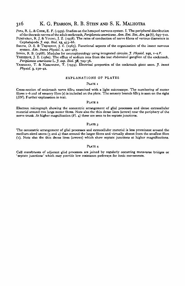
Histology. Only the cockroach nerve was examined histologically. Fixative (3%glutaraldehyde in o-i M phosphate buffer at pH 7'2-7'4) was poured on the nervein situ. After fixation, the nerve was removed, dehydrated in ethanol and embedded inAraldite. One micron sections were cut starting from the peripheral end of the nerveuntil after the point where a sensory branch (nerve 6Br3) separated from the rest ofthe nerve. Sections were then stained with methylene blue and examined under alight microscope. The sensory branch can be seen on the right side of PI. 1. At thelevel of this section, it had nearly separated from the larger motor nerve 6Br4. Thiswas close to the point where electrical recording was normally made after removingthe sensory branch. The various fibres were identified using criteria given in thetext.
Thin sections were cut and routinely stained with an aqueous solution of uranylacetate and then in lead citrate before examination in the electron microscope. Fibrediameters were calculated from measurements of cross-sectional areas in electronmicrographs as described in the text. These were considerably more accurate, par-ticularly for small fibres which were difficult to measure from the light micrographs.Agreement between light micrographs and electron micrographs of various magnifica-tions was generally better than 20%.
302 K. G. PEARSON, R. B. STEIN AND S. K. MALHOTRA
RESULTS
Cockroach nerve
In an intact cockroach there are three or four different-sized fibres in nerve 6Br4of the metathoracic segment which are continuously spontaneously active. In addition,a number of larger fibres tend to fire together in large bursts of activity. Summationand interaction of these fibres makes it difficult to distinguish single fibres accurately.If the abdominal and thoracic connectives are cut out to neurally isolate the segment,generally only six motor fibres are active even with strong mechanical stimulation.The amplitude of these six fibres recorded triphasically from an intact nerve variesover a wide range. Also the discharge pattern of each fibre is different (Pearson &Bergman, 1969) so each fibre can be readily distinguished from animal to animal.This ability to recognize corresponding fibres in a number of experimental animalsis a great advantage. Results can then be averaged from a number of experiments todetermine more accurately the properties of each motor nerve fibre. We have numberedthese fibres from 1 to 6 in order of increasing amplitude and shall refer to them bynumber in the rest of the paper.
0-2 mV 1 mV
1 mV 1 mV
0-5 ms
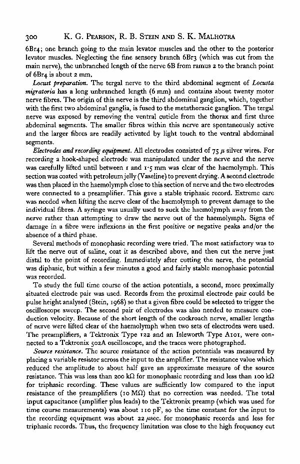
Text-fig. 1. Spontaneously occurring nerve action potentials recorded triphasically from anintact cockroach nerve in the animal's haemolymph (a) and as increasing lengths are lifted outof the haemolymph (b-d). Beyond a certain length the amplitudes of the first two phases donot increase though the waveform continues to spread out in time.
Amplitude. In a freshly dissected animal, the nerve lies in a thin layer of haemo-lymph. The potentials that can be recorded are small (Text-fig. 1 a) but as one liftsthe nerve free of the haemolymph, they rapidly increase in size and spread out in time(Text-fig. 1 b and c). Beyond a certain length out of solution the amplitude does notincrease further (Text-fig. 1 d) although the time course continues to lengthen andthe third phase continues to increase. The saturated values of the first two peaks arein the ratio 1:2 as predicted (Pearson, 1969) and indeed the shapes of the actionpotentials show good qualitative agreement with computed values.
If a long length of nerve is pulled out so that the maximum amplitudes are approached,
Insect motor nerve fibres 303
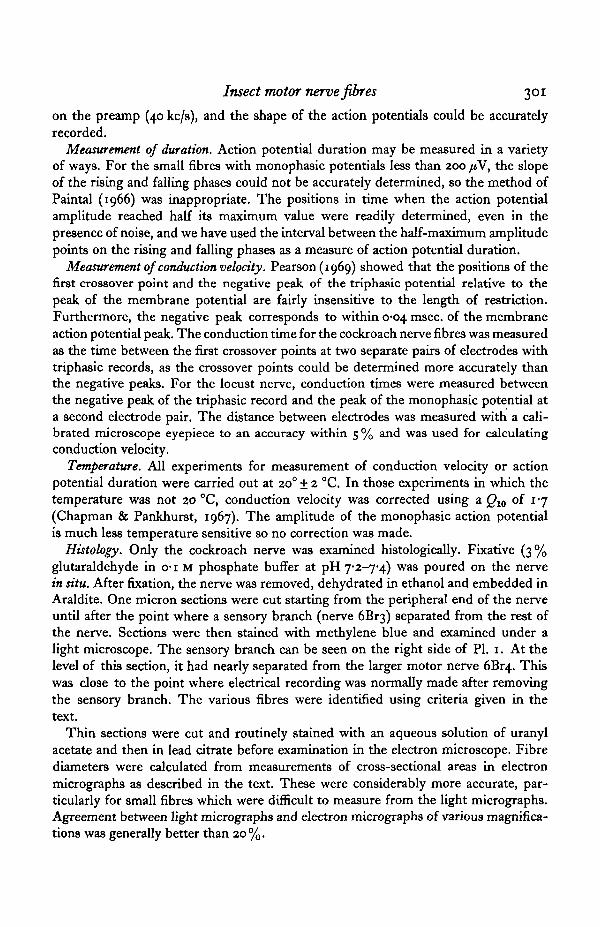
and one is careful not to injure the nerve, the amplitude of each fibre is quite repro-ducible (the standard deviation from animal to animal is about 15% of the mean).After coating the nerve with petroleum jelly (Vaseline) to prevent drying, stablerecords can be obtained for hours without the need for any saline. Cutting the nervedistal to the electrode changes the record from triphasic (Text-fig. 2 a) to diphasic(Text-fig. 26). The positive peak of the diphasic record is equal in size to the negativepeak of the triphasic record and thus twice the first positive peak of the triphasic record.If the nerve is cut close to the electrode, the amplitude of the positive peak remainsconstant while the negative peak decreases in size and lengthens out. Soon, a goodmonophasic potential remains (Text-fig, zc) which is an extracellular replica of themembrane action potential. Monophasic amplitudes w$re slightly more reproduciblethan triphasic amplitudes (the standard deviation frotn animal to animal was onlyabout 10-15% °f the mean amplitude), though they .are not as stable as triphasicrecords. After 10 or 15 min the monophasic action potentials lengthen and thengradually decline in size. This process takes longer the greater the length of nerve,and is presumably due to the nerve 'running down' as it loses potassium and othersubstances from the cut nerve end.
1 miText-fig. 2. Comparison of waveforms obtained (a) from an intact cockroach nerve (triphasicrecord), (6) immediately after cutting the nerve distal to the recording electrode (diphasicrecord), and (c) a few minutes later (monophasic record).
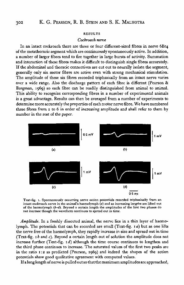
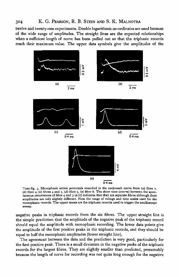
Text-fig. 3 shows monophasic records of the six efferent fibres which discharge withsegment neurally isolated. A second pair of electrodes was placed proximally to triggerthe oscilloscope so that the whole time course of the action potential could be seen.
For recording fibres 3-6 in Text-fig. 3, only a short length of nerve was pulled outat the proximal electrode to minimize interaction between the two pairs of electrodesover this short length of nerve. This means that the triphasic potentials were smalland one cannot easily trigger off fibres 1 and 2. In another experiment, a longer lengthwas pulled out for triphasic recording (and a shorter length for monophasic recording)to give records for fibres 1 and 2. The temperature in the first experiment was 3 °Chigher than in the second.
The amplitudes of fibres 3 and 4 were sometimes indistinguishable. When theywere different in size, fibres 3 and 4 could be identified by their spontaneous dischargepatterns and response to mechanical stimulation. Also, fibre 3 is a branch of thecommon inhibitor (Pearson & Bergman, 1969) and can be readily distinguished if onerecords simultaneously from another of its branches.
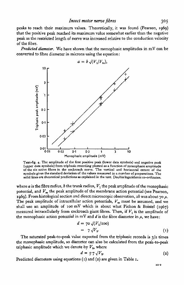
Text-fig. 4 shows the relation between monophasic amplitude and the amplitudeof both the first positive peak and the negative peak of the triphasic waveform. Theextent of each symbol gives the standard deviation of the measurements from between
20 E X B 53
304 K. G. PEARSON, R. B. STEIN AND S. K. MALHOTRA
twelve and twenty-one experiments. Double logarithmic co-ordinates are used becauseof the wide range of amplitudes. The straight lines are the expected relationshipswhen a sufficient length of nerve has been pulled out so that the triphasic recordsreach their maximum value. The upper data symbols give the amplitudes of the
2 ml 2 ms
(M ms <M ms
0-4 mi
Text-fig. 3. Monophasic action potentials recorded in the cockroach nerve from (a) fibre 1,(6) fibre 3, (c) fibres 4 and 3, (d) fibre 5, («) fibre 6. The short time interval between the spon-taneous occurrences of fibre 4 and 3 in (c) indicates that they are separate fibres although theiramplitudes are only slightly different. Note the range of voltage and time scales used for themonophasic records. The upper traces are the triphasic records used to trigger the oscilloscopesweep.
negative peaks in triphasic records from the six fibres. The upper straight line isthe simple prediction that the amplitude of the negative peak of the triphasic recordshould equal the amplitude with monophasic recording. The lower data points givethe amplitude of the first positive peaks in the triphasic records, and they should beequal to half the monophasic amplitudes (lower straight line).
The agreement between the data and the prediction is very good, particularly forthe first positive peak. There is a small deviation in the negative peaks of the triphasicrecords for the largest fibres. They are slightly smaller than predicted, presumablybecause the length of nerve for recording was not quite long enough for the negative
Insect motor nerve fibres 305
peaks to reach their maximum values. Theoretically, it was found (Pearson, 1969)that the positive peak reached its maximum value somewhat earlier than the negativepeak as the restricted length of nerve was increased relative to the conduction velocityof the fibre.
Predicted diameter. We have shown that the monophasic amplitudes in mV can beconverted to fibre diameter in microns using the equation:
10 r
01•o
E0-3
0-1
003
001001 003 0-1 0-3 1
Monophasic amplitude (mV)10
Text-fig. 4. The amplitude of the first positive peak (lower data symbols) and negative peak(upper data symbols) from triphasic recording plotted as a function of monophasic amplitudeof the six active fibres in the cockroach nerve. The vertical and horizontal extent of thesymbols gives the standard deviation of the values measured in a number of preparations. Thesolid lines are theoretical predictions as explained in the text. Double logarithmic co-ordinates.
where a is the fibre radius, b the trunk radius, Vx the peak amplitude of the monophasicpotential, and Vm the peak amplitude of the membrane action potential (see Pearson,1969). From histological section and direct microscopic observation, ib was about 70 ji.The peak amplitude of intracellular action potentials, Vm must be assumed, and weshall use an amplitude of 100 mV which is about what Pichon & Boistel (1967)measured intracellularly from cockroach giant fibres. Then, if V± is the amplitude ofthe monophasic action potential in mV and d is the fibre diameter in fi, we have:
= 7^i- (1)
The saturated peak-to-peak value expected from the triphasic records is 3/2 timesthe monophasic amplitude, so diameter can also be calculated from the peak-to-peaktriphasic amplitude which we denote by Vz, where
<*=57Vn- (2)Predicted diameters using equations (1) and (2) are given in Table 1.
306 K. G. PEARSON, R. B. STEIN AND S. K. MALHOTRA
Histological measurement of diameter. Both light and electron micrographs from fivefresh cockroach preparations were examined in detail, sometimes at more than onepoint along the nerve. Although one cannot equate with absolute certainty, the fibresobserved electrically with those seen in fixed sections, a number of criteria wereavailable to reliably identify the fibres in cross sections such as PI. i.
(1) Experiments using both reflex and electrical stimulation of intact animalsindicate the presence of a number of fibres larger than the largest one (fibre 6) whichshows activity with the segment neurally isolated. At least four such fibres have beenrecorded in single experiments so that most of the largest fibres do not correspond toany observed electrically.
(2) In no experiment was any motor fibre found which had an amplitude inter-mediate in size between the six fibres which show activity in the neurally isolatedsegment. One and only one sensory fibre which was intermediate in size betweenmotor fibres 2 and 3 was regularly seen. If we call this fibre S, there should be a sequenceof fibres of steadily increasing size in the order 1, 2, S, 3, 4, 5, 6.
(3) The amplitudes of electrical records from fibres 3 and 4 are very similar in size,and so their diameters are presumably of similar size. The only two medium-sizedfibres with comparable diameters are those at about 5 /i in PI. 1. Having picked outthese two fibres, it is straightforward to identify successively larger or smaller fibresand number them as indicated on PI. 1.
Table 1
eon
1
2
3456
Monophasicamplitude (mV)
00430-085o-880-962-9
7-4
Predicteddiameter (/t)
i - 52 - 0
6-66 9
1 1 9
i9'0
Tnphasic p/pamplitude (mV)
00730 1 31-2
1 34 19-7
Predicteddiameter (ji)
' • 52 - i
6-26-5
" • 517-7
Measureddiameter (ji)
1 61 9
5'i5 7
10-416-1
This labelling was confirmed from low power electron micrographs and higherpower electron micrographs were taken of the fibres of interest. The cross-sectionalareas were measured with a planimeter and average diameter was then calculated bydividing the cross-sectional area by \n and taking the square root. The averagemeasured diameters from five cockroach nerves are shown in Table 1, and the predicteddiameters from spike amplitude measurements agree to within about 20% of thesemeasured diameters. This is reasonably good agreement considering the inaccuraciesinherent in both methods. However, the measured diameters are mainly smaller thanpredicted. Shrinkage during fixation could account for part of this difference sincethe average nerve diameter measured from the total cross-sectional area of the fixednerves was 64 fi, whereas the diameter measured from direct observation of the livingnerve was 70 fi. There could also have been systematic differences between the twogroups of animals used or systematic errors resulting from the choice of constants inequations (1) and (2). Nonetheless, our results indicate that a good estimate of fibrediameter can be obtained simply by measuring the heights of extracellularly recordedaction potentials from fibres in small nerves, and directly observing the diameter ofthe nerve in situ.
Insect motor nerve fibres 307
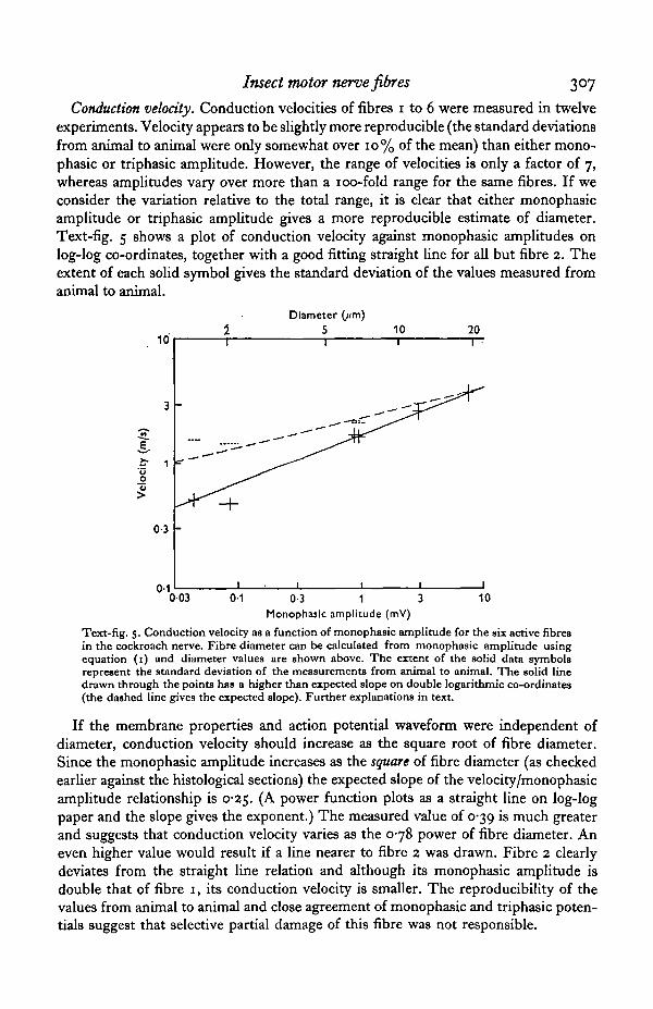
Conduction velocity. Conduction velocities of fibres 1 to 6 were measured in twelveexperiments. Velocity appears to be slightly more reproducible (the standard deviationsfrom animal to animal were only somewhat over 10% of the mean) than either mono-phasic or triphasic amplitude. However, the range of velocities is only a factor of 7,whereas amplitudes vary over more than a 100-fold range for the same fibres. If weconsider the variation relative to the total range, it is clear that either monophasicamplitude or triphasic amplitude gives a more reproducible estimate of diameter.Text-fig. 5 shows a plot of conduction velocity against monophasic amplitudes onlog-log co-ordinates, together with a good fitting straight line for all but fibre 2. Theextent of each solid symbol gives the standard deviation of the values measured fromanimal to animal.
10
3
1
0-3
n.1
11
-
Diameter51
1
(jtm)
—ftitr'
10 20
1 1
003 01 0-3 1 10Monophasic amplitude (mV)
Text-fig. 5. Conduction velocity as a function of monophasic amplitude for the six active fibresin the cockroach nerve. Fibre diameter can be calculated from monophasic amplitude usingequation (i) and diameter values are shown above. The extent of the solid data symbolsrepresent the standard deviation of the measurements from animal to animal. The solid linedrawn through the points has a higher than expected slope on double logarithmic co-ordinates(the dashed line gives the expected slope). Further explanations in text.
If the membrane properties and action potential waveform were independent ofdiameter, conduction velocity should increase as the square root of fibre diameter.Since the monophasic amplitude increases as the square of fibre diameter (as checkedearlier against the histological sections) the expected slope of the velocity/monophasicamplitude relationship is 0*25. (A power function plots as a straight line on log-logpaper and the slope gives the exponent.) The measured value of 0-39 is much greaterand suggests that conduction velocity varies as the 0-78 power of fibre diameter. Aneven higher value would result if a line nearer to fibre 2 was drawn. Fibre 2 clearlydeviates from the straight line relation and although its monophasic amplitude isdouble that of fibre 1, its conduction velocity is smaller. The reproducibihty of thevalues from animal to animal and close agreement of monophasic and triphasic poten-tials suggest that selective partial damage of this fibre was not responsible.
308 K. G. PEARSON, R. B. STEIN AND S. K. MALHOTRA
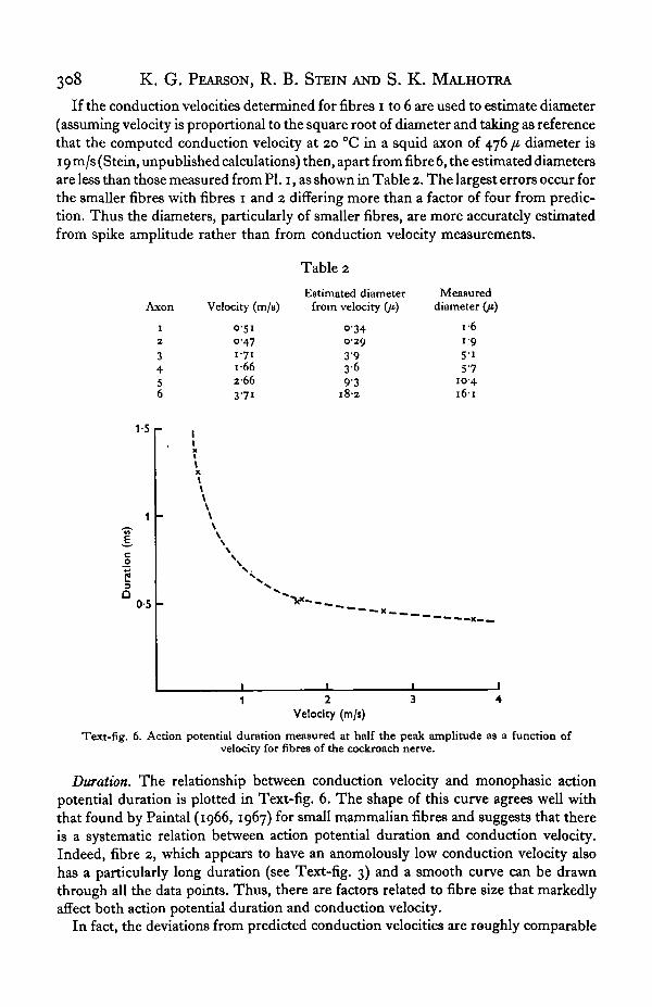
If the conduction velocities determined for fibres i to 6 are used to estimate diameter(assuming velocity is proportional to the square root of diameter and taking as referencethat the computed conduction velocity at 20 °C in a squid axon of 476 fi diameter is19 m/s (Stein, unpublished calculations) then, apart from fibre 6, the estimated diametersare less than those measured from PI. 1, as shown in Table 2. The largest errors occur forthe smaller fibres with fibres 1 and 2 differing more than a factor of four from predic-tion. Thus the diameters, particularly of smaller fibres, are more accurately estimatedfrom spike amplitude rather than from conduction velocity measurements.
Table 2
son
1
2
3456
Velocity (m/s)
0 5 10-471 7 11 6 62663 7 1
Estimated diameterfrom velocity (ji)
0'340-293 93 69 3
1 8 2
Measureddiameter (/*)
1 61-9
5 15"7
10-41 6 1
1-5 r
cjo
C
a0-5
Velocity (m/s)
Text-fig. 6. Action potential duration measured at half the peak amplitude as a function ofvelocity for fibres of the cockroach nerve.
Duration. The relationship between conduction velocity and monophasic actionpotential duration is plotted in Text-fig. 6. The shape of this curve agrees well withthat found by Paintal (1966, 1967) for small mammalian fibres and suggests that thereis a systematic relation between action potential duration and conduction velocity.Indeed, fibre 2, which appears to have an anomolously low conduction velocity alsohas a particularly long duration (see Text-fig. 3) and a smooth curve can be drawnthrough all the data points. Thus, there are factors related to fibre size that markedlyaffect both action potential duration and conduction velocity.
In fact, the deviations from predicted conduction velocities are roughly comparable
Insect motor nerve fibres 309
to the increase in durations. This can be shown by multiplying the conduction velocityof fibres 1 to 5 by an amount equal to the ratio of the duration of each fibre's actionpotential relative to that of fibre 6. These new values are represented as horizontalinterrupted lines in Text-fig. 5 with the same lateral extent as previously. They lieclose to the expected relation between conduction velocity and amplitude (an inter-rupted line with slope 0-25 has been drawn through the value for fibre 6).
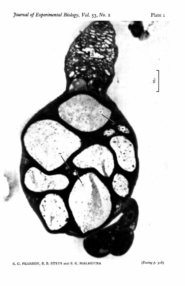
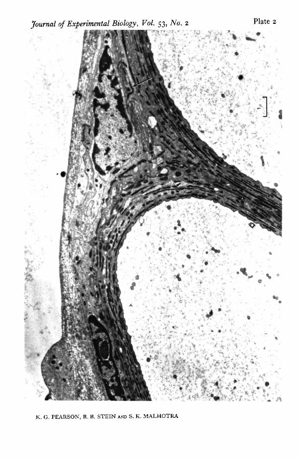
Structural features. Electron micrographs were examined for systematic differencesbetween large and small fibres which might account for their different electricalproperties. One striking difference is seen in PI. 2 and 3. Plate 2 shows two very largefibres while PI. 3 shows fibres 1, 3, 4 and 6. The largest fibres have a prominent con-centric arrangement containing half a dozen layers of glial processes. Between theselayers is found a dense material thought to be an acid muco-polysaccharide (Smith &Treherne, 1963) which is stained very well by uranyl acetate and gives the nerves acharacteristic appearance. This arrangement of glial processes and dense extracellularmaterial is less prominent around the medium-sized fibres (2 to 3 layers) and virtuallyabsent from the smallest fibres.
The intricate arrangement of glial cells also raises a question concerning the inter-pretation of recordings taken with electrodes external to the nerve, for these cells mayalter the extracellular current pathways. The electron micrograph indicates that theperipheral organization is very similar to that described by Maddrell & Treherne (1967)in the neural connectives and ganglia of Periplaneta. Underlying the connective tissuesheath is a region of perineurium with numerous dense lines toward its inner border.At higher magnification (PI. 4) these are seen to be composed of regularly spacedtransverse bridges across the space between adjacent glial cell membranes. Similartransverse bridges, known as septate junctions have been described in the salivarygland of Ckironomus and Drosophila (Bullivant & Lowenstein, 1968; Malhotra, 1969)where they have been shown to provide low resistance connexions between cells. If asimilar function were present here, the effect of the glia on the electrical records wasprobably small. Also the outer sheath probably had little effect for Treherne (1961)found that even with the much thicker sheath surrounding central ganglia in thecockroach, removal of the sheath does not greatly affect the passive efflux of sodiumions from the central nervous system.
In conclusion, the properties of nerve action potentials vary systematically withfibre size, and there are important systematic differences in structure, although, aswill be discussed later, the two cannot yet be correlated with certainty. These resultsare based on rather few fibres, so a second insect nerve was investigated.
Locust nerve
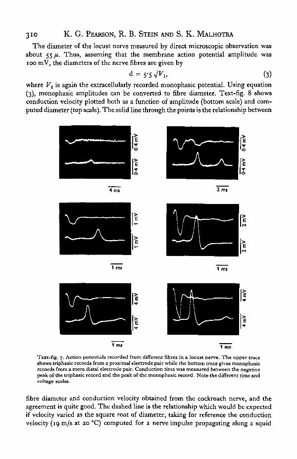
The tergal nerve to the third abdominal segment of the locust contains a numberof spontaneously active fibres. Further fibres can be activated by graded mechanicalstimuli to the abdomen or the cercus. With this number corresponding fibres are noteasily identified from animal to animal. However, this preparation has the advantageover the cockroach preparation that the nerve trunk is long enough to allow mono-phasic amplitude, duration and conduction velocity to be measured simultaneouslyfor any particular fibre. Text-fig. 7 shows records from a number of different fibresin one experiment.
310 K. G. PEARSON, R. B. STEIN AND S. K. MALHOTRA
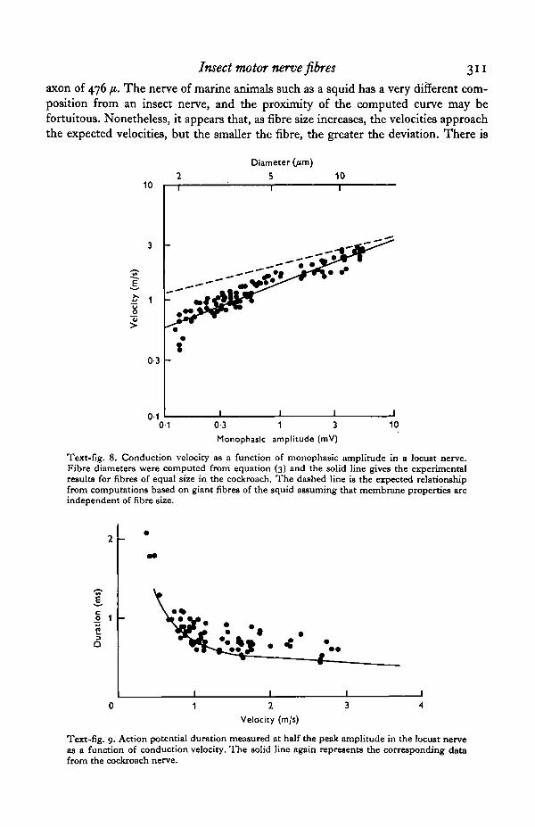
The diameter of the locust nerve measured by direct microscopic observation wasabout 55 /i. Thus, assuming that the membrane action potential amplitude wasioo mV, the diameters of the nerve fibres are given by
d = S-SV^i, (3)where Vx is again the extracellularly recorded monophasic potential. Using equation(3), monophasic amplitudes can be converted to fibre diameter. Text-fig. 8 showsconduction velocity plotted both as a function of amplitude (bottom scale) and com-puted diameter (top scale). The solid line through the points is the relationship between
4 mi 2 mi
1 ms 1 ms
1 ms 1 ms-
Text-fig. 7. Action potentials recorded from different fibres in a locust nerve. The upper traceshows triphasic records from a proximal electrode pair while the bottom trace gives monophasicrecords from a more distal electrode pair. Conduction time was measured between the negativepeak of the triphasic record and the peak of the monophasic record. Note the different time andvoltage scales.
fibre diameter and conduction velocity obtained from the cockroach nerve, and theagreement is quite good. The dashed line is the relationship which would be expectedif velocity varied as the square root of diameter, taking for reference the conductionvelocity (19 m/s at 20 °C) computed for a nerve impulse propagating along a squid
Insect motor nerve fibres 311
axon of 476 fi. The nerve of marine animals such as a squid has a very different com-position from an insect nerve, and the proximity of the computed curve may befortuitous. Nonetheless, it appears that, as fibre size increases, the velocities approachthe expected velocities, but the smaller the fibre, the greater the deviation. There is
10
Diameter (/im)
S 10
8
0-3
0-10-1 0-3 1 3
Monophajlc amplitude (mV)10
Text-fig. 8. Conduction velocity as a function of monophasic amplitude in a locust nerve.Fibre diameters were computed from equation (3) and the solid line gives the experimentalresults for fibres of equal size in the cockroach. The dashed line is the expected relationshipfrom computations based on giant fibres of the squid assuming that membrane properties areindependent of fibre size.
co
C
Q
Velocity (m/s)
Text-fig. 9. Action potential duration measured at half the peak amplitude in the locust nerveas a function of conduction velocity. The solid line again represents the corresponding datafrom the cockroach nerve.
312 K. G. PEARSON, R. B. STEIN AND S. K. MALHOTRA
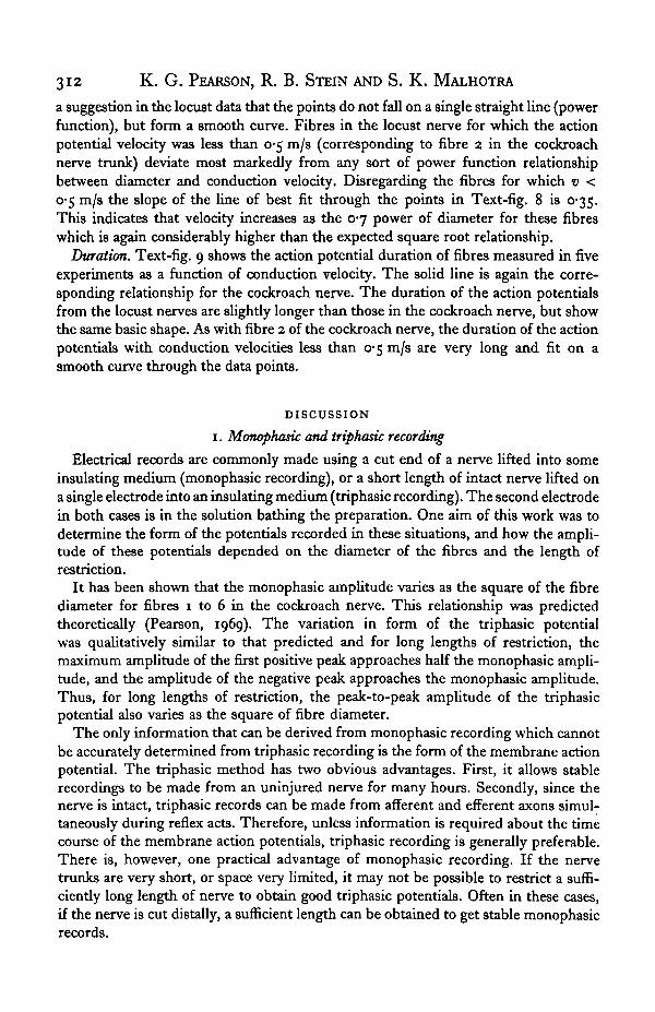
a suggestion in the locust data that the points do not fall on a single straight line (powerfunction), but form a smooth curve. Fibres in the locust nerve for which the actionpotential velocity was less than 0-5 m/s (corresponding to fibre 2 in the cockroachnerve trunk) deviate most markedly from any sort of power function relationshipbetween diameter and conduction velocity. Disregarding the fibres for which v <0-5 m/s the slope of the line of best fit through the points in Text-fig. 8 is 0-35.This indicates that velocity increases as the 0-7 power of diameter for these fibreswhich is again considerably higher than the expected square root relationship.
Duration. Text-fig. 9 shows the action potential duration of fibres measured in fiveexperiments as a function of conduction velocity. The solid line is again the corre-sponding relationship for the cockroach nerve. The duration of the action potentialsfrom the locust nerves are slightly longer than those in the cockroach nerve, but showthe same basic shape. As with fibre 2 of the cockroach nerve, the duration of the actionpotentials with conduction velocities less than 0-5 m/s are very long and fit on asmooth curve through the data points.
DISCUSSION
1. Monophasic and triphasic recording
Electrical records are commonly made using a cut end of a nerve lifted into someinsulating medium (monophasic recording), or a short length of intact nerve lifted ona single electrode into an insulating medium (triphasic recording). The second electrodein both cases is in the solution bathing the preparation. One aim of this work was todetermine the form of the potentials recorded in these situations, and how the ampli-tude of these potentials depended on the diameter of the fibres and the length ofrestriction.
It has been shown that the monophasic amplitude varies as the square of the fibrediameter for fibres 1 to 6 in the cockroach nerve. This relationship was predictedtheoretically (Pearson, 1969). The variation in form of the triphasic potentialwas qualitatively similar to that predicted and for long lengths of restriction, themaximum amplitude of the first positive peak approaches half the monophasic ampli-tude, and the amplitude of the negative peak approaches the monophasic amplitude.Thus, for long lengths of restriction, the peak-to-peak amplitude of the triphasicpotential also varies as the square of fibre diameter.
The only information that can be derived from monophasic recording which cannotbe accurately determined from triphasic recording is the form of the membrane actionpotential. The triphasic method has two obvious advantages. First, it allows stablerecordings to be made from an uninjured nerve for many hours. Secondly, since thenerve is intact, triphasic records can be made from afferent and efferent axons simul-taneously during reflex acts. Therefore, unless information is required about the timecourse of the membrane action potentials, triphasic recording is generally preferable.There is, however, one practical advantage of monophasic recording. If the nervetrunks are very short, or space very limited, it may not be possible to restrict a suffi-ciently long length of nerve to obtain good triphasic potentials. Often in these cases,if the nerve is cut distally, a sufficient length can be obtained to get stable monophasicrecords.
Insect motor nerve fibres 313
2. Axon diameters
The determination of fibre diameter from the action potential conduction velocityin unmyelinated nerve fibres contained in small nerve trunks is unsuitable for anumber of theoretical and practical reasons. A procedure for estimating the diameter ofunmyelinated fibres from measurements of the amplitude of their extracellularpotentials is therefore desirable. A method for doing this from monophasic ampli-tude measurements was suggested by Pearson (1969) (equation (1) of this paper),and has been used in this experimental investigation with reasonable success (forlong lengths of restriction triphasic amplitude is proportional to monophasic ampli-tude ; therefore a simular formula can be used to estimate axon diameterfrom triphasicrecords, e.g. equation (2)).
The estimated diameters from amplitude measurements for fibres 1 to 6 in thecockroach nerve were in reasonably good agreement with histological measure,although histologically measured diameters tended to be somewhat smaller thanpredicted. For the smaller fibres, these estimates were considerably more accuratethan those made from conduction velocity, the latter underestimating the fibrediameters by more than a factor of 4. We concluded from the observations made onthe cockroach levator nerve that, for short nerves containing small unmyelinatedfibres, the simplest and most accurate method for estimating fibre diameter is fromthe amplitude of the extracellular potentials, rather than from conduction velocity.
One further point of interest is the diameters of fibres 1 and 2 in the cockroachnerve. It has sometimes been assumed (Guthrie, 1962) that peripheral cockroach nervefibres larger than 5 fi are motor and those smaller than 5 fi are sensory. The aboveobservation on the diameters of fibres 1 and 2 indicates that some motor fibres aremuch smaller than this value, and Chapman & Pankhurst (1967) have shown thatsome sensory fibres have diameters double this value.
3. Conduction velocity and duration
Another aim of this work was to use extracellular recording techniques to studysome of the properties of action potentials in small unmyelinated fibres. The twoproperties of particular interest were action potential conduction velocity and actionpotential duration.
At present there is little direct experimental information on the conduction velocity-diameter relationship for small unmyelinated nerve fibres. Pumphrey & Young (1938)showed for large unmyelinated axons of the squid (> 30 /i) that conduction speed variesas the o-6i power of diameter which is significantly larger than the expected value of 0-5.More recently, Burrows et al. (1965) studied this relationship in octopus, squid andsepia, extending the range of diameters and found the best fitting power function overthe range 2-480 /i had an exponent of 0-57, although there was no precise determinationof fibre diameter in the 2-22 fi range which was approximately the range of interesthere. The results presented above for the cockroach fibres (neglecting axon 2) showedthat on a log-log plot of velocity against diameter the slope was 0-78, and was evenlarger if fibre 2 was included. The slope of the best fitting straight line for the locusttergal nerve is 0-70. However, the results for the locust suggest that there may be nosingle power function relating velocity and diameter, but that the slope of a log-log
314 K. G. PEARSON, R. B. STEIN AND S. K. MALHOTRA
plot increases continuously for the smaller axons. Thus, there appears to be con-siderable evidence that conduction velocity varies more rapidly than the square rootof diameter in unmyelinated fibres, and that the deviations are more extreme thesmaller the fibre.
In both the cockroach nerve and the locust tergal nerve, it was found that withdecreasing fibre diameter, the duration of the action potential increased by an amountapproximately proportional to the deviation of the conduction velocity from theexpected square root relationship with diameter.
The explanation of these results is uncertain. Stein (unpublished calculations) showedthat a decrease in the specific conductance of a unit membrane area to all ions increasedaction potential duration and conduction time by roughly equal amounts, in agreementwith the experimental results. Changes in membrane capacity affect conductionvelocity much more than duration, while changing the rate constants in the Hodgkin-Huxley equations affects conduction velocity much less than duration. Therefore,changes in membrane capacitance or rate constants alone could not account for the exp-erimental results. Maddrell & Treherne (1967) suggest that the function of the concen-tric arrangement of glial processes and extracellular spaces containing an acid muco-polysaccharide is to regulate the ionic environment around the nerve cell. This wouldpermit the larger cells which contain these structures to function despite large changesin ionic concentrations elsewhere in the extracellular space, but it would also permitthese cells to function under normal conditions with the higher conductances per unitmembrane area which our results suggest they possess.
However, this is not the only possible explanation. The concentric arrangement ofglial processes suggests that the effective resistance of the larger nerve cells might beincreased and the effective membrane capacitance decreased. This combined effecton resistance and capacitance might also account for the roughly proportional increasein the duration and deviation in the conduction velocity of nerve impulses. Thisexplanation is difficult to reconcile with the results of Yamasaki & Narahashi (1959)who calculated an extremely high value of membrane capacity for giant fibres in thecockroach. However, their results depend critically on assumptions about axoplasmicresistivity and fibre diameter, neither of which was measured directly.
Paintal (1967) found similar changes in the duration of action potentials in mam-malian axons of slowest conduction velocity, and he has evidence for structural changeswith size (personal communication, 1968) which suggests that the effects of axon sizeon the properties of action potentials may be quite general. There are now also anumber of studies indicating that other neuronal properties change systematicallywith size (Bullock, 1953; Henneman, Somjen & Carpenter, 1965; Kernell, 1965;Kennedy, 1967). This evidence must eventually be fitted into a general theory of therelation between neuronal size and function.
SUMMARY
1. The properties of nerve action potentials in small insect motor nerves werestudied using extracellular recording electrodes.
2. A length of nerve was lifted out of solution and recordings were made with
Insect motor nerve fibres 315
respect to the solution either from an intact nerve (triphasic recording) or from neara cut end of the nerve (monophasic recording).
3. In a cockroach nerve, the number of spontaneously active fibres was smallenough that corresponding nerve fibres could be identified in each preparation bytheir action potential amplitude and their pattern of activity. Under controlled condi-tions, the absolute amplitudes of either monophasic or triphasic records were repro-ducible and could be used to calculate fibre diameter. The calculations were confirmedfrom histological sections of the nerve.
4. Conduction velocity varied approximately as the 0-78 power of fibre diameterin a cockroach nerve and as 0-7 power of fibre diameter in a locust nerve. These valuesare considerably larger than the square root relation predicted if membrane propertiesare independent of fibre diameter.
5. Membrane properties probably vary with fibre diameter since the action potentialduration increases dramatically for fibres below 5 /i in diameter.
6. For the cockroach nerve systematic structural differences between fibres ofdifferent sizes are also seen with the electron microscope and the relation of these tothe functional differences is considered.
This work was supported in part by grants from the Medical Research Counciland the National Research Council of Canada.
REFERENCES
BULLIVANT, S. & LOEWBNSTEIN, W. R. (1968). Structure of coupled and uncoupled cell junctions.J. cell Biol. 37,621-32.
BULLOCK, T. H. (1953). Comparative aspects of some biological transducers. Fed. Proc. ia, 666-72.BURROWS, T. M. O., CAMPBELL, I. A., HOWE, E. J. & YOUNG, J. Z. (1965). Conduction velocity and
diameter of nerve fibres of cephalopods. J. Phytiol. 179, 39-40P.CARBONELL, C. S. (1947). The thoracic muscles of the cockroach, Periplaneta americana (L.). Smith-
sonian Misc. Coll. 107 (2), 1-23.CHAPMAN, K. M. & PANKHURST, J. H. (1967). Conduction velocities and their temperature coefficients
in sensory nerve fibres of cockroach legs. J. exp. Biol. 46, 63-84.GUTHRIE, D. M. (1962). Regenerative growth in insect nerve axons. J. Insect Pkysiol. 8, 79-92.HENNBMAN, E., SOMJEN, G. & CARPENTER, D. O. (1965). Functional significance of cell size in spinal
motoneurons. J. Neurophysiol. a8, 560-80.KENNEDY, D. (1967). The reflex control of muscle. In Invertebrate Nervous Systems. Ed. C. A. G.
Wiersma.KBRNELL, D. (1965). The limits of firing frequency of cat lumbosacral motoneurons possessing different
time course of afterhyperpolarization. Acta Physiol. Scand. 65, 87-100.MADDRELL, S. H. P. & TREHERNE, J. E. (1967). The ultrastructure of the perineurium in two insect
species, Carausius morosus and Periplaneta americana. J. cell Sci. a, 119—28.MALHOTRA, S. K. (1969). Organization of the cellular membranes. Prog. Biophys. Molec. Biol. ai , 67-
131-PAINTAL, A. S. (1966). The influence of diameter of medullated nerve fibres of cats on the rising and
falling phases of the spike and its recovery. J. Physiol. 184, 791-811.PAINTAL, A. S. (1967). A comparison of the nerve impulses of mammalian nonmedullated nerve fibres
with those of the smallest diameter medullated fibres. J. Physiol. 193, 523-33.PEARSON, K. G. (1969). Electrophysiological studies on insect motoneurones. D.Phil. Thesis. Oxford.PEARSON, K. G. & BERGMAN, S. J. (1969). Common inhibitory motoneurones in insects. J. exp. Biol.
S°, 445-73-PEARSON, K. G. & ILES, J. F. (1970). Discharge patterns in coxal levator and depressor motoneurones
of the cockroach, Periplaneta americana. J. exp. Biol. 5a, 139-65.PICHON, Y. & BOISTEL, J. (1967). Microelectrode study of the resting and action potentials of the cock-
roach giant axon with special reference to the role played by the nerve sheath. J. exp. Biol. 47, 357-72.
316 K. G. PEARSON, R. B. STEIN AND S. K. MALHOTRA
PIPA, R. L. & COOK, E. F. (1959). Studies on the hexapod nervous system. I. The peripheral distributionof the thoracic nerves of the adult cockroach, Periplaneta americana. Ann. Ent. Soc. Am. 5a (6), 693-710.
PUMPHREY, R. J. & YOUNG, J. Z. (1938). The rates of conduction of nerve fibres of various diameters inCephalapods. J. exp. Biol. 15, 453-66.
SMITH, D. S. & TREHERNE, J. E. (1963). Functional aspects of the organization of the insect nervoussystem. Adv. Insect Physiol. 1, 401-465.
STEIN, R. B. (1968). Modules for neurophysiology using integrated circuits. J . Physiol. 197, 1-2P.TREHERNE, J. E. (i960). The efflux of sodium ions from the last abdominal ganglion of the cockroach,
Periplaneta americana h.jf. exp. Biol. 38, 729-36.YAMASAKI, T. & NARAHASHI, T. (1959). Electrical properties of the cockroach giant axon. J. insect
Pkysiol. 3, 230-42.
EXPLANATIONS OF PLATES
PLATE I
Cross-section of cockroach nerve 6Br4 examined with a light microscope. The numbering of motorfibres 1-6 and of sensory fibre (1) is included on the plate. The sensory branch 6Br3 is seen on the right(SN). Further explanation in text.
PLATE 2
Electron micrograph showing the concentric arrangement of glial processes and dense extracellularmaterial around two large motor fibres. Note also the thin dense lines (arrow) near the periphery of thenerve trunk. At higher magnification (PI. 4) these are seen to be septate junctions.
PLATE 3
The concentric arrangement of glial processes and extracellular material is less prominent around themedium-sized axons (3 and 4) than around the larger fibres and virtually absent from the smallest fibre(1). Note also the thin dense lines (arrows) which show septate junctions at higher magnifications.
PLATE 4
Cell membranes of adjacent glial processes are joined by regularly occurring transverse bridges or' septate junctions' which may provide low resistance pathways for ionic movements.
Journal of Experimental Biology, Vol. 53, No. 2 Plate 1
K. G. PEARSON, R. D. STEIN AND S. K. MALHOTRA (Facing p. 316)
Journal of Experimental Biology, Vol. 53, No. 2 Plate 2
c '
K. G. PEARSON, R. B. STEIN AND S. K. MALHOTRA

Journal of Experimental Biology, Vol. 53, No. 2 Plate 3
• : » *
K. C. PEARSON, R. B. STEIN AND S. K. MALHOTRA
Journal of Experimental Biology, Vol. 53, No. 2 Plate 4
K. G. PEARSON, R. B. STEIN AND S. K. MALHOTRA


![STUDIES ON THE MORPHOLOGY OF GANGLION CELLS IN THE … · studies on the morphology of ganglion cells in the rabbit. i. the normal nerve cells. ii. c~a~ges ix ti~e nerve c]blls ii~](https://img.pdfslide.us/doc/110x75/5e8656a8ba60b45598552db9/studies-on-the-morphology-of-ganglion-cells-in-the-studies-on-the-morphology-of.jpg)


![[832] TRANSMISSION THROUG THH E LAST ABDOMINAL …jeb.biologists.org/content/jexbio/37/4/832.full.pdf · TRANSMISSION THROUG THH E LAST ABDOMINAL GANGLION OF TH DRAGONFLE Y NYMPH,](https://img.pdfslide.us/doc/110x75/5be6d03209d3f2ea1a8db4b0/832-transmission-throug-thh-e-last-abdominal-jeb-transmission-throug-thh.jpg)