Embed Size (px)
Citation preview
FP6-016039
C IL IA Customized Intelligent Life-Inspired Arrays
Integrated Project
Information Society Technolgies
Future & Emerging Technologies Proactive Initiative BIO-I3
DELIVERABLE: D2.2.7 - Public C R I C K E T F U N C T I O N A L A N A T O M Y O F T H E C E R C A L
E S C A P E S Y S T E M
Actual submission date: February 28, 2007
Start day of project: September 1st, 2005 Duration: 48 months
Copyright © Members of the CILIA Consortium. 2008. See http://www.cilia-bionics.org/partners/ for details on the copyright holders. CILIA (“Customized Intelligent Life-Inspired Arrays”) is a project funded by the European Union. For more information on the project, its partners and contributors please see http://www.cilia-bionics.org/ . The information contained in this document represents the views of CILIA as of the date they are published. CILIA does not guarantee that any information contained herein is error-free, or up to date. CILIA MAKES NO WARRANTIES, EXPRESS, IMPLIED, OR STATUTORY, BY PUBLISHING THIS DOCUMENT.
Doc. Identifier:CILIA-D2.2.7-publicCRICKET FUNCTIONAL ANATOMY OF THE
CERCAL ESCAPE SYSTEM Date: February 28, 2007
FP6-016039 © Members of CILIA consortium PUBLIC 2 / 27
CONTENT
1. EXECUTIVE SUMMARY..................................................................................................................... 3
2. THE TERMINAL ABDOMINAL GANGLION OF THE WOOD CRICKET .......................................... 4 2.1. INTRODUCTION................................................................................................................................ 4 2.2. MATERIALS AND METHODS .............................................................................................................. 5 2.3. RESULTS ........................................................................................................................................ 8
2.3.1. The cerci ................................................................................................................................ 8 2.3.2. The central nervous system ................................................................................................ 10 2.3.3. The Terminal Abdominal Ganglion (TAG)........................................................................... 10 2.3.4. The giant ascending interneurons ....................................................................................... 13 2.3.5. The projection of sensory axons ......................................................................................... 17
2.4. DISCUSSION ................................................................................................................................. 19 2.4.1. The arrangement of sensory axons in the TAG .................................................................. 20 2.4.2. Segmental homologies of GINs........................................................................................... 21 2.4.3. The GINs fibres ................................................................................................................... 22 2.4.4. Considerations on the GINs size......................................................................................... 23
3. REFERENCES .................................................................................................................................. 25
Doc. Identifier:CILIA-D2.2.7-publicCRICKET FUNCTIONAL ANATOMY OF THE
CERCAL ESCAPE SYSTEM Date: February 28, 2007
FP6-016039 © Members of CILIA consortium PUBLIC 3 / 27
1. EXECUTIVE SUMMARY The abdominal cerci of the wood cricket, Nemobius sylvestris, are covered by a variety of
hairs which differ in length, thickness and articulation. Fillings from the cercal nerves with
cobalt chloride and fluorescent dyes revealed the projection of sensory axons into the
terminal abdominal ganglion (TAG) of the ventral nerve chain. Two projection areas on each
side of the TAG midline could be identified: a posterior cercal glomerulus and an anterior
bristle neuropil. Axons from some cercal sensilla ascend through the connectives to reach
the metathoracic ganglionic mass. As their axons pass through each segmental abdominal
ganglion, they project medial arborisation. Cross sections of the TAG and retrograde fills with
cobalt chloride and fluorescent dyes from connectives revealed several small cells and 7
pairs of giant ascending interneurons (GINs) organized symmetrically. Their somata are
located contralateral to their axons (diameters between 20 and 45 µm). The cercal
projections overlap extensively with the dendritic fields of the GINs. In the TAG we identified
nine longitudinal tracts, two major tracts and seven smaller ones. The functional implications
of the neuranatomical organization of the system are discussed on a comparative basis.
Keywords: cricket, neuroanatomy, giant interneurons, mechanoreceptor projections,
terminal abdominal ganglion.
Doc. Identifier:CILIA-D2.2.7-publicCRICKET FUNCTIONAL ANATOMY OF THE
CERCAL ESCAPE SYSTEM Date: February 28, 2007
FP6-016039 © Members of CILIA consortium PUBLIC 4 / 27
2. THE TERMINAL ABDOMINAL GANGLION OF THE WOOD CRICKET
2.1. INTRODUCTION
Insects such as cockroaches, locusts, crickets and others posses a pair of cerci
bearing air-sensitive filiform hairs at the end of the abdomen. When stimulated by the air-
flow produced by the attack of a predator, this mechanosensitive system triggers jumping or
running, in order to move away from the danger. The filiform wind-receptors are among the
most sensitive animal mechanoreceptors. Cercal sensory inputs and motor units are usually
connected by a reduced number of interneurons bearing axons of large diameters, allowing
the information to be quickly transmitted to command units (Ritzmann, 1984; Camhi, 1980;
Boyan and Ball, 1986, 1989; Edwards and Palka, 1974; Edwards and Williams, 1891; Palka
et al., 1977).
The wind-activated escape system of crickets constitutes a classical model in
neuroethology (e.g., Bacon and Murphey, 1984; Jacobs et al., 1986; Miller et al., 1991;
Jacobs and Theunissen, 1996, 2000; Ogawa et al., 1999, 2004; Paydar et al. 1999; cf.
Dangles et al., 2006b). The most detailed description of the organization of the nervous
elements associated to this system in any Orthoptera is the analysis by Seabrook (1970) of
the terminal ganglionic mass of the locust, Schistocerca gregaria. Descriptions of the
organization of sensory afferents and giant interneurons into the terminal abdominal
ganglion (TAG) are also available for the crickets Acheta and Gryllus and the cockroach
Periplaneta, (e.g., Edwards and Palka, 1974; Mendenhall and Murphey, 1974; Sasira Babu
and Subhashini, 1981; Blagburn et al. 1984; Boyan et al., 1989; Blagburn and Thompson,
1990).
Doc. Identifier:CILIA-D2.2.7-publicCRICKET FUNCTIONAL ANATOMY OF THE
CERCAL ESCAPE SYSTEM Date: February 28, 2007
FP6-016039 © Members of CILIA consortium PUBLIC 5 / 27
Recently, a significant amount knowledge has been gained on the biology of the
wood cricket Nemobius sylvestris (Orthoptera: Gryllidae), in particular concerning how they
make use of mechanoreceptive information to escape from the attack of their predators in
their natural environment. This offers a unique opportunity to integrate, for a single and
same species, neuroethology in a natural context (Dangles et al., 2005) and the physical
basis of air-flow production and detection (Magal et al., 2006; Dangles et al., 2006a;
Steinman et al., 2006). The wood-cricket, N. sylvestris, is a particularly suitabl$e model for
integrative physiology since its biology can be easily studied in the field in contrast to other
cricket species classically investigated (e.g. Dangles et al, 2005, 2006a, b; Coolen et al.,
2005; Steinmann et al., 2006). The comprehension of how air-currents are perceived and
coded by the nervous system is relevant not only to understand the adaptive value and
evolution of this kind of sensory system, but also as a source of inspiration for the
development of new technologies, such as micro-electrical-mechanical-systems (MEMS,
Dijkstra et al., 2005). The aim of the present work is thus to describe the gross anatomical
structure of the terminal abdominal ganglion in the wood cricket and to establish a basis for
subsequent physiological studies both, in the laboratory and in the field.
2.2. MATERIALS AND METHODS
Animals. Adult crickets, Nemobius sylvestris (Bosc, 1792) of both sexes, sampled in
surrounding woodland areas of Tours (France) and maintained in the laboratory during the
winter months, were used throughout this study.
Doc. Identifier:CILIA-D2.2.7-publicCRICKET FUNCTIONAL ANATOMY OF THE
CERCAL ESCAPE SYSTEM Date: February 28, 2007
FP6-016039 © Members of CILIA consortium PUBLIC 6 / 27
Scanning electron microscopy. The hair canopy structure was examined by means of
scanning electron microscopy (SEM, DSM 982 GEMINI, LEO Microscopie) of cerci that had
been dissected from crickets, dehydrated and sputter coated with platinum.
Afferent staining. The projection of sensory neurons of cercal hairs within the TAG was
determined by backfilling with cobalt chloride (CoCl2). The insects were immobilised ventral
side up with double sided tape on a plastic Petri dish. In some cases, a single hair or group
of hairs was pulled out and a drop of 2.5% cobalt chloride placed on the hair base, which
was then covered with Vaseline. In other cases, a portion of the cercal nerve was dissected
out and cut, and the cut end was placed in 2.5% cobalt chloride for a mass backfilling.
Preparations were left overnight at 10° C in a humid chamber or for 4-5 hours at 21° C. The
TAG was thereafter dissected out and the cobalt precipitated by treating the ganglion with a
freshly prepared solution of ammonium sulphide in saline. The preparations were then fixed
in glacial acetic acid/ethanol/formalin fixative and intensified with silver by Bacon and
Altman’s (1977) wholemount Timms’s procedure. This was followed by dehydration, clearing
in methyl salicylate and mounting in Permount. The central arborisation of successfully
stained neurons were drawn from wholemounts and photographed.
The same kind of anterograde staining has been performed using fluorescent dyes. A
solution of 1% Neurobiotin (Vector) in 0.25 M KCl was applied to the cercal nerve using the
same procedure as described for cobalt fillings. The dissected ganglion was fixed overnight
at room temperature in 4% paraformaldehyde in Milloning phosphate buffer at pH 7.2. The
ganglion was dehydrated and rehydrated, and then incubated in a 0.025 % phosphate
buffered Oregon Green-avidin solution (NeutrAvidin, Oregon Green 488 conjugate, Molecular
Probes, A6374) containing 1% bovine serum albumin and 0.25% Triton X- 100 for 12 h at 4
Doc. Identifier:CILIA-D2.2.7-publicCRICKET FUNCTIONAL ANATOMY OF THE
CERCAL ESCAPE SYSTEM Date: February 28, 2007
FP6-016039 © Members of CILIA consortium PUBLIC 7 / 27
°C. The ganglion was then dehydrated, cleared in methyl salicylate and mounted in
Permount (Merck) as wholemount. The preparations were examined using an Olympus
FluoView 500 confocal laser-scanning microscope, equipped with lasers: Ar 488 nm and 514
nm, and HeNe 544 nm and 633 nm. Stacks of optical sections were analyzed using ImageJ
software.
Interneurons staining. The GIN were stained with cobalt chloride by retrograde axonal
diffusion. The ventral cuticle of the second abdominal segment was stripped away, exposing
the abdominal ventral cord. The connectives anterior to the third abdominal ganglion were
transected and a Vaseline well was built around, which isolated the ganglion from the
abdominal nerve cord. A drop of 2.5% cobalt chloride was placed in the well, which was
sealed over with Vaseline. The preparation was left at room temperature for 4-5 hours or
overnight at 10° C in a humid chamber. The procedure was then the same as that described
previously.
Two different fluorescent dyes were used to label the GINs and the sensory axons of
the cerci simultaneously. A 5% dextran tetramethylrhodamine solution (Molecular Probes,
D3308) was applied in the connectives using the same procedure as described for cobalt
labeling, and neurobiotin-oregon green was applied to the cercal nerve using the same
tracing technique as described above.
Light Microscopy was performed on crickets nervous system processed following the
technique described by Ribi (1987). Briefly, the distal part of the abdomen was sectioned and
then fixed for 3-4 hours in a mixture of 2.5 % glutaraldehyde and 2.0% paraformaldehyde in
phosphate buffer (pH 7.3) with sucrose and CaCl2 added. Subsequently, the pieces were
postfixed with buffered 1% osmium tetroxide for 1-2 hours. After dehydration, they were
embedded via propylene oxide in Durcupan. The blocks were serially sectioned at 2-5 µm
Doc. Identifier:CILIA-D2.2.7-publicCRICKET FUNCTIONAL ANATOMY OF THE
CERCAL ESCAPE SYSTEM Date: February 28, 2007
FP6-016039 © Members of CILIA consortium PUBLIC 8 / 27
using glass knives. The sections were stained on a hot plate with Toluidine Blue - Basic
Fuchsin.
To elucidate nerve tracts and neuropilar areas within the TAG, a modification of the
osmium tetroxide and ethyl gallate methods of fixing and staining described by Wigglesworth
(1957) was used. Ganglia were removed from live animals, fixed for 3 hours in a mixture of
2.5 % glutaraldehyde and 2.0% paraformaldehyde in phosphate buffer (pH 7.3) with sucrose
and CaCl2 added. They were then rinsed in buffer and postfixed with phosphate- buffered 1%
osmium tetroxide in the dark at 4o C for 2h on a rotator, and for an additional 30 min at room
temperature. After another rinse in buffer, they were stained in fresh supersaturated ethyl
gallate solution overnight at 4° C, dehydrated, and embedded in epoxy resin (Durcupan
Fluka). Horizontal, transverse, and sagittal sections, 10 µm thick, were then cut, mounted
with Permount, and examined with a light microscope. Photographs and drawings were
made from appropriate sections.
The terminology for the tracts of the TAG is based on that employed for locust by
Tyrer and Gregory (1982). The GIN are named according to Jacobs and Murphey (1987).
2.3. RESULTS
2.3.1. The cerci The cercal system comprises: the external structures (cerci) with their complement of
innervated sensory hairs, the projections of sensory axons into the TAG of the ventral
nervous chain and the postsynaptic cells activated by cercal sensory inputs. The cerci of
Nemobius sylvestris are covered by a variety of hairs which differ in length, thickness and
type of socket. Five different types of sensilla have been identified. These are: (1) filiform
Doc. Identifier:CILIA-D2.2.7-publicCRICKET FUNCTIONAL ANATOMY OF THE
CERCAL ESCAPE SYSTEM Date: February 28, 2007
FP6-016039 © Members of CILIA consortium PUBLIC 9 / 27
hairs, (2) dome-shaped campaniform sensilla, (3) clavate hairs, (4) long slender bristles and
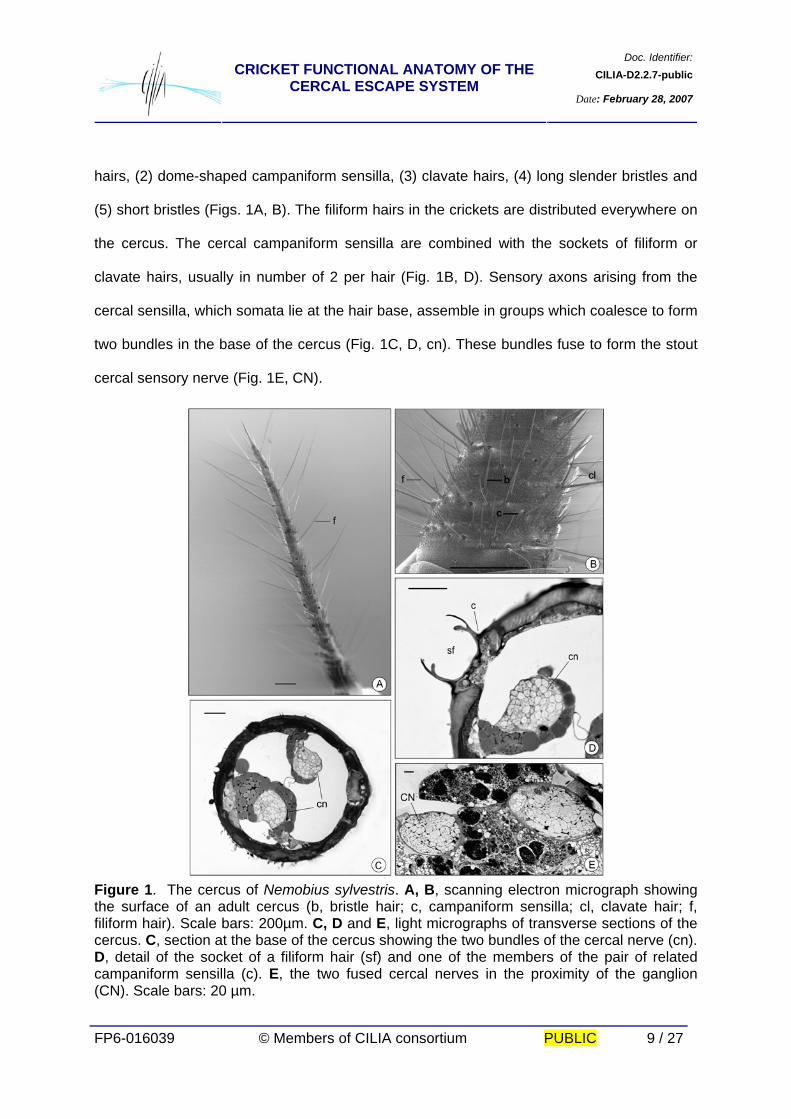
(5) short bristles (Figs. 1A, B). The filiform hairs in the crickets are distributed everywhere on
the cercus. The cercal campaniform sensilla are combined with the sockets of filiform or
clavate hairs, usually in number of 2 per hair (Fig. 1B, D). Sensory axons arising from the
cercal sensilla, which somata lie at the hair base, assemble in groups which coalesce to form
two bundles in the base of the cercus (Fig. 1C, D, cn). These bundles fuse to form the stout
cercal sensory nerve (Fig. 1E, CN).
Figure 1. The cercus of Nemobius sylvestris. A, B, scanning electron micrograph showing the surface of an adult cercus (b, bristle hair; c, campaniform sensilla; cl, clavate hair; f, filiform hair). Scale bars: 200µm. C, D and E, light micrographs of transverse sections of the cercus. C, section at the base of the cercus showing the two bundles of the cercal nerve (cn). D, detail of the socket of a filiform hair (sf) and one of the members of the pair of related campaniform sensilla (c). E, the two fused cercal nerves in the proximity of the ganglion (CN). Scale bars: 20 µm.
Doc. Identifier:CILIA-D2.2.7-publicCRICKET FUNCTIONAL ANATOMY OF THE
CERCAL ESCAPE SYSTEM Date: February 28, 2007
FP6-016039 © Members of CILIA consortium PUBLIC 10 / 27
2.3.2. The central nervous system The gross anatomy of the central nervous system of Nemobius sylvestris is similar to
that described for other crickets. It consists of a dorsal anterior brain, circumenteric
connectives (which encircle the oesophagus to unite ventral to the gut at the subesophageal
ganglion), and a ventral nerve cord with segmental ganglia and segmental peripheral nerves.
The ventral nerve cord includes three large segmental thoracic ganglia, one for each
segment of the thorax, but the metathoracic ganglion also includes the ganglia of the first two
abdominal segments. Five smaller ganglia are spaced along the nerve cord into the
abdomen. The fifth one, the TAG, is formed by the complete fusion of the 7th to 11th
segmental ganglia.
2.3.3. The Terminal Abdominal Ganglion (TAG) The TAG of Nemobius sylvestris has four paired ventral and dorsal nerves which
innervate the abdominal segments 7 to 10. The cercal nerves (dorsal n10) contain the bundle
of cercal sensory axons, which enter the posterolateral corners of the terminal ganglion (Fig.
2).
Doc. Identifier:CILIA-D2.2.7-publicCRICKET FUNCTIONAL ANATOMY OF THE
CERCAL ESCAPE SYSTEM Date: February 28, 2007
FP6-016039 © Members of CILIA consortium PUBLIC 11 / 27
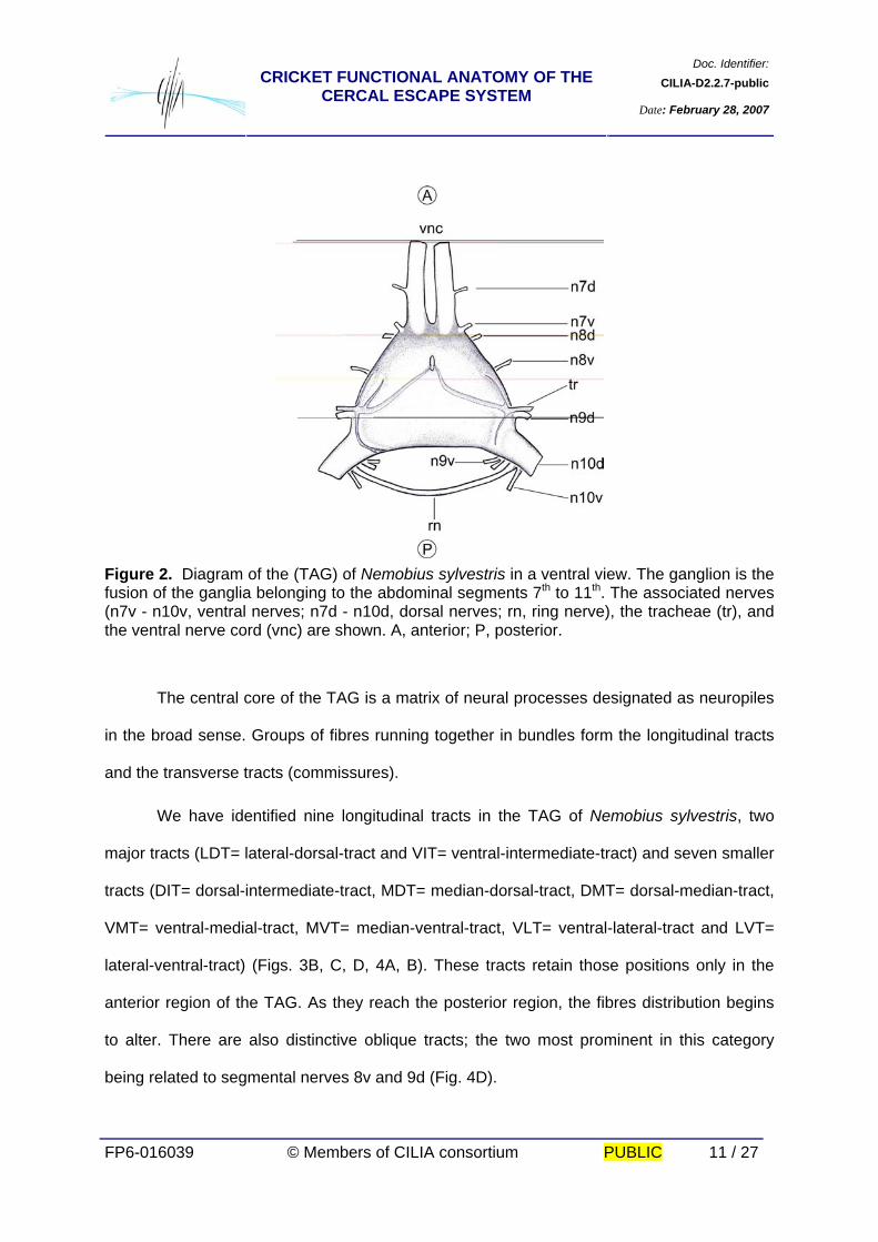
Figure 2. Diagram of the (TAG) of Nemobius sylvestris in a ventral view. The ganglion is the fusion of the ganglia belonging to the abdominal segments 7th to 11th. The associated nerves (n7v - n10v, ventral nerves; n7d - n10d, dorsal nerves; rn, ring nerve), the tracheae (tr), and the ventral nerve cord (vnc) are shown. A, anterior; P, posterior.
The central core of the TAG is a matrix of neural processes designated as neuropiles
in the broad sense. Groups of fibres running together in bundles form the longitudinal tracts
and the transverse tracts (commissures).
We have identified nine longitudinal tracts in the TAG of Nemobius sylvestris, two
major tracts (LDT= lateral-dorsal-tract and VIT= ventral-intermediate-tract) and seven smaller
tracts (DIT= dorsal-intermediate-tract, MDT= median-dorsal-tract, DMT= dorsal-median-tract,
VMT= ventral-medial-tract, MVT= median-ventral-tract, VLT= ventral-lateral-tract and LVT=
lateral-ventral-tract) (Figs. 3B, C, D, 4A, B). These tracts retain those positions only in the
anterior region of the TAG. As they reach the posterior region, the fibres distribution begins
to alter. There are also distinctive oblique tracts; the two most prominent in this category
being related to segmental nerves 8v and 9d (Fig. 4D).

Doc. Identifier:CILIA-D2.2.7-publicCRICKET FUNCTIONAL ANATOMY OF THE
CERCAL ESCAPE SYSTEM Date: February 28, 2007
FP6-016039 © Members of CILIA consortium PUBLIC 12 / 27
Figure 3. A, light micrograph of a transverse sec$tion of one connective of the ventral nerve cord, close to the TAG. B, transverse section of the anterior region of the TAG; arrows indicate the two longitudinal tracts were the giant axons are located. C, horizontal section of the TAG, showing the giant cell bodies (GIN) located in the periphery of the ganglion, the tracts (LDT: lateral dorsal tract) and commissures (ac, anterior commissure; pc, posterior commissure), and the entrance of the cercal sensory axons via the cercal nerves (CN). D, schematic diagram of a transversal section of the TAG showing the tracts and the giant axons located in the lateral dorsal tract (LDT), and the ventral intermediate tract (VIT). A, anterior; P, posterior; DIT, dorsal intermediate tract; DMT, dorsal medial tract; LVT, lateral ventral tract; MDT, medial dorsal tract; MVT, medial ventral tract; VLT, ventral lateral tract; VMT, ventral medial tract. Scale bars: 50 µm. The neuronal cell bodies are located at the periphery of the ganglion, in the lateral
region of the anterior part of the TAG, and laterodorsal in the posterior part of the TAG. The
dorsal and ventral central regions of the TAG are mostly free of somata (Figs. 3B, C).
Doc. Identifier:CILIA-D2.2.7-publicCRICKET FUNCTIONAL ANATOMY OF THE
CERCAL ESCAPE SYSTEM Date: February 28, 2007
FP6-016039 © Members of CILIA consortium PUBLIC 13 / 27
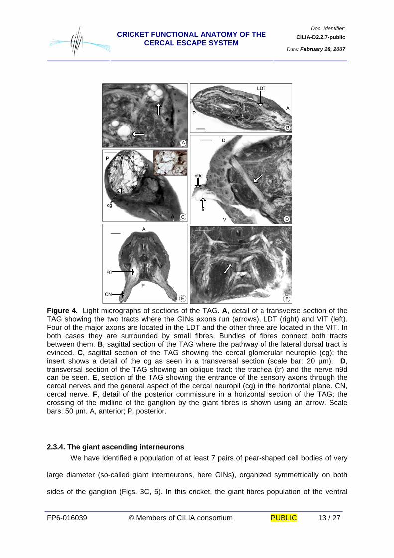
Figure 4. Light micrographs of sections of the TAG. A, detail of a transverse section of the TAG showing the two tracts where the GINs axons run (arrows), LDT (right) and VIT (left). Four of the major axons are located in the LDT and the other three are located in the VIT. In both cases they are surrounded by small fibres. Bundles of fibres connect both tracts between them. B, sagittal section of the TAG where the pathway of the lateral dorsal tract is evinced. C, sagittal section of the TAG showing the cercal glomerular neuropile (cg); the insert shows a detail of the cg as seen in a transversal section (scale bar: 20 µm). D, transversal section of the TAG showing an oblique tract; the trachea (tr) and the nerve n9d can be seen. E, section of the TAG showing the entrance of the sensory axons through the cercal nerves and the general aspect of the cercal neuropil (cg) in the horizontal plane. CN, cercal nerve. F, detail of the posterior commissure in a horizontal section of the TAG; the crossing of the midline of the ganglion by the giant fibres is shown using an arrow. Scale bars: 50 µm. A, anterior; P, posterior.
2.3.4. The giant ascending interneurons We have identified a population of at least 7 pairs of pear-shaped cell bodies of very
large diameter (so-called giant interneurons, here GINs), organized symmetrically on both
sides of the ganglion (Figs. 3C, 5). In this cricket, the giant fibres population of the ventral
Doc. Identifier:CILIA-D2.2.7-publicCRICKET FUNCTIONAL ANATOMY OF THE
CERCAL ESCAPE SYSTEM Date: February 28, 2007
FP6-016039 © Members of CILIA consortium PUBLIC 14 / 27
nerve cord is composed of a group of seven axons with diameters comprised between 20
and 45 µm (measured at the interganglionar connective) (Fig. 3A). The neuropile of the
ganglion is dominated by the processes of these giant interneurons, which lie in the two
major tracts LDT and VIT. Four of the largest axons are located superficially in the LTD,
accompanied by a group of smaller fibres. The fifth largest axon is located in the VIT together
with the other two smaller GINs and a group of fibres of small size (Figs. 3B, C, D, 4A, B).
The tracts of GINs are interconnected by two commissures. The anterior one is loosely
organized. The posterior one has many fibres crossing the midline of the ganglion in close
apposition (Figs. 3C, 4F). A glomerulus-like structure is evinced in the dense neuropile of the
posterior region of the TAG. It receives the sensory fibres of cercal nerves and the terminal
processes of GINs (Figs. 4C, E).
Mass retrograde cobalt fills from connectives linking the TAG to the sixth abdominal
ganglion confirmed the existence of the population of 7 pairs of cells of very large diameters,
organized symmetrically on both sides of the ganglion (Fig. 5).
Doc. Identifier:CILIA-D2.2.7-publicCRICKET FUNCTIONAL ANATOMY OF THE
CERCAL ESCAPE SYSTEM Date: February 28, 2007
FP6-016039 © Members of CILIA consortium PUBLIC 15 / 27
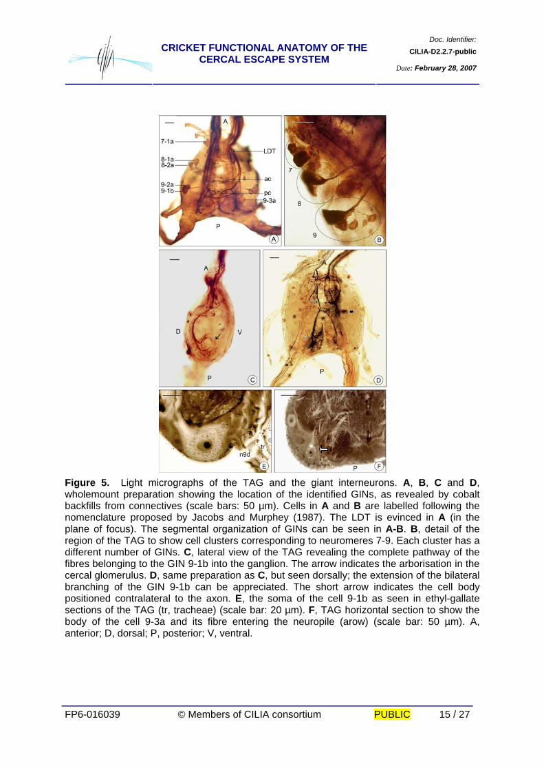
Figure 5. Light micrographs of the TAG and the giant interneurons. A, B, C and D, wholemount preparation showing the location of the identified GINs, as revealed by cobalt backfills from connectives (scale bars: 50 µm). Cells in A and B are labelled following the nomenclature proposed by Jacobs and Murphey (1987). The LDT is evinced in A (in the plane of focus). The segmental organization of GINs can be seen in A-B. B, detail of the region of the TAG to show cell clusters corresponding to neuromeres 7-9. Each cluster has a different number of GINs. C, lateral view of the TAG revealing the complete pathway of the fibres belonging to the GIN 9-1b into the ganglion. The arrow indicates the arborisation in the cercal glomerulus. D, same preparation as C, but seen dorsally; the extension of the bilateral branching of the GIN 9-1b can be appreciated. The short arrow indicates the cell body positioned contralateral to the axon. E, the soma of the cell 9-1b as seen in ethyl-gallate sections of the TAG (tr, tracheae) (scale bar: 20 µm). F, TAG horizontal section to show the body of the cell 9-3a and its fibre entering the neuropile (arow) (scale bar: 50 µm). A, anterior; D, dorsal; P, posterior; V, ventral.
Doc. Identifier:CILIA-D2.2.7-publicCRICKET FUNCTIONAL ANATOMY OF THE
CERCAL ESCAPE SYSTEM Date: February 28, 2007
FP6-016039 © Members of CILIA consortium PUBLIC 16 / 27
Each GIN is characterized by the position of its soma and by a specific branching
pattern. This group of cells maintains a constant position in the TAG. The somata of all
identified GINs are located contralateral to their axons, and are positioned sequentially along
the periphery of the ganglion. The GINs are named according to the number corresponding
to the original segment of the fused ganglion (Jacobs and Murphey, 1987. (Figs. 5A, B, C, D,
E, F). We have identified a cell whose body is located in the 7th segment, the 7-1a, two cells
located in the 8th segment, the 8-1a and 8-2a, three cells located in the 9th segment, 9-2a,
9-1b and 9-3a and a cell located in the 10th segment, the 10-3a. Figure 6 depicts a
schematic representation of the relative position of the GINs into the TAG, compiled from
series of sections of ethyl-gallate preparations in the three planes and several cobalt fillings.
The most useful landmark for identifying the cells body position is the midline trachea which
goes to the centre of the ganglion (Figs. 2, 4D, 5E). Cells 9-1b and 9-2a are located near the
conjunction of the tracheae with the nerve 9d. Groups 7 and 8 are located anteriorly and the
group 10 posteriorly.
Doc. Identifier:CILIA-D2.2.7-publicCRICKET FUNCTIONAL ANATOMY OF THE
CERCAL ESCAPE SYSTEM Date: February 28, 2007
FP6-016039 © Members of CILIA consortium PUBLIC 17 / 27
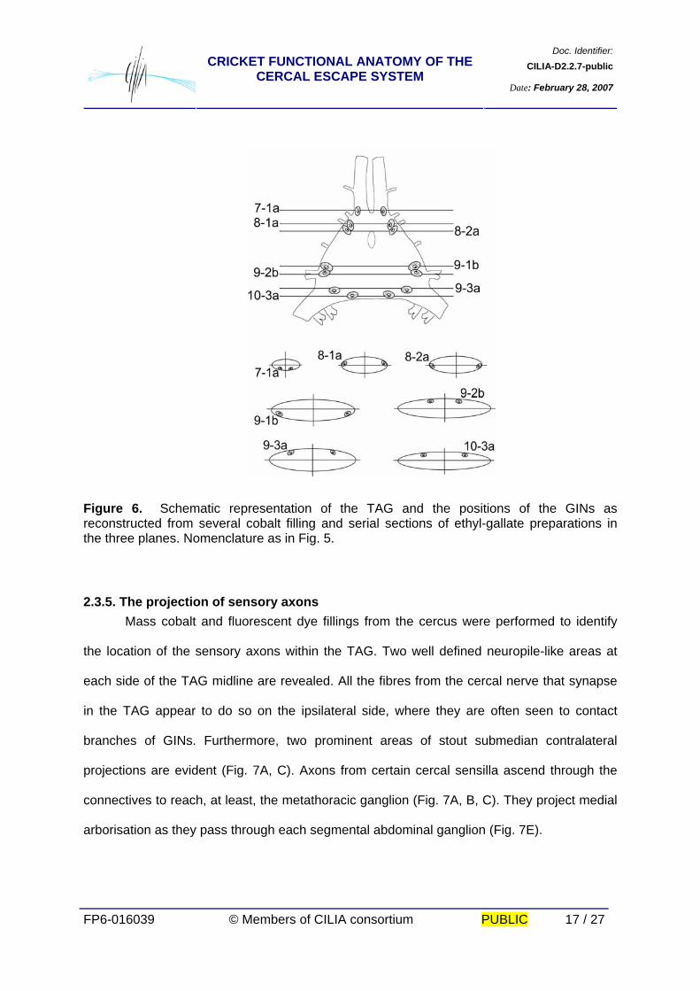
Figure 6. Schematic representation of the TAG and the positions of the GINs as reconstructed from several cobalt filling and serial sections of ethyl-gallate preparations in the three planes. Nomenclature as in Fig. 5.
2.3.5. The projection of sensory axons Mass cobalt and fluorescent dye fillings from the cercus were performed to identify
the location of the sensory axons within the TAG. Two well defined neuropile-like areas at
each side of the TAG midline are revealed. All the fibres from the cercal nerve that synapse
in the TAG appear to do so on the ipsilateral side, where they are often seen to contact
branches of GINs. Furthermore, two prominent areas of stout submedian contralateral
projections are evident (Fig. 7A, C). Axons from certain cercal sensilla ascend through the
connectives to reach, at least, the metathoracic ganglion (Fig. 7A, B, C). They project medial
arborisation as they pass through each segmental abdominal ganglion (Fig. 7E).
Doc. Identifier:CILIA-D2.2.7-publicCRICKET FUNCTIONAL ANATOMY OF THE
CERCAL ESCAPE SYSTEM Date: February 28, 2007
FP6-016039 © Members of CILIA consortium PUBLIC 18 / 27
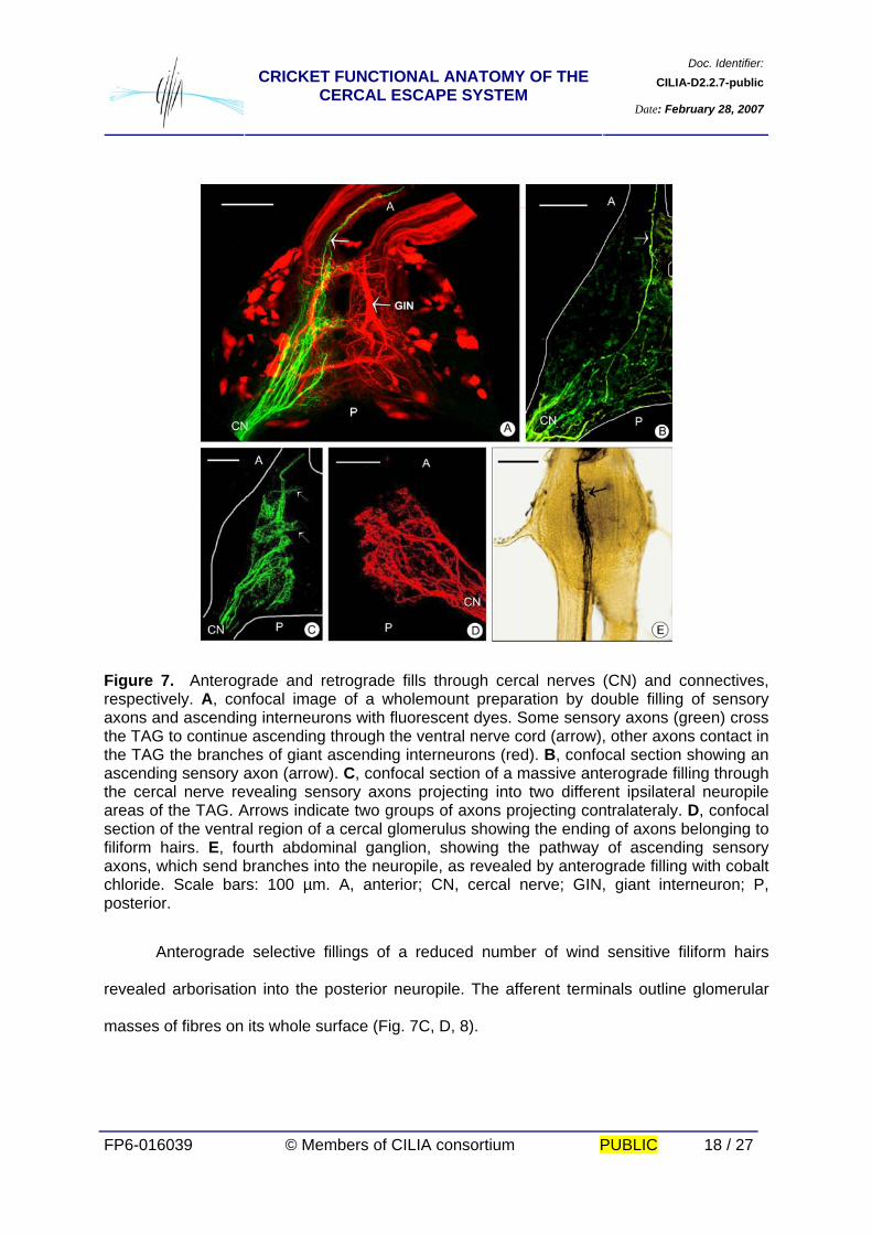
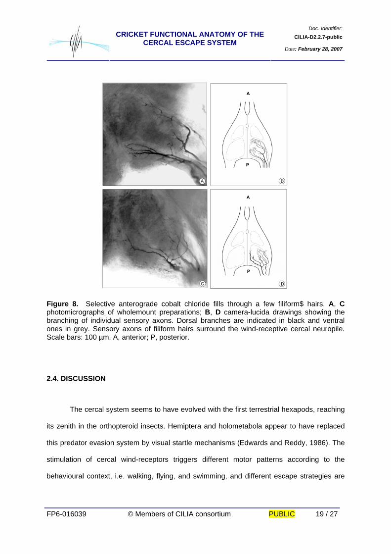
Figure 7. Anterograde and retrograde fills through cercal nerves (CN) and connectives, respectively. A, confocal image of a wholemount preparation by double filling of sensory axons and ascending interneurons with fluorescent dyes. Some sensory axons (green) cross the TAG to continue ascending through the ventral nerve cord (arrow), other axons contact in the TAG the branches of giant ascending interneurons (red). B, confocal section showing an ascending sensory axon (arrow). C, confocal section of a massive anterograde filling through the cercal nerve revealing sensory axons projecting into two different ipsilateral neuropile areas of the TAG. Arrows indicate two groups of axons projecting contralateraly. D, confocal section of the ventral region of a cercal glomerulus showing the ending of axons belonging to filiform hairs. E, fourth abdominal ganglion, showing the pathway of ascending sensory axons, which send branches into the neuropile, as revealed by anterograde filling with cobalt chloride. Scale bars: 100 µm. A, anterior; CN, cercal nerve; GIN, giant interneuron; P, posterior. Anterograde selective fillings of a reduced number of wind sensitive filiform hairs
revealed arborisation into the posterior neuropile. The afferent terminals outline glomerular
masses of fibres on its whole surface (Fig. 7C, D, 8).
Doc. Identifier:CILIA-D2.2.7-publicCRICKET FUNCTIONAL ANATOMY OF THE
CERCAL ESCAPE SYSTEM Date: February 28, 2007
FP6-016039 © Members of CILIA consortium PUBLIC 19 / 27
Figure 8. Selective anterograde cobalt chloride fills through a few filiform$ hairs. A, C photomicrographs of wholemount preparations; B, D camera-lucida drawings showing the branching of individual sensory axons. Dorsal branches are indicated in black and ventral ones in grey. Sensory axons of filiform hairs surround the wind-receptive cercal neuropile. Scale bars: 100 µm. A, anterior; P, posterior.
2.4. DISCUSSION
The cercal system seems to have evolved with the first terrestrial hexapods, reaching
its zenith in the orthopteroid insects. Hemiptera and holometabola appear to have replaced
this predator evasion system by visual startle mechanisms (Edwards and Reddy, 1986). The
stimulation of cercal wind-receptors triggers different motor patterns according to the
behavioural context, i.e. walking, flying, and swimming, and different escape strategies are
Doc. Identifier:CILIA-D2.2.7-publicCRICKET FUNCTIONAL ANATOMY OF THE
CERCAL ESCAPE SYSTEM Date: February 28, 2007
FP6-016039 © Members of CILIA consortium PUBLIC 20 / 27
executed, even in closely related species (Hirota et al. 1993, Matsuda et al. 2002, Kanou et
al., 2006).
2.4.1. The arrangement of sensory axons in the TAG The anatomical organization of the TAG of Nemobius sylvestris shares some
characteristics with that of other cricket species, but differences also occur. Two neuropilar
areas were revealed on each side of the TAG of N. sylvestris by means of massive injections
of dyes through the cercal nerve and histology. The largest area corresponds to the posterior
cercal glomerulus and the smaller anterior region to the bristle neuropil of other cricket
species (Murphey, 1981; Bacon and Murphey, 1984; Murphey, 1985). Cercal filiform hairs of
N. sylvestris arborise into the first one. The selective injection of the dye into sensory
neurons of the filiform hairs revealed that they arborise over the whole surface of the cercal
glomerulus, outlining it. In Acheta domesticus, there are two principal planes of insertion for
the filiform hairs, one allowing vibration on the longitudinal cercal axis on wind movement
and another transversely (Edwards and Palka, 1974; Bacon and Murphey, 1984). In this
species, each physiological type of hair arborises in a different region of the cercal
glomerulus (Bacon and Murphey, 1984). In the case of N. sylvestris, further studies would be
necessary to verify whether this is the case or not. Murphey (1985) described in A.
domesticus a system of hair-like receptors distributed over the cercus that he named
“bristles”. Sensory neurons from bristles project into a common area of the TAG (segments 7
and 8) that this author describes as the “bristle neuropil”. Collaterals of several of these
bristles leave the TAG to arborise on the ventral region of every abdominal segment.
Furthermore, Heusslein and Gnatzy (1987) showed that in Gryllus bimaculatus, A.
domesticus and Periplaneta americana, the sensory axons of campaniform sensilla
associated to filiform hairs also ascend as “through fibres” from the TAG to reach the sixth

Doc. Identifier:CILIA-D2.2.7-publicCRICKET FUNCTIONAL ANATOMY OF THE
CERCAL ESCAPE SYSTEM Date: February 28, 2007
FP6-016039 © Members of CILIA consortium PUBLIC 21 / 27
abdominal ganglion. In N. sylvestris, a high number of thin fibres ascend from the TAG via
the connectives, arborise in each abdominal segmental ganglion, and finally reach the
metathoracic fused mass. While the occurrence of ascending sensory fibres was therefore
confirmed in our species by retrograde injection, our results do not allow us to elucidate
which type of receptor ascends, at least, up to the thorax.
In Acheta domesticus adults, sensory axons project in the ipsilateral neuropiles of the
TAG, no contralateral terminations being present in normal adults (Edwards and Palka,
1974). In this species, contralateral interactions are mediated by interneurons (Palka and
Olberg, 1977). In Nemobius sylvestris, we have found two groups of contralateral projections
in the anterior and median region of the TAG (Fig. 7A). This pattern has also been shown in
Grylloblatta (three areas) and in the praying mantid Archimantis brunneriana (two
projections) by Edwards and Mann (1981) and by Ball et al. (1982), respectively. According
to the last authors, mantids also evince smaller and more sharply defined neuropile than
crickets. Indeed, Edwards and Palka (1974) described glomerular neuropiles of A.
domesticus as loosely organized. Nevertheless, neuropiles of N. sylvestris appear as quite
compact, resembling those of mantids, rather than those of other cricket species. In addition,
the number of sensory axons ascending through the nerve cord seems to be higher in
mantids than in A. domesticus (Ball et al., 1982). In N. sylvestris, as mentioned above, an
important number of sensory axons projects beyond the TAG. Nevertheless, we lack data
concerning the other species to allow precise comparisons.
2.4.2. Segmental homologies of GINs Because the early patterning of neurogenesis is similar in different segments of the
same insect and is even conserved among different insects, it is sometimes possible to
Doc. Identifier:CILIA-D2.2.7-publicCRICKET FUNCTIONAL ANATOMY OF THE
CERCAL ESCAPE SYSTEM Date: February 28, 2007
FP6-016039 © Members of CILIA consortium PUBLIC 22 / 27
recognize homologous neurons in different ganglia and even in different insect species
despite differences arising postembryonically as a consequence of functional differentiation.
For example, specific interneurons in different neuromeres that have similar arrangement of
their axons and arborisation develop from precisely homologous cells during early
development (Chapman, 1998). The GINs of Nemobius sylvestris can be ascribed to
neuromeres corresponding to the abdominal segments 7th to 10th, based both on the
comparative analysis with other cricket species and their position in the TAG. The somata of
GINs of N. sylvestris are positioned sequentially along the periphery of the ganglion. The
Acheta domesticus GINs retain the original segmental position into the fused neuromeres
which form the TAG, according to Mendenhall and Murphey (1974). Jacobs and Murphy
(1987) identified some of the GINs of the adult Gryllus assimilus and A. domesticus in the
embryo, confirming this hypothesis. Thus, we can conclude that the occurrence and
segmental position of GINs are highly conserved characteristics among the different cricket
species. The reason why different segments kept different numbers of GINs (1, 2, 3 and 1
GIN at each side in the 7th, 8th and 9th segment, respectively) remains nevertheless to be
clarified. Either only some cells of each segment differentiate in a giant one, or the original
number is equal along segments at the beginning, but subsequently undergoes a reduction
in certain segments due to cellular death. The available information does not allow us to
discern between these two possibilities.
2.4.3. The GINs fibres In contrast to the very small diameters of most neurons, some insects have “giant”
axons of varying size. These fibres can be descending, as in Musca, Calliphora and
Drosophila (Bacon and Strausfeld, 1986) or ascending units, as in cockroaches and crickets.
According to Edwards and Palka (1974), 10 giant axons can be seen in the ventral nerve
Doc. Identifier:CILIA-D2.2.7-publicCRICKET FUNCTIONAL ANATOMY OF THE
CERCAL ESCAPE SYSTEM Date: February 28, 2007
FP6-016039 © Members of CILIA consortium PUBLIC 23 / 27
chain of the TAG of Acheta domesticus. In Nemobius sylvestris 7 pairs of GINs, with their
corresponding axons, leaves the TAG to ascend thought the ventral cord. In Gryllus
assimilus and A. domesticus, axons of GINs are distributed into two (DIT and VIT; Jacobs
and Murphy, 1987) or three (dorso-lateral, ventro-medial and ventro-lateral; Mendenhal and
Murphy, 1974) longitudinal tracts. In N. sylvestris they clearly occupate two tracts (LDT and
VIT). Thus, the number and pathway organisation of GINs seems to vary among cricket
species.
2.4.4. Considerations on the GINs size The giant fibres are not the largest calibre axons. They are considered as giant if they
are discontinuously larger than the next largest fibres in that species, the absolute diameter
being not considered crucial (Bullock and Horridge, 1965). The axons diameter of the GINs
vary according to species, being 20-60µm in P. americana, 8-15 µm in Locusta migratoria,
12-16 µm in Anax spp. and 5-15 µm in Tettigonia cantans (Bullock and Horridge, 1965;
Nation, 2002, Jun-Xian Shen, 1983). In Nemobius sylvestris, the seven pairs of neurons here
considered as GINs posses axons of diameters ranging between 20 and 45 µm. This is
larger than the diameters of axons in other cricket species of bigger size (e.g. A.
domesticus= body size 16-20 mm, giant axons 10-40 µm diameter compared to 7-10 mm
and 20-45 µm in N. sylvestris). Two questions arise from this comparison. The first one
concerns the subjective criterion to consider a cell as giant or non-giant. Here, we applied a
quite conservative criterion, following Edwards and Palka (1974), but no operational or
statistical definition of “giant”, relative to the diameter of other axons in the nervous system,
has been proposed so far. As can be noted in figure 3A, several other smaller fibres, not
considered as GINs here, appear much larger than most axons of the nervous system.
Therefore, the GINs described here represent only “the quickest among the quickest”
Doc. Identifier:CILIA-D2.2.7-publicCRICKET FUNCTIONAL ANATOMY OF THE
CERCAL ESCAPE SYSTEM Date: February 28, 2007
FP6-016039 © Members of CILIA consortium PUBLIC 24 / 27
ascending mechanosensory pathways. The second question is that of the ratio between
GINs and body size. Provided that the velocity of conduction is directly proportional to the
diameter of the axons, the escape reaction might be quicker in N. sylvestris than in other
crickets for two reasons: the axons have larger diameters and ganglia are closer. Further
electrophysiological work in our laboratory should raise some light on this question. This
work provides the neuroanatomical frame for such physiological studies.

Doc. Identifier:CILIA-D2.2.7-publicCRICKET FUNCTIONAL ANATOMY OF THE
CERCAL ESCAPE SYSTEM Date: February 28, 2007
FP6-016039 © Members of CILIA consortium PUBLIC 25 / 27
3. REFERENCES Bacon JP, Altman JS. 1977. A silver intensification method for cobalt-filled neurons in
wholemount preparations. Brain Res 138:359-363. Bacon JP, Murphey RK. 1984. Receptive fields of cricket (Acheta domesticus) interneurons
are related to their dendritic structure. J Physiol 352:601-623. Bacon JP, Strausfeld NJ. 1986. The dipteran ‘Giant fibre’ pathway: neurons and signals. J
Comp Physiol A 158:529-548. Ball EE, Boyan GS, Stone RC. 1982. The cercal receptor system of the praying mantid,
Arcimantis brunneriana Sauss. II. Cercal nerve structure and projection and electrophysiological responses of individual receptor. Cell Tissue Res 224:71-80.
Blagburn JM and Thompson KSJ. 1990. Specificity of filiform hair afferent synapses onto giant interneurons in Periplaneta americana: anatomy is not a sufficient determinant. J Comp Neurol 302:255-271.
Blagburn JM, Beadle DJ and Sattelle DB. 1984. Synapses between an identified giant interneurone and a filiform hair sensory neurone in the terminal ganglion of first instar cockroaches (Periplaneta americana L.). J Exp Biol 113:477-481.
Boyan GS, Ball EE. 1986. Wind-sensitive interneurones in the terminal ganglion of praying mantids. J Comp Physiol A 159:773-789.
Boyan GS, Ball EE. 1989. The wind-sensitive cercal receptor/giant interneurone system of the locust, Locusta migratoria. II. Physiology of giant interneurones. J Comp Physiol A 165:511-521.
Boyan GS, Williams JLD, Ball EE. 1989. The wind-sensitive cercal receptor: giant/interneurone system of the locust, Locusta migratoria. I Anatomy of the system. J Comp Physiol A 165:495-510
Bullock TH, Horridge GA. 1965. Structure and Function in the Invertebrate Nervous System. San Francisco, London: W. H. Freeman & Co. 1719 pp.
Camhi JM. 1980. The escape system of the cockroach. Sci Am 243, 144-157. Chapman RF. 1998. The insects. Structure and function. United Kingdom: Cambridge
University Press. 770 pp. Coolen I, Dangles O, Casas J. 2005. Social learning in non colonial insects?. Curr Biol.15
(21):1931-1935. Dangles O, Magal C, Pierre D, Olivier A, Casas J. 2005. Variation in morphology and
performance of predator-sensing system in wild cricket populations. J Exp Biol 208:461-468.
Dangles O, Casas J, Coolen I. 2006a. Textbook cricket goes to the field: the ecological scene of the neuroethological play. J Exp Biol 209:393-398.
Dangles O, Ory O, Steinmann T, Christides JP, Casas J. 2006b. Spider’s attack vs. cricket’s escape: velocity modes determine success. Anim Behav 72:603-610.
Dijkstra M, van Baar J, Wiegerink R, Lammerink T, de Boer J, Krijnen G. 2005. Artificial sensory hairs based on the flow sensitive receptor hairs of crickets. J Micromech Microengineer 15:132-138.
Edwards JS, Mann D. 1981. The structure of the cercal sensory system and ventral nerve cord of Grylloblatta. A comparative study. Cell Tissue Res 217:177-188.

Doc. Identifier:CILIA-D2.2.7-publicCRICKET FUNCTIONAL ANATOMY OF THE
CERCAL ESCAPE SYSTEM Date: February 28, 2007
FP6-016039 © Members of CILIA consortium PUBLIC 26 / 27
Edwards JS, Palka J. 1974. The cerci and abdominal giant fibres of the house cricket Acheta domesticus. I. Anatomy and physiology of normal adults. Proc R Soc Lond B 185, 83-103.
Edwards JS, Reddy GR. 1986. Mechanosensory appendages and giant interneurons in the firebrat (Thermobia domestica, Thysanura): a prototype system for terrestrial predator evasion. J Comp Neurol 243:535-546.
Edwards JS, Williams L. 1891. Anterior-most projections of giant interneurones in Acheta domesticus terminate in mechano-receptor neuropile of the brain. Soc Neurosci Abs 7:252.
Heusslein R, Gnatzy W. 1987. Central projections of campaniform sensilla on the cerci of crickets and cockroaches. Cell Tissue Res 247:591-598.
Hirota K, Sonoda Y, Baba Y, Yamaguchi T. 1993. Distinction in morphology and behavioral role between dorsal and ventral groups of cricket giant interneurons. Zool Sci 10, 705-709.
Jacobs GA, Miller JP, Murphey RK. 1986. Integrative mechanisms controlling directional sensitivity of an identified sensory interneuron. J Neurosci. 6:2298-2311.
Jacobs GA, Murphey RK. 1987. Segmental origins of the cricket giant interneuron system. J Comp Neurol 256:145-157.
Jacobs GA, Theunissen FE. 1996. Functional organization of a neural map in the cricket cercal sensory system. J Neurosci 16:769-784.
Jacobs GA, Theunissen FE. 2000. Extraction of sensory parameters from a neural map by primary sensory interneurons. J Neurosci 20:2934-2943.
Jun-Xian Shen. 1983. The cercus-to-giant interneuron system in the bushcricket Tettigonia cantans: Morphology and response to low-frequency sound. J Comp Physiol A 151(4):449-459.
Kanou M, Nawae M, Kuroishi H. 2006. Cercal sensory system and giant interneurons in Gryllodes sigillatus. Zool Sci 23:365-373.
Magal C, Dangles O, Caparroy P, Casas J. 2006. Hair canopy of cricket sensory system tuned to predator signals. J Theor Biol 241:459-466.
Matsuda T, Kanou M, Yamaguchi T. 2002. Motor program initiation and selection in crickets, with special reference to swimming and flying behaviour. J Comp Physiol A 187:987-995.
Mendenhall B, Murphey RK. 1974. The morphology of cricket giant interneurons. J Neurobiol 5:565-580.
Miller JP, Jacobs GA, Theunissen, FE. 1991. Representation of sensory information in the cricket cercal sensory system. I: Response properties of the primary interneurons. J Neurophysiol 66 (5):1680-1689.
Murphey RK. 1981. The structure and development of a somatotopic map in crickets: The cercal afferent projection. Dev Biol 88:236-246.
Murphey RK. 1985. A second cricket sensory system: bristle hairs and the interneurons they activate. J Comp Physiol A 156:357-367.
Nation JL. 2002. Insect Physiology and Biochemistry. Boca Raton, London, New York, Washington D.C : CRC Press.
Doc. Identifier:CILIA-D2.2.7-publicCRICKET FUNCTIONAL ANATOMY OF THE
CERCAL ESCAPE SYSTEM Date: February 28, 2007
FP6-016039 © Members of CILIA consortium PUBLIC 27 / 27
Ogawa H, Baba Y, Oka K. 1999. Dendritic Ca2+ transient increase evoked by wind stimulus in the cricket giant interneuron. Neurosci Lett 275:61-64.
Ogawa H, Baba Y, Oka K. 2004. Directional sensitivity of dendritic calcium responses to wind stimuli in the cricket giant interneuron. Neurosci Lett 358:185-188.
Palka J, Levine R, Schubiger M. 1977. The cercus-to-giant interneuron system of crickets. I. Some attributes of the sensory cells. J Comp Physiol 119:267-283.
Palka J, Olberg R. 1977. The cercus-to-giant interneuron system of crickets. III. Receptive field organization. J Comp Physiol 119:301-317.
Paydar S, Doan CA, Jacobs, GA. 1999. Neural mapping of direction and frequency in the cricket cercal system. J Neurosci 19:1771-1781.
Ribi WA. 1987. Biological Electron Microscopy. A Handbook in Biological Electron Microscopy. Max-Planck-Institüt und Universität Tübingen. 106 pp.
Ritzmann RE. 1984. The cockroach escape response. In: Neural Mechanisms of Startle Behavior, pp. 93-131. New York, Plenum: Eaton RC.
Sasira Babu K, Subhashini K. 1981. Morphology of soma and dendrites of the giant fiber system in the sixth abdominal ganglion of the cockroach. J Morphol 169 (3):351-355.
Seabrook WD. 1970. The structure of the terminal ganglionic mass of locust, Schistocerca gregaria (Forskäl). J Comp Neurol 138:63-86.
Steinmann T, Casas J, Krijnen G, Dangles O. 2006. Air-flow sensitive hairs: boundary layers in oscillatory flows around arthropod appendages. J Exp Biol 209:4398-4408.
Tyrer NM, Gregory GE. 1982. A guide to the neuroanatomy of locust subesophageal and thoracic ganglia. Philos Trans R Soc Lond B 297:91-124.
Wigglesworth VB. 1957. The use of osmium in the fixation and staining of tissues. Proc R Soc Lond B 147:185-199.