Embed Size (px)
Citation preview
Proneural clusters of achaete-scute expression and the generation of sensory organs in the Drosophila imaginal wing disc
Pilar Cubas, Jos6-Fdlix de Cells , Sonsoles Campuzano , and Juan Modole l l
Centro de Biologia Molecular, Consejo Superior de Investigaciones Cientificas and Universidad Aut6noma de Madrid, 28049 Madrid, Spain
The proneural genes a c h a e t e (ac) and s c u t e (sc) confer to D r o s o p h i l a epidermal cells the ability to become sensory mother cells (SMCs). In imaginal discs, a c - s c are expressed in groups of cells, the proneural clusters, which are thought to delimit the areas where SMCs arise. We have visualized with the resolution of single cells the initial stages of sensory organ development by following the evolving pattern of proneural clusters and the emergence of SMCs. At reproducible positions within clusters, a small number of cells accumulate increased amounts of a c - s c protein. Subsequently, one of these cells, the SMC, accumulates the highest amount. Later, at least some SMCs become surrounded by cells with reduced a c - s c expression, a phenomenon probably related to lateral inhibition. Genetic mosaic analyses of cells with different doses of a c - s c genes, the sc expression in sc mutants, and the above findings show that the levels of a c - s c products are most important for SMC singling-out and SMC state maintenance. These products do not intervene in the differentiation of SMC descendants. The e x t r a m a c r o c h a e t a e gene, an antagonist of proneural genes, negatively regulates sc expression, probably by interfering with activators of this gene.
[Key W o r d s : Sensory organ precursors; a c h a e t e ; s c u t e ; pattern formation; proneural cluster]
Received January 15, 1991; revised version accepted March 5, 1991.
Morphogenesis in multicellular organisms results from organized cell proliferation coupled with the differenti- ation of various cell types according to spatial arrange- ments. The adult cuticle of D r o s o p h i l a contains thou- sands of sensory organs (SOs, chaetae, and sensilla of other kinds) whose type and position form a stereotyped pattern. Each SO is formed by the progeny of a single precursor cell, the sensory mother cell (SMC), which, during the third-instar larva or early pupa stages, is sin- gled out from the population of imaginal disc cells or abdominal histoblasts (Garcia-Bellido and Merriam 1971). A current model (Ghysen and Dambly-Chaudihre 1989; Jan and Jan 1990) proposes that the generation of the pattern of SOs is a multistep process that begins when clusters of ectodermal cells, defined by pre-exist- ing cues, acquire a proneural state. Later, by means of cell-cell interactions, an SMC is singled out from the cells of the proneural cluster and prevents its neighbors from following the same fate (lateral inhibition; Wiggles- worth 1940). SMCs then undergo two differential divi- sions to yield four cells that will differentiate into the SO (Lawrence 1966; Bate 1978; Hartenstein and Posakony 1989).
Genetic analysis has identified a number of genes that act on one or several of these steps (Lindsley and Grell
1968; Jan and Jan 1990). Among them, the a c h a e t e (ac) and s c u t e (sc) genes, two members of the a c - s c complex (AS-C; Garcia-Bellido 1979; Campuzano et al. 1985), are expressed in clusters of cells which, according to the fate map of the imaginal wing disc (Bryant 1975), roughly correspond to areas where SMCs should arise (Romani et al. 1989). Moreover, since hypomorphic ac or sc muta- tions cause the loss of SOs from allele-specific positions (Garcia-Bellido 1979), while generalized expression of ac and/or sc (Campuzano et al. 1986; Garcia-Alonso and Garcia-Bellido 1986; Balcells et al. 1988; Rodriguez et al. 1990) induces the formation of extra SOs in ectopic po- sitions, it has been proposed that a c - s c confer to cells the capacity to become neural precursors. Their expres- sion in groups of cells would define the proneural clus- ters, from which one or a few cells would be recruited to become SMCs (for review, see Ghysen and Dambly- Chaudihre 1989; Jan and Jan 1990).
The ac and sc proteins contain the basic helix-loop- helix domain (bHLH) characteristic of a family of tran- scriptional regulators (Villares and Cabrera 1987; Murre et al. 1989a). This domain allows these proteins to dimerize with themselves and/or with other members of the family and to bind to specific DNA sequences (Murre et al. 1989b). This suggests that ac and sc are transcrip-
996 GENES & DEVELOPMENT 5:996-1008 © 1991 by Cold Spring Harbor Laboratory ISSN 0890-9369/91 $3.00
Cold Spring Harbor Laboratory Press on August 30, 2018 - Published by genesdev.cshlp.orgDownloaded from
Proneural clusters in the imaginal wing disc
tional regulators that control the activity of genes in- volved in SO differentiation, probably in combination with other HLH proteins, like that encoded by the gene d a u g h t e r l e s s ( d a ) ( C a u d y et al. 1988; Dambly-Chaudihre et al. 1988). The ability of HLH proteins to form het- erodimers may also account for the antagonistic effect of e x t r a m a c r o c h a e t a e (emc) and h a i r y (h) proteins on the proneural activity of ac and sc (Botas et al. 1982; Mos- coso del Prado and Garcia-Bellido 1984; Garcia-Alonso and Garcia-Bellido 1988). Both e m c and h contain the HLH motif but lack (emc) or have an altered basic region (Rushlow et al. 1989; Ellis et al. 1990; Garrell and Mod- olell 1990). So, they may sequester ac and sc proteins in complexes inefficient for DNA interaction.
To analyze, at single-cell resolution, the relationship between the generation of the clusters of ac and sc ex- pression and the emergence of SMCs, we have performed in situ hybridizations to whole discs using digoxigenin (D1G)-labeled probes (Tautz and Pfeifle 1989) in the A101.1F3 transformant line of flies, which allows early detection of SMCs by expressing the bacterial l a c Z gene in these cells (Huang et al. 1991 ). With the help of anti-ac and anti-sc antibodies (Skeath and Carroll 1991 ), we have also analyzed the fine structure of the ac and sc expres- sion clusters and their modifications in mutants. In ad- dition, we have examined by genetic mosaic analysis the temporal requirement of a c - s c functions for the devel- opment of each notum macrochaeta. We have found an intimate relationship between the spatial and temporal evolution of the a c - s c clusters and the emergence of SMCs. Thus, SMCs appear at specific positions within clusters that are defined by increased levels of a c - s c
products, a c - s c intervene in the process of SMC sin- gling-out and SMC state maintenance, but not in the differentiation of the SMC descendants. Analysis of the patterns of a c - s c expression in mutants underline the relevance of the levels of a c - s c products for these devel- opmental processes. Some of the results presented have also been independently obtained by Skeath and Carroll (this issue), there being essential agreement between their conclusions and ours.
R e s u l t s
ac a n d sc e x p r e s s i o n p r e c e d e s t h e a p p e a r a n c e o f S M C s
We have examined, in wing imaginal discs, the spatial and temporal relationship between the patterns of a c - s c
expression and the appearance of SMCs. Discs from A101.1F3 larvae were stained with X-gal, to reveal SMCs, and hybridized with ac or sc DIG-labeled probes, to show the distribution of ac or sc mRNA. Both mRNAs have similar distributions (not shown, and Romani et al. 1989), due to a reciprocal gene activation between ac and sc (Martinez and Modolell 1991; Skeath and Carroll, this issue). Figure 1 shows examples of increasingly older discs hybridized with the sc probe. The main finding is that SMCs appear in a sequence (for a more detailed de- scription, see Huang et al. 1991), which is temporally preceded and spatially prefigured by the expression of ac
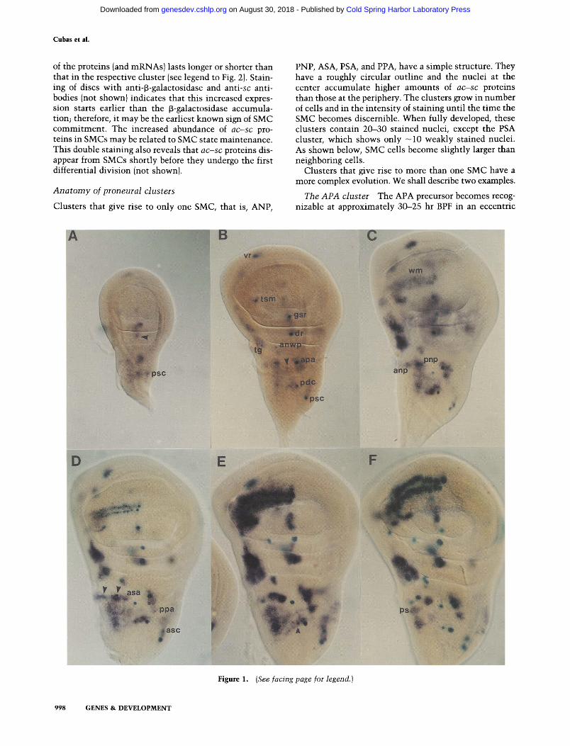
and sc. Thus, at -35 hr before puparium formation (BPF), sc is expressed in clusters of cells, but no or very few SMCs are detectable (Fig. 1A). Approximately 10 hr later (Fig. 1B), several SMCs, like those for the PDC and APA macrochaetae, have become visible within clusters of sc-expressing cells. In the middle of the presumptive no- tum, a cell, which originates in a small and weakly ex- pressing cluster, shows an increased sc expression (Fig. 1B, arrowhead). It corresponds to the PSA macrochaeta precursor, which later will express l a c Z (Fig. 1C). In the following hours, some clusters become more prominent and SMCs emerge in them, such as those for the ANP and PNP macrochaetae (Fig. 1C, D). Also, new clusters appear, like those at the presumptive wing margin (Fig. 1C) and PPA and ASA regions (Fig. 1D). The correspond- ing precursors will later become visible, either at - 1 0 hr BPF (wing margin, Fig. 1D) or several hours after pupar- ium formation (APF) (PPA and ASA; not shown, and Huang et al. 1991). The ADC and ASC macrochaetae precursors appear several hours BPF within the clusters that previously gave rise to their posterior counterparts (Fig. 1E). Around puparium formation, sc (and ac) expres- sion disappear from many clusters (Fig. IF). Those re- maining will give rise to additional SMCs. Moreover, additional sites of a c - s c expression appear AFP (not shown). It should be stressed that expression of ac and sc
is a prerequisite for SMC development: I n ( 1 ) s c l ° l ;
A101.1F3 wing discs, which lack functional a c - s c genes (Garcia-Bellido 1979; Campuzano et al. 1985; Villares and Cabrera 1987), are devoid of l a c Z - e x p r e s s i n g cells (not shown).
The shape, size, and position of the proneural clusters are very reproducible when comparing discs of approxi- mately the same age. Moreover, since SMCs become de- tectable, they are located in characteristic positions within clusters that in some cases (APA, trl, PDC) are clearly eccentric. This led us to examine, with higher resolution, the configuration of clusters using antibodies specific for the ac and sc proteins.
S M C s p r e f e r e n t i a l l y a c c u m u l a t e ac-sc p r o t e i n s
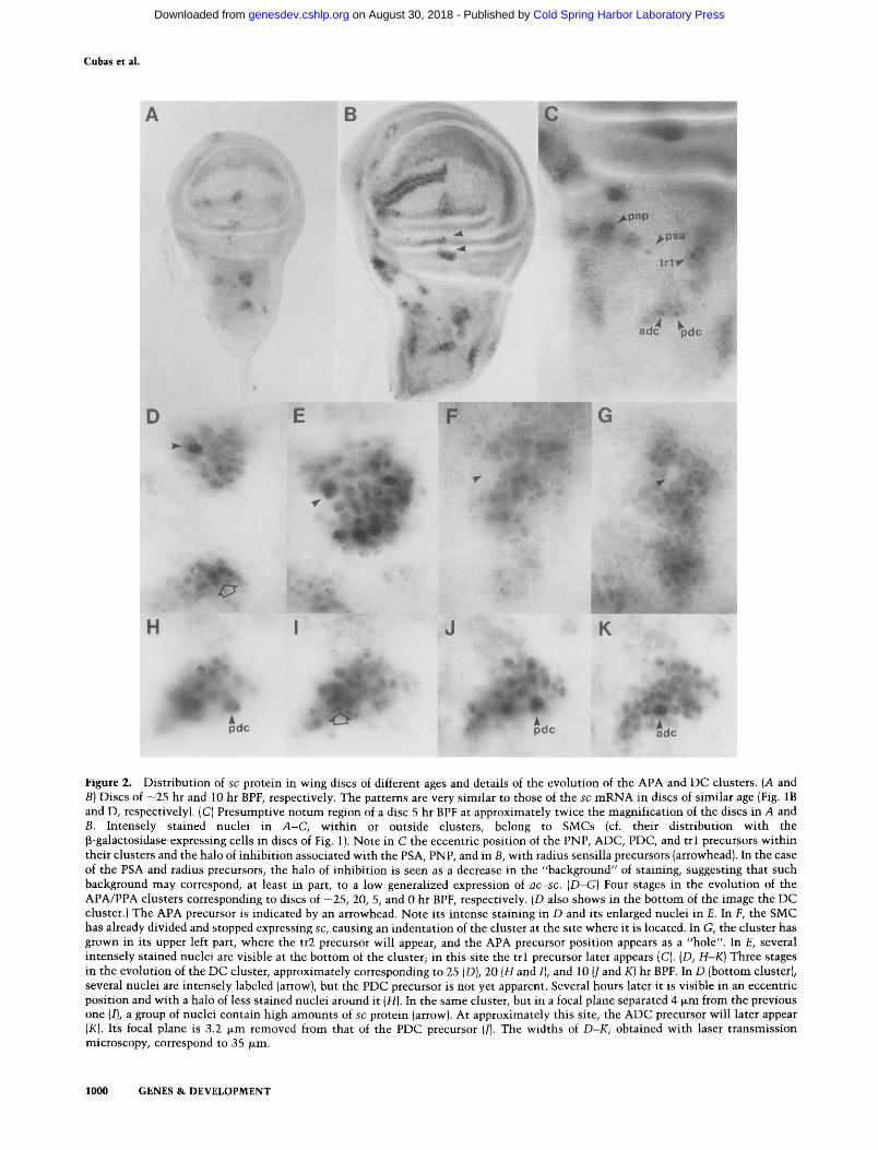
The patterns of ac and sc protein accumulation are very similar (Skeath and Carroll, this issue; P. Cubas el al., unpubl.) and strongly resemble those of the correspond- ing mRNAs (cf. Fig. 1B and E with Fig. 2A and B). The proteins are unambigously detected in discs as early as 40 hr BPF and are restricted to cell clusters (not shown). As expected from their putative role as transcriptional regulators, the a c - s c proteins are localized in nuclei. Within many proneural clusters, and depending on the developmental age, there is an intensely stained nucleus (Fig. 2A-C). It belongs to the SMC, as its position exactly matches that of the l a c Z - e x p r e s s i n g cells in the A101.1F3 discs. The enhanced abundance of a c - s c pro- teins in SMCs is due, at least in part, to a system of specific activation of a c - s c transcription in these cells (Martinez and Modolell 1991). This transcription is in- dependently regulated from the expression in other cells of the cluster since, depending on the SMC, the presence
GENES & DEVELOPMENT 997
Cold Spring Harbor Laboratory Press on August 30, 2018 - Published by genesdev.cshlp.orgDownloaded from
Cubas et al.
of the proteins (and mRNAs) lasts longer or shorter than that in the respective cluster (see legend to Fig. 2). Stain- ing of discs with anti-f~-galactosidase and anti-sc anti- bodies (not shown) indicates that this increased expres- sion starts earlier than the f~-galactosidase accumula- tion; therefore, it may be the earliest known sign of SMC commitment. The increased abundance of ac-sc pro- teins in SMCs may be related to SMC state maintenance. This double staining also reveals that ac-sc proteins dis- appear from SMCs shortly before they undergo the first differential division {not shown).
A n a t o m y of proneural clusters
Clusters that give rise to only one SMC, that is, ANP,
PNP, ASA, PSA, and PPA, have a simple structure. They have a roughly circular outline and the nuclei at the center accumulate higher amounts of ac-sc proteins than those at the periphery. The clusters grow in number of cells and in the intensity of staining until the time the SMC becomes discernible. When fully developed, these clusters contain 20-30 stained nuclei, except the PSA cluster, which shows only - 1 0 weakly stained nuclei. As shown below, SMC cells become slightly larger than neighboring cells.
Clusters that give rise to more than one SMC have a more complex evolution. We shall describe two examples.
The APA cluster The APA precursor becomes recog- nizable at approximately 30-25 hr BPF in an eccentric
Figure 1. (See facing page for legend.)
998 GENES & DEVELOPMENT
Cold Spring Harbor Laboratory Press on August 30, 2018 - Published by genesdev.cshlp.orgDownloaded from

Proneural clusters in the imaginal wing disc
position wi th in the cluster (Fig. 2D). Five to 10 hr later, the cluster has enlarged, and the APA precursor is lo- cated at its anterior (left) edge (Fig. 2E). At the bottom edge, several nuclei are stained more intensely; one of them will probably become the tr l precursor (Fig. 2C). Later, at approximately the t ime this precursor becomes visible, a c - s c expression ceases in the APA precursor. Its position is revealed by an indentat ion in the cluster (Fig. 2F). At the t ime of pupar ium formation, additional cells at the top of the cluster express a c - s c (Fig. 2G) and one of them will become the tr2 precursor.
T h e D C c l u s t e r In its early stages, this cluster is roughly elliptical (Figs. 1A and 2A). The PDC precursor is not yet detectable, and several centrally located nuclei are more intensely stained than others (open arrow, Fig. 2D). When the PDC precursor is distinguishable, the cluster is larger and the precursor is reproducibly located at the bottom right side of the cluster (Fig. 2H). Intensely stained cells are then found more anteriorly (toward the left) at the place where the ADC precursor later appears, also off this center (Fig. 2I). Note that when both the ADC and PDC nuclei are visible, they are located at different planes (Fig. 2J and K).
Some t ime after the PDC, PSA, APA, and PNP SMCs and several relatively isolated sensilla campaniformia precursors at the dorsal wing radius become recognizable by their intensely stained nuclei, they appear surrounded by cells not expressing or expressing lower amounts of a c - s c than the rest of the cluster (Fig. 2B, C, H, and J). These halo-like zones, one to two cell diameters in width, are also seen in discs stained for [3-galactosidase and sc m R N A [Fig. 1C (PDC precursor) and Fig. 1D (PNP precursor)]. In the case of the APA precursor, some t ime after this cell has divided and stopped expressing a c - s c ,
the area of nonexpression is reduced to approximately the size of the SMC daughters, suggesting that the halo has disappeared (Fig. 2F and G). The possibili ty that the
halo could be an artifact due to oversized SMC cells was ruled out by staining the outlines of cells. Confocal im- ages of different SMCs, like those in Figure 3, showed that although SMCs may be slightly larger than neigh- boring cells, the size difference cannot account for the inhibi t ion zones. We do not know whether these zones accompany all SMCs in some period of their develop- ment. Halos may be masked in clusters sequential ly giv- ing rise to many SMCs, like those of the tegula, ventral radius, and wing margin, and difficult to detect in clus- ters where sc expression is extinguished soon after the appearance of the SMC (ADC and ASC). The existence of halos around the pupar ium stage-appearing ASA, PPA, and PS precursors has not been examined.
T e m p o r a l r e q u i r e m e n t o f ac-sc f u n c t i o n for i n d i v i d u a l
S M C d e t e r m i n a t i o n
We have shown that SMCs arise among cells of pre-ex- isting proneural clusters. To analyze the temporal re- quirement of a c - s c for the development of macrochaeta precursors, we have generated homozygous ac +sc + and a c - s c - cells in a c + s c + / a c - s c - heterozygous larvae by mitot ic recombination. The abili ty of these cells to be- come SOs was compared wi th that of homozygous ac +sc + cells in wild-type larvae. This analysis has been performed for each macrochaetae, in contrast to an ear- lier study where all macrochaeta were considered to- gether (Garcia-Bellido and Santamaria 1978). Represen- tative examples are shown in Figure 4. Homozygous a c - s c - cells resulting from irradiation early in the third larval instar do not develop as macrochaetae. Homozy- gosity induced later allows some a c - s c - cells to become SOs, and after a developmental t ime characteristic for each SO, a c - s c - chaetae appear as frequently as in the respective control of each SO. From this t ime on, the a c - s c functions are no longer required in the descen- dants of the irradiated cell to form an SO. For most mac-
Figure 1. Development of the patterns of sc expression (purple) and SO precursors [deep green). Site nomenclature and SMC assign- ment are according to Bryant (1975), Huang et al. (1991), and our own observations. (A) Disc of -35 hr BPF. Note the sc expression in clusters of cells and that only the PSC macrochaeta precursor and that for a dorsal radius sensillum (out of focus, arrowhead) have begun expressing lacZ. [B} Disc of -25 hr BPF. Several SMCs express lacZ. They correspond to the PDC and APA macrochaetae, the GSR sensillum, one sensillum of the tegula (tg), the anterior notal wing process [anwp), and ventral radius (vr), respectively, and one of the twin sensilla campaniformia of the wing margin (tsm). Note the increased sc expression in a single cell that corresponds to the PSA macrochaeta precursor (arrowhead). (C) Disc of -20 hr BPF. The PSA precursor has started expressing lacZ. sc expression has started in the presumptive anterior wing margin (wm), in the clusters for the ANP and PNP and other sites where SMCs will appear. (D) Disc of -10 hr BPF. The wing margin SMCs are detectable by X-gal staining. Note that at the wing margin, both sc and lacZ expressions are first visible in a cluster of cells and a single SMC, respectively, that mark the right (distal/end of the top row (B and C). SMC development then progresses leftward up to the proximal end. These precursors most likely correspond to the recurved, multiply innervated bristles of the wing margin since both the precursors and these bristles, but not the straight bristles of the margin, are absent in In(1)sc 1°1 flies inot shown). Moreover, the recurved bristles start their final differentiation -10 hr earlier than the other margin bristles [Hartenstein and Posakony 1989). At -10 hr BPF, the precursors for the PNP, ANP [arrowhead), and ASC macrochaetae and the trl sensillum trichoideum below the APA precursor have become visible. Also, within the presumptive notum, the ASA and PPA clusters are already developing. In E, -5 hr BPF, they have become larger, with more intense sc expression. The corresponding SMCs start expressing l acZ several hours APF (not shown). The ADC macrochaeta precursor has become visible (arrowhead). (F) Disc just before puparium formation. The PS precursor becomes detectable slightly later within an area of intense sc expression (ps). sc (and ac) expression has disappeared from many clusters. Those remaining will give rise to additional SMCs. Note that throughout the 35 hr of development examined, ac-sc are expressed in the prospective anterior and mid-notum, a region that develops -125 regularly spaced microchaetae. Their SMCs appear at -10 hr APF (not shown). All discs are reproduced at the same scale.
GENES & DEVELOPMENT 999
Cold Spring Harbor Laboratory Press on August 30, 2018 - Published by genesdev.cshlp.orgDownloaded from
Cubas et al.
i, ii ii!ii!iiiii !i i ̧ , ....
D ~i ~ iili E
H ! J K
Figure 2. Distribution of sc protein in wing discs of different ages and details of the evolution of the APA and DC clusters. (A and B) Discs of - 2 5 hr and 10 hr BPF, respectively. The patterns are very similar to those of the sc mRNA in discs of similar age (Fig. 1B and D, respectively). (C) Presumptive notum region of a disc 5 hr BPF at approximately twice the magnification of the discs in A and B. Intensely stained nuclei in A - C , within or outside clusters, belong to SMCs (cf. their distribution with the [3-galactosidase-expressing cells in discs of Fig. 1). Note in C the eccentric position of the PNP, ADC, PDC, and trl precursors within their clusters and the halo of inhibit ion associated with the PSA, PNP, and in B, with radius sensilla precursors (arrowhead). In the case of the PSA and radius precursors, the halo of inhibit ion is seen as a decrease in the "background" of staining, suggesting that such background may correspond, at least in part, to a low generalized expression of ac-sc. (D-G) Four stages in the evolution of the APA/PPA clusters corresponding to discs of -25, 20, 5, and 0 hr BPF, respectively. (D also shows in the bottom of the image the DC cluster.) The APA precursor is indicated by an arrowhead. Note its intense staining in D and its enlarged nuclei in E. In F, the SMC has already divided and stopped expressing sc, causing an indentation of the cluster at the site where it is located. In G, the cluster has grown in its upper left part, where the tr2 precursor will appear, and the APA precursor position appears as a "hole". In E, several intensely stained nuclei are visible at the bottom of the cluster; in this site the trl precursor later appears (C). (D, H-K) Three stages in the evolution of the DC cluster, approximately corresponding to 25 (D), 20 (H and I), and 10 (J and K) hr BPF. In D (bottom cluster), several nuclei are intensely labeled (arrow), but the PDC precursor is not yet apparent. Several hours later it is visible in an eccentric position and with a halo of less stained nuclei around it (H). In the same cluster, but in a focal plane separated 4 ~m from the previous one (I), a group of nuclei contain high amounts of sc protein (arrow). At approximately this site, the ADC precursor will later appear (K). Its focal plane is 3.2 ~m removed from that of the PDC precursor (J). The widths of D-K, obtained with laser transmission microscopy, correspond to 35 ~m.
1000 GENES & DEVELOPMENT
Cold Spring Harbor Laboratory Press on August 30, 2018 - Published by genesdev.cshlp.orgDownloaded from
Proneural clusters in the imaginal wing disc
rochaetae (Fig. 4, bottom), this occurs when a c - s c are already expressed in the presumptive region (Fig. 1) but 10-25 hr before the time when SMCs become visible by the enhanced expression of a c - s c . Exceptionally, for the APA precursor, the time of a c - s c function dispensability occurs approximately when the precursor becomes de- tectable.
Figure 4 also shows that before the time a c - s c func- tions appear to become dispensable, the frequency with which homozygous a c + s c + cells become SMCs in a a c + s c + / a c - s c - heterozygous background is two to four times higher than in a homozygous a c + s c + background. This indicates that among a c - s c - e x p r e s s i n g cells, those
dx
f ( so - ) , • J . / ' ~ ' - - - - I ~ l t I J I I
e' ~' e' ~' e" ~' ÷' u Z ~ ,~ h bpf
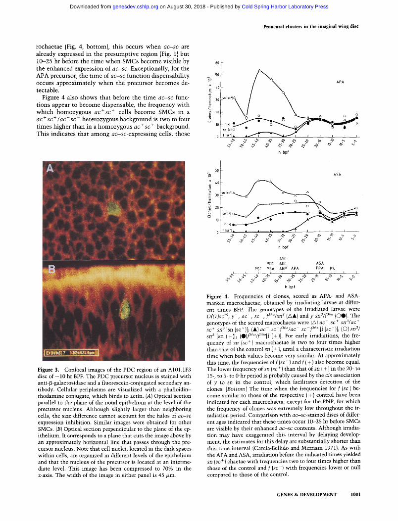
Figure 3. Confocal images of the PDC region of an A101.1F3 disc of -10 hr BPF. The PDC precursor nucleus is stained with anti-13-galactosidase and a fluorescein-conjugated secondary an- tibody. Cellular periplasms are visualized with a phalloidin- rhodamine conjugate, which binds to actin. (A) Optical section parallel to the plane of the notal epithelium at the level of the precursor nucleus. Although slightly larger than neighboring cells, the size difference cannot account for the halos of a c - s c expression inhibition. Similar images were obtained for other SMCs. {B) Optical section perpendicular to the plane of the ep- ithelium. It corresponds to a plane that cuts the image above by an approximately horizontal line that passes through the pre- cursor nucleus. Note that cell nuclei, located in the dark spaces within cells, are organized in different levels of the epithelium and that the nucleus of the precursor is located at an interme- diate level. This image has been compressed to 70% in the z-axis. The width of the image in either panel is 45 ~m.
50 %,
x z,0
I 301 E
20 g
10
ASA
" ( s c ' l a ~ a / ~ " " A - - - ~
o - - - - - o - ' o - - ° % o
h bpf
ASC PDC ADC ASA
PSE PSA ANP APA PPA
h bpf
PS
Figure 4. Frequencies of clones, scored as APA- and ASA- marked macrochaetae, obtained by irradiating larvae at differ- ent times BFP. The genotypes of the irradiated larvae were D f ( 1 ) s c zg, y - , a c - , s c - , f36~/sn3 (/x&) and y s n 3 / f 36~ (OO). The genotypes of the scored macrochaeta were (G) ac + sc + s n S / a c ÷ sc + sn 3 [sn (sc+)]; (&) a c - sc f36a/ac s c - f 36a [f (so)]j (O)8113/ sn 3 [sn (+)1; (O)f36"/f3~a[f (+)1. For early irradiations, the fre- quency of sn (sc +) macrochaetae is two to four times higher than that of the control sn ( + ), until a characteristic irradiation time when both values become very similar. At approximately this time, the frequencies of f ( s c - ) and f ( + ) also become equal. The lower frequency of sn (sc + ) than that of sn ( + ) in the 20- to 15-, to 5- to 0-hr period is probably caused by the cis association of y to sn in the control, which facilitates detection of the clones. ( B o t t o m ) The time when the frequencies for f (sc-) be- come similar to those of the respective (+) control have been indicated for each macrochaeta, except for the PNP, for which the frequency of clones was extremely low throughout the ir- radiation period. Comparison with a c - s c - s t a i n e d discs of differ- ent ages indicated that these times occur 10--25 hr before SMCs are visible by their enhanced a c - s c contents. Although irradia- tion may have exaggerated this interval by delaying develop- ment, the estimates for this delay are substantially shorter than this time interval (Garcia-Bellido and Merriam 1971}. As with the APA and ASA, irradiation before the indicated times yielded sn (sc + ) chaetae with frequencies two to four times higher than those of the control and f ( s c - ) with frequencies lower or null compared to those of the control.
GENES & DEVELOPMENT 1001
Cold Spring Harbor Laboratory Press on August 30, 2018 - Published by genesdev.cshlp.orgDownloaded from
Cubas et al.
with more doses of the a c + s c + genes, and presumably wi th the highest level of a c - s c products, have an in- creased probability to be singled out for becoming SMCs.
sc e x p r e s s i o n i n sc m u t a n t s
To evaluate the significance of the expression pattern of s c for SMC determination, we have examined its modi- fications in s c mutan t discs and compared them with the mutan t phenotypes.
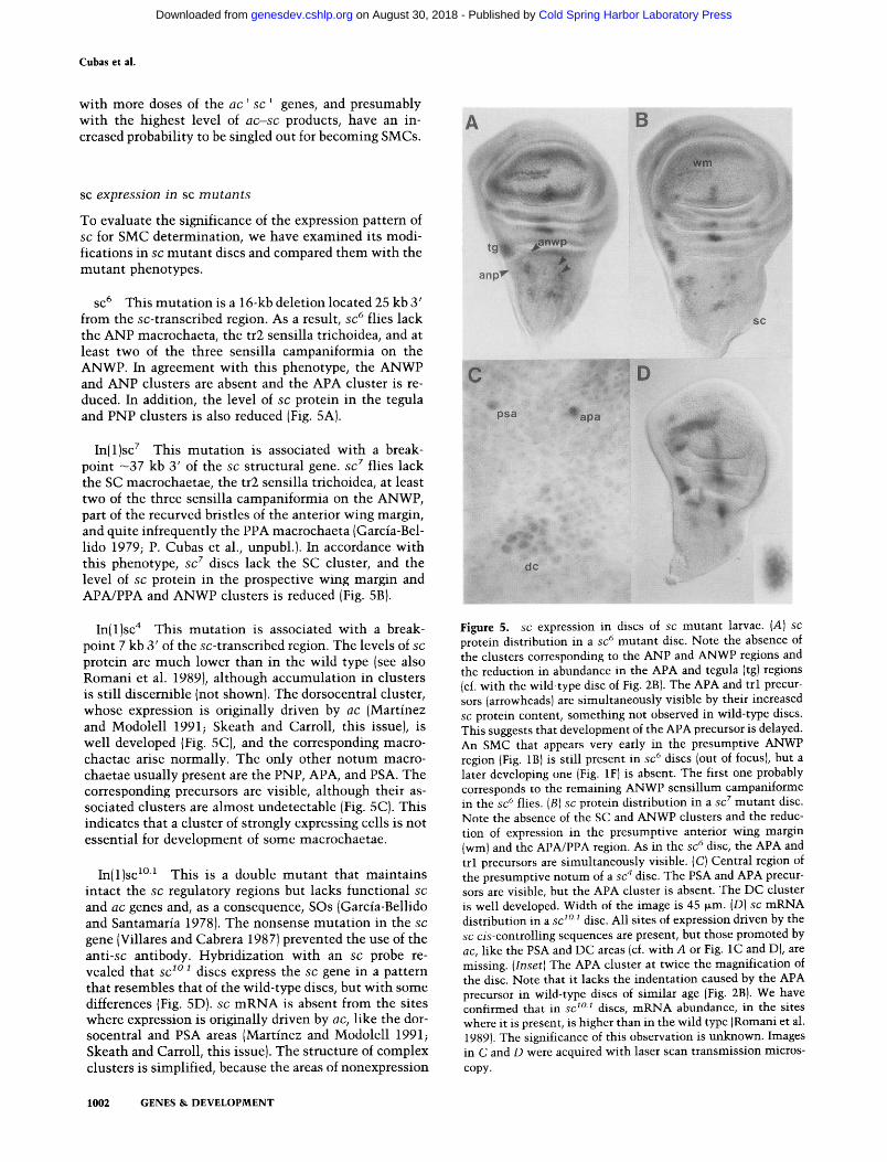
SC 6 This muta t ion is a 16-kb deletion located 25 kb 3' from the sc-transcribed region. As a result, s c 6 flies lack the ANP macrochaeta, the tr2 sensilla trichoidea, and at least two of the three sensilla campaniformia on the ANWP. In agreement wi th this phenotype, the ANWP and ANP clusters are absent and the APA cluster is re- duced. In addition, the level of s c protein in the tegula and PNP clusters is also reduced (Fig. 5A).
In(1)sc 7 This muta t ion is associated with a break- point ~37 kb 3' of the s c structural gene. s c 7 flies lack the SC macrochaetae, the tr2 sensilla trichoidea, at least two of the three sensilla campaniformia on the ANWP, part of the recurved bristles of the anterior wing margin, and quite infrequently the PPA macrochaeta (Garcia-Bel- lido 1979; P. Cubas et al., unpubl.). In accordance with this phenotype, s c z discs lack the SC cluster, and the level of s c protein in the prospective wing margin and APA/PPA and ANWP clusters is reduced (Fig. 5B).
In(1)sc 4 This muta t ion is associated with a break- point 7 kb 3' of the sc-transcribed region. The levels of s c
protein are much lower than in the wild type (see also Romani et al. 1989), al though accumulat ion in clusters is still discernible (not shown). The dorsocentral cluster, whose expression is originally driven by a c (Martinez and Modolell 1991; Skeath and Carroll, this issue), is well developed (Fig. 5C), and the corresponding macro- chaetae arise normally. The only other no tum macro- chaetae usually present are the PNP, APA, and PSA. The corresponding precursors are visible, al though their as- sociated clusters are almost undetectable (Fig. 5C). This indicates that a cluster of strongly expressing cells is not essential for development of some macrochaetae.
In(1)sc ~°~ This is a double mutan t that mainta ins intact the s c regulatory regions but lacks functional s c
and a c genes and, as a consequence, SOs (Garcia-Bellido and Santamaria 1978). The nonsense muta t ion in the s c
gene (Villares and Cabrera 1987) prevented the use of the a n t i - s c antibody. Hybridization with an s c probe re- vealed that s c s ° s discs express the s c gene in a pattern that resembles that of the wild-type discs, but with some differences (Fig. 5D). s c m R N A is absent from the sites where expression is originally driven by a c , like the dor- socentral and PSA areas (Martinez and Modolell 1991; Skeath and Carroll, this issue). The structure of complex clusters is simplified, because the areas of nonexpression
S. - < . ,
Figure 5. s c expression in discs of s c mutant larvae. (A) s c
protein distribution in a s c 6 mutant disc. Note the absence of the clusters corresponding to the ANP and ANWP regions and the reduction in abundance in the APA and tegula (tg) regions (cf. with the wild-type disc of Fig. 2B). The APA and trl precur- sors (arrowheads) are simultaneously visible by their increased sc protein content, something not observed in wild-type discs. This suggests that development of the APA precursor is delayed. An SMC that appears very early in the presumptive ANWP region (Fig. 1B) is still present in s c 6 discs (out of focus), but a later developing one (Fig. 1F) is absent. The first one probably corresponds to the remaining ANWP sensillum campaniforme in the s c 6 flies. (B) s c protein distribution in a s c 7 mutant disc. Note the absence of the SC and ANWP clusters and the reduc- tion of expression in the presumptive anterior wing margin (wm) and the APA/PPA region. As in the s c 6 disc, the APA and trl precursors are simultaneously visible. (C) Central region of the presumptive notum of a s c 4 disc. The PSA and APA precur- sors are visible, but the APA cluster is absent. The DC cluster is well developed. Width of the image is 45 ~m. (D) s c mRNA distribution in a s c 1°1 disc. All sites of expression driven by the s c c i s - c o n t r o l l i n g sequences are present, but those promoted by a c , like the PSA and DC areas (cf. with A or Fig. 1C and D), are missing. ( I n s e t ) The APA cluster at twice the magnification of the disc. Note that it lacks the indentation caused by the APA precursor in wild-type discs of similar age (Fig. 2B). We have confirmed that in s c 1°1 discs, mRNA abundance, in the sites where it is present, is higher than in the wild type (Romani et al. 1989). The significance of this observation is unknown. Images in C and D were acquired with laser scan transmission micros- copy.
1002 GENES & DEVELOPMENT
Cold Spring Harbor Laboratory Press on August 30, 2018 - Published by genesdev.cshlp.orgDownloaded from
Proneural clusters in the imaginal wing disc
caused by SMCs and associated halos are absent. Thus, the APA cluster has an elliptical shape (Fig. 5D, inset), which lacks the prominent indentat ion where the APA precursor and its inhibi t ion halo are normal ly located (Fig. 2F).
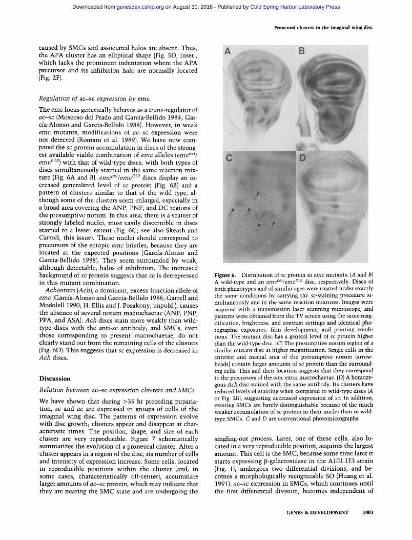
R e g u l a t i o n o f ac-sc e x p r e s s i o n b y emc
The e m c locus genetically behaves as a t r a n s - r e g u l a t o r of a c - s c (Moscoso del Prado and Garcia-Bellido 1984; Gar- cia-Alonso and Garcia-Bellido 1988). However, in weak e m c mutants , modifications of a c - s c expression were not detected (Romani et al. 1989). We have now com- pared the sc protein accumulat ion in discs of the strong- est available viable combinat ion of e m c alleles (emcpel /
e m c ~I2) with that of wild-type discs, with both types of discs s imul taneously stained in the same reaction mix- ture (Fig. 6A and B). e m c P @ e m c ~ 2 discs display an in- creased generalized level of sc protein (Fig. 6B) and a pattern of clusters s imilar to that of the wild type, al- though some of the clusters seem enlarged, especially in a broad area covering the ANP, PNP, and DC regions of the presumptive notum. In this area, there is a scatter of strongly labeled nuclei, most easily discernible in discs stained to a lesser extent (Fig. 6C; see also Skeath and Carroll, this issue). These nuclei should correspond to precursors of the ectopic e m c bristles, because they are located at the expected positions (Garcia-Alonso and Garcia-Bellido 1988). They seem surrounded by weak, although detectable, halos of inhibit ion. The increased background of sc protein suggests that sc is derrepressed in this mutan t combination.
A c h a e t o u s (Ach) , a dominant, excess-function allele of e m c (Garcia-Alonso and Garcia-Bellido 1988; Garrell and Modolell 1990; H. Ellis and J. Posakony, unpubl.), causes the absence of several no tum macrochaetae (ANP, PNP, PPA, and ASA). A c h discs stain more weakly than wild- type discs wi th the anti-sc antibody; and SMCs, even those corresponding to present macrochaetae, do not clearly stand out from the remaining cells of the clusters (Fig. 6D). This suggests that sc expression is decreased in A c h discs.
D i s c u s s i o n
R e l a t i o n b e t w e e n ac-sc e x p r e s s i o n c l u s t e r s a n d S M C s
We have shown that during >35 hr preceding puparia- tion, sc and ac are expressed in groups of cells of the imaginal wing disc. The patterns of expression evolve wi th disc growth; clusters appear and disappear at char- acteristic times. The position, shape, and size of each cluster are very reproducible. Figure 7 schematical ly summarizes the evolution of a proneural cluster. After a cluster appears in a region of the disc, its number of cells and in tens i ty of expression increase. Some cells, located in reproducible positions wi th in the cluster (and, in some cases, characteristically off-center), accumulate larger amounts of a c - s c protein, which may indicate that they are nearing the SMC state and are undergoing the
Figure 6. Distribution of sc protein in e m c mutants. (A and B) A wild-type and an emcpel/emc e12 disc, respectively. Discs of both phenotypes and of similar ages were treated under exactly the same conditions by carrying the sc-staining procedure si- multaneously and in the same reaction mixtures. Images were acquired with a transmission laser scanning microscope, and pictures were obtained from the TV screen using the same mag- nification, brightness, and contrast settings and identical pho- tographic exposures, film development, and printing condi- tions. The mutant disc has a general level of sc protein higher than the wild-type disc. (C) The presumptive notum region of a similar mutant disc at higher magnification. Single cells in the anterior and medial area of the presumptive notum (arrow- heads) contain larger amounts of sc protein than the surround- ing cells. This and their location suggests that they correspond to the precursors of the emc extra macrochaetae. (D) A homozy- gous Ach disc stained with the same antibody. Its clusters have reduced levels of staining when compared to wild-type discs (A or Fig. 2B), suggesting decreased expression of sc. In addition, existing SMCs are barely distinguishable because of the much weaker accumulation of sc protein in their nuclei than in wild- type SMCs. C and D are conventional photomicrographs.
singling-out process. Later, one of these cells, also lo- cated in a very reproducible position, acquires the largest amount. This cell is the SMC, because some t ime later it starts expressing ~-galactosidase in the A101.1F3 strain (Fig. 1), undergoes two differential divisions, and be- comes a morphologically recognizable SO (Huang et al. 1991). a c - s c expression in SMCs, which continues unt i l the first differential division, becomes independent of
GENES & DEVELOPMENT 1003
Cold Spring Harbor Laboratory Press on August 30, 2018 - Published by genesdev.cshlp.orgDownloaded from
Cubas et al.
a @
1
SMC singling
out
SMC maintenance (specific gene
activation)
Lateral inhibition
Delayed mitosis
t e @
1 ( ~ SMC
division Schematic summary of the development of a proneu- Figure 7.
ral cluster and the emergence of an SMC: initially (a), a group of cells start expressing ac-sc (light shading); subsequently (b), the cluster grows in the number of cells and in the intensity of expression. It is unknown whether cluster growth occurs by cell recruitment or by division of cells expressing ac-sc. Some cells, in a reproducible position within the cluster and sometimes off the center, contain higher amounts of ac-sc proteins (darker shading). A cell within this territory is selected to become an SMC and starts expressing ac-sc at the highest levels {O in c). Later, in at least some clusters, ac-sc expression is reduced or absent in the SMC neighbors (d). The cluster eventually ceases expressing ac-sc, although expression continues in the SMC for some time (e), until it divides (f). (Right) Some developmental operations involved in SMC generation and their tentative tem- poral relation with the evolution of the proneural cluster are indicated, emc would be involved in sc down-regulation and in antagonizing the proneural effects of the sc product all along the development of dusters and SMCs.
expression in the cluster, probably as a result of a specific system of a c - s c activation in these ceils (Martinez and Modolell 19911. Thus, SMCs arise wi th in pre-existing ac - sc -expres s ing clusters, in a stereotyped sequence that is in t imate ly related to the evolution of the clusters.
Proneural c lus ters a n d the express ion o f ac-sc
It has been proposed that each proneural cluster, as de- fined by the expression of ac-sc , is an "equivalence group," which, according to the number of chaetae aris- ing in clones mutan t for genes involved in cell-to-cell communicat ion, like Del ta (DI) and N o t c h (N), would be composed of six or seven cells (Simpson 1990a). How- ever, we find that a mature, average cluster that gives rise to a single macrochaeta contains from 20 to 30 a c - sc-expressing cells. This discrepancy suggests that not all cells wi th in a cluster of a c - s c expression are strictly equivalent. The very high reproducibili ty of the position of an SMC wi th in a cluster suggests that not all of its cells have the same probabili ty to become an SMC in wild-type conditions. This may be due to the different amounts of a c - s c products in the cells, neural izat ion be- ing possible only above a certain threshold. In support of this interpretation, extra macrochaetae near the extant ones arise when sc is overexpressed in late larval stages (Rodriguez et al. 1990), suggesting that under these con- ditions more than one SMC is generated from a cluster. Within clusters, additional heterogeneities, independent from the distr ibution of a c - s c products, modulate the abili ty of cells to respond to the neuralizing effect of a c - s c (Rodriguez et al. 1990). Evidently, another expla- nat ion for the discrepancy could be that the absence of N or D1 does not totally abolish lateral inhibit ion, or the number of chaetae underest imates the number of SMCs due to the transformation of the external components of the SO into neural cells (Dietrich and Campos-Ortega 1984; Hartenstein and Posakony 1990).
Effects of sc m u t a t i o n s in the pa t t e rn o f sc express ion and in SO genera t ion
By examining the sc expression in sc mutants and com- paring it wi th the mutan t phenotypes, we have found that, in general, there is a good correlation between the absence of an SO and a strong reduction of sc expression in the corresponding cluster. Moreover, SMCs do not develop in the affected positions, as indicated by absence of the corresponding [3-galactosidase-expressing cells in A l O l . l F 3 s c ; mutant discs {not shownl. The results also reveal that positions vary in their quanti tat ive require- ments of sc product for developing SOs. Thus, strong reductions in the SC, ANP, ANWP, ASA, and PPA clus- ters, but not in the APA and PNP clusters, prevent de- velopment of the corresponding SOs. In all cases, the development of no tum macrochaetae is associated wi th increased expression of a c - s c in the corresponding SMCs, suggesting that these products are important for starting the developmental program that the precursors will subsequently undergo {Fig. 7).
1004 GENES & DEVELOPMENT
Cold Spring Harbor Laboratory Press on August 30, 2018 - Published by genesdev.cshlp.orgDownloaded from
Proneural clusters in the imaginal wing disc
It has been proposed that c i s - a c t i n g elements harbored within the regions 5 kb upstream and 50 kb downstream of the sc-transcribed region respond to local cues and activate the gene in specific sites of the wing disc (Ruiz- G6mez and Modolell 1987; Leyns et al. 1989). The pat- terns of cluster suppression in sc 6 and I n ( 1 ) s c z support this model. Moreover, because the removal of most of the downstream regulatory region in I n ( 1 ) s c 4 discs still allows weak expression in many clusters, at least part of the elements regulating the basic pattern of sc expres- sion should be located in the neighborhood of the tran- scribed region. The far removed c i s - a c t i n g sequences would function as site-specific enhancers.
In the ac and sc null mutant I n ( 1 ) s c 1°1 (Campuzano et al. 1985; Villares and Cabrera 1987), the gross distribu- tion of sc mRNA is similar to that of the wild type, except for the absence of the mRNA in the clusters where expression is driven by ac (Martinez and Modolell 1991). The appearance of the ASA and PPA clusters and the extinction of the ANP cluster also seem to occur at the normal developmental time (not shown). Thus, we suggest that the overall temporal and spatial regulation of sc expression in clusters is independent of the pres- ence of the a c - s c proteins. On the other hand, in the wild type, the fine structure of clusters is affected by the pres- ence of these proteins due to the generation of SMCs and their inhibitory halos (see below). The cross-stimulation between ac and sc and the postulated self-activation of these genes may help to refine the structure of clusters (Garcia-Alonso and Garcia-Bellido 1986; Martinez and Modolell 1991; Skeath and Carroll, this issue).
S M C g e n e r a t i o n h a s an e a r l y r e q u i r e m e n t for ac-sc f u n c t i o n
Genetic mosaics of a c - s c - cells in ac + sc + / a c - s c - lar- vae had shown that a c - s c functions begin to be dispens- able for macrochaetae differentiation as early as 48 hr BPF (Garcia-Bellido and Santamaria 1978). We have now found that for each particular notum macrochaeta, there is a characteristic time after which irradiation yields a c + s c + and a c - s c - chaetae with frequencies equal to those of the controls (marked a c + s c + c h a e t a e in wild- type larvae; Fig. 4). Mitotic recombination induces a change of genotype in the daughters of the irradiated cell. Assuming that the half-life of the a c - s c products is short compared with the 8.5-hr average mitotic cycle in the wing disc {Garcia-Bellido and Merriam 1971), which seems reasonable given the rapid degradation of sc
mRNA (Rodriguez et al. 1990) and the relatively fast changes in the expression patterns of a c - s c mRNA and proteins (Figs. 1 and 2), the daughters of the recombinant cell should behave according to their new genotype, a c -
sc are expressed in SMCs but not in their descendants, suggesting that their functions are required in the former but not in the latter. Therefore, when irradiation yields frequencies of appearance of a c - s c - and a c + s c + mac- rochaetae equal to those of the controls, recombination has most likely taken place in the SMC or in the cell that will later become an SMC. Note, however, that the time
of dispensability for each macrochaeta starts 10--25 hr before the corresponding SMC is visible by an increased accumulation of ac-sc protein. This may indicate that the singling-out of a macrochaeta precursor occurs from 10 to 25 hr before this accumulation begins. Alterna- tively, these results may be explained if mitosis is stopped or delayed in a group of cells at the sites where SMCs will emerge (Hartenstein and Posakony 1989; see also Huang et al. 1991) and the decision to single out one of them is taken later (Fig. 7). Because a c - s c expression in clusters is already present at the time of dispensabil- ity, perhaps the a c - s c products contribute to delay mi- tosis. A late singling-out may be supported by the in- creased levels of ac-sc proteins in several cells at the sites where SMCs later appear and by the inference of a possible reversible "pre-SMC" state visualized by weak l a c Z expression in two or more cells at the places where the strongly staining SMC will subsequently emerge (Huang et al. 1991). It is of interest that MyoD, a mam- malian protein that shares with a c - s c proteins the bHLH domain and promotes myogenesis, can inhibit cell pro- liferation independently of myogenic differentiation (Sorrentino et al. 1990). Perhaps the disappearance of a c -
sc protein from the SMC is a prerequisite for its division. Irradiation before the time of dispensability yields
a c - s c - bristles with lower or null frequencies and a c + s c + chaetae with frequencies two to four times higher than the controls. Thus, cells with two doses of a c + s c + in a background of cells with one dose of a c + s c +
have a high probability to become an SMC, suggesting that the levels of a c - s c products are critical for SMC selection. Within a group of cells, the one with the high- est levels of a c - s c proteins is favored to become an SMC. This fact could be related to the preferential acquisition of SMC fate of cells with fewer doses of the N gene than their neighbors (de Celis et al. 1991a; Heitzler and Sim- pson 1991) and to the postulated down-regulation of a c -
sc functions by N and D1 (Brand and Campos-Ortega 1989; de Celis et al. 1991b).
as-sc e x p r e s s i o n is r e d u c e d m ce l l s a d j a c e n t to S M C s
We have seen for several positions that after SMCs have preferentially accumulated a c - s c proteins, the surround- ing cells, up to a distance of one or two cell diameters, do not express a c - s c or express it at lower levels than those of the other cells of the cluster (Fig. 7). Although we do not know whether this phenomenon occurs for most or all SMCs, it may be related to lateral inhibition, a mech- anism postulated to prevent most cells of the cluster from becoming SMCs (Wigglesworth 1940; for review, see Simpson 1990b). Still, because mosaic analysis of cells with different doses of a c - s c or N (see above) indi- cates that lateral inhibition already operates in the pro- cess of SMC singling-out, that is, before the appearance of the precursor, the late occurrence of the "halo" and the increased levels of a c - s c proteins in the precursor may reflect the maintenance by cell interactions of the non-SMC and SMC cellular states reached earlier. In the case of the APA precursor, after this cell has ceased ex-
GENES & DEVELOPMENT 1005
Cold Spring Harbor Laboratory Press on August 30, 2018 - Published by genesdev.cshlp.orgDownloaded from
Cubas et al.
pressing a c - s c , the sur rounding halo disappears (Fig. 2F, G), suggesting tha t the halo is a consequence of the increased a c - s c expression in the SMC. Candidates to med ia te this i nh ib i t i on are the "neu rogen ic" genes (Ar- tavanis -Tsakonas 1988; Campos-Ortega 1988). ne ura l -
i z e d is specif ical ly expressed in SMCs (G. Boulianne, A. de la Concha, J.A. Campos-Ortega, L.Y. Jan, and Y.N. Jan, pers. comm.). Ano the r candidate recent ly proposed to be involved in lateral i nh ib i t i on is s c a b r o u s , a gene tha t encodes a secreted peptide and whose expression, cont ro l led by a c - s c , is m a x i m a l in SMCs (Mlodzik et al. 1990).
emc r e g u l a t e s sc e x p r e s s i o n a n d f u n c t i o n
In discs carrying a strong but viable loss-of-funct ion e m c
heteroal le l ic combina t ion , the a m o u n t of sc protein is ub iqu i tous ly increased and at least some clusters of sc
expression are somewha t enlarged. In contrast , discs car- rying the e m c excess-funct ion A c h allele show the op- posi te effects. These resul ts provide direct evidence for e m c regula t ion of sc expression, as inferred previously from genetic analyses (Moscoso del Prado and Garcia- Bellido 1984; Garcia-Alonso and Garcia-Bellido 1988), and support the proposal tha t e m c part ic ipates in the genera t ion of the pa t te rn of sc expression (Moscoso del Prado and Garcia-Bellido 1984). However, the s t ructure of the e m c product, an HLH prote in tha t lacks the basic region impor t an t for D N A in te rac t ion (Ellis et al. 1990; Garrel l and Modole l l 1990), makes un l ike ly a direct reg- u l a t ion of sc t ranscr ip t ion by this protein. Rather, it may interfere w i th sc ac t iva t ion p romoted by o ther HLH pro- te ins (ac, sc, da, etc . ) (Garcia-Alonso and Garcia- Bellido 1986; Dambly-Chaud ih re et al. 1988; Mar t inez and Mo- dolell 1991; Skeath and Carroll, this issue) by sequester- ing t h e m in inac t ive heterodimers . Consider ing tha t e m c
is ub iqu i tous ly expressed (Ellis et al. 1990; Garrell and Modolel l 1990), its product would s imu l t aneous ly pre- ven t generalyzed sc ac t iva t ion and help refine the con- f igurat ion of clusters, by a l lowing sc expression only above a cer ta in threshold of HLH activators. We should stress tha t the e m c prote in probably also interferes w i th the proneural ac t iv i ty of a c - s c proteins by complexing t h e m and prevent ing thei r in te rac t ion wi th target genes involved in SMC deve lopment (Ellis et al. 1990; Garrell and Modole l l 1990). Thus, we conclude tha t the ernc
func t ion in chaetae pa t te rn fo rmat ion is mos t l ike ly car- ried out both by cont ro l l ing sc t ranscr ip t ion and by l im- i t ing the a m o u n t of sc prote in available for proneural act ion.
M a t e r i a l s and m e t h o d s
Drosophila stocks
The genetic and molecular descriptions of the emc alleles used (emc pel, emc ~z2, Ach) are found in Garc/a-Alonso and Garcia- Bellido (1988) and Garrell and Modolell (1990). To obtain em- cPel/emc E12 larvae, emc pel homozygous females were crossed to Df(3L)emc~le/TM6 b, Hu, Tb males and Tb + larvae were se- lected.
The A101.1F3 stock is a transformant line obtained by Hugo Bellen and co-workers in W. Gehring's laboratory. It expresses the lacZ gene in SMCs (Huang et al. 1991) and was kindly pro- vided by H. Bellen. Other stocks used are from the collection of A. Garc/a-Bellido and are described in Lindsley and Grell (1968). Molecular descriptions of sc mutants can be found in Cam- puzano et al. (1985) and Villares and Cabrera (1987).
Larval age determinat ion
To select individuals of specific ages, Drosophila melanogaster (Oregon-R or A101.1F3) flies, which were laying well, were al- lowed to lay eggs for 2 hr in standard 1-pint food bottles. Adults were removed and, after 72 hr or more of development at 25°C, part of the larvae were removed from a large piece of the food pellet and wing discs were dissected out. The age of these larvae, in hours BPF, was determined by comparison with the time when their siblings reached the white pupae stage [at 120 + 4 hr after egg laying). For late wandering larvae, a group was removed from the culture bottles; part of the group was dissected, and the rest was allowed to pupariate. Groups for age determination contained at least 50 individuals.
In situ hybr id izat ion in imaginal disc whole m o u n t s
Unless otherwise indicated, all solutions were made in PBS. Imaginal discs were dissected and fixed by treatment in 4% paraformaldehyde for 20 min at room temperature, washed in PBS, inmersed in 0.5% glutaraldehyde for 2 min on ice, washed again, and treated for 20 min at room temperature with 4% paraformaldehyde, 0.1% Triton X- 100, and 0.1% deoxycholate. After a 5-min wash and two washes in 0.1% Tween [PBT), discs were digested with Boehringer proteinase K (50 ~g/ml in PBTI for 2 min. Following digestion, discs were washed, refixed in 4% paraformaldehyde, prehybridized, and hybridized at 45°C, as de- scribed by Tautz and Pfeifle {1989), using DIG-labeled ac or sc cDNAs as probes. These probes were prepared according to the manufacturer's instructions (Boehringer Manheim) and used at -60 ng/ml. After hybridization, discs were washed at 45°C and incubated for 1 hr at room temperature in a --!-1 dilution of anti-DIG antibody conjugated to alkaline phosp~]tase. Imme- diately before using, the antibody was preabsorbed for 1 hr with fixed larval heads. After alkaline phosphatase activity detec- tion, discs were washed in PBT, dehydrated, and mounted in Canada balsam.
To double-label discs by X-gal staining and in situ hybridiza- tion, discs were incubated, after the glutaraldehyde fixation and a 5-min wash in PBS, at 37°C for 2 hr in the X-gal staining solution described in Romani et al. {1989), supplemented with 0.3% Triton X-100. After washing with PBS and PBT, the hy- bridization protocol was then continued from the proteinase K digestion step, as described above.
A n t i b o d y staining
Oregon-R imaginal discs were hand-dissected in PBS and fixed as described in Romani et al. {1989). After fixation and washing in 0.3% Triton X-100 in PBS {PB-Tritonl, the tissue was blocked for 1 hr at room temperature in PB-Triton containing 10 mg/ml of bovine serum albumin (PBTBI. Following an ovemight incu- bation at 4°C with a monoclonal anti-ac antibody, diluted 1 : 20, or a rabbit anti-sc antibody (Skeath and Carroll, this is- sue), diluted 1 : 1000 in PBTB, discs were washed three times in PB-Triton, blocked again in PBTB for 1 hr, and incubated with the appropriate peroxidase-conjugated secondary antibodies [sheep anti-mouse IgG--HRP {Amersham) or sheep anti-rabbit
1006 GENES & DEVELOPMENT
Cold Spring Harbor Laboratory Press on August 30, 2018 - Published by genesdev.cshlp.orgDownloaded from
Proneural clusters in the imaginai wing disc
IgG-HRP {Boehringer)] for 2 hr at room temperature. Discs were washed again with three changes of PB-Triton, and the perox- idase signal was developed in 0.5 mg/ml of diaminobenzidine in PBS containing 0.03% Co 2+ and Ni 2+ and 0.02% H202. After staining, discs were dehydrated, cleared in xylene, and mounted in Canada balsam. Double staining with phalloidin-rhodamine conjugate (Sigma) and anti-f~-galactosidase antibody was carried out by the simultaneous overnight incubation, at 4°C and in the dark, of discs, prepared as described above, with 0.05 mg/ml of phalloidin-rhodamine and rabbit anti-B-galactosidase anti- body (diluted 1 : 2000) in PBTB. After washing, discs were incu- bated with swine anti-rabbit IgG-FITC conjugate (Dakopatts), washed, and mounted in N-propyl-gallate. Where indicated, im- ages where acquired with a Zeiss laser scan microscope (LSM), either in the transmission (HRP staining) or confocal modes (fluorescent staining).
Clonal analysis
Mitotic recombination was induced by X-rays at a dose of 1000 rads (Philips M6-15/Be tube, 100 kV, 15 mA, with a 2-mm A1 filter, at 20 cm focal distance). Larvae resulting from 24 hr of egg laying by 40 females were grown at 25°C in 1-pint bottles con- taining an agar, yeast, flour, and brown sugar medium. These conditions prevented overcrowding. After irradiation, cultures were left to age at 25°C, all pupae were collected every 5 hr, and each group of pupae were left to develop in separate culture tubes. Irradiated larvae were of two genotypes: Df(1)sc 19, y - , ac-, so-, f36a/sn3, obtained from a cross of females Df(1)sc 19, f36~/FM6 x males sn 3, and y sn3/fa6~; mwh jvZ°b/ +, prepared from a cross of females y sn 3 x males f~6~; mwh jv z°b. Marked macrochaetae were scored under a dissecting microscope. Be- tween 500 and 800 heminota were examined for each genotype and each developmental interval.
A c k n o w l e d g m e n t s
We are most grateful to S. Carroll, J. Skeath, F. Jim6nez, A. Garcia-Bellido, and colleagues in our laboratories for illuminat- ing discussions and constructive suggestions on this paper; to S. Carroll and J. Skeath for gifts of the anti-ac and anti-sc antibod- ies and for sharing their findings with us; and to J. Garrell for help in computer drawing. P.C. and J.F. de C. are predoctoral and postdoctoral fellows of the Comunidad Aut6noma of Madrid and Ministerio de Educaci6n y Ciencia, respectively. This work was supported by grant PB87-0433-C02-01 from Direcci6n Gen- eral de Investigaci6n Cientifica y T4cnica (DGICYT) and an institutional grant from Fundaci6n Ram6n Areces to the Centro de Biologia Molecular.
The publication costs of this article were defrayed in part by payment of page charges. This article must therefore be hereby marked "advertisement" in accordance with 18 USC section 1734 solely to indicate this fact.
R e f e r e n c e s
Artavanis-Tsakonas, S. 1988. The molecular biology of the Notch locus and the fine tuning of differentiation in Droso- phila. Trends Genet. 4: 95-100.
Balcells, L., J. Modolell, and M. Ruiz-G6mez. 1988. A unitary basis for different Hairy-wing mutations of Drosophila mel- anogaster. EMBO J. 7: 3899-3906.
Bate, M. 1978. Development of sensory systems in arthropods. In Handbook of sensory physiology. Springer-Verlag, Berlin.
Botas, J., J. Moscoso del Prado, and A. Garcia-Bellido. 1982.
Gene-dose titration analysis in the search of trans-regulatory genes in Drosophila. EMBO J. 1: 307-310.
Brand, M. and J.A. Campos-Ortega. 1989. Two groups of inter- related genes regulate early neurogenesis in Drosophila mel- anogaster. Wilhelm Roux's Arch. Dev. Biol. 197: 457-470.
Bryant, P.J. 1975. Pattern formation in the imaginal wing disc of Drosophila melanogaster: Fate map, regeneration and dupli- cation. I. Exp. Zool. 193: 49-78.
Campos-Ortega, J.A. 1988. Cellular interactions during early neurogenesis in Drosophila melanogaster. Trends Neurosci. 11: 400-405.
Campuzano, S., L. Carramolino, C.V. Cabrera, M. Ruiz-G6mez, R. Villares, A. Boronat, and J. Modolell. 1985. Molecular ge- netics of the achaete-scute gene complex of D. melano- gaster. Cell 40: 327-338.
Campuzano, S., L. Balcells, R. Villares, L. Carramolino, L. Gar- cia-Alonso, and J. Modolell. 1986. Excess function Hairy- wing mutations caused by gypsy and copia insertions within structural genes of the achaete-scute locus of Drosophila. Cell 44: 303-312.
Candy, M., H. Vfissin, M. Brand, R. Tuma, L.Y. Jan, and Y.N. Jan. 1988. daughterless, a Drosophila gene essential for both neurogenesis and sex determination, has sequence similari- ties to myc and the achaete-scute complex. Cell 55: 1061- 1067.
Dambly-Chaudi6re, C., A. Ghysen, L.Y. Jan, and Y.N. Jan. 1988. The determination of sense organs in Drosophila: Interac- tion of scute with daughterless. Wilhelm Roux's Arch. Dev. Biol. 197: 419-423.
de Celis, J.F., M. Mari-Beffa, and A. Garcia-Bellido. 1991a. Cell autonomous role of the Notch gene, an epidermal growth factor homolog, in sensory organ differentiation in Droso- phila. Proc. Natl. Acad. Sci. 88: 632-636.
de Celis, J.F., M. Mari-Beffa, and A. Garcia-Bellido. 1991b. Func- tion of trans-acting genes of the achaete-scute complex in sensory organ patterning in the mesonotum of Drosophila. Wilhelm Roux's Arch. Dev. Biol. (in press).
Dietrich, U. and J.A. Campos-Ortega. 1984. The expression of neurogenic loci in imaginal epidermal cells of Drosophila melanogaster. 1. Neurogenet. 1: 315-332.
Ellis, H.M., D.R. Spann, and J.W. Posakony. 1990. extramacro- chaetae, a negative regulator of sensory organ development in Drosophila, defines a new class of helix-loop-helix pro- teins. Cell 61: 27-38.
Garcia-Alonso, L. and A. Garcia-Bellido. 1986. Genetic analysis of Hairy-wing mutations. Wilhelm Roux's Arch. Dev. Biol. 195: 259-264.
1988. extramacrochaetae, a trans-acting gene of the achaete-scute complex of Drosophila involved in cell com- munication. Wilhelm Roux's Arch. Dev. Biol. 197: 328-338.
Garcia-Bellido, A. 1979. Genetic analysis of the achaete-scute system of Drosophila melanogaster. Genetics 91: 491-520.
Garcia-Bellido, A. and J. Merriam. 1971. Parameters of the wing imaginal disc development of Drosophila melanogaster. Dev. Biol. 24: 61-87.
Garcia-Bellido, A. and P. Santamaria. 1978. Developmental analysis of the achaete-scute system of Drosophila melano- gaster. Genetics 88: 469-486.
Garrell, J. and J. Modolell. 1990. The Drosophila extramacro- chaetae locus, an antagonist of proneural genes that, like these genes, encodes a helix-loop-helix protein. Cell 61: 39- 48.
Ghysen, A. and C. Dambly-Chaudi6re. 1989. Genesis of the Drosophila peripheral nervous system. Trends Genet. 5: 251-255.
Hartenstein, V. and J.W. Posakony. 1989. Development of adult
GENES & DEVELOPMENT 1007
Cold Spring Harbor Laboratory Press on August 30, 2018 - Published by genesdev.cshlp.orgDownloaded from
Cubas et al.
sensilla on the wing and notum of Drosophila melanogaster. Development 107: 389-405.
• 1990. A dual function of the Notch gene in Drosophila sensillum development. Dev. Biol. 142: 13-30.
Heitzler, P. and P. Simpson. 1991. The choice of cell fate in the epidermis of Drosophila. Cell 64: 1083--1092.
Huang, F., C. Dambly-Chaudihre, and A. Ghysen. 1991. The emergence of sense organs in the wing disc of Drosophila. Development {in press).
Jan, Y.N. and L.Y. Jan. 1990. Genes required for specifying cell fates in Drosophila embryonic nervous system• Trends Neu- rosci. 13: 493--498.
Lawrence, P.A. 1966. Development and determination of hairs and bristles in the milkweed bug Oncopeltus fasciatus (Lyg- aeidae, Hemiptera). J. Cell. Sci. 1: 475.
Leyns, L., C. Dambly-Chaudihre, and A. Ghysen. 1989. Two different sets of cis elements regulate scute to establish two different sensory patterns. Wilhelm Roux's Arch. Dev. Biol. 198: 227-232.
Lindsley, D.L. and E.H. Grell. 1968. Genetic variations of Drosophila melanogaster. Carnegie Inst. Wash. Publ.
Martinez, C. and I. Modolell. 1991. Cross-regulatory interac- tions between the proneural achaete and scute genes of Drosophila. Science 251: 1485-1487.
Mlodzik, M., N.E. Baker, and G.M. Rubin. 1990. Isolation and expression of scabrous, a gene regulating neurogenesis in Drosophila. Genes & Dev. 4: 1848-1861.
Moscoso del Prado, J. and A. Garcia-Bellido. 1984. Genetic reg- ulation of the achaete-scute complex of Drosophila melano- gaster. Wilhelm Roux's Arch. Dev. Biol. 193: 242-245.
Murre, C., P.S. McCaw, and D. Baltimore. 1989a. A new DNA binding and dimerization motif in immunoglobin enhancer binding, daughterless, MyoD and myc proteins. Cell 56: 777-783.
Murre, C., P.S. McCaw, H. Vaessin, M. Caudy, L.Y. Jan, Y.N. Jan, C.V. Cabrera, J.N. Buskin, S.D. Hauschka, A.B. Lassar, H. Weintraub, and D. Baltimore. 1989b. Interactions be- tween heterologous helix-loop-helix proteins generate com- plexes that bind specifically to a common DNA sequence. Cell 58: 537-544.
Rodriguez, I., R. Hernandez, J. Modolell, and M. Ruiz-G6mez. 1990. Competence to develop sensory organs is temporally and spatially regulated in Drosophila epidermal primordia. EMBO J. 11: 3583-3592.
Romani, S., S. Campuzano, E. Macagno, and J. Modolell. 1989. Expression of achaete and scute genes in Drosophila imagi- nal discs and their function in sensory organ development. Genes & Dev. 3: 997-1007.
Ruiz-G6mez, M. and J. Modolell. 1987. Deletion analysis of the achaete-scute locus of D. melanogaster. Genes & Dev. 1: 1238-1246.
Rushlow, C.A., A. Hogan, S.M. Pinchin, K.M. Howe, M. Lardelli, and D. Ish-Horowicz. 1989. The Drosophila hairy protein acts in both segmentation and bristle patterning and shows homology to N-myc. EMBO J. 8" 3095-3103.
Simpson, P. 1990a. Lateral inhibition and the development of the sensory bristles of the adult peripheral nervous system of Drosophila. Development 109:509-519.
~ . 1990b. Notch and the choice of cell fate in Drosophila neuroepithelium. Trends Genet. 6" 343-345.
Sorrentino, V., R. Pepperkok, R.L. Davis, W. Ansorge, and L. Philipson. 1990. Cell proliferation inhibited by MyoD1 in- dependently of myogenic differentiation. Nature 345: 813-- 815.
Tautz, D. and C. Pfeifle. 1989. A non-radioactive in situ hybrid- ization method for the localization of specific RNAs in
Drosophila embryos reveals translational control of the seg- mentation gene hunchback. Chromosoma 98:81-85.
Villares, R. and C.V. Cabrera. 1987. The achaete-scute gene complex of D. melanogaster: Conserved domains in a subset of genes required for neurogenesis and their homology to myc. Cell 50: 415-424.
Wigglesworth, V.B. 1940. Local and general factors in the devel- opment of "pattern" in Rhodnius prolixus (Hemiptera). J. Exp. Zool. 17: 180-220.
1008 GENES & DEVELOPMENT
Cold Spring Harbor Laboratory Press on August 30, 2018 - Published by genesdev.cshlp.orgDownloaded from

10.1101/gad.5.6.996Access the most recent version at doi: 5:1991, Genes Dev.
P Cubas, J F de Celis, S Campuzano, et al. sensory organs in the Drosophila imaginal wing disc.Proneural clusters of achaete-scute expression and the generation of
References
http://genesdev.cshlp.org/content/5/6/996.full.html#ref-list-1
This article cites 41 articles, 10 of which can be accessed free at:
License
ServiceEmail Alerting
click here.right corner of the article or
Receive free email alerts when new articles cite this article - sign up in the box at the top
Copyright © Cold Spring Harbor Laboratory Press
Cold Spring Harbor Laboratory Press on August 30, 2018 - Published by genesdev.cshlp.orgDownloaded from