Embed Size (px)
Citation preview
DEVELO
PMENT
3349RESEARCH ARTICLE
INTRODUCTIONLike many unicellular eukaryotes, the social soil amoebaDictyostelium forms spores in response to starvation (Kessin, 2001).Solitary amoebae aggregate to form migratory slugs consisting ofmultiple pre-stalk cell types and pre-spore cells. Sensory mechanismsacting in the anterior pre-stalk A cells guide the slug from its relativelydark, humid and cool subterranean habitat to the soil surface, where itexecutes a slug-to-fruit switch and undergoes culmination. This yieldsa sessile fruiting body consisting of spores supported aerially on topof a vertical stalk, which facilitates spore dispersal. The slug-to-fruitswitch, thought to be mediated by the anterior tip (Smith andWilliams, 1980), is subject to regulation by NH3, light, O2 and otherexternal cues by mechanisms that involve at least ten genes (Schindlerand Sussman, 1977; Newell et al., 1969; Sandona et al., 1995; Newelland Ross, 1982). Genetic and biochemical studies suggest that anNH3-sensor/transporter, 2-component His kinases (DhkA, DhkC andDhkK), a cAMP phosphodiesterase (RegA), protein kinase A, anE3(SCFFbxA)-Ub ligase, transcription factor-like proteins (STATa,STATc, CudA, MybC), and cytoplasmic red-ox proteins are involvedin transducing the signals (Harwood et al., 1992; Mohanty et al., 2001;Kirsten et al., 2005; Singleton et al., 2006; Choi et al., 2006;Thomason et al., 2006).
Culmination appears to be exquisitely O2-dependent inDictyostelium, requiring O2 at a level of greater than 10% (Sandonaet al., 1995), whereas growth and early development are relativelynormal, down to 2.5% (normoxia=21%). Many acute and chronicsensors and signaling pathways for O2 availability have beenidentified in prokaryotes and eukaryotes. Recent studies have
revealed a major role for cytoplasmic/nuclear prolyl 4-hydroxylases(PHDs, HPHs or EGLNs) as mediators of hypoxic sensing inanimals (Kaelin, 2005b; Dann and Bruick, 2005). PHDs, encodedby three genes in humans, depend on physiological levels of �-ketoglutarate (�KG) and ascorbate for optimal activity, and are rate-limited atmospheric levels of O2. PHDs were discovered asdestabilizers of HIF(1-3)�, a family of basic helix-loop-helix-typetranscription factor subunits that mediate activation of hypoxia-associated genes. The PHDs modify two Pro residues in HIF�.The resulting 4-HyPro residues render HIF� a target forpolyubiquitylation by the E3VHLUb-ligase, resulting in itsdegradation by the 26S-proteasome. Hypoxia limits PHD activity,which spares HIF�, allowing it to dimerize with HIF� and activateappropriate target gene transcription. PHD may also have otherhydroxylation substrates (Kuznetsova et al., 2003). Hypoxia mightregulate PHD activity directly via O2 starvation, or indirectly viareactive oxygen species (Kaelin, 2005a) or effects on otherregulatory factors and substrates, such as Krebs cycle intermediates(Briere et al., 2005; Lee et al., 2005).
Dictyostelium possesses an ortholog of the animal HIF1�-typePHDs. P4H1 was discovered as a factor required for theglycosylation of Skp1 (van der Wel et al., 2005), a small cytoplasmicprotein known best as an adaptor of the SCF-class of E3-Ub-ligases(Zheng et al., 2002; Willems et al., 2004) that is evolutionarilyrelated to the VHL class described above. The HyPro residue onSkp1 is subsequently capped by an �GlcNAc residue (van der Welet al., 2002b), which is then extended by a series of four moreglycosylation reactions (West et al., 2004), resulting in the formationof a HyPro-linked pentasaccharide (Teng-umnuay et al., 1998). Thismodification appears to be conserved in selected other protists (Westet al., 2004; West et al., 2006). Dictyostelium lacks basic helix-loop-helix-type transcriptional factors (Eichinger et al., 2005) such asHIF�, which are the best-known targets of PHDs in mammals. Aphenotypic analysis of the P4H1-null strain used to establish therequirement of P4H1 for Skp1 glycosylation (van der Wel et al.,2005) revealed a specific effect on culmination, as occurs in hypoxia.
Prolyl 4-hydroxylase-1 mediates O2 signaling duringdevelopment of DictyosteliumChristopher M. West*, Hanke van der Wel and Zhuo A. Wang
Development in multicellular organisms is subject to both environmental and internal signals. In Dictyostelium, starvation inducesamoebae to form migratory slugs that translocate from subterranean areas to exposed sites, where they culminate to form sessilefruiting bodies. Culmination, thought to be regulated by anterior tip cells, is selectively suppressed by mild hypoxia by a mechanismthat can be partially overridden by another environmental signal, overhead light, or genetic activation of protein kinase A.Dictyostelium expresses, in all cells, an O2-dependent prolyl 4-hydroxylase (P4H1) required for O-glycosylation of Skp1, a subunit ofE3SCF-Ub-ligases. P4H1-null cells differentiate the basic pre-stalk and pre-spore cell types but exhibit a selectively increased O2
requirement for culmination, from ~12% to near or above ambient (21%) levels. Overexpression of P4H1 reduces the O2
requirement to <5%. The requirement for P4H1 can be met by forced expression of the active enzyme in either pre-stalk (anterior)or pre-spore (posterior) cells, or replaced by protein kinase A activation or addition of small numbers of wild-type cells. P4H1-expressing cells accumulate at the anterior end, suggesting that P4H1 enables transcellular signaling by the tip. The evidenceprovides novel genetic support for the animal-derived O2-sensor model of prolyl 4-hydroxylase function, in an organism that lacksthe canonical HIF� transcriptional factor subunit substrate target that is a feature of animal hypoxic signaling.
KEY WORDS: Prolyl hydroxylase, Hypoxia, Oxygen, Dictyostelium, Cytoplasmic glycosylation, Skp1
Development 134, 3349-3358 (2007) doi:10.1242/dev.000893
Department of Biochemistry and Molecular Biology and the Oklahoma Center forMedical Glycobiology, University of Oklahoma Health Sciences Center, OklahomaCity, OK, 73104 USA
*Author for correspondence (e-mail: [email protected])
Accepted 5 July 2007
DEVELO
PMENT
3350
Evidence presented here suggests that PHDs mediate anunexpectedly ancient mechanism of O2 sensing, predating animals,that in Dictyostelium regulates culmination non-cell autonomouslyfrom the tip of the slug.
MATERIALS AND METHODSCell culture and analysisStrains listed in Table 1 were grown in association with Klebsiella aerogenesor axenically in HL-5 medium at 22°C (Sassi et al., 2001). For development,HL-5 grown cells were washed by centrifugation and deposited on Milliporefilters, non-nutrient agar or water agar (for migrating slugs). Plates wereincubated under overhead (ceiling) fluorescent lighting in humid chambersat 22°C in ambient atmosphere, or in sealed plastic boxes through whichprepared mixtures of humidified O2 and N2 (Airgas) flowed. Some trialswere conducted under strong overhead light (two 40 W fluorescent bulbs at50 cm) or in the dark. Strain NC-4 slugs and pre-culminants were dissectedas described previously (West and McMahon, 1979), lysed by freeze-thawin 20 mM Tris-HCl (pH 8.0), and centrifuged at 17,000 g for 30 minutes toyield a soluble S17 fraction. Cellulose stalk tubes and slime sheath wereobserved using 0.1% Calcofluor White ST (American Cyanamid) in 10 mMpotassium phosphate (pH 6.5). lacZ expression was analyzed cytochemicallyas described (Ferguson et al., 1994). RFP-fluorescence was recorded fromslugs partially flattened under a supported coverslip. Spore number wasdetermined in a hemacytometer after vortexing filters in 15 ml polyethylenetubes containing 0.5% NP-40, 5 mM EDTA (pH 8.0). Phase contrast andfluorescence images were photographed digitally and processed in AdobePhotoshop to display the image over the full 28-step range, without changingcontrast. Fluorescence intensity was quantified using the Histogram functionin Photoshop.
Expression plasmidsThe integrating pVS4-derived plasmid for expressing P4H1-myc undercontrol of the discoidin 1� promoter (pVS-P4H1) (van der Wel et al., 2005)was modified by removing its discoidin promoter using XhoI and KpnI, andreplacement by new promoter DNA amplified with primers containing XhoIor KpnI sites in their 5� tails. P4H1 promoter DNA was amplified fromgenomic DNA using primers 5�-AACTCGAGAACTCTTGTTTCTGCTT -AATTAAATTATTAAACT-3� and 5�-TTGGTACCTAATACCCGAAA G -TAGTTCTTACCACACC-3� (restriction sites in italics) in a PCR reaction,and cloned into pCR4TOPO (Invitrogen). This includes the entire 740 nt ofintergenic region between P4H1 and the stop codon of the adjacent,upstream, forward-oriented gene. ecmA promoter DNA, referred to asecmA(FL), was amplified from genomic DNA using 5�-TTCTCGAG -TTTTTTTATTTTTTATTTTTTTAACTTTACAAAGC-3� and 5�-TTG -GTACCTTTCAACGTTATAATTTTTAAACTAATGATTTTAG-3�. Thisincludes 1832 nt upstream of the ecmA start codon that matches the full-length pDd63 promoter construct described previously (Early et al., 1993).cotB promoter DNA was amplified from cotBGal17 (Fosnaugh and Loomis,1993) using 5�-TTCTCGAGGACATTGTGTTATTATTTGTGTGA AAA -AAA-3� and 5�-TTGGTACCTTTATTACTGGTACTTTTACTAT ATTA A -TGGTATATGTATA-3�, which includes 1714 nt upstream of the cotB startcodon as described previously. PCR-amplified DNA was identical tocorresponding sequences at dictyBase (http://www.dictybase.org). Theresulting plasmids are pVSP-P4H1 (phyA), pVSE-P4H1 (ecmA) and pVSC-P4H1 (cotB).
Point mutations were introduced as previously described (van der Wel etal., 2002a). Primers for the D132N mutation, 5�-GGATCCATA GAA -ATTCAAATAGTAGAATTCAAGATA-3� and 5�-CTATTTGAATT TCT -ATGGATCCATTGAATATAATCAC-3�, resulted in a silent mutation,A387C, which creates a new BamH1 site, and G394A, the desired missense
RESEARCH ARTICLE Development 134 (18)
Table 1. Normal and mutant Dictyostelium strainsName Parent Genotype (reference)
NC-4 Wild-typeAx3 NC-4 axeA/B/CAx3-RFP Ax3 act15::RFPHW285 Ax3 phyA+, dscB::phyA-mycHW288 Ax3 phyA–(=P4H1), clone 1a (van der Wel et al., 2005)HW289 Ax3 phyA–, clone 1b (van der Wel et al., 2005)P4H1–-RFP HW288 phyA–, act15::RFPHW401 HW288 phyA–, dscB::phyA-myc (semi-constitutive)HW402 HW288 phyA–, phyA::phyA-mycHW403 HW288 phyA–, cotB::phyA-myc (prespore)HW404 HW288 phyA–, ecmA(FL)::phyA-myc (prestalk)HW405 HW288 phyA–, pspA::lacZ (prespore)HW406 HW288 phyA–, ecmA(construct O)::lacZ (PstA) (Early et al., 1993)HW407 HW288 phyA–, ecmA(construct S)::lacZ (PstA) (Zhukovskaya et al., 2006)HW408 HW288 phyA–, ecmA0::lacZ (PstA & 0) (Early et al., 1993)HW409 HW288 phyA–, ecm0::lacZ (Pst0) (Early et al., 1993)HW410 HW288 phyA–, pkaC::pkaC (Anjard et al., 1992)HW415 Ax3 phyA::RFPHW421 Ax3 dscB::phyA-myc (semi-constitutive)HW422 Ax3 phyA::phyA-mycHW423 Ax3 cotB::phyA-myc (prespore)HW424 Ax3 ecmA(FL)::phyA-myc (prestalk)HW425 Ax3 pspA::lacZ (prespore)HW426 Ax3 ecmA(construct O)::lacZ (PstA) (Early et al., 1993)HW427 Ax3 ecmA(construct S)::lacZ (PstA) (Zhukovskaya et al., 2006)HW428 Ax3 ecmA0::lacZ (PstA & 0) (Early et al., 1993)HW429 Ax3 ecm0::lacZ (Pst0) (Early et al., 1993)HW430 Ax3 pkaC::pkaC (Anjard et al., 1992)HW260 Ax3 pgtA– (van der Wel et al., 2002a)HW263 HW260 pgtA–, cotB::phyAHW264 HW260 pgtA–, ecmA(FL)::phyAHW265 HW260 pgtA–, act15::RFPHR6 Ax3 fbxA– (Ennis et al., 2000)
A series analogous to HW401-404 was established in HW289 as HW411-414. Independent clones were established and analyzed for other strains. Under genotype is listedpromoter::coding region.
DEVELO
PMENT
mutation. Numbering starts at ATG of the start codon and changed nts arein bold. Primers for the R276A mutation, 5�-TAATTTCGAA CCA -GCTATTGCAATTACAACTTGGATTTATAG-3� and 5�-TAATTGCA -ATAGCTGGTTCGAAATTACATTGTAAAACTTCAT-3�, resulted in asilent mutation, T819C, which creates a new BstB1 site, andA826G/G827C/A828T, the desired missense mutation.
Gal (lacZ)-containing reporter plasmids (Early et al., 1993) and pTX-RFP,derived from pTX-GFP (Levi et al., 2000) by replacement of GFP by RFPfrom mRFP-mars, were from the Dictyostelium Stock Center(http://dictybase.org/StockCenter/StockCenter.html). EcmA(construct S)-Gal (Zhukovskaya et al., 2006) was from J. G. Williams. The A7-Neo vectorfor overexpression of the catalytic subunit of PKA (Anjard et al., 1992) wasfrom C. Anjard.
The phyA::RFP expression plasmid was created by amplifying the RFPcoding region of mRFPmars (Fischer et al., 2006) using 5�-AAG -GTACCAAAATAAAATAAAAAAATGGCATCATCAGAAGATGTTAT -TAAAG-3� and 5�-AAAGATCTTGCACCTGTTGAATGTCTACCTTCT-3�, by PCR and cloning into pCR4TOPO. The KpnI site in codon T127 ofRFP was eliminated by site-directed mutagenesis that did not alter the aminoacid sequence, using primers 5�-AAATTAAGAGGTACTAATTT TCCA -TCAGATGGT-3� and 5�-TGATGGAAAATTAGTACCTC TTAATT TAA -CTTTATAAAT-3�, as above. The coding region was excised with KpnI andBglII, and cloned into similarly digested pVSP, yielding pVSP-RFP. Forexpression of labile GFP, the coding region was excised from pV18-I-S65T-GFP (from the Dictyostelium Stock Center) and cloned into pVSP-RFP(T127T) digested with SacI and BamHI, yielding pVSP-lGFP.
Plasmids were electroporated into growing Dictyostelium, and G418-resistant cells were selected at 20 or 120 �g/ml G418 to enrich forchromosomally integrated low- or high-level expressors, and cloned onbacteria plates (van der Wel et al., 2005). Strains are listed in Table 1.
anti-P4H1 antiserumRecombinant P4H1 was purified from Escherichia coli as described (van derWel et al., 2005), mixed with Freund’s complete/incomplete adjuvant, andinjected into rabbits (21st Century Biochemicals). Preimmune serum andantiserum from the seventh bleed were used at 1:100.
Protein expression analysisWhole cells (1-2�106) or soluble S17 or S100 fractions were subjected toSDS-PAGE and western blotting (van der Wel et al., 2005). Skp1 wasanalyzed on 15-20% polyacrylamide gradient gels. Blots were blocked in3% bovine serum albumin or 5% nonfat dry milk in Tris-buffered saline, andantibodies were prepared in the same solutions. Alexa 680-labeledsecondary antibodies (Molecular Probes) were used at 1:10,000, and blotswere imaged in a Li-Cor Odyssey infrared scanner.
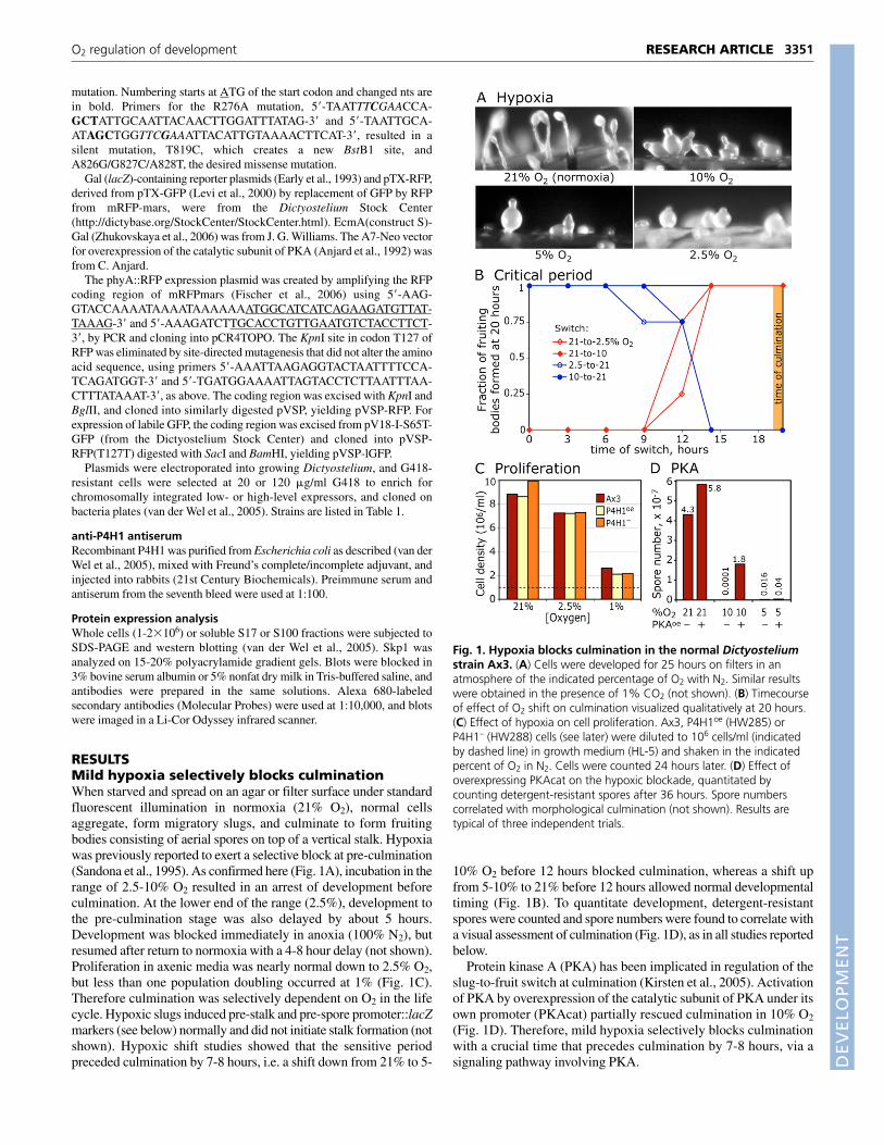
RESULTSMild hypoxia selectively blocks culminationWhen starved and spread on an agar or filter surface under standardfluorescent illumination in normoxia (21% O2), normal cellsaggregate, form migratory slugs, and culminate to form fruitingbodies consisting of aerial spores on top of a vertical stalk. Hypoxiawas previously reported to exert a selective block at pre-culmination(Sandona et al., 1995). As confirmed here (Fig. 1A), incubation in therange of 2.5-10% O2 resulted in an arrest of development beforeculmination. At the lower end of the range (2.5%), development tothe pre-culmination stage was also delayed by about 5 hours.Development was blocked immediately in anoxia (100% N2), butresumed after return to normoxia with a 4-8 hour delay (not shown).Proliferation in axenic media was nearly normal down to 2.5% O2,but less than one population doubling occurred at 1% (Fig. 1C).Therefore culmination was selectively dependent on O2 in the lifecycle. Hypoxic slugs induced pre-stalk and pre-spore promoter::lacZmarkers (see below) normally and did not initiate stalk formation (notshown). Hypoxic shift studies showed that the sensitive periodpreceded culmination by 7-8 hours, i.e. a shift down from 21% to 5-
10% O2 before 12 hours blocked culmination, whereas a shift upfrom 5-10% to 21% before 12 hours allowed normal developmentaltiming (Fig. 1B). To quantitate development, detergent-resistantspores were counted and spore numbers were found to correlate witha visual assessment of culmination (Fig. 1D), as in all studies reportedbelow.
Protein kinase A (PKA) has been implicated in regulation of theslug-to-fruit switch at culmination (Kirsten et al., 2005). Activationof PKA by overexpression of the catalytic subunit of PKA under itsown promoter (PKAcat) partially rescued culmination in 10% O2
(Fig. 1D). Therefore, mild hypoxia selectively blocks culminationwith a crucial time that precedes culmination by 7-8 hours, via asignaling pathway involving PKA.
3351RESEARCH ARTICLEO2 regulation of development
Fig. 1. Hypoxia blocks culmination in the normal Dictyosteliumstrain Ax3. (A) Cells were developed for 25 hours on filters in anatmosphere of the indicated percentage of O2 with N2. Similar resultswere obtained in the presence of 1% CO2 (not shown). (B) Timecourseof effect of O2 shift on culmination visualized qualitatively at 20 hours.(C) Effect of hypoxia on cell proliferation. Ax3, P4H1oe (HW285) orP4H1– (HW288) cells (see later) were diluted to 106 cells/ml (indicatedby dashed line) in growth medium (HL-5) and shaken in the indicatedpercent of O2 in N2. Cells were counted 24 hours later. (D) Effect ofoverexpressing PKAcat on the hypoxic blockade, quantitated bycounting detergent-resistant spores after 36 hours. Spore numberscorrelated with morphological culmination (not shown). Results aretypical of three independent trials.
DEVELO
PMENT
3352
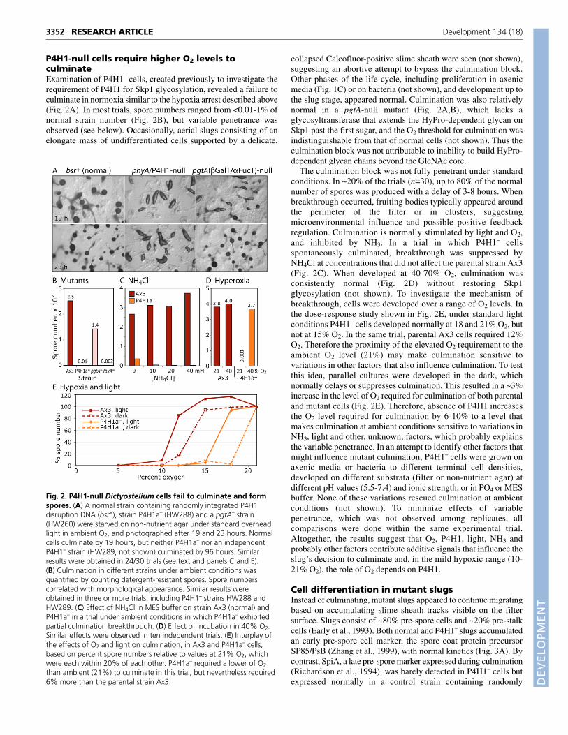
P4H1-null cells require higher O2 levels toculminateExamination of P4H1– cells, created previously to investigate therequirement of P4H1 for Skp1 glycosylation, revealed a failure toculminate in normoxia similar to the hypoxia arrest described above(Fig. 2A). In most trials, spore numbers ranged from <0.01-1% ofnormal strain number (Fig. 2B), but variable penetrance wasobserved (see below). Occasionally, aerial slugs consisting of anelongate mass of undifferentiated cells supported by a delicate,
collapsed Calcofluor-positive slime sheath were seen (not shown),suggesting an abortive attempt to bypass the culmination block.Other phases of the life cycle, including proliferation in axenicmedia (Fig. 1C) or on bacteria (not shown), and development up tothe slug stage, appeared normal. Culmination was also relativelynormal in a pgtA-null mutant (Fig. 2A,B), which lacks aglycosyltransferase that extends the HyPro-dependent glycan onSkp1 past the first sugar, and the O2 threshold for culmination wasindistinguishable from that of normal cells (not shown). Thus theculmination block was not attributable to inability to build HyPro-dependent glycan chains beyond the GlcNAc core.
The culmination block was not fully penetrant under standardconditions. In ~20% of the trials (n=30), up to 80% of the normalnumber of spores was produced with a delay of 3-8 hours. Whenbreakthrough occurred, fruiting bodies typically appeared aroundthe perimeter of the filter or in clusters, suggestingmicroenvironmental influence and possible positive feedbackregulation. Culmination is normally stimulated by light and O2,and inhibited by NH3. In a trial in which P4H1– cellsspontaneously culminated, breakthrough was suppressed byNH4Cl at concentrations that did not affect the parental strain Ax3(Fig. 2C). When developed at 40-70% O2, culmination wasconsistently normal (Fig. 2D) without restoring Skp1glycosylation (not shown). To investigate the mechanism ofbreakthrough, cells were developed over a range of O2 levels. Inthe dose-response study shown in Fig. 2E, under standard lightconditions P4H1– cells developed normally at 18 and 21% O2, butnot at 15% O2. In the same trial, parental Ax3 cells required 12%O2. Therefore the proximity of the elevated O2 requirement to theambient O2 level (21%) may make culmination sensitive tovariations in other factors that also influence culmination. To testthis idea, parallel cultures were developed in the dark, whichnormally delays or suppresses culmination. This resulted in a ~3%increase in the level of O2 required for culmination of both parentaland mutant cells (Fig. 2E). Therefore, absence of P4H1 increasesthe O2 level required for culmination by 6-10% to a level thatmakes culmination at ambient conditions sensitive to variations inNH3, light and other, unknown, factors, which probably explainsthe variable penetrance. In an attempt to identify other factors thatmight influence mutant culmination, P4H1– cells were grown onaxenic media or bacteria to different terminal cell densities,developed on different substrata (filter or non-nutrient agar) atdifferent pH values (5.5-7.4) and ionic strength, or in PO4 or MESbuffer. None of these variations rescued culmination at ambientconditions (not shown). To minimize effects of variablepenetrance, which was not observed among replicates, allcomparisons were done within the same experimental trial.Altogether, the results suggest that O2, P4H1, light, NH3 andprobably other factors contribute additive signals that influence theslug’s decision to culminate and, in the mild hypoxic range (10-21% O2), the role of O2 depends on P4H1.
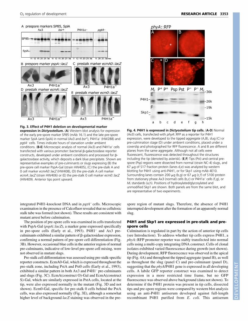
Cell differentiation in mutant slugsInstead of culminating, mutant slugs appeared to continue migratingbased on accumulating slime sheath tracks visible on the filtersurface. Slugs consist of ~80% pre-spore cells and ~20% pre-stalkcells (Early et al., 1993). Both normal and P4H1– slugs accumulatedan early pre-spore cell marker, the spore coat protein precursorSP85/PsB (Zhang et al., 1999), with normal kinetics (Fig. 3A). Bycontrast, SpiA, a late pre-spore marker expressed during culmination(Richardson et al., 1994), was barely detected in P4H1– cells butexpressed normally in a control strain containing randomly
RESEARCH ARTICLE Development 134 (18)
Fig. 2. P4H1-null Dictyostelium cells fail to culminate and formspores. (A) A normal strain containing randomly integrated P4H1disruption DNA (bsr+), strain P4H1a– (HW288) and a pgtA– strain(HW260) were starved on non-nutrient agar under standard overheadlight in ambient O2, and photographed after 19 and 23 hours. Normalcells culminate by 19 hours, but neither P4H1a– nor an independentP4H1– strain (HW289, not shown) culminated by 96 hours. Similarresults were obtained in 24/30 trials (see text and panels C and E).(B) Culmination in different strains under ambient conditions wasquantified by counting detergent-resistant spores. Spore numberscorrelated with morphological appearance. Similar results wereobtained in three or more trials, including P4H1– strains HW288 andHW289. (C) Effect of NH4Cl in MES buffer on strain Ax3 (normal) andP4H1a– in a trial under ambient conditions in which P4H1a– exhibitedpartial culmination breakthrough. (D) Effect of incubation in 40% O2.Similar effects were observed in ten independent trials. (E) Interplay ofthe effects of O2 and light on culmination, in Ax3 and P4H1a– cells,based on percent spore numbers relative to values at 21% O2, whichwere each within 20% of each other. P4H1a– required a lower of O2
than ambient (21%) to culminate in this trial, but nevertheless required6% more than the parental strain Ax3.
DEVELO
PMENT
integrated P4H1-knockout DNA and in pgtA– cells. Microscopicexamination in the presence of Calcofluor revealed that no cellulosicstalk tube was formed (not shown). These results are consistent withmutant arrest before culmination.
The position of pre-spore cells was examined in cells transfectedwith PspA-Gal (pspA::lacZ), a marker gene expressed specificallyin pre-spore cells (Early et al., 1993). P4H1– and Ax3 pre-culminants exhibited a similar pattern of �-galactosidase expression,confirming a normal pattern of pre-spore cell differentiation (Fig.3B). However, occasional blue cells in the anterior region of normalpre-culminants, indicative of low-level pre-spore cell mixing, werenot observed in mutant slugs.
Pre-stalk cell differentiation was assessed using pre-stalk-specificreporter constructs. EcmA0-Gal, which is expressed throughout thepre-stalk zone, including PstA and Pst0 cells (Early et al., 1993),exhibited a similar pattern in both Ax3 and P4H1– pre-culminantsand slugs (Fig. 3C). EcmA(construct O)-Gal and EcmA(constructS)-Gal, which are similarly expressed in PstA cells, located at thetip, were also expressed normally in the mutant (Fig. 3D and notshown). Ecm0-Gal, specific for pre-stalk 0 cells behind the PstAcells, was also expressed normally (Fig. 3E), although a somewhathigher level of background lacZ staining was observed in the pre-
spore region of mutant slugs. Therefore, the absence of P4H1interrupted development after the formation of an apparently normalslug.
P4H1 and Skp1 are expressed in pre-stalk and pre-spore cellsCulmination is regulated in part by the action of anterior tip cells(see Introduction). To address whether tip cells express P4H1, aphyA::RFP promoter reporter was stably transfected into normalcells using a multi-copy integrating DNA construct. Cells of clonalisolates exhibited varied fluorescence during growth (not shown).During development, RFP fluorescence was observed in the apicaltip (Fig. 4A) and throughout the tipped aggregate (panel B), as wellas throughout the slug (panel C) and pre-culminant (panel D),suggesting that the phyA/P4H1 gene is expressed in all developingcells. A labile GFP reporter construct was examined to detectexpression in a more restricted time frame, but no GFPfluorescence was observed above background (data not shown). Todetermine if the P4H1 protein was present in tip cells, dissectedtips and pre-spore regions were compared by western blot analysisusing an anti-P4H1 antiserum developed against full-lengthrecombinant P4H1 purified from E. coli. This antiserum
3353RESEARCH ARTICLEO2 regulation of development
Fig. 3. Effect of P4H1 deletion on developmental markerexpression in Dictyostelium. (A) Western blot analysis for expressionof the early pre-spore marker SP85 (mAb 16.1) and the late pre-sporemarker SpiA (anti-SpiA) in normal (Ax3 and bsr+), P4H1a– (HW288) andpgtA– cells. Times indicate hours of starvation under ambientconditions. (B-E) Microscopic analysis of normal (Ax3) and P4H1a– cellstransfected with various promoter::bacterial �-galactosidase reporterconstructs, developed under ambient conditions and processed for �-galactosidase activity, which deposits a dark blue precipitate. Shown arerepresentative examples of pre-culminants or slugs expressing (B) thepre-spore cell marker PspA-Gal (strain HW405), (C) the pre-stalk A and0 cell marker ecmA0::lacZ (HW408), (D) the pre-stalk A cell markerecmA::lacZ (strain HW406) or (E) the pre-stalk 0 cell marker ecm0::lacZ(HW409). Anterior tips point upward.
Fig. 4. P4H1 is expressed in Dictyostelium tip cells. (A-D) Normal(Ax3) cells, transfected with phyA::RFP as a reporter for P4H1expression, were developed to the tipped aggregate (A,B), slug (C) orpre-culmination stage (D) under ambient conditions, placed under acoverslip and photographed for RFP fluorescence. A and B are differentplanes from the same aggregate. Although not all cells werefluorescent, fluorescence was detected throughout the structuresincluding the tip (denoted by asterisk). (E,F) Tips (Pst) and central pre-spore (Psp) regions were dissected from normal (strain NC-4) slugs, and67 �g of S17 fraction protein (lanes d,e) was analyzed by westernblotting for P4H1 using anti-P4H1, or for Skp1 using mAb 4E10.Surrounding lanes contain 200 �g (b,g) or 67 �g (c,f) of S100 proteinfrom stationary phase Ax3 (normal) cells (b,c) or P4H1a– cells (f,g), orMr standards (a,h). Positions of hydroxylated/glycosylated andunmodified Skp1 are shown. Both panels are from the same blot, andare representative of two experiments.
DEVELO
PMENT
3354
recognizes P4H1 in extracts of normal stationary stage cells, but isnot reactive, except for a faint presumably cross-reacting band, inP4H1– cells (panel E). Preimmune serum was negative (data notshown). Similar levels of P4H1 were detected in both regions ofthe slug, suggesting that the P4H1 protein is constitutivelyexpressed. To address whether P4H1 is active, Skp1 was examinedby western blotting. Skp1 was present in similar levels in bothregions of the slug, and exhibited a mobility similar to that ofnormal Skp1 rather than that of Skp1 from P4H1– cells, which isnot hydroxylated and glycosylated (panel F). Altogether, the datastrongly suggest that P4H1 is expressed and active in the tip andthroughout the slug.
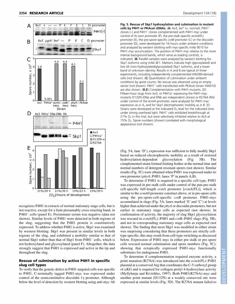
Rescue of culmination by active P4H1 in specificslug cell typesTo verify that the genetic defect in P4H1-targeted cells was specificto P4H1, C-terminally tagged P4H1-myc was expressed undercontrol of the semiconstitutive discoidin 1� promoter. Althoughbelow the level of detection by western blotting using anti-myc Ab
(Fig. 5A, lane ‘D’), expression was sufficient to fully modify Skp1based on reduced electrophoretic mobility as a result of restoredhydroxylation-dependent glycosylation (Fig. 5B). Thecomplemented strain formed fruiting bodies at the normal time andnormal numbers of detergent-resistant spores (not shown). Similarresults (Fig. 5C) were obtained when P4H1 was expressed under itsown promoter (phyA::P4H1; lanes ‘P’ in panels A,B).
To determine if P4H1 is required in a specific cell type, P4H1was expressed in pre-stalk cells under control of the pan-pre-stalkcell-specific full-length ecmA promoter [ecmA(FL)], which issimilar to the ecmA0 promoter construct above, or in pre-spore cellsusing the pre-spore-cell-specific cotB promoter. P4H1-mycaccumulated in slugs (Fig. 5A, lanes marked ‘E’ and ‘C’) at levelshigher than achieved under the phyA or discoidin promoters, but notearlier in stationary stage cells as expected (not shown). Inconfirmation of activity, the majority of slug Skp1 glycosylationwas rescued in ecmA(FL)::P4H1 and cotB::P4H1 slugs (Fig. 5B),and not in corresponding stationary stage cells as expected (notshown). The finding that most Skp1 was modified in either strainwas surprising considering that these promoters are strictly cell-type specific; this may result from cell-type switching as discussedbelow. Expression of P4H1-myc in either pre-stalk or pre-sporecells rescued normal culmination and spore numbers (Fig. 5C),showing that ectopically expressed P4H1-myc functionallysubstitutes for endogenous P4H1.
To determine if complementation required enzyme activity, apoint mutation (R276A) was introduced into the ecmA(FL)::P4H1plasmid at a conserved Arg that coordinates the C-5 carboxyl groupof �KG and is required for collagen prolyl 4-hydroxylase activity(Myllyharju and Kivirikko, 1997). Both P4H1(R276A)-myc andanother point mutant (D132N) at a weakly conserved site wereexpressed at similar levels (Fig. 5D). The R276A mutant failed to
RESEARCH ARTICLE Development 134 (18)
Fig. 5. Rescue of Skp1 hydroxylation and culmination in mutantcells by P4H1 or PKAcat cDNAs. (A) Ax3, bsr+ (+, normal), P4H1–
clones (–) and P4H1– clones complemented with P4H1-myc undercontrol of its own promoter (P), the pre-stalk-specific ecmA(FL)promoter (E), the pre-spore-specific cotB promoter (C) or the discoidinpromoter (D), were developed for 16 hours under ambient conditionsand analyzed by western blotting with myc-specific mAb 9E10 forP4H1-myc accumulation. The position of P4H1-myc relative to the moreintense background bands, which serve as loading controls, isindicated. (B) Parallel samples were analyzed by western blotting forSkp1 isoforms using mAb 4E1. Markers indicate high (glycosylated) andlow Mr (non-hydroxylated/glycosylated) Skp1 isoforms, and a lowerband of unknown identity. Results in A and B are typical of threeexperiments, including independently complemented HW289-derivedcells (not shown). (C) Quantitation of culmination under ambientconditions by spore counts. No rescue was observed using an emptyvector (not shown). P4H1– cells transfected with PKAcat (strain HW410)are also shown. (D,E) Complementation with P4H1-mutants. (D)Fifteen-hour slugs from Ax3, or P4H1a– expressing the P4H1-mycmutants D132N (DNa and DNb are independent clones) or R276A (RA)under control of the ecmA-promoter, were analyzed for P4H1-mycexpression as in A, and for Skp1 electrophoretic mobility as in B. (E)Strains were developed at the indicated O2 level for the indicated timeunder strong overhead light. P4H1– cells exhibited breakthrough at21% O2 in this trial, but were selectively inhibited relative to Ax3 at15% O2. Spore numbers (shown) correlated with morphologicalappearance (not shown).
DEVELO
PMENT
modify the electrophoretic mobility of Skp1 consistent with theexpected absence of P4H1 activity. These cells did not culminate(Fig. 5E), indicating that the prolyl 4-hydroxylase activity of P4H1is required for complementation. By contrast, the D132N mutant,which partially restored Skp1 hydroxylation/glycosylationconsistent with expression in only pre-stalk cells, fully supportedculmination. Therefore P4H1 requires its prolyl 4-hydroxylaseactivity to complement its deficiency.
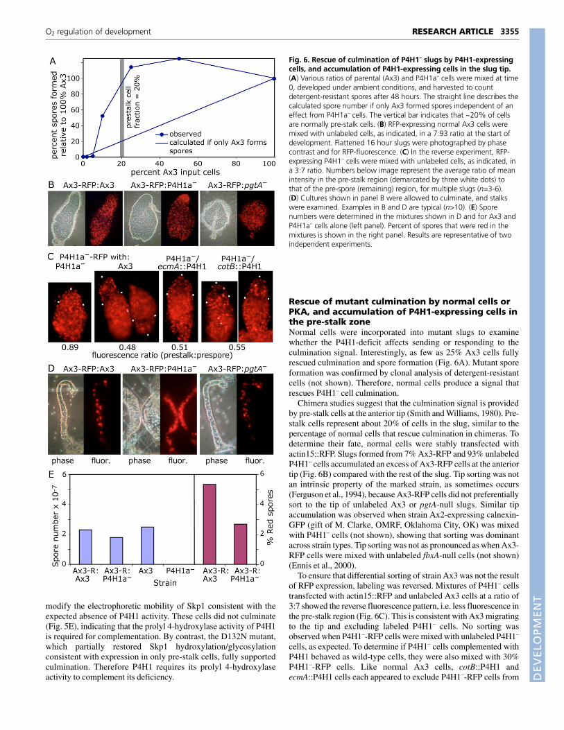
Rescue of mutant culmination by normal cells orPKA, and accumulation of P4H1-expressing cells inthe pre-stalk zoneNormal cells were incorporated into mutant slugs to examinewhether the P4H1-deficit affects sending or responding to theculmination signal. Interestingly, as few as 25% Ax3 cells fullyrescued culmination and spore formation (Fig. 6A). Mutant sporeformation was confirmed by clonal analysis of detergent-resistantcells (not shown). Therefore, normal cells produce a signal thatrescues P4H1– cell culmination.
Chimera studies suggest that the culmination signal is providedby pre-stalk cells at the anterior tip (Smith and Williams, 1980). Pre-stalk cells represent about 20% of cells in the slug, similar to thepercentage of normal cells that rescue culmination in chimeras. Todetermine their fate, normal cells were stably transfected withactin15::RFP. Slugs formed from 7% Ax3-RFP and 93% unlabeledP4H1– cells accumulated an excess of Ax3-RFP cells at the anteriortip (Fig. 6B) compared with the rest of the slug. Tip sorting was notan intrinsic property of the marked strain, as sometimes occurs(Ferguson et al., 1994), because Ax3-RFP cells did not preferentiallysort to the tip of unlabeled Ax3 or pgtA-null slugs. Similar tipaccumulation was observed when strain Ax2-expressing calnexin-GFP (gift of M. Clarke, OMRF, Oklahoma City, OK) was mixedwith P4H1– cells (not shown), showing that sorting was dominantacross strain types. Tip sorting was not as pronounced as when Ax3-RFP cells were mixed with unlabeled fbxA-null cells (not shown)(Ennis et al., 2000).
To ensure that differential sorting of strain Ax3 was not the resultof RFP expression, labeling was reversed. Mixtures of P4H1– cellstransfected with actin15::RFP and unlabeled Ax3 cells at a ratio of3:7 showed the reverse fluorescence pattern, i.e. less fluorescence inthe pre-stalk region (Fig. 6C). This is consistent with Ax3 migratingto the tip and excluding labeled P4H1– cells. No sorting wasobserved when P4H1–-RFP cells were mixed with unlabeled P4H1–
cells, as expected. To determine if P4H1– cells complemented withP4H1 behaved as wild-type cells, they were also mixed with 30%P4H1–-RFP cells. Like normal Ax3 cells, cotB::P4H1 andecmA::P4H1 cells each appeared to exclude P4H1–-RFP cells from
3355RESEARCH ARTICLEO2 regulation of development
Fig. 6. Rescue of culmination of P4H1– slugs by P4H1-expressingcells, and accumulation of P4H1-expressing cells in the slug tip.(A) Various ratios of parental (Ax3) and P4H1a– cells were mixed at time0, developed under ambient conditions, and harvested to countdetergent-resistant spores after 48 hours. The straight line describes thecalculated spore number if only Ax3 formed spores independent of aneffect from P4H1a– cells. The vertical bar indicates that ~20% of cellsare normally pre-stalk cells. (B) RFP-expressing normal Ax3 cells weremixed with unlabeled cells, as indicated, in a 7:93 ratio at the start ofdevelopment. Flattened 16 hour slugs were photographed by phasecontrast and for RFP-fluorescence. (C) In the reverse experiment, RFP-expressing P4H1– cells were mixed with unlabeled cells, as indicated, ina 3:7 ratio. Numbers below image represent the average ratio of meanintensity in the pre-stalk region (demarcated by three white dots) tothat of the pre-spore (remaining) region, for multiple slugs (n=3-6).(D) Cultures shown in panel B were allowed to culminate, and stalkswere examined. Examples in B and D are typical (n>10). (E) Sporenumbers were determined in the mixtures shown in D and for Ax3 andP4H1a– cells alone (left panel). Percent of spores that were red in themixtures is shown in the right panel. Results are representative of twoindependent experiments.
DEVELO
PMENT
3356
the pre-stalk zone, as shown in Fig. 6C. P4H1– cells expressingcotB::P4H1 therefore must migrate anteriorly after first expressingP4H1 posteriorly in the pre-spore zone, where cotB is active. Thisbehavior is consistent with their signaling culmination from the tip,as inferred from the similar behavior of wild-type cells and whenP4H1 is expressed in pre-stalk cells by means of the ecmA promoter.
To determine if the tip-associated Ax3-RFP cells differentiate aspre-stalk A (PstA) cells, which are precursors of the cellular stalk,their fate in the fruiting body was examined. Ax3-RFP cells wereenriched in the stalk formed from mixtures with P4H1– relative tomixtures formed with unlabeled Ax3 or pgtA-null cells (Fig. 6D). Inaddition, a smaller fraction of spores was fluorescent in chimeras ofAx3-RFP with unlabeled P4H1– cells compared with unlabeled Ax3cells (Fig. 6E). The differentiation of tip-associated Ax3-RFP cellsas stalk cells confirms their conversion to functional PstA cells in theslug, consistent with their proposed ability to signal culminationfrom the anterior tip.
Overexpression of PKAcat under its own promoter in P4H1– cellspartially rescued culmination (Fig. 5C). As in O2 signaling (Fig. 1D),P4H1 therefore appears to function upstream of PKA, although thepossibility that PKA functions as a bypass suppressor cannot beexcluded.
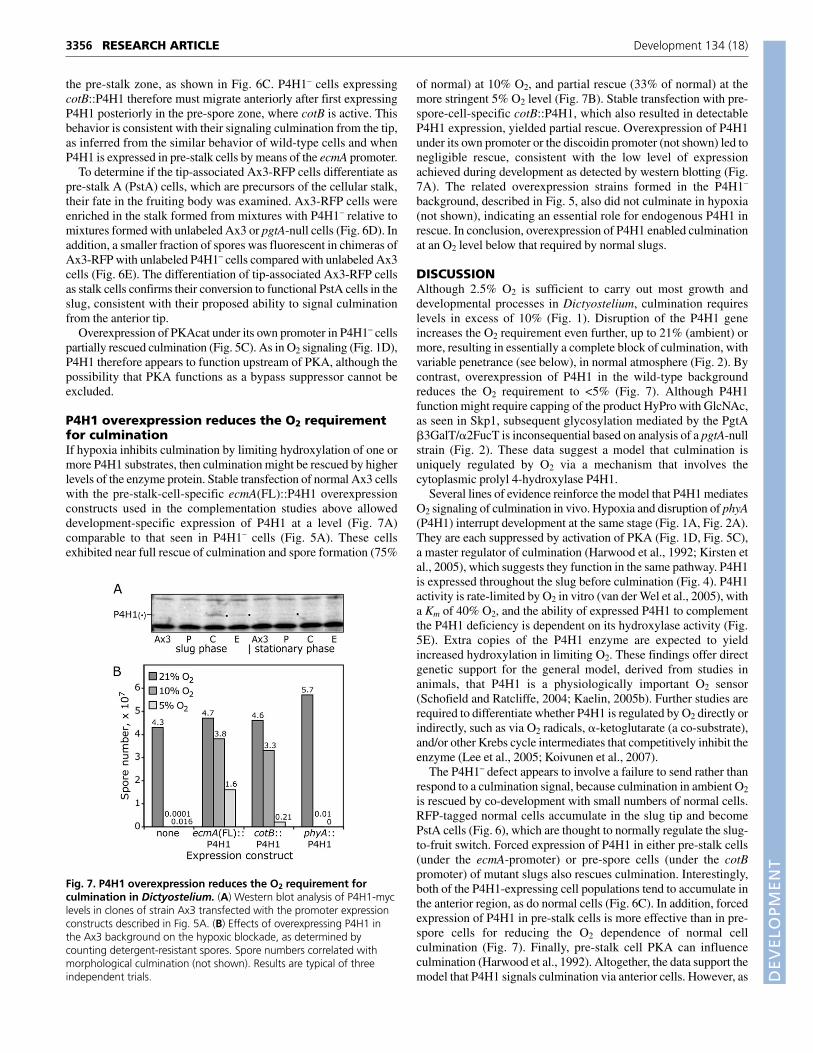
P4H1 overexpression reduces the O2 requirementfor culminationIf hypoxia inhibits culmination by limiting hydroxylation of one ormore P4H1 substrates, then culmination might be rescued by higherlevels of the enzyme protein. Stable transfection of normal Ax3 cellswith the pre-stalk-cell-specific ecmA(FL)::P4H1 overexpressionconstructs used in the complementation studies above alloweddevelopment-specific expression of P4H1 at a level (Fig. 7A)comparable to that seen in P4H1– cells (Fig. 5A). These cellsexhibited near full rescue of culmination and spore formation (75%
of normal) at 10% O2, and partial rescue (33% of normal) at themore stringent 5% O2 level (Fig. 7B). Stable transfection with pre-spore-cell-specific cotB::P4H1, which also resulted in detectableP4H1 expression, yielded partial rescue. Overexpression of P4H1under its own promoter or the discoidin promoter (not shown) led tonegligible rescue, consistent with the low level of expressionachieved during development as detected by western blotting (Fig.7A). The related overexpression strains formed in the P4H1–
background, described in Fig. 5, also did not culminate in hypoxia(not shown), indicating an essential role for endogenous P4H1 inrescue. In conclusion, overexpression of P4H1 enabled culminationat an O2 level below that required by normal slugs.
DISCUSSIONAlthough 2.5% O2 is sufficient to carry out most growth anddevelopmental processes in Dictyostelium, culmination requireslevels in excess of 10% (Fig. 1). Disruption of the P4H1 geneincreases the O2 requirement even further, up to 21% (ambient) ormore, resulting in essentially a complete block of culmination, withvariable penetrance (see below), in normal atmosphere (Fig. 2). Bycontrast, overexpression of P4H1 in the wild-type backgroundreduces the O2 requirement to <5% (Fig. 7). Although P4H1function might require capping of the product HyPro with GlcNAc,as seen in Skp1, subsequent glycosylation mediated by the PgtA�3GalT/�2FucT is inconsequential based on analysis of a pgtA-nullstrain (Fig. 2). These data suggest a model that culmination isuniquely regulated by O2 via a mechanism that involves thecytoplasmic prolyl 4-hydroxylase P4H1.
Several lines of evidence reinforce the model that P4H1 mediatesO2 signaling of culmination in vivo. Hypoxia and disruption of phyA(P4H1) interrupt development at the same stage (Fig. 1A, Fig. 2A).They are each suppressed by activation of PKA (Fig. 1D, Fig. 5C),a master regulator of culmination (Harwood et al., 1992; Kirsten etal., 2005), which suggests they function in the same pathway. P4H1is expressed throughout the slug before culmination (Fig. 4). P4H1activity is rate-limited by O2 in vitro (van der Wel et al., 2005), witha Km of 40% O2, and the ability of expressed P4H1 to complementthe P4H1 deficiency is dependent on its hydroxylase activity (Fig.5E). Extra copies of the P4H1 enzyme are expected to yieldincreased hydroxylation in limiting O2. These findings offer directgenetic support for the general model, derived from studies inanimals, that P4H1 is a physiologically important O2 sensor(Schofield and Ratcliffe, 2004; Kaelin, 2005b). Further studies arerequired to differentiate whether P4H1 is regulated by O2 directly orindirectly, such as via O2 radicals, �-ketoglutarate (a co-substrate),and/or other Krebs cycle intermediates that competitively inhibit theenzyme (Lee et al., 2005; Koivunen et al., 2007).
The P4H1– defect appears to involve a failure to send rather thanrespond to a culmination signal, because culmination in ambient O2
is rescued by co-development with small numbers of normal cells.RFP-tagged normal cells accumulate in the slug tip and becomePstA cells (Fig. 6), which are thought to normally regulate the slug-to-fruit switch. Forced expression of P4H1 in either pre-stalk cells(under the ecmA-promoter) or pre-spore cells (under the cotBpromoter) of mutant slugs also rescues culmination. Interestingly,both of the P4H1-expressing cell populations tend to accumulate inthe anterior region, as do normal cells (Fig. 6C). In addition, forcedexpression of P4H1 in pre-stalk cells is more effective than in pre-spore cells for reducing the O2 dependence of normal cellculmination (Fig. 7). Finally, pre-stalk cell PKA can influenceculmination (Harwood et al., 1992). Altogether, the data support themodel that P4H1 signals culmination via anterior cells. However, as
RESEARCH ARTICLE Development 134 (18)
Fig. 7. P4H1 overexpression reduces the O2 requirement forculmination in Dictyostelium. (A) Western blot analysis of P4H1-myclevels in clones of strain Ax3 transfected with the promoter expressionconstructs described in Fig. 5A. (B) Effects of overexpressing P4H1 inthe Ax3 background on the hypoxic blockade, as determined bycounting detergent-resistant spores. Spore numbers correlated withmorphological culmination (not shown). Results are typical of threeindependent trials.
DEVELO
PMENT
sorting of pre-spore cells expressing P4H1 is not absolute and P4H1is expressed in all slug cells (Fig. 4), a role for pre-spore cell P4H1in culmination is not excluded.
P4H1 appears to influence culmination jointly with otherenvironmental factors. O2 and P4H1 function synergistically withoverhead light (Newell et al., 1969) to regulate culmination. The O2
requirement of both normal and mutant cells is decreased by 2-3%when cells are developed in overhead light compared with darkness(Fig. 2E). Previous studies suggested that overhead light attracts thetip aerially, which promotes culmination indirectly (Bonner et al.,1982), although in the present study of stalled pre-culminants, tipsappeared to be elevated from the substratum in either light or darkconditions (not shown). The slug-to-fruit switch is also normallyregulated by NH3 (Kirsten et al., 2005), a catabolic by-product ofslug metabolism. NH3 inhibits culmination in P4H1– cells at lowerconcentrations than those required to affect normal cells (Fig. 2C),as described for other slugger mutants (Gee et al., 1994), suggestingthat O2 and NH3 signaling interact. Evidence indicates that NH3, O2
and P4H1 each influence culmination via PKA. Geneticmanipulations that affect superoxide levels and the cellular redoxbalance also affect culmination (Garcia et al., 2003; Jeong et al.,2006; Choi et al., 2006) and, as PHD function depends on O2,reduced Fe+2 and ascorbate, which may be affected by superoxides(Kaelin, 2005a; Kaelin, 2005b), these agents might also signal viaP4H1. In addition, hyperoxia rescues P4H1– culmination (Fig. 2D),which suggests a second mechanism by which O2 regulatesculmination. This might involve one of the other four PHD-likegenes in the Dictyostelium genome (West et al., 2004). The slug-to-fruit switch is evidently regulated by a complex pattern ofextracellular signals, and subtle variations, in concert withmicroenvironmental effects and potential positive feedbackinteractions between slugs suggested by edge and group effects, mayexplain the 18-21+% range of O2 required for culmination indifferent trials.
A candidate target of P4H1 action is Skp1, the glycosylation ofwhich requires P4H1 in Dictyostelium and which was used tomonitor P4H1 activity in the mutants. However, Skp1 isglycosylated normally, within the resolution of western blotanalysis, in slugs of Ax3, and even HW403 (cotB::phyA in P4H1–
background) or HW404 (corresponding ecmA::phyA expressionstrain), formed in 5-10% O2 (our unpublished data). In addition,Skp1 is not glycosylated in 40% O2 (not shown), which overridesinhibition of culmination in P4H1– cells. Although these resultsshow that hydroxylation of the bulk pool of Skp1 is not regulatedin parallel with culmination, a role for Skp1 is not excluded, as acritical subpool (e.g. nascent, nuclear or pre-stalk) might bedifferentially hydroxylated. Skp1 is the only substrate thataccumulates in P4H1– cells based on an indirect assay with rP4H1and rGnT1 (our unpublished data). A role for Skp1 is furthersupported by evidence that CulA and FbxA, proteins thatphysically associate with Skp1 in SCF-type E3 Ub-ligasecomplexes (Willems et al., 2004), are also required for multipledevelopmental steps, including culmination (Mohanty et al., 2001;Nelson et al., 2000; Ennis et al., 2000). However, P4H1– slugs donot accumulate the E3(SCFFbxA)Ub-ligase substrate RegA (notshown), as occurs in culA- and fbxA-null cells (Mohanty et al.,2001). Yet it is intriguing that the SCF class of E3-Ub-ligases,which contains HyPro-Skp1, is evolutionarily related to the VHLclass that recognizes HyPro-HIF� in animals. Additional studiesare needed to evaluate the O2 dependence of the hydroxylation ofSkp1, which is encoded by two genes, in the slug tip, and the roleof GnT1, which mediates addition of the GlcNAc cap to HyPro-
Skp1 (van der Wel et al., 2002b). The best-known substrate ofanimal PHDs, HIF1� and the E3VHLUb-ligase that recognizesHyPro-HIF1�, are apparently absent from the Dictyosteliumgenome based on bioinformatics studies (not shown), but otherpotential targets (Kuznetsova et al., 2003) occur.
Hypoxic regulation of culmination may provide a selectiveadvantage to Dictyostelium in its normal habitat. Hypoxiaencountered by developing cells in confined or water-saturatedmicroenvironments may signal delay of culmination. In this scheme,O2 synergizes with NH3 depletion and light, which can override asuboptimal level of O2 (Fig. 2E). The crucial period for O2 regulationis many hours in advance of culmination (Fig. 1B), and cells havingthe highest P4H1 activity tend to migrate to the slug tip (Fig. 6),suggesting complexities that remain to be explored. Anotherfunction for O2 in Dictyostelium, cytochrome oxidase subunit VIIswitching (Sandona et al., 1995), does not involve P4H1 based onDNA microarray studies (L. Eichinger and C.M.W., unpublished).The role of P4H1 in O2-dependent slug polarization and guidance ofslug migration (Sternfeld and Bonner, 1977; Sternfeld and David,1981) remains to be examined. Because hypoxic regulation isconnected with sporulation, Dictyostelium offers a unique geneticopportunity to investigate the mechanism of P4H1 action, includingits role as a direct O2 sensor and the identification of upstream anddownstream regulatory genes.
This work was supported by NIH grant GM-37539. We thank C. Anjard, W. F.Loomis, M. Clarke, J. G. Williams, D. Traynor, R. Kay, and the DictyosteliumStock Center for plasmids, strains and antisera.
ReferencesAnjard, C., Pinaud, S., Kay, R. R. and Reymond, C. D. (1992). Overexpression of
Dd PK2 protein kinase causes rapid development and affects the intracellularcAMP pathway of Dictyostelium discoideum. Development 115, 785-790.
Bonner, J. T., Davidowski, T. A., Hsu, W.-L., Lapeyrolerie, D. A. and Suthers,H. L. B. (1982). The role of surface water and light on differentiation in thecellular slime molds. Differentiation 21, 123-126.
Briere, J. J., Favier, J., Benit, P., Ghouzzi, V. E., Lorenzato, A., Rabier, D., DiRenzo, M. F., Gimenez-Roqueplo, A. P. and Rustin, P. (2005). Mitochondrialsuccinate is instrumental for HIF1� nuclear translocation in SDHA-mutantfibroblasts under normoxic conditions. Hum. Mol. Genet. 14, 3263-3269.
Choi, C. H., Kim, B. J., Jeong, S. Y., Lee, C. H., Kim, J. S., Park, S. J., Yim, H. S.and Kang, S. O. (2006). Reduced glutathione levels affect the culmination andcell fate decision in Dictyostelium discoideum. Dev. Biol. 295, 523-533.
Dann, C. E., 3rd and Bruick, R. K. (2005). Dioxygenases as O2-dependentregulators of the hypoxic response pathway. Biochem. Biophys. Res. Commun.338, 639-647.
Early, A. E., Gaskell, M. J., Traynor, D. and Williams, J. G. (1993). Two distinctpopulations of prestalk cells within the tip of the migratory Dictyostelium slugwith differing fates at culmination. Development 118, 353-362.
Eichinger, L., Pachebat, J. A., Glockner, G., Rajandream, M. A., Sucgang, R.,Berriman, M., Song, J., Olsen, R., Szafranski, K., Xu, Q. et al. (2005). Thegenome of the social amoeba Dictyostelium discoideum. Nature 435, 43-57.
Ennis, H. L., Dao, D. N., Pukatzki, S. U. and Kessin, R. H. (2000). Dictyosteliumamoebae lacking an F-box protein form spores rather than stalk in chimeras withwild type. Proc. Natl. Acad. Sci. USA 97, 3292-3297.
Ferguson, T. F., Vozenilek, J. and West, C. M. (1994). The differentiation of acell sorting mutant of Dictyostelium discoideum. Dev. Growth Differ. 36, 597-604.
Fischer, M., Haase, I., Wiesner, S. and Muller-Taubenberger, A. (2006).Visualizing cytoskeleton dynamics in mammalian cells using a humanized variantof monomeric red fluorescent protein. FEBS Lett. 580, 2495-2502.
Fosnaugh, K. L. and Loomis, W. F. (1993). Enhancer regions responsible fortemporal and cell-type-specific expression of a spore coat gene in Dictyostelium.Dev. Biol. 157, 38-48.
Garcia, M. X., Alexander, H., Mahadeo, D., Cotter, D. A. and Alexander, S.(2003). The Dictyostelium discoideum prespore-specific catalase B functions tocontrol late development and to protect spore viability. Biochim. Biophys. Acta1641, 55-64.
Gee, K., Russell, F. and Gross, J. D. (1994). Ammonia hypersensitivity of sluggermutants of D. discoideum. J. Cell Sci. 107, 701-708.
Harwood, A. J., Hopper, N. A., Simon, M. N., Driscoll, D. M., Veron, M. andWilliams, J. G. (1992). Culmination in Dictyostelium is regulated by the cAMP-dependent protein kinase. Cell 69, 615-624.
3357RESEARCH ARTICLEO2 regulation of development
DEVELO
PMENT
3358
Jeong, S. Y., Choi, C. H., Kim, J. S., Park, S. J. and Kang, S. O. (2006).Thioredoxin reductase is required for growth and regulates entry intoculmination of Dictyostelium discoideum. Mol. Microbiol. 61, 1443-1456.
Kaelin, W. G., Jr (2005a). ROS: really involved in oxygen sensing. Cell Metab. 1,357-358.
Kaelin, W. G., Jr (2005b). Proline hydroxylation and gene expression. Annu. Rev.Biochem. 74, 115-128.
Kessin, R. H. (2001). Dictyostelium: Evolution, Cell Biology, and the Developmentof Multicellularity. Cambridge: Cambridge University Press.
Kirsten, J. H., Xiong, Y., Dunbar, A. J., Rai, M. and Singleton, C. K. (2005).Ammonium transporter C of Dictyostelium discoideum is required for correctprestalk gene expression and for regulating the choice between slug migrationand culmination. Dev. Biol. 287, 146-156.
Koivunen, P., Hirsila, M., Remes, A. M., Hassinen, I. E., Kivirikko, K. I. andMyllyharju, J. (2007). Inhibition of hypoxia-inducible factor (HIF) hydroxylasesby citric acid cycle intermediates: possible links between cell metabolism andstabilization of HIF. J. Biol. Chem. 282, 4524-4532.
Kuznetsova, A. V., Meller, J., Schnell, P. O., Nash, J. A., Ignacak, M. L.,Sanchez, Y., Conaway, J. W., Conaway, R. C. and Czyzyk-Krzeska, M. F.(2003). von-Hippel-Lindau protein binds hyperphosphorylated large subunit ofRNA polymerase II through a proline hydroxylation motif and targets it forubiquitination. Proc. Natl. Acad. Sci. USA 100, 2706-2711.
Lee, S., Nakamura, E., Yang, H., Wei, W., Linggi, M. S., Sajan, M. P., Farese,R. V., Freeman, R. S., Carter, B. D., Kaelin, W. G., Jr et al. (2005). Neuronalapoptosis linked to EglN3 prolyl hydroxylase and familial pheochromocytomagenes: developmental culling and cancer. Cancer Cell 8, 155-167.
Levi, S., Polyakov, M. and Egelhoff, T. T. (2000). Green fluorescent protein andepitope tag fusion vectors for Dictyostelium discoideum. Plasmid 44, 231-238.
Mohanty, S., Lee, S., Yadava, N., Dealy, M. J., Johnson, R. S. and Firtel, R. A.(2001). Regulated protein degradation controls PKA function and cell-typedifferentiation in Dictyostelium. Genes Dev. 15, 1435-1448.
Myllyharju, J. and Kivirikko, K. I. (1997). Characterization of the iron- and 2-oxoglutarate-binding sites of human prolyl 4-hydroxylase. EMBO J. 16, 1173-1180.
Nelson, M. K., Clark, A., Abe, T., Nomura, A., Yadava, N., Funair, C. J.,Jermyn, K. A., Mohanty, S., Firtel, R. A. and Williams, J. G. (2000). An F-Box/WD40 repeat-containing protein important for Dictyostelium cell-typeproportioning, slug behaviour, and culmination. Dev. Biol. 224, 42-59.
Newell, P. C. and Ross, F. M. (1982). Genetic analysis of the slug stage ofDictyostelium discoideum. J. Gen. Microbiol. 128, 1639-1652.
Newell, P. C., Telser, A. and Sussman, M. (1969). Alternative developmentalpathways determined by environmental conditions in the cellular slime moldDictyostelium discoideum. J. Bacteriol. 100, 763-768.
Richardson, D. L., Loomis, W. F. and Kimmel, A. R. (1994). Progression of aninductive signal activates sporulation in Dictyostelium discoideum. Development120, 2891-2900.
Sandona, D., Gastaldello, S., Rizzuto, R. and Bisson, R. (1995). Expression ofcytochrome c oxidase during growth and development of Dictyostelium. J. Biol.Chem. 270, 5587-5593.
Sassi, S., Sweetinburgh, M., Erogul, J., Zhang, P., Teng-umnuay, P. and West,C. M. (2001). Analysis of Skp1 glycosylation and nuclear enrichment inDictyostelium. Glycobiology 11, 283-295.
Schindler, J. and Sussman, M. (1977). Ammonia determines the choice ofmorphogenetic pathways in Dictyostelium discoideum. J. Mol. Biol. 116, 161-169.
Schofield, C. J. and Ratcliffe, P. J. (2004). Oxygen sensing by HIF hydroxylases.Nat. Rev. Mol. Cell Biol. 5, 343-354.
Singleton, C. K., Kirsten, J. H. and Dinsmore, C. J. (2006). Function ofammonium transporter A in the initiation of culmination of development inDictyostelium discoideum. Eukaryotic Cell 5, 991-996.
Smith, E. and Williams, K. L. (1980). Evidence for tip control of the ‘slug/fruit’switch in slugs of Dictyostelium discoideum. J. Embryol. Exp. Morphol. 57, 233-240.
Sternfeld, J. and Bonner, J. T. (1977). Cell differentiation in Dictyostelium undersubmerged conditions. Proc. Natl. Acad. Sci. USA 74, 268-271.
Sternfeld, J. and David, C. N. (1981). Oxygen gradients cause pattern orientationin Dictyostelium cell clumps. J. Cell Sci. 50, 9-17.
Teng-umnuay, P., Morris, H. R., Dell, A., Panico, M., Paxton, T. and West,C. M. (1998). The cytoplasmic F-box binding protein Skp1 contains a novelpentasaccharide linked to hydroxyproline in Dictyostelium. J. Biol. Chem. 273,18242-18249.
Thomason, P. A., Sawai, S., Stock, J. B. and Cox, E. C. (2006). The histidinekinase homologue DhkK/Sombrero controls morphogenesis in Dictyostelium.Dev. Biol. 292, 358-370.
van der Wel, H., Fisher, S. Z. and West, C. M. (2002a). A bifunctionaldiglycosyltransferase forms the Fuc�1,2Gal�3-disaccharide on Skp1 in thecytoplasm of Dictyostelium. J. Biol. Chem. 277, 46527-46534.
van der Wel, H., Morris, H. R., Panico, M., Paxton, T., Dell, A., Kaplan, L. andWest, C. M. (2002b). Molecular cloning and expression of a UDP-GlcNAc:hydroxyproline polypeptide GlcNAc-transferase that modifies Skp1 inthe cytoplasm of Dictyostelium. J. Biol. Chem. 277, 46328-46337.
van der Wel, H., Ercan, A. and West, C. M. (2005). The Skp1 prolyl hydroxylaseof Dictyostelium is related to the HIF�-class of animal prolyl 4-hydroxylases. J.Biol. Chem. 280, 14645-14655.
West, C. M. and McMahon, D. (1979). The axial distribution of plasmamembrane molecules in pseudoplasmodia of Dictyostelium discoideum. Exp. CellRes. 124, 393-401.
West, C. M., van der Wel, H., Sassi, S. and Gaucher, E. A. (2004). Cytoplasmicglycosylation of protein-hydroxyproline and its relationship to other glycosylationpathways. Biochim. Biophys. Acta 1673, 29-44.
West, C. M., van der Wel, H. and Blader, I. J. (2006). Detection of cytoplasmicglycosylation associated with hydroxyproline. Meth. Enzymol. 417, 385-400.
Willems, A. R., Schwab, M. and Tyers, M. (2004). A hitchhiker’s guide to thecullin ubiquitin ligases: SCF and its kin. Biochim. Biophys. Acta 1695, 133-170.
Zhang, Y., Zhang, P. and West, C. M. (1999). A linking function for the cellulose-binding protein SP85 in the spore coat of Dictyostelium discoideum. J. Cell Sci.112, 4367-4377.
Zheng, N., Schulman, B. A., Song, L., Miller, J. J., Jeffrey, P. D., Wang, P.,Chu, C., Koepp, D. M., Elledge, S. J., Pagano, M. et al. (2002). Structure ofthe Cul1-Rbx1-Skp1-F boxSkp2 SCF ubiquitin ligase complex. Nature 416, 703-709.
Zhukovskaya, N. V., Fukuzawa, M., Yamada, Y., Araki, T. and Williams, J. G.(2006). The Dictyostelium bZIP transcription factor DimB regulates prestalk-specific gene expression. Development 133, 439-448.
RESEARCH ARTICLE Development 134 (18)