Embed Size (px)
Citation preview
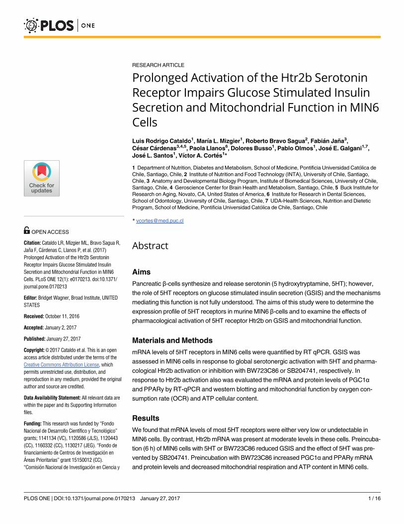
RESEARCH ARTICLE
Prolonged Activation of the Htr2b Serotonin
Receptor Impairs Glucose Stimulated Insulin
Secretion and Mitochondrial Function in MIN6
Cells
Luis Rodrigo Cataldo1, Marıa L. Mizgier1, Roberto Bravo Sagua2, Fabian Jana3,
Cesar Cardenas3,4,5, Paola Llanos6, Dolores Busso1, Pablo Olmos1, Jose E. Galgani1,7,
Jose L. Santos1, Vıctor A. Cortes1*
1 Department of Nutrition, Diabetes and Metabolism, School of Medicine, Pontificia Universidad Catolica de
Chile, Santiago, Chile, 2 Institute of Nutrition and Food Technology (INTA), University of Chile, Santiago,
Chile, 3 Anatomy and Developmental Biology Program, Institute of Biomedical Sciences, University of Chile,
Santiago, Chile, 4 Geroscience Center for Brain Health and Metabolism, Santiago, Chile, 5 Buck Institute for
Research on Aging, Novato, CA, United States of America, 6 Institute for Research in Dental Sciences,
School of Odontology, University of Chile, Santiago, Chile, 7 UDA-Health Sciences, Nutrition and Dietetic
Program, School of Medicine, Pontificia Universidad Catolica de Chile, Santiago, Chile
Abstract
Aims
Pancreatic β-cells synthesize and release serotonin (5 hydroxytryptamine, 5HT); however,
the role of 5HT receptors on glucose stimulated insulin secretion (GSIS) and the mechanisms
mediating this function is not fully understood. The aims of this study were to determine the
expression profile of 5HT receptors in murine MIN6 β-cells and to examine the effects of
pharmacological activation of 5HT receptor Htr2b on GSIS and mitochondrial function.
Materials and Methods
mRNA levels of 5HT receptors in MIN6 cells were quantified by RT qPCR. GSIS was
assessed in MIN6 cells in response to global serotonergic activation with 5HT and pharma-
cological Htr2b activation or inhibition with BW723C86 or SB204741, respectively. In
response to Htr2b activation also was evaluated the mRNA and protein levels of PGC1αand PPARy by RT-qPCR and western blotting and mitochondrial function by oxygen con-
sumption rate (OCR) and ATP cellular content.
Results
We found that mRNA levels of most 5HT receptors were either very low or undetectable in
MIN6 cells. By contrast, Htr2b mRNA was present at moderate levels in these cells. Preincuba-
tion (6 h) of MIN6 cells with 5HT or BW723C86 reduced GSIS and the effect of 5HT was pre-
vented by SB204741. Preincubation with BW723C86 increased PGC1α and PPARy mRNA
and protein levels and decreased mitochondrial respiration and ATP content in MIN6 cells.
PLOS ONE | DOI:10.1371/journal.pone.0170213 January 27, 2017 1 / 16
a1111111111
a1111111111
a1111111111
a1111111111
a1111111111
OPENACCESS
Citation: Cataldo LR, Mizgier ML, Bravo Sagua R,
Jana F, Cardenas C, Llanos P, et al. (2017)
Prolonged Activation of the Htr2b Serotonin
Receptor Impairs Glucose Stimulated Insulin
Secretion and Mitochondrial Function in MIN6
Cells. PLoS ONE 12(1): e0170213. doi:10.1371/
journal.pone.0170213
Editor: Bridget Wagner, Broad Institute, UNITED
STATES
Received: October 11, 2016
Accepted: January 2, 2017
Published: January 27, 2017
Copyright: © 2017 Cataldo et al. This is an open
access article distributed under the terms of the
Creative Commons Attribution License, which
permits unrestricted use, distribution, and
reproduction in any medium, provided the original
author and source are credited.
Data Availability Statement: All relevant data are
within the paper and its Supporting Information
files.
Funding: This research was funded by “Fondo
Nacional de Desarrollo Cientıfico y Tecnologico”
grants; 1141134 (VC), 1120586 (JLS), 1120443
(CC), 1160332 (CC), 1130217 (JEG). “Fondo de
financiamiento de Centros de Investigacion en
Areas Prioritarias” grant 15150012 (CC).
“Comision Nacional de Investigacion en Ciencia y
Conclusions
Our results indicate that prolonged Htr2b activation in murine β-cells decreases glucose-
stimulated insulin secretion and mitochondrial activity by mechanisms likely dependent on
enhanced PGC1α/PPARy expression.
Introduction
Although most studies on serotonin (5-hydroxytryptamine, 5HT) focus on its role as a
central neurotransmitter, the bulk of 5HT (>90%) is synthetized by enteroendocrine cells,
secreted to systemic circulation and stored in platelets [1]. Additional peripheral tissues
are also able to synthesize and release small amounts of 5HT [1, 2]. These microserotoni-
nergic systems have been involved in auto and paracrine regulatory circuits. Nowadays,
pancreatic β-cells are recognized as a bona fide microserotonergic system, able to synthe-
size, store and release 5HT in response to glucose stimulation [3–6]. Whether 5HT has
physiological or pathophysiological implications on glucose-stimulated insulin secretion
(GSIS) is still controversial.
Current evidence indicates that extracellular 5HT modulates GSIS depending on the spe-
cific 5HT receptors expressed by β-cells as well as the extent of serotoninergic stimulation [7–
9]. Nonetheless, a systematic exploration of the identity and function of 5HT receptors in pan-
creatic β-cells is still lacking [10]. Recent studies have shed light on the role of 5HT receptors
in GSIS. Gene deletion of ionotropic Htr3a receptor results in gestational diabetes in mice [8]
and glucose intolerance upon feeding a high-fat diet [9]. Accordingly, acute Htr3 stimulation
increases GSIS in states of high metabolic demand such as pregnancy and obesity [8, 9]. By
contrast, prolonged exposure of pancreatic β-cells and murine islets to 5HT decreases GSIS
[11, 12]. This effect is replicated by prolonged pharmacological activation of Htr2c receptor in
mouse pancreatic islets and MIN6 cells [13]. Nonetheless, although Htr2b receptor mediates
insulin opposite actions of gut-derived 5HT in adipocytes and hepatocytes [14, 15], the role of
Htr2b in β-cells regulating GSIS has been poorly studied.
Pancreatic β-cells requires normal mitochondrial function for glucose stimulated ATP pro-
duction and insulin secretion [16, 17]. PPARy co-activator 1α (PGC1α) is a key regulator of
mitochondrial biogenesis. Although its exact role in β-cells function has not been elucidated,
PGC1α seems to negatively affect the β-cell differentiation and insulin secretion. Fetal PGC1αoverexpression determines pancreatic β-cell dysfunction during the adulthood through inhibi-
tion of Pdx1 expression [18] and the overexpression of PGC1α in pancreatic islets decreases
GSIS in rats [19]. Interestingly, PGC1α levels are higher in pancreatic islets of diabetic mice in
comparison with non-diabetic animals [19], suggesting a role for excessive PGC1α activity on
beta cell dysfunction in diabetes.
It has been shown that the pharmacological activation of Htr2 receptors increases the tran-
scriptional activity of PGC1α promotor and its mRNA and protein expression levels in epithe-
lial renal cells [20]. Furthermore, overexpression of Htr2b in heart of mice leads to abnormal
mitochondrial function in cardiomyocytes [21]. Whether prolonged Htr2b activation in β-
cells impairs GSIS and what are its effects on mitochondrial functioning and PGC1α levels
remains unknown.
Herein, we show that 5HT receptor Htr2b is expressed in MIN6 β-cells and mouse pancre-
atic islets and its prolonged activation decreases GSIS in association with increased PGC1αexpression and decreased mitochondrial function.
Prolonged Htr2b Activation Decreases Insulin Secretion
PLOS ONE | DOI:10.1371/journal.pone.0170213 January 27, 2017 2 / 16
Tecnologıa”; doctoral scholarship 21140087
(LRC), postdoctoral fellowship 3160226 (RBS),
postdoctoral fellowship 3140458 (FJ). The funders
had no role in study design, data collection and
analysis, decision to publish, or preparation of the
manuscript.
Competing Interests: The authors have declared
that no competing interests exist.
Material and Methods
MIN6 cells
MIN6 cells, a mouse pancreatic β-cell line stablished by Miyazaki et al. [22] were donated by
Dr. Francisco Perez-Bravo (University of Chile, Santiago, Chile). Cells were maintained at
37˚C in a 5% CO2 atmosphere in Dulbecco’s modified Eagle medium (DMEM) containing
10% fetal bovine serum (FBS), 25 mmol/l glucose, 3.7 g/L sodium bicarbonate, 100 U/ml peni-
cillin and 100 mg/ml streptomycin. All culture solutions were purchased from Thermo Fisher
Scientific (Waltham, MA).
GSIS assay
MIN6 cells and pancreatic islets were incubated in glucose-free or low glucose (2.8 mM)
Krebs-Ringer HEPES buffer (KRH; NaCl 130 mmol/L, KH2PO4 1.25 mmol/L, MgSO4 1.25
mmol/L, CaCl2 2.68 mmol/L, NaHCO3 5.26 mmol/L, HEPES 10 mmol/L) respectively, for
30 min, to increase glucose sensitivity. Then, cells and islets were incubated with glucose-free
or low glucose (2.8 mM) and with high-glucose (20 or 16.7 mM) KRH buffer 0.5% BSA, res-
pectively, for 1 h. Assay buffers were then centrifuged for 10 min at 5000 rpm at 4˚C and stored
at –20˚C. Insulin was measured with mouse/rat insulin ELISA kit (Merck Millipore, Billerica,
MA). GSIS in cells and islets was expressed as insulin concentration normalized to total cellu-
lar proteins or total insulin content, respectively, and relativized as the Stimulation Index (SI).
GSIS in MIN6 cells was repeated three times independently and each measurement was per-
formed in triplicate. GSIS in mice islets was performed only once in a single experiment with 5
islets per well/condition. Although the measurement of each was replicated four times we pres-
ent the data as a single value, corresponding to the average of these determinations.
5HT measurement
MIN6 cells were incubated with 5HT precursor 5-hydroxytryptophan (5HTP) (500 μM) or
vehicle in DMEM medium without FBS for 6 h and then 5HT concentration was quantified in
the conditioned medium by HPLC. Proteins in the media were precipitated with 3.4 M per-
chloric acid and centrifuged at 13,000 rpm for 5 min. 20 μl of supernatant were used for HPLC
analysis. The HPLC system consisted in an Ultrasphere 5-μm ODS column (HiChrom, Theale,
UK), Rheodyne manual injector (Sigma-Aldrich, St. Louis, MO), and Waters 515 HPLC pump
(Waters, Milford, MA). HPLC mobile phase was 0.1 M sodium acetate at 1 ml/min. Detection
was performed with a Waters 464 electrochemical detector set at 500 mV, current 10 nA and
latency of 5 s, using EMPOWER software (Waters, Milford, MA) for analysis. 5HT mass was
estimated by interpolation into a calibration curve.
Animals
Wild-type and db/db mice (C57BL6/J genetic background in both cases) were obtained from
Jackson Laboratories (Bar Harbor, ME) and housed in colony cages (3–4 per cage). Animals
were maintained on a 12 h light cycle with controlled humidity and temperature and with free
access to standard chow diet and water. Animal procedures were approved by the Bioethical
and Animal Welfare Committee from the School of Medicine, Pontificia Universidad Catolica
de Chile (Permit Number 14–011).
Pancreatic islets isolation
Only male 16–20 weeks old WT (24–30 g) and db/db (55–60 g) mice were used in this study.
Pancreatic islets were isolated as previously reported [23, 24]. Briefly, animals were sacrificed
Prolonged Htr2b Activation Decreases Insulin Secretion
PLOS ONE | DOI:10.1371/journal.pone.0170213 January 27, 2017 3 / 16
with ketamine/xylazine overdose (100/10 mg/kg) and the pancreas was digested in situ by per-
fusion with 0.21 mg/ml Liberase TL Research Grade collagenase (Roche, Basel, Switzerland).
Pancreas from three mice were pooled in every islets isolation assay and further incubated for
14 min at 37˚C with collagenase solution. The digestion was stopped with RPMI medium 10%
FBS. After rinsing steps, the tissue suspension was filtered through a 250 μm wire mesh and
isolated islets were separated by Histopaque1077 (Sigma-Aldrich, St. Louis, MO). Based on
morphological criteria, healthy islets were manually selected for both GSIS assays and gene
expression profiling. Islets used for RNA extraction were stored in Ambion PureLink lysis
buffer (Thermo Fisher Scientific, Waltham, MA) at -80˚C.
mRNA levels analysis
Total RNA was extracted from MIN6 cells or pooled mice islets using Ambion PureLink affin-
ity columns (Thermo Fisher Scientific, Waltham, MA). For 5HT receptors genes, cDNA was
obtained from 600 ng of RNA using RT2 first strand system and analyzed with a RT-Profiler
Custom PCR-Array (Qiagen, Hilden, Germany). For PGC1α, PPARy and mitochondrial com-
plexes mRNA analysis, cDNA was obtained from 1000 ng of RNA using AffinityScript qPCR
cDNA Synthesis Kit (Agilent Technologies, Santa Clara, CA) and amplified with previously
validated primers [25] (S1 Table). Gene expression in MIN6 cells was analyzed from three
independent experiments. The relative abundance of 5HT receptors mRNA was expressed as
2−ΔCt 5HT receptors/2−ΔCt InsR, where ΔCt = average Ct of target genes (5HT receptors or insulin
receptor) minus the average Ct of housekeeping genes β-Actin, Glyceraldehyde-3-phosphate
dehydrogenase and β-Glucuronidase. InsR was selected as a comparator because the absolute
abundance of its mRNA was similar (Ct = 25) with some 5HT receptors evaluated in the PCR-
array. For gene expression analysis in mice pancreatic islets, the relative mRNA abundance
was expressed as 2−ΔCt db/db/2−ΔCt WT.
PGC1α overexpression
400,000 MIN6 cells/well were seeded in 6-well plates and maintained for 48 h to reach ~70%
confluence. Cells were transiently transfected with 3 μg of plasmid DNA (pCDNA3.1-PGC1αor pCDNA3.1-mock) using Lipofectamine 2000, following manufacturer´s recommendations
(Thermo Fisher Scientific, Waltham, MA).
Immunoblot
MIN6 cells were washed with cold PBS (pH 7.4) and lysed with RIPA buffer containing prote-
ase inhibitor cocktail (Thermo Fisher Scientific, Waltham, MA). Lysates were centrifuged at
14,000 g for 15 min at 4˚C, and the supernatant was stored at –20˚C. Protein concentration
was quantified with Pierce BCA Protein Assay Kit (Thermo Fisher Scientific, Waltham, MA).
20–30 μg of the protein extracts were denatured with Laemmli buffer (SDS 10%, glycerol 50%,
DTT 6 M and bromophenol blue 1.2%), SDS-PAGE separated and electrotransferred onto
nitrocellulose membranes (350 mA for 60 min). Membranes were blocked with 5% fat-free
milk in TBS-T buffer (50 mM Tris-HCl, pH 7.6, 150 mM NaCl, 0.1% Tween 20) for 90 min
and incubated overnight at 4˚C with primary antibodies. A primary antibody against Htr2b
(1:1000) was obtained from Aviva Systems Biology (code: OAAB07368). Antibodies against
PGC1α (1:500), Gapdh (1:600) and histone H3 (1:1500) were obtained from Santa Cruz Bio-
technology (codes: sc-13067, sc-25778 and 4499P, respectively). After rinsing with TBS-T,
membranes were incubated with a HRP-conjugated anti-rabbit IgG secondary antibody (code:
7074; Cell Signaling, Danvers, MA) for 60 min. Immunoblots were visualized with Pierce ECL
Prolonged Htr2b Activation Decreases Insulin Secretion
PLOS ONE | DOI:10.1371/journal.pone.0170213 January 27, 2017 4 / 16
kit (Thermo Fisher Scientific, Waltham, MA). Intensity of bands was quantified with ImageJ
software (NIH, https://imagej.nih.gov/ij).
ATP quantitation
MIN6 cells were lysed with ice-cold reaction buffer (200 mM Tris, pH 7.5; 2 M NaCl; 20 mM
EDTA; 0.2% Triton X-100). Total ATP content was quantified with ATP Determination Kit
(Thermo Fisher Scientific, Waltham, MA). Protein concentration was determined with Pierce
BCA Protein Assay Kit for normalization.
Mitochondrial respiration assay
Mitochondrial oxygen consumption rate (OCR) was evaluated with XFe96 extracellular flux
analyzer (Agilent Technologies, CA, USA). MIN6 cells were seeded at 40000 cells/well on
96-well XFe96 cell culture microplates and cultured for 48 h. Cells were treated with Htr2b
selective agonist BW723C86 (10 μM) or vehicle (DMEM) for 6 h. For respiration assay, cells
were incubated in a CO2-free environment for 1 h and OCR was measured every 3 minutes for
the next 90 min. First, OCR was quantified in basal condition (20 mM glucose), then with
1 μM oligomycin (ATP Synthase inhibitor), then with 0.125 μM FCCP (mitochondrial respira-
tion uncoupler), and finally with 1 μM Rotenone/Antimycin A (Complex I and III inhibitors,
respectively). OCR data was normalized to total protein content and analyzed with Wave Sea-
horse Software. Non-mitochondrial respiration was subtracted to the other parameters.
Intracellular calcium imaging
400,000 MIN6 cells/well were seeded on glass coverslips in 6-well plates, then loaded for 30 min
at 37˚C with 5 μM Fluo-4-AM (Thermo Fisher Scientific, Waltham, MA) dissolved in glucose-
free KRH buffer with 0.02% Pluronic acid. After washing, intracellular calcium was recorded
with an Axiovert 200 LSM 5 Pascal confocal microscope (Carl Zeiss, Oberkochen, Germany).
Basal fluorescence was recorded for 120 s. Afterward, the cells were stimulated with 10 μM
BW723C86. As a positive control, MIN6 cells were treated with 30 μM Carbachol. Different
regions of interest (ROI) were analyzed for each experimental condition.
Mitochondrial DNA copy number
Mitochondrial DNA copy number was estimated as previously described [26]. Briefly, MIN6
cells were seeded at 400,000 cells/well on 6-well plates and maintained for 48 h. Following experi-
mental treatment, cells were washed with cold PBS (pH 7.4) and total DNA was extracted with
QIAamp DNA Blood kit (Qiagen, Hilden, Germany). The copy number of mitochondrial gene
ND5 and nuclear gene GAPDH was quantified in 50 ng of total DNA by real-time PCR using the
following primers: ND5, forward primer: 5´-CTGGCAGACGAACAAGAC-3´, reverse primer:
5´-GAGGCTTCCGATTACTAGG-3; GADPH, forward primer: 5´-CAATGTGTCCGTCGTGGATCT-3´, reverse primer: 5´-GTCCTCAGTGTAGCCCAAGAT-3´. Relative quantification of
ND5/GAPDH was calculated by the ΔCt method (ΔCt = CtND5 –CtGAPDH). Final result was mul-
tiplied by 2 (2�2-ΔCt), due to the diploid abundance of GAPDH.
Mitochondrial superoxide quantitation
40,000 MIN6 cells/well were seeded in a black 96-well plate and maintained in standard culture
conditions for 48 h. Cells were treated with Htr2b selective agonist BW723C86 (10 μM) or vehi-
cle (DMEM) for 6 h. Cells were washed and incubated for 30 min with glucose-free KRH buffer.
Later, cells were incubated for 15 minutes with 20 mM glucose KRH buffer supplemented with
Prolonged Htr2b Activation Decreases Insulin Secretion
PLOS ONE | DOI:10.1371/journal.pone.0170213 January 27, 2017 5 / 16
5 μM MitoSOX (Thermo Fisher Scientific, code: M36008). After rising with glucose-free KRH,
cellular fluorescence (excitation 530/25 nm and emission 590/35 nm and a set gain of 80) was
quantified with a microplate reader (Biotek, Synergy HT. Afterward, cells were lysed and whole
cell protein concentration was measured with Pierce BCA Protein Assay. Relative fluorescence
units (RFU) were normalized to total protein content (RFU/mg of protein).
Statistical analysis
Data of studies with MIN6 cells was expressed as mean ± standard error (SEM) of at least 3
independent experiments. Student’s t test or one-way ANOVA were used for simple or multi-
ple comparisons, respectively. Statistical significance was set at p < 0.05. Data of studies with
mice pooled pancreatic islets is expressed as the mean of 3 or 4 replicated determinations.
Prism 6.0 (Graph Pad) was used for statistical analysis and graphics generation.
Results
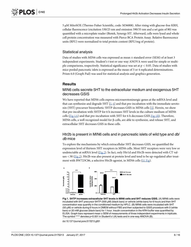
MIN6 cells secrete 5HT to the extracellular medium and exogenous 5HT
decreases GSIS
We have reported that MIN6 cells express microserotoninergic genes at the mRNA level and
that can synthesize and degrade 5HT [5, 6] and that pre-incubation with the immediate seroto-
nin (5HT) precursor biosynthetic 5HTP decreases GSIS in MIN6 cells [5]. Herein, we show
that pre-incubation with 5HTP for 6 h increases 5HT levels in the culture medium of MIN6
cells (Fig 1A) and that pre-incubation with 5HT for 6 h decreases GSIS Fig 1B). Therefore,
MIN6 cells, a well recognized model for β-cells, are able to synthesize, and release 5HT, and
extracellular 5HT decreases GSIS in these cells.
Htr2b is present in MIN6 cells and in pancreatic islets of wild type and db/
db mice
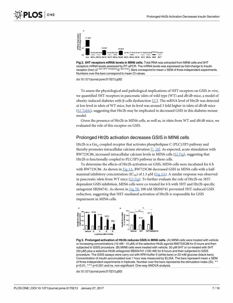
To explore the mechanisms by which extracellular 5HT decreases GSIS, we quantified the
expression level of thirteen 5HT receptors in MIN6 cells. Most 5HT receptors were very low or
undetectable at mRNA level (Fig 2). In fact, only Htr1d and Htr2b were detected with CT val-
ues<30 (Fig 2). Htr2b was also present at protein level and tend to be up-regulated after treat-
ment with BW723C86, a selective Htr2b agonist, in MIN6 cells (S1 Fig).
Fig 1. 5HTP increases extracellular 5HT levels in MIN6 cells and 5HT reduces GSIS. (A) MIN6 cells were
incubated with 5HT precursor 5HTP (500 μM) (black bars) or vehicle (white bars) for 6 hours and then 5HT
concentration was quantify in the conditioned medium by HPLC. (B) MIN6 cells were incubated with 5HT
(50 μM) or vehicle during 6 hours in DMEM without FBS and then subjected to GSIS procedure with 0 (white
bars) or 20 mM glucose (black bars) for 1 hour. Insulin concentration in the KRH buffer was quantified by
ELISA. Graph bars represent mean ± SEM of measurements of three independent experiments in triplicate.
The symbol *** denotes p<0.001 in Student’s t (A) tests and in one-way ANOVA (B).
doi:10.1371/journal.pone.0170213.g001
Prolonged Htr2b Activation Decreases Insulin Secretion
PLOS ONE | DOI:10.1371/journal.pone.0170213 January 27, 2017 6 / 16
To assess the physiological and pathological implications of 5HT receptors on GSIS in vivo,
we quantified 5HT receptors in pancreatic islets of wild type (WT) and db/db mice, a model of
obesity-induced diabetes with β-cells dysfunction [27]. The mRNA level of Htr2b was detected
at low level in islets of WT mice, but its level was around 3 fold higher in islets of db/db mice
(S2 Table), suggesting that Htr2b may be implicated in decreased GSIS in this diabetes mouse
model.
Given the presence of Htr2b in MIN6 cells, as well as, in islets from WT and db/db mice, we
evaluated the role of this receptor on GSIS.
Prolonged Htr2b activation decreases GSIS in MIN6 cells
Htr2b is a Gαq-coupled receptor that activates phospholipase C (PLC)/IP3 pathway and
thereby promotes intracellular calcium elevation [7, 28]. As expected, acute stimulation with
BW723C86, increased intracellular calcium levels in MIN6 cells (S2 Fig), suggesting that
Htr2b is functionally coupled to PLC/IP3 pathway in these cells.
To determine the effects of Htr2b activation on GSIS, MIN6 cells were incubated for 6 h
with BW723C86. As shown in Fig 3A, BW723C86 decreased GSIS in MIN6 cells with a half-
maximal inhibitory concentration (IC50) of 1.3 μM (Fig 3A). A similar response was observed
in pancreatic islets from WT mice (S3 Fig). To further evaluate the role of Htr2b on 5HT-
dependent GSIS inhibition, MIN6 cells were co-treated for 6 h with 5HT and Htr2b specific
antagonist SB204741. As shown in Fig 3B, 100 nM SB204741 prevented 5HT-induced GSIS
reduction, suggesting that 5HT-mediated activation of Htr2b is responsible for GSIS
impairment in MIN6 cells.
Fig 2. 5HT receptors mRNA levels in MIN6 cells. Total RNA was extracted from MIN6 cells and 5HT
receptors mRNA levels assessed by RT-qPCR. The mRNA levels was expressed as fold-change to Insulin
receptor (Insr) (2−ΔCt 5HT receptors/2−ΔCt Insr). Bars correspond to mean ±SEM of three independent experiments.
Numbers over the bars correspond to mean Ct values.
doi:10.1371/journal.pone.0170213.g002
Fig 3. Prolonged activation of Htr2b reduces GSIS in MIN6 cells. (A) MIN6 cells were treated with vehicle
or increasing concentrations (10 nM– 10 μM) of the selective Htr2b agonist BW723C86 for 6 hours and then
subjected to GSIS procedure. (B) MIN6 cells were treated with vehicle, 50 μM 5HT or co-treated with 5HT
(50 μM) plus a selective Htr2b antagonist SB204741 (100 nM) for 6 hours and then subjected to GSIS
procedure. The GSIS assays were carry out with KRH buffer 0 (white bars) or 20 mM glucose (black bars).
Concentration of insulin accumulated over 1 hour was measured by ELISA. The bars represent mean ± SEM
of three independent experiments in triplicate. Number over the bars represents the stimulation index (SI). **p<0.01, *** p<0.001 and ns, non-significant. One-way ANOVA analysis.
doi:10.1371/journal.pone.0170213.g003
Prolonged Htr2b Activation Decreases Insulin Secretion
PLOS ONE | DOI:10.1371/journal.pone.0170213 January 27, 2017 7 / 16
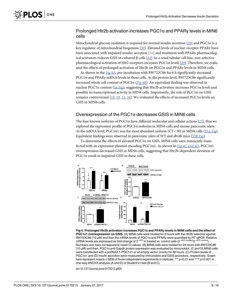
Prolonged Htr2b activation increases PGC1α and PPARy levels in MIN6
cells
Mitochondrial glucose oxidation is required for normal insulin secretion [29] and PGC1α is a
key regulator of mitochondrial biogenesis [30]. Elevated levels of nuclear receptor PPARy have
been associated with impaired insulin secretion [31] and treatment with PPARy pharmacolog-
ical activators reduces GSIS in cultured β-cells [32]. In a renal tubular cell line, non-selective
pharmacological activation of Htr2 receptors increases PGC1α levels [20]. Therefore, we evalu-
ated the effects of prolonged activation of Htr2b on PGC1α and PPARy levels in MIN6 cells.
As shown in the Fig 4A, pre-incubation with BW723C86 for 6 h significantly increased
PGC1α and PPARy mRNA levels in these cells. At the protein level, BW723C86 significantly
increased whole cell content of PGC1α (Fig 4B). An equivalent finding was observed in
nuclear PGC1α content (S4 Fig), suggesting that Htr2b activation increases PGC1α levels and
possibly its transcriptional activity in MIN6 cells. Importantly, the role of PGC1α on GSIS
remains controversial [18, 19, 33, 34]. We evaluated the effects of increased PGC1α levels on
GSIS in MIN6 cells.
Overexpression of the PGC1α decreases GSIS in MIN6 cells
The four known isoforms of PGC1α have different molecular and cellular actions [25], thus we
explored the expression profile of PGC1α isoforms in MIN6 cells and mouse pancreatic islets.
At the mRNA level, PGC1α1 was the most abundant isoform (CT<30) in MIN6 cells (S5A Fig).
Equivalent findings were observed in pancreatic islets of WT and db/db mice (S5B Fig).
To determine the effects of elevated PGC1α on GSIS, MIN6 cells were transiently trans-
fected with an expression plasmid encoding PGC1α1. As shown in Fig 4C and 4D, PGC1α1
overexpression decreased GSIS in MIN6 cells, suggesting that Htr2b-dependent elevation of
PGC1α result in impaired GSIS in these cells.
Fig 4. Prolonged Htr2b activation increases PGC1α and PPARy levels in MIN6 cells and the effect of
PGC1α1 overexpression on GSIS. (A) MIN6 cells were treated for 6 hours with the Htr2b selective agonist
BW723C86 (10 μM) and then the mRNA levels of PGC1α and PPARy were quantified by RT-qPCR. Relative
mRNA levels are expressed as fold-change of 2−ΔCt in treated vs. control cells (2−ΔCt treated/2−ΔCt control).
Numbers over bars correspond to mean Ct values. (B) MIN6 cells were treated for 24 hours with BW723C86
(10 μM) and then, PGC1α and Gapdh protein expression was evaluated by immunoblot. (C and D) MIN6 cells
were transfected with a pcDNA3.1-PGC1α1 or an empty vector (mock) for 48 hours. (C) Protein levels of
PGC1α1 and (D) insulin secretion were measured by immunoblot and GSIS procedure, respectively. Graph
bars represent means ± SEM of three independent experiments in triplicate. ** p<0.01 and *** p<0.001 in
one-way ANOVA analysis (A and D) or Student’s t test (B and C).
doi:10.1371/journal.pone.0170213.g004
Prolonged Htr2b Activation Decreases Insulin Secretion
PLOS ONE | DOI:10.1371/journal.pone.0170213 January 27, 2017 8 / 16
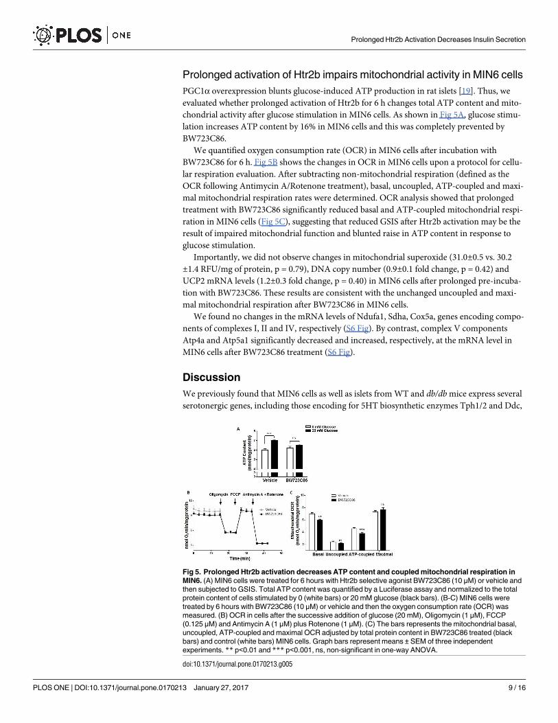
Prolonged activation of Htr2b impairs mitochondrial activity in MIN6 cells
PGC1α overexpression blunts glucose-induced ATP production in rat islets [19]. Thus, we
evaluated whether prolonged activation of Htr2b for 6 h changes total ATP content and mito-
chondrial activity after glucose stimulation in MIN6 cells. As shown in Fig 5A, glucose stimu-
lation increases ATP content by 16% in MIN6 cells and this was completely prevented by
BW723C86.
We quantified oxygen consumption rate (OCR) in MIN6 cells after incubation with
BW723C86 for 6 h. Fig 5B shows the changes in OCR in MIN6 cells upon a protocol for cellu-
lar respiration evaluation. After subtracting non-mitochondrial respiration (defined as the
OCR following Antimycin A/Rotenone treatment), basal, uncoupled, ATP-coupled and maxi-
mal mitochondrial respiration rates were determined. OCR analysis showed that prolonged
treatment with BW723C86 significantly reduced basal and ATP-coupled mitochondrial respi-
ration in MIN6 cells (Fig 5C), suggesting that reduced GSIS after Htr2b activation may be the
result of impaired mitochondrial function and blunted raise in ATP content in response to
glucose stimulation.
Importantly, we did not observe changes in mitochondrial superoxide (31.0±0.5 vs. 30.2
±1.4 RFU/mg of protein, p = 0.79), DNA copy number (0.9±0.1 fold change, p = 0.42) and
UCP2 mRNA levels (1.2±0.3 fold change, p = 0.40) in MIN6 cells after prolonged pre-incuba-
tion with BW723C86. These results are consistent with the unchanged uncoupled and maxi-
mal mitochondrial respiration after BW723C86 in MIN6 cells.
We found no changes in the mRNA levels of Ndufa1, Sdha, Cox5a, genes encoding compo-
nents of complexes I, II and IV, respectively (S6 Fig). By contrast, complex V components
Atp4a and Atp5a1 significantly decreased and increased, respectively, at the mRNA level in
MIN6 cells after BW723C86 treatment (S6 Fig).
Discussion
We previously found that MIN6 cells as well as islets from WT and db/db mice express several
serotonergic genes, including those encoding for 5HT biosynthetic enzymes Tph1/2 and Ddc,
Fig 5. Prolonged Htr2b activation decreases ATP content and coupled mitochondrial respiration in
MIN6. (A) MIN6 cells were treated for 6 hours with Htr2b selective agonist BW723C86 (10 μM) or vehicle and
then subjected to GSIS. Total ATP content was quantified by a Luciferase assay and normalized to the total
protein content of cells stimulated by 0 (white bars) or 20 mM glucose (black bars). (B-C) MIN6 cells were
treated by 6 hours with BW723C86 (10 μM) or vehicle and then the oxygen consumption rate (OCR) was
measured. (B) OCR in cells after the successive addition of glucose (20 mM), Oligomycin (1 μM), FCCP
(0.125 μM) and Antimycin A (1 μM) plus Rotenone (1 μM). (C) The bars represents the mitochondrial basal,
uncoupled, ATP-coupled and maximal OCR adjusted by total protein content in BW723C86 treated (black
bars) and control (white bars) MIN6 cells. Graph bars represent means ± SEM of three independent
experiments. ** p<0.01 and *** p<0.001, ns, non-significant in one-way ANOVA.
doi:10.1371/journal.pone.0170213.g005
Prolonged Htr2b Activation Decreases Insulin Secretion
PLOS ONE | DOI:10.1371/journal.pone.0170213 January 27, 2017 9 / 16
vesicle transporters Vmat1/2 and catabolic enzymes Maoa/b [5, 6]. Others have described
equivalent findings in mouse islets [3, 4]. Additionally, in this and previous work [5], we have
shown that 5HT precursor 5HTP and Mao inhibitor Pargyline, increases 5HT content and
extracellular 5HT levels in MIN6 cells, indicating these cells synthesize, degrade, store and
secrete 5HT, constituting a functional microserotonergic system.
In our previous work [5] as well as in this current manuscript, we report that 5HTP pre-
incubation increases extracellular 5HT in MIN6 cells and that it is associated with decreases
GSIS. Similar to our findings, Paulmann et al. [12] showed a decrease in GSIS after 5HT pre-
incubation in MIN6 cells as well as in rat β-cell lines INS1 and RINm5f. Interestingly, they
report that 5HTP pre-incubation increases GSIS in RINm5f cells, suggesting differential
actions for intracellular and extracellular 5HT pools on insulin secretion. Nonetheless, these
authors do not show nor mention the effects of 5HTP incubation on GSIS in MIN6 cells.
In this study, we found that MIN6 cells and mouse pancreatic islets express Htr2b 5HT
receptor. Interestingly, in islets of db/db mice, a model of diabetes and β-cells dysfunction [27],
the mRNA level of Htr2b was *3-fold higher compared to islets of WT mice. We also found
that prolonged incubation with extracellular 5HT or a selective Htr2b agonist reduces GSIS.
On the contrary, pharmacological antagonism of Htr2b prevents 5HT inhibitory actions on
GSIS, suggesting that prolonged activation of this receptor mediates serotoninergic inhibition
of insulin secretion. Of note, the inhibitory effect of 5HT is less potent than the one exerted by
Htr2b agonist, BW723C86. Importantly, although the physiological and pathological implica-
tions of 5HT on GSIS still remain debated, the inhibitory actions of extracellular 5HT on insu-
lin secretion are consistent finding in the literature [5, 11–13, 35–37]. It is possible that, by
itself, 5HT actions have a low functional impact on GSIS, but in combination with other dysre-
gulations present in the diabetic milieu, could have a relevant pathological meaning. For exam-
ple, we have found that Htr2b protein expression tend to be up-regulated by its own activation
in MIN6 cells as well as others have shown that its mRNA levels are regulated by its own acti-
vation or inhibition in INS1(832/13) cells [7], suggesting that pharmacologic interventions on
specific 5HT receptors may represent a clinically relevant therapeutic innovation in diabetes.
At the mechanistic level, we found that prolonged Htr2b activation in MIN6 cells decreases
glucose-induced ATP content and basal and ATP-coupled mitochondrial respiration without
modification in mitochondrial DNA copy number, mitochondrial superoxide content as well
as no changes in mRNA levels of UCP2 and some genes of complex I, II and IV. All these facts
led us to propose that prolonged Htr2b activation reduces GSIS by decreasing mitochondrial
function and ATP production but without changing in mitochondrial uncoupling, biogenesis
and superoxide production.
On the other hand, we found that prolonged Htr2b activation in MIN6 cells decrease and
increase the mRNA levels of ATP synthase complex genes Atp4a and Atp5a1, respectively.
These findings altogether with reduced basal and ATP-coupled OCR and glucose-stimulated
ATP content, suggest that prolonged Htr2b activation leads to defects in ATP synthase activity.
In line with this proposition, Htr2b overexpression in mice cardiomyocytes has been associ-
ated with reduced adenine nucleotide translocator and abnormal mitochondrial function [21].
Increased PGC1α expression appears to determine different metabolic outcomes depending
on what specific tissue is targeted [38]. In adipose tissue, high PGC1α expression participates in
induced-browning and activation of thermogenesis increasing the uncoupling mitochondrial
respiration [38], while in skeletal muscle and myocardial tissues, PGC1α upregulation results in
gene reprograming from glycolytic to glucose and fatty acids oxidative metabolism which is
accompanied by enhancement of mitochondrial biogenesis and respiration as well as ATP pro-
duction [38]. By contrast, in liver and pancreas PGC1α activation promotes hepatic glucose
production and inhibits GSIS, respectively [38]. Here we found that prolonged Htr2b activation
Prolonged Htr2b Activation Decreases Insulin Secretion
PLOS ONE | DOI:10.1371/journal.pone.0170213 January 27, 2017 10 / 16
increases PGC1α and PPARy levels and that PGC1α overexpression blunts GSIS in MIN6 cells.
Our results agree with the proposed role of PGC1α on suppression of β-cell energy metabolism
and GSIS. In fact, overexpression of PGC1α to levels similar to those present in islets of diabet-
ics rodents leads to a marked decreases of GSIS as consequences of alterations in expression of
genes that participates in glucose sensing and metabolism and reduces glucose-induced ATP
production in rat pancreatic islets [19]. These reported roles of PGC1α in β-cells are coherent
with previous studies showing higher PGC1α levels in pancreatic islets of diabetic mice [19] as
with the association between specific haplotypes in PPARGC1A, the gene encoding PGC1α,
with indices of beta cell function in humans [39].
Elevated glucocorticoids (GCs) during the fetal period promote pancreatic β-cell dysfunction
in the adulthood, possibly by a GCs-dependent elevation of PGC1α levels and secondary down-
regulation of pancreatic duodenal homebox 1 (Pdx1) in β-cells [18]. Notably, the forced overex-
pression of PGC1α in β-cells during the adulthood fails to induce β-cell dysfunction [18]. These
findings suggest that elevated levels of PGC1α in β-cells are possibly associated with alterations
in β-cells development during fetal stages rather than with primary defects in GSIS in terminally
differentiated β-cells. The implications in vivo of our observed deleterious effects of Htr2b and
PGC1α-dependent reduction of GSIS in MIN6 cells, remain to be determined.
Previous studies have shown that extracellular 5HT reduces GSIS in different clonal β-cell
lines as well as in mouse and rat pancreatic islets [5, 11–13, 35–37]. Importantly, it has been
reported that 5HT perfusion in vivo is associated with decreased insulin plasma levels in mice
[12, 35]. In humans, 5HT plasma levels are reduced after glucose intake [40], suggesting a
functional link between circulating 5HT, GSIS and glucose homeostasis. This idea is further
supported by the fact that dietary-induced obese mice have seven times higher serum 5HT
than lean mice [41] and that plasma levels of 5HT and 5HT metabolite 5-hydroxyindole-3-ace-
tic acid are elevated in diabetic subjects [42, 43]. Furthermore, there are reported cases of glu-
cose intolerance and low insulin levels in patients with carcinoid syndrome, characterized by
excessive systemic 5HT levels [44, 45].
Only one previous study has reported GSIS inhibition by a specific 5HT receptor [13].
After finding that islets from db/db have increased level of Htr2c, Zhang et al. showed that
pharmacological activation of Htr2c was associated with decreased GSIS in MIN6 cells [13]. In
our studies, Htr2c was undetectable in both MIN6 cells and mice pancreatic islets. We do not
know the reason for this discrepancy, however, others have also failed to find Htr2c in human
and mouse islets or in MIN6 cells [3, 7]. Here we report that Htr2b is present in MIN6 cells at
the mRNA and protein level, that its protein level tend to be up-regulated by its own activation
in MIN6 cells and that its mRNA is higher in islets of db/db in comparison with WT mice. In
agreement with us, it was recently reported that Htr2b is expressed in mouse and human islets
and, interestingly, its expression level is regulated by its own pharmacological activation or
inhibition [7].
Our assertion that prolonged activation of Htr2b decreases GSIS is grounded on pharmaco-
logical evidence. It is unlikely that BW723C86 may activate other Htr2 subtypes in the concentra-
tions we used in this study. In fact, BW723C86 affinity is three hundred times higher for Htr2b
than for Htr2a. In addition, we found that Htr2a and Htr2c were not expressed in MIN6 cells
and mouse pancreatic islets. In spite of that, the direct assessment of the actions of BW723C86 in
islets of Htr2b deficient mice will provide conclusive functional evidence of the role of Htr2b on
GSIS.
Bennet et al. recently showed that simultaneous Htr2b activation and glucose stimulation
leads to increased insulin secretion by β-cells [7]. That study is, at the first glance, contradic-
tory with our results; however, we believe that these discrepancies might result from the differ-
ent duration of Htr2b activation between the two studies. In fact, equivalent dual biological
Prolonged Htr2b Activation Decreases Insulin Secretion
PLOS ONE | DOI:10.1371/journal.pone.0170213 January 27, 2017 11 / 16

effects have been reported for another Gαq-coupled receptor, the M3 cholinergic receptor
[46]. In rat islets, short term co-stimulation with glucose and Carbachol increased GSIS in
comparison with glucose-stimulation only. By contrast, prolonged M3 activation (3.5 hours)
with Carbachol followed by glucose stimulation, virtually abolished GSIS [46]. Interestingly, it
has been reported that pretreatment of rat islets with 5HT for 3 hours also impairs GSIS by
altering glucose-induced activation of PLC activity [11].
In conclusion, in this study we show that prolonged Htr2b activation with BW723C86
reduces GSIS in MIN6 cells possibly as a result of impaired mitochondrial activity and ATP
production by mechanisms likely dependent on enhanced PGC1α/PPARy levels (Fig 6).
Supporting Information
S1 Fig. Htr2b expression in MIN6 cells after prolonged Htr2b activation. MIN6 cells were
treated for 24 hours with vehicle or BW723C86 (10 μM) and then, Htr2b and Gapdh protein
expression was evaluated by immunoblot. Graph bars represent means ± SEM of relative
expression of Htr2b/Gapdh in treated versus control cells in three independent experiments.
Student’s t test was used for statistical analysis.
(DOCX)
S2 Fig. Calcium signals in MIN6 cells acutely stimulated with Htr2b agonist. MIN6 cells
were cultured in standard conditions and charged with the calcium sensitive fluorescence dye
Fluo4-AM (5 μM) in KRH buffer (0 mM glucose) by 30 min. The fluorescence signal (488 nm)
was registered during 800 seconds by a confocal microscope (Carl Zeiss, Axiovert 200, LSM 5
Pascal) measured before and after the acute stimulation with (A) glucose (20 mM) and Carba-
chol (30 μM) or (B) the Htr2b selective agonist BW723C86 (10 μM). (C) The area under the
curve (AUC) of fluorescence intensity adjusted by time was calculated before (white bars) and
after (black bars) acute stimulus. The symbol �� denotes p<0.01 and ��� p<0.001 in a Student’s
t tests.
(DOCX)
Fig 6. Proposed model for the inhibitory effect of prolonged Htr2b activation on GSIS in MIN6 cells.
MIN6 cells constitute a functional microserotoninergic system, with the ability to synthesis 5HT from its
precursors (tryptophan or 5HTP), storage, degrade and release this monoamine. The extracellular 5HT may
decrease GSIS through the activation of the Htr2b receptor, the most abundantly 5HT receptor expressed in
MIN6 cells. Prolonged Htr2b activation is accompanied with calcium release from endoplasmic reticulum and
its inhibitory effects on GSIS is likely the result of enhanced PGC1α/PPARy expression and mitochondrial
dysfunction with decreased glucose-stimulated ATP synthesis.
doi:10.1371/journal.pone.0170213.g006
Prolonged Htr2b Activation Decreases Insulin Secretion
PLOS ONE | DOI:10.1371/journal.pone.0170213 January 27, 2017 12 / 16
S3 Fig. Prolonged activation of Htr2b reduces GSIS in mouse pancreatic islets. Pooled islets
from WT mice were treated with 50 μM 5HT or 10 μM BW723C86 for 6 h and then subjected
to GSIS procedures. The GSIS assays were carry out with KRH buffer 2.8 (white bars) or 16.7
mM glucose (black bars). Concentration of insulin accumulated over 1 hour was measured by
ELISA. Bars represent the mean of a single experiment (5 islets/well and 4 wells/condition), for
that reason no statistical analysis was done. Number over the bars represents the stimulation
index (SI).
(DOCX)
S4 Fig. Prolonged Htr2b activation increases PGC1α levels in MIN6 cells. MIN6 cells were
treated for 24 hours with BW723C86 (10 μM) and then the cytosolic and nuclear fractions
were separated. PGC1α, Histone H3 (nuclear marker) and Gapdh (cytosolic marker) proteins
were quantified by immunoblot.
(DOCX)
S5 Fig. PGC1α isoforms levels in MIN6 cells and mouse islets. Total RNA was extracted
from MIN6 cells (A) or pooled mouse pancreatic islets (n = 11 for WT and n = 12 for db/dbmice) (B) with standard procedures. The mRNA levels of total PGC1α and its four isoforms
were quantified by RT-qPCR and expressed as the fold change of 2−ΔCt of isoforms vs. total
PGC1α (2−ΔCt isoform/2−ΔCt total). The graph bars represent mean ± SEM of three independent
experiments for MIN6 cells (A) and the unique values for mRNA expression in mice islets (B).
Numbers over the bars correspond to mean Ct value.
(DOCX)
S6 Fig. mRNA expression of mitochondrial complex genes in MIN6 cells after Htr2b pro-
longed activation. MIN6 cells were treated for 6 h with the Htr2b selective agonist BW723C86
(10 μM) or vehicle. Then, total RNA was extracted from MIN6 cells and the relative mRNA
levels of mitochondrial complex genes were quantified by RT-qPCR and expressed as fold-
change of 2−ΔCt in treated vs. control cells (2−ΔCt treated/2−ΔCt control). Numbers over bars corre-
spond to mean Ct values in control condition. The graph bars represent mean ± SEM of three
independent experiments.
(DOCX)
S1 Table. List of primers used to evaluate the mRNA expression of PGC1α isoforms,
PPARy and mitochondrial complexes genes.
(DOCX)
S2 Table. 5HT receptors mRNA levels in pancreatic islets of WT and db/db mice. Total
RNA was extracted from a pool of 335 islets from 11 WT mice or 312 islets from 12 db/dbmice. 5HT receptors mRNA level was expressed as specific Ct values for every gene in WT and
db/db mice islets and as fold-change of db/db to WT mice (2−ΔCt db/db/2−ΔCt WT). Data corre-
spond to uniplicate values derived from pooled mouse islets.
(DOCX)
Acknowledgments
We acknowledge Paula Mennickent of the Pontificia Universidad Catolica de Chile, who
kindly drew the Fig 6.
Author Contributions
Conceptualization: LRC JLS VAC.
Prolonged Htr2b Activation Decreases Insulin Secretion
PLOS ONE | DOI:10.1371/journal.pone.0170213 January 27, 2017 13 / 16
Data curation: LRC RBS FJ PLl MLM.
Formal analysis: LRC RBS FJ PLl MLM.
Funding acquisition: JLS VAC JEG CC.
Investigation: LRC MLM RBS PLl FJ.
Methodology: LRC RBS MLM PLl CC FJ.
Project administration: JLS VAC.
Resources: JLS VAC CC JEG DB PLl.
Software: FJ PLl LRC.
Supervision: CC VAC JLS PLl RBS JEG DB.
Validation: CC VAC PLl RBS FJ DB PO.
Visualization: LRC PLl RBS CC VAC JLS.
Writing – original draft: LRC VAC JLS.
Writing – review & editing: VAC LRC CC RBS PLl JLS.
References1. Berger M, Gray JA, Roth BL. The expanded biology of serotonin. Annu Rev Med. 2009; 60:355–66. doi:
10.1146/annurev.med.60.042307.110802 PMID: 19630576
2. Amireault P, Sibon D, Cote F. Life without peripheral serotonin: insights from tryptophan hydroxylase 1
knockout mice reveal the existence of paracrine/autocrine serotonergic networks. ACS Chem Neurosci.
2013; 4(1):64–71. doi: 10.1021/cn300154j PMID: 23336045
3. Kim H, Toyofuku Y, Lynn FC, Chak E, Uchida T, Mizukami H, et al. Serotonin regulates pancreatic beta
cell mass during pregnancy. Nat Med. 2010; 16(7):804–8. doi: 10.1038/nm.2173 PMID: 20581837
4. Ohta Y, Kosaka Y, Kishimoto N, Wang J, Smith SB, Honig G, et al. Convergence of the insulin and sero-
tonin programs in the pancreatic beta-cell. Diabetes. 2011; 60(12):3208–16. doi: 10.2337/db10-1192
PMID: 22013016
5. Cataldo LR, Cortes VA, Mizgier ML, Aranda E, Mezzano D, Olmos P, et al. Fluoxetine impairs insulin
secretion without modifying extracellular serotonin levels in MIN6 beta-cells. Experimental and clinical
endocrinology & diabetes: official journal, German Society of Endocrinology [and] German Diabetes
Association. 2015; 123(8):473–8.
6. Cataldo LR, Mizgier ML, Busso D, Olmos P, Galgani JE, Valenzuela R, et al. Serotonin- and Dopamine-
Related Gene Expression in db/db Mice Islets and in MIN6 beta-Cells Treated with Palmitate and Ole-
ate. J Diabetes Res. 2016; 2016:3793781. doi: 10.1155/2016/3793781 PMID: 27366756
7. Bennet H, Mollet IG, Balhuizen A, Medina A, Nagorny C, Bagge A, et al. Serotonin (5-HT) receptor 2b
activation augments glucose-stimulated insulin secretion in human and mouse islets of Langerhans.
Diabetologia. 2016.
8. Ohara-Imaizumi M, Kim H, Yoshida M, Fujiwara T, Aoyagi K, Toyofuku Y, et al. Serotonin regulates glu-
cose-stimulated insulin secretion from pancreatic beta cells during pregnancy. Proc Natl Acad Sci U S
A. 2013; 110(48):19420–5. doi: 10.1073/pnas.1310953110 PMID: 24218571
9. Kim K, Oh CM, Ohara-Imaizumi M, Park S, Namkung J, Yadav VK, et al. Functional role of serotonin in
insulin secretion in a diet-induced insulin-resistant state. Endocrinology. 2014:en20141687.
10. Hoyer D, Clarke DE, Fozard JR, Hartig PR, Martin GR, Mylecharane EJ, et al. International Union of
Pharmacology classification of receptors for 5-hydroxytryptamine (Serotonin). Pharmacol Rev. 1994;
46(2):157–203. PMID: 7938165
11. Zawalich WS, Tesz GJ, Zawalich KC. Effects of prior 5-hydroxytryptamine exposure on rat islet insulin
secretory and phospholipase C responses. Endocrine. 2004; 23(1):11–6. doi: 10.1385/ENDO:23:1:11
PMID: 15034191
Prolonged Htr2b Activation Decreases Insulin Secretion
PLOS ONE | DOI:10.1371/journal.pone.0170213 January 27, 2017 14 / 16
12. Paulmann N, Grohmann M, Voigt JP, Bert B, Vowinckel J, Bader M, et al. Intracellular serotonin modu-
lates insulin secretion from pancreatic beta-cells by protein serotonylation. PLoS Biol. 2009; 7(10):
e1000229. doi: 10.1371/journal.pbio.1000229 PMID: 19859528
13. Zhang Q, Zhu Y, Zhou W, Gao L, Yuan L, Han X. Serotonin Receptor 2C and Insulin Secretion. PLoS
One. 2013; 8(1):e54250. doi: 10.1371/journal.pone.0054250 PMID: 23349838
14. Sumara G, Sumara O, Kim JK, Karsenty G. Gut-derived serotonin is a multifunctional determinant to
fasting adaptation. Cell Metab. 2012; 16(5):588–600. doi: 10.1016/j.cmet.2012.09.014 PMID:
23085101
15. El-Merahbi R, Loffler M, Mayer A, Sumara G. The roles of peripheral serotonin in metabolic homeosta-
sis. FEBS Lett. 2015.
16. Soejima A, Inoue K, Takai D, Kaneko M, Ishihara H, Oka Y, et al. Mitochondrial DNA is required for reg-
ulation of glucose-stimulated insulin secretion in a mouse pancreatic beta cell line, MIN6. J Biol Chem.
1996; 271(42):26194–9. PMID: 8824267
17. Tsuruzoe K, Araki E, Furukawa N, Shirotani T, Matsumoto K, Kaneko K, et al. Creation and characteri-
zation of a mitochondrial DNA-depleted pancreatic beta-cell line: impaired insulin secretion induced by
glucose, leucine, and sulfonylureas. Diabetes. 1998; 47(4):621–31. PMID: 9568696
18. Valtat B, Riveline JP, Zhang P, Singh-Estivalet A, Armanet M, Venteclef N, et al. Fetal PGC-1alpha
Overexpression Programs Adult Pancreatic beta-cell Dysfunction. Diabetes. 2012
19. Yoon JC, Xu G, Deeney JT, Yang SN, Rhee J, Puigserver P, et al. Suppression of beta cell energy
metabolism and insulin release by PGC-1alpha. Dev Cell. 2003; 5(1):73–83. PMID: 12852853
20. Rasbach KA, Funk JA, Jayavelu T, Green PT, Schnellmann RG. 5-hydroxytryptamine receptor stimula-
tion of mitochondrial biogenesis. J Pharmacol Exp Ther. 2010; 332(2):632–9. doi: 10.1124/jpet.109.
159947 PMID: 19875674
21. Nebigil CG, Jaffre F, Messaddeq N, Hickel P, Monassier L, Launay JM, et al. Overexpression of the
serotonin 5-HT2B receptor in heart leads to abnormal mitochondrial function and cardiac hypertrophy.
Circulation. 2003; 107(25):3223–9. doi: 10.1161/01.CIR.0000074224.57016.01 PMID: 12810613
22. Miyazaki J, Araki K, Yamato E, Ikegami H, Asano T, Shibasaki Y, et al. Establishment of a pancreatic
beta cell line that retains glucose-inducible insulin secretion: special reference to expression of glucose
transporter isoforms. Endocrinology. 1990; 127(1):126–32. doi: 10.1210/endo-127-1-126 PMID:
2163307
23. Kulkarni RN, Wang ZL, Wang RM, Hurley JD, Smith DM, Ghatei MA, et al. Leptin rapidly suppresses
insulin release from insulinoma cells, rat and human islets and, in vivo, in mice. J Clin Invest. 1997; 100
(11):2729–36. doi: 10.1172/JCI119818 PMID: 9389736
24. Li DS, Yuan YH, Tu HJ, Liang QL, Dai LJ. A protocol for islet isolation from mouse pancreas. Nat Protoc.
2009; 4(11):1649–52. doi: 10.1038/nprot.2009.150 PMID: 19876025
25. Ruas JL, White JP, Rao RR, Kleiner S, Brannan KT, Harrison BC, et al. A PGC-1alpha isoform induced
by resistance training regulates skeletal muscle hypertrophy. Cell. 2012; 151(6):1319–31. doi: 10.1016/
j.cell.2012.10.050 PMID: 23217713
26. Nile DL, Brown AE, Kumaheri MA, Blair HR, Heggie A, Miwa S, et al. Age-related mitochondrial DNA
depletion and the impact on pancreatic Beta cell function. PLoS One. 2014; 9(12):e115433. doi: 10.
1371/journal.pone.0115433 PMID: 25532126
27. Dalboge LS, Almholt DL, Neerup TS, Vassiliadis E, Vrang N, Pedersen L, et al. Characterisation of
Age-Dependent Beta Cell Dynamics in the Male db/db Mice. PLoS One. 2013; 8(12):e82813. doi: 10.
1371/journal.pone.0082813 PMID: 24324833
28. Kursar JD, Nelson DL, Wainscott DB, Cohen ML, Baez M. Molecular cloning, functional expression,
and pharmacological characterization of a novel serotonin receptor (5-hydroxytryptamine2F) from rat
stomach fundus. Mol Pharmacol. 1992; 42(4):549–57. PMID: 1331748
29. Kohler M, Norgren S, Berggren PO, Fredholm BB, Larsson O, Rhodes CJ, et al. Changes in cyto-
plasmic ATP concentration parallels changes in ATP-regulated K+-channel activity in insulin-secreting
cells. FEBS Lett. 1998; 441(1):97–102. PMID: 9877173
30. Puigserver P, Spiegelman BM. Peroxisome proliferator-activated receptor-gamma coactivator 1 alpha
(PGC-1 alpha): transcriptional coactivator and metabolic regulator. Endocr Rev. 2003; 24(1):78–90.
doi: 10.1210/er.2002-0012 PMID: 12588810
31. Patane G, Anello M, Piro S, Vigneri R, Purrello F, Rabuazzo AM. Role of ATP production and uncoupl-
ing protein-2 in the insulin secretory defect induced by chronic exposure to high glucose or free fatty
acids and effects of peroxisome proliferator-activated receptor-gamma inhibition. Diabetes. 2002; 51
(9):2749–56. PMID: 12196468
Prolonged Htr2b Activation Decreases Insulin Secretion
PLOS ONE | DOI:10.1371/journal.pone.0170213 January 27, 2017 15 / 16
32. Nakamichi Y, Kikuta T, Ito E, Ohara-Imaizumi M, Nishiwaki C, Ishida H, et al. PPAR-gamma overex-
pression suppresses glucose-induced proinsulin biosynthesis and insulin release synergistically with
pioglitazone in MIN6 cells. Biochem Biophys Res Commun. 2003; 306(4):832–6. PMID: 12821117
33. Oropeza D, Jouvet N, Bouyakdan K, Perron G, Ringuette LJ, Philipson LH, et al. PGC-1 coactivators in
beta-cells regulate lipid metabolism and are essential for insulin secretion coupled to fatty acids. Molec-
ular metabolism. 2015; 4(11):811–22. doi: 10.1016/j.molmet.2015.08.001 PMID: 26629405
34. Ling C, Del Guerra S, Lupi R, Ronn T, Granhall C, Luthman H, et al. Epigenetic regulation of
PPARGC1A in human type 2 diabetic islets and effect on insulin secretion. Diabetologia. 2008; 51
(4):615–22. doi: 10.1007/s00125-007-0916-5 PMID: 18270681
35. Lernmark A. The significance of 5-hydroxytryptamine for insulin secretion in the mouse. Horm Metab
Res. 1971; 3(5):305–9. doi: 10.1055/s-0028-1094131 PMID: 4944450
36. Lundquist I, Ekholm R, Ericson LE. Monoamines in the pancreatic islets of the mouse. 5-hydroxytrypta-
mine as an intracellular modifier of insulin secretion, and the hypoglycaemic action of monoamine oxi-
dase inhibitors. Diabetologia. 1971; 7(6):414–22. PMID: 5004176
37. Feldman JM, Lebovitz HE. Serotonin inhibition of in vitro insulin release from golden hamster pancreas.
Endocrinology. 1970; 86(1):66–70. doi: 10.1210/endo-86-1-66 PMID: 4311193
38. Besseiche A, Riveline JP, Gautier JF, Breant B, Blondeau B. Metabolic roles of PGC-1alpha and its
implications for type 2 diabetes. Diabetes & metabolism. 2015.
39. Oberkofler H, Linnemayr V, Weitgasser R, Klein K, Xie M, Iglseder B, et al. Complex haplotypes of the
PGC-1alpha gene are associated with carbohydrate metabolism and type 2 diabetes. Diabetes. 2004;
53(5):1385–93. PMID: 15111510
40. Ho JE, Larson MG, Vasan RS, Ghorbani A, Cheng S, Rhee EP, et al. Metabolite profiles during oral glu-
cose challenge. Diabetes. 2013; 62(8):2689–98. doi: 10.2337/db12-0754 PMID: 23382451
41. Kim HJ, Kim JH, Noh S, Hur HJ, Sung MJ, Hwang JT, et al. Metabolomic analysis of livers and serum
from high-fat diet induced obese mice. J Proteome Res. 2011; 10(2):722–31. doi: 10.1021/pr100892r
PMID: 21047143
42. Barradas MA, Gill DS, Fonseca VA, Mikhailidis DP, Dandona P. Intraplatelet serotonin in patients with
diabetes mellitus and peripheral vascular disease. Eur J Clin Invest. 1988; 18(4):399–404. PMID:
3139426
43. Fukui M, Tanaka M, Toda H, Asano M, Yamazaki M, Hasegawa G, et al. High plasma 5-hydroxyindole-
3-acetic acid concentrations in subjects with metabolic syndrome. Diabetes Care. 2012; 35(1):163–7.
doi: 10.2337/dc11-1619 PMID: 22100965
44. Feldman JM, Marecek RL, Quickel KE Jr., Lebovitz HE. Glucose metabolism and insulin secretion in
the carcinoid syndrome. J Clin Endocrinol Metab. 1972; 35(2):307–11. doi: 10.1210/jcem-35-2-307
PMID: 4262845
45. Sirek A, Sirek OV. Serotonin: a review. Can Med Assoc J. 1970; 102(8):846–9. PMID: 4910705
46. Zawalich WS, Zawalich KC, Kelley GG. Time-dependent effects of cholinergic stimulation on beta cell
responsiveness. Pflugers Arch. 1996; 432(4):589–96. PMID: 8764958
Prolonged Htr2b Activation Decreases Insulin Secretion
PLOS ONE | DOI:10.1371/journal.pone.0170213 January 27, 2017 16 / 16