Embed Size (px)
Citation preview
HAL Id: inserm-00195963https://www.hal.inserm.fr/inserm-00195963
Submitted on 11 Dec 2007
HAL is a multi-disciplinary open accessarchive for the deposit and dissemination of sci-entific research documents, whether they are pub-lished or not. The documents may come fromteaching and research institutions in France orabroad, or from public or private research centers.
L’archive ouverte pluridisciplinaire HAL, estdestinée au dépôt et à la diffusion de documentsscientifiques de niveau recherche, publiés ou non,émanant des établissements d’enseignement et derecherche français ou étrangers, des laboratoirespublics ou privés.
Progressive motor neuronopathy: a critical role of thetubulin chaperone TBCE in axonal tubulin routing from
the Golgi apparatus.Michael Schaefer, Henning Schmalbruch, Emmanuelle Buhler, Catherine
Lopez, Natalia Martin, Jean-Louis Guénet, Georg Haase
To cite this version:Michael Schaefer, Henning Schmalbruch, Emmanuelle Buhler, Catherine Lopez, Natalia Martin, etal.. Progressive motor neuronopathy: a critical role of the tubulin chaperone TBCE in axonal tubulinrouting from the Golgi apparatus.. Journal of Neuroscience, Society for Neuroscience, 2007, 27 (33),pp.8779-89. �10.1523/JNEUROSCI.1599-07.2007�. �inserm-00195963�
Neurobiology of Disease
Progressive Motor Neuronopathy: A Critical Role of theTubulin Chaperone TBCE in Axonal Tubulin Routing fromthe Golgi Apparatus
Michael K. E. Schaefer,1,2 Henning Schmalbruch,3 Emmanuelle Buhler,1,2 Catherine Lopez,1,2 Natalia Martin,4
Jean-Louis Guenet,4 and Georg Haase1,2
1Inserm, Unite 29, Equipe Avenir, 13273 Marseille, France, 2Aix Marseille Universite, Institut de Neurobiologie de la Mediterranee, 13284 Marseille, France,3Panum Institute, University of Copenhagen, DK-2200 Copenhagen, Denmark, and 4Institut Pasteur, 75015 Paris, France
Axonal degeneration represents one of the earliest pathological features in motor neuron diseases. We here studied the underlyingmolecular mechanisms in progressive motor neuronopathy ( pmn) mice mutated in the tubulin-specific chaperone TBCE. We demon-strate that TBCE is a peripheral membrane-associated protein that accumulates at the Golgi apparatus. In pmn mice, TBCE is destabilizedand disappears from the Golgi apparatus of motor neurons, and microtubules are lost in distal axons. The axonal microtubule lossproceeds retrogradely in parallel with the axonal dying back process. These degenerative changes are inhibited in a dose-dependentmanner by transgenic TBCE complementation that restores TBCE expression at the Golgi apparatus. In cultured motor neurons, the pmnmutation, interference RNA-mediated TBCE depletion, and brefeldin A-mediated Golgi disruption all compromise axonal tubulin rout-ing. We conclude that motor axons critically depend on axonal tubulin routing from the Golgi apparatus, a process that involves TBCE andpossibly other tubulin chaperones.
Key words: motor neuron disease; ALS; axon degeneration; tubulin chaperone; microtubules; Golgi apparatus
IntroductionHuman motor neuron diseases such as amyotrophic lateral scle-rosis (ALS) and spinal muscular atrophy (SMA) are incurableand fatal disorders characterized by loss of motor neuron cellbodies, axonal degeneration, and skeletal muscle denervation(for review, see Boillee et al., 2006; Pasinelli and Brown, 2006). Inseveral ALS and SMA mouse models, motor axonal degenerationoccurs weeks to months before cell body loss and displays numer-ous features of a retrograde “dying back” process. In transgenicmutant superoxide dismutase 1 G93A mice, a model of familialALS1, denervation of endplates appears at presymptomatic stage,loss of ventral root axons at disease onset, and loss of spinal motorneuron cell bodies only at end stage (Fischer et al., 2004) (see alsoKennel et al., 1996a; Frey et al., 2000; Pun et al., 2006). SMAmodel mice deficient in neuronal SMN (survival motor neuron)display extensive muscle denervation and motor axon loss at amoment when the number of spinal motor neuron cell bodies is
only modestly reduced (Cifuentes-Diaz et al., 2002). Alsin/ALS2knock-out mice show retrograde axonal degeneration in the dor-solateral spinal cord tract but no loss of cell bodies in the motorcortex (Devon et al., 2006; Yamanaka et al., 2006). It remainsunclear however whether axonal dying back is triggered in theaxon itself or whether it is a consequence of degenerative changesoriginating in the cell body (Fischer et al., 2004; Conforti et al.,2007).
We here studied this question in mice with progressive motorneuronopathy ( pmn). Homozygous pmn mice suffer from a se-vere motor neuron disease characterized by axonal dying back(Schmalbruch et al., 1991) and progressive loss of motor units(Kennel et al., 1996b). Axonal degeneration and clinical diseasecourse in pmn mice can be attenuated by neurotrophic factorgene therapy (Sendtner et al., 1992; Sagot et al., 1995a; Haase etal., 1997) or expression of the axonoprotective Wld S protein(Ferri et al., 2003) but not by overexpression of the antiapoptoticprotein Bcl-2 (Sagot et al., 1995b). We and others showed thatpmn mice are mutated in TBCE (Bommel et al., 2002; Martin etal., 2002), one of five tubulin-specific chaperones (TBCA–TBCE)involved in tubulin folding and dimerization (Tian et al., 1996,1997). The pmn mutation, a tryptophan to glycine exchange atthe C terminus of TBCE, causes axonal microtubule loss in vivo(Martin et al., 2002) and, according to Bommel et al. (2002), alsoimpedes motor axon growth in vitro.
We identify TBCE as a tubulin chaperone that accumulates atthe cis-Golgi apparatus and demonstrate its requirement for ax-onal tubulin routing. In spinal motor neurons of early symptom-
Received April 10, 2007; revised May 26, 2007; accepted June 21, 2007.This work was funded by grants from Inserm (Avenir Program), Conseil General des Bouches du Rhone, Associ-
ation Francaise contre les Myopathies, Federation pour la Recherche sur le Cerveau, and the Amyotrophic LateralSclerosis Association. M.S. received a postdoctoral fellowship from Inserm and Deutsche Forschungsgemeinschaft(Grant DFG 1261/1-1). We thank M. Szatanek (Institut Pasteur, Paris, France) and S. Corby (Inserm, Marseille, France)for genotyping, M. Bjaerg (University of Copenhagen, Copenhagen, Denmark) for expert help in histology, and A.Andrieux and D. Job (Commissariat a l’Energie Atomique, Grenoble, France), F. Lotfi, C. Faivre-Sarrailh, and C. Raoul(Inserm, Marseille, France) for helpful discussions.
Correspondence should be addressed to Dr. Georg Haase, Institut de Neurobiologie de la Mediterranee, EquipeAvenir, 13288 Marseille cedex 09, France. E-mail: [email protected].
DOI:10.1523/JNEUROSCI.1599-07.2007Copyright © 2007 Society for Neuroscience 0270-6474/07/278779-11$15.00/0
The Journal of Neuroscience, August 15, 2007 • 27(33):8779 – 8789 • 8779
atic pmn mice, the TBCE protein is destabilized, leading to adrastic reduction in tubulin levels and microtubule densities indistal axons. Axonal microtubule loss progresses from distal toproximal, correlates with axonal degeneration, and is inhibitedby transgenic TBCE protein expression. These data help to ex-plain the axonal dying back process in pmn mice and provide amechanistic link between motor axon maintenance and TBCEfunction at the Golgi apparatus.
Materials and MethodsAntibodies and reagents. Antiserum against TBCE (SA53) was generatedby immunizing a rabbit with a mixture of two peptides corresponding toamino acids 87–102 and 389 – 402 of mouse TBCE (Eurogentec, Liege,Belgium). A second anti-TBCE antiserum (GP52) was generated againstthe same peptides. In control experiments, antisera were preabsorbedwith antigenic peptides or recombinant green fluorescent protein (GF-P)–TBCE protein (G. Haase and A. Elmarjou, unpublished observation).Antibodies and their dilutions in immunocytochemistry were as follows:TBCE (1:300), �-tubulin (1:1000; Sigma, St. Louis, MO), �III-tubulin(TuJ1; 1:2000; Babco, Richmond, CA), GFP (monoclonal antibody,1:1000; Roche, Indianapolis, IN), myelin basic protein (1:500; Chemi-con, Temecula, CA), neurofilament medium chain (NN18, 1:2000;Sigma), S-100 (SH-B1, 1:1000; Sigma), and GM130, p115, Vti1a, andVti1b (1:250; Becton Dickinson, Walton, KY). Fluorochrome- or horseradish peroxidase-conjugated secondary antibodies were from Invitro-gen (Carlsbad, CA) or Jackson ImmunoResearch (West Grove, PA).
Reagents were from the following suppliers: PBS, HBSS, HAM F-10,trypsin, culture media, and supplements (Invitrogen); Hibernate E(BrainBits, Springfield, IL); DNase I, polyornithin, laminin, glial cellline-derived neurotrophic factor (GDNF), taxol, and nocodazole(Sigma), CNTF and BDNF (R & D Systems, Minneapolis, MN),Vectashield– 4�,6�-diamidino-2-phenylindole (Vector Laboratories,Burlingame, CA); complete protease inhibitors and Dormicum (Roche);Hypnorm (Janssen, Beerse, Belgium); bisbenzimide and brefeldin A(BFA) (Fluka, Saint Quentin Fallavier, France); and Fluoromount(Southern Biotechnology, Birmingham, AL).
Mouse lines and genotyping. The pmn mice were maintained as �/pmnmice and Xt �/� pmn (“Xt/pmn”) mice (Brunialti et al., 1995) on amixed background (C57BL/6; 129/SvJ) using intercrosses (more thanF5). Lines of transgenic neuron-specific enolase (NSE):TBCE mice (Mar-tin et al., 2002) were crossed with Xt/pmn mice and maintained as Xt/pmn TBCE PA/� and Xt/pmn TBCE PC/� stocks on the same genetic back-ground. Thy1–yellow fluorescent protein (YFP) line 16 mice (Feng et al.,2000) were used for the analysis of TBCE localization in peripheralnerves. Mice were housed in animal facilities licensed by the FrenchMinistry of Agriculture (agreement A 75485), and experiments wereperformed in strict compliance with European legislation.
Mouse genotyping was done as follows. The pmn and wild-type alleleswere detected using primers 5�-GTCTTACTGCTCCCTACTTG (C45F)and 5�-GTGAAAACAGAAAGGGCAGAG (C45R) and 35 cycles of am-plification (95°C, 40 s; 58°C, 40 s; 72°C, 40 s), followed by DNA purifi-cation (QiaQuick; Qiagen, Hilden, Germany), 2 h incubation with therestriction enzyme MnlI (New England Biolabs, Ipswich, MA), and gelelectrophoresis (3% Metaphor agarose; Tebu, Le Perray en Yvelines,France). The NSE:TBCE transgene was detected by PCR using primers5�-CCAAGGAGATCGACTCTAGAG (NSE-7F), 5�-AAGCGACTGT-TCATATTC (TB-7R), 5�-GATCATGACCGCCGTAGG (X1), and 5�-CATGAACTTGTCCCAGGCTT (X2), 35 cycles (92°C, 40 s; 58°C, 40 s;72°C, 1 min) and 2% agarose gel electrophoresis.
Western blots and immunohistochemistry. Tissues were dissected fromdeeply anesthetized mice. Proximal and distal sciatic nerve segments of 4mm length were dissected out, respectively, at the hip level and the kneelevel. Mouse spinal cords and sciatic nerves were homogenized in lysisbuffer (150 mM NaCl, 50 mM Tris-HCl, 2 mM EDTA, 1% Triton X-100,0.1% SDS, pH 7.4, and protease inhibitors). NSC34 cells were directlylysed in Laemli’s buffer, subjected to SDS-PAGE, Western blotting, in-cubated with antibodies, and revealed with the ECL plus system (GE
Healthcare, Little Chalfont, UK). Western blots were scanned and ana-lyzed by densitometry (NIH ImageJ). For immunohistochemistry,deeply anesthetized mice were perfused with 4% paraformaldehyde, andspinal cords, sciatic nerves, and phrenic nerves were dissected, postfixedovernight, and cryoprotected in 30% sucrose for 48 h. After tissue em-bedding, 14 �m transverse or 20 �m frontal sections of the spinal cordand 10 �m cross sections of nerves were cut on a cryostat and collected onglass slides. Sections were blocked, incubated overnight with primaryantibodies, washed, incubated with fluorochrome-conjugated secondaryantibodies, reacted with 0.1 �g/ml bisbenzimide, and mounted inFluoromount.
Subcellular fractionation. Spinal cords or NSC34 cells were homoge-nized in buffer (50 mM Tris-HCl, pH 7.5, 2 mM EGTA, 2 mM EDTA, 5%sucrose, 0.1 mM DTT, and protease inhibitors) and centrifuged at 900 �g for 10 min at 4°C to remove nuclei and cell debris. The postnuclearsupernatant was centrifuged for 1.5 h at 4°C and 60,000 rpm in a TLA-100 rotor (Beckman Coulter, Fullerton, CA) to obtain cytosol and crudemembrane fractions. The membrane pellet was dissolved in homogeni-zation buffer containing 0.5% Triton X-100 and incubated for 1 h on ice.Equal volumes of subcellular fractions were subjected to SDS-PAGE andprocessed as outlined above.
Retrograde labeling of motor neurons. Mice were anesthetized withHypnorm/Dormicum (fentanyl at 0.5 mg/ml, fluanisone at 2.5 mg/ml,and midazolam at 1.25 mg/ml; 6 ml/kg body weight, s.c.). Under a ste-reomicroscope, a ventral incision was made on the right side of the neck,the cervical plexus was isolated, and the phrenic nerve was identified. Thephrenic nerve was cut without opening the pleural cavity, and a piece ofhemostatic sponge (Spongostan; Ferrosan, Soeborg, Denmark) 1 � 1 �1 mm in size was soaked in 10% Fluoro-ruby (Chemicon) in Ringer’ssolution with 2% DMSO, and applied to the proximal stump. Labeling bydextran tracers is restricted to cut axons (Richmond et al., 1994), and carewas taken not to injure other branches of the cervical plexus or musclesinnervated by it. The wound was closed in two layers with 9-0 and 6-0sutures.
Fluorescent, light, and electron microscopy. Confocal microscopy wasperformed with upright or inverted LSM 510 microscopes (Zeiss,Oberkochen, Germany) using 63�, 40�, or 10� objectives. For lightand electron microscopy, glutaraldehyde-fixed nerves and roots werepostfixed with osmic tetroxide and embedded in epoxy resin. For lightmicroscopy, cross sections 3 �m thick were stained withp-phenylenediamine, photographed with a 40� oil immersion objective,and printed to 1200 times. Thin cross sections for electron microscopywere stained with uranyl acetate and lead citrate. To facilitate identifyingmicrotubules, the sections were tilted in the electron microscope(CM100; Philips, Eindhoven, The Netherlands) by means of a goniom-eter to obtain exact cross sections. The magnification of the microscopewas calibrated against a replica of a diffraction grating (2160 lines/mm).With the aid of a digitizer tablet, all apparently intact myelinated axonswere counted on light micrographs of the phrenic nerves, axonal areaswere measured, and microtubules were counted on electron micro-graphs printed to �60,000 times. The investigator (H.S.) was blindedwith respect to the genotype of the animals.
Expression plasmids and small interference RNAs. Expression vectorsfor GFP–TBCE, hemagglutinin–TBCE, and FLAG–TBCE were gener-ated by subcloning mouse wild-type TBCE cDNA using pCAGGS–GFP(Jacquier et al., 2006) or pCMV–Tag1 vectors (Stratagene, La Jolla, CA)as backbone. The GFP–�-tubulin expression vector was generated bysubcloning a fragment from pAcGFP1–tubulin (Clontech) intopCAGGS (Jacquier et al., 2006). Small interference RNAs (siRNAs)against luciferase or TBCE were from Dharmacon (Chicago, IL). Tar-geted regions of mouse TBCE (AY082332) were as follows: siTBCE 1,nucleotides 118 –137; siTBCE 2, nucleotides 242–262; siTBCE 3, nucle-otides 792– 812; siTBCE 4, nucleotides 1108 –1126; and siTBCE pool,equimolar mix of siTBCE 1– 4.
Cell cultures and in vitro assays. Motor neurons were prepared fromembryonic day 12 spinal cords, electroporated with DNA plasmidsand/or siRNAs, and cultured in the presence of the neurotrophic factorsBDNF, CNTF, and GDNF (Raoul et al., 2002; Jacquier et al., 2006). Forimmunocytochemistry, cells were fixed by adding an equal volume of 8%
8780 • J. Neurosci., August 15, 2007 • 27(33):8779 – 8789 Schaefer et al. • TBCE in pmn Mice
formaldehyde for 20 min at room temperature, blocked for 30 min inPBS containing 5% goat serum, 1% BSA, and 0.5% Triton X-100, andimmunostained. NSC34 cells (Cashman et al., 1992) were cultured inDMEM supplemented with 10% fetal calf serum and transfected withDNA plasmids and lipofectamine (Invitrogen). To induce Golgi disas-sembly, cells were treated with 10 �M brefeldin A for the indicated times,and reassembly was induced by washout for 30 min in culture medium.
For quantification of GFP–�-tubulin and Discosoma red (DsRed) flu-orescence, images were acquired in electroporated motor neurons cul-tured for 2 or 3 d in vitro (DIV) from wild-type and pmn embryos ortransduced with siRNAs, respectively. Images from cell bodies or distalaxons were obtained by confocal microscopy in optical sections of 2.8�m using identical acquisition parameters in each experiment and ana-lyzed by MetaMorph (Universal Imaging, Downingtown, PA). Ratios ofGFP–�-tubulin to DsRed were calculated from mean fluorescent inten-sities of each condition and experiment.
Microtubule growth assays were performed as described by Ahmadand Baas (1995) with the following modifications. Primary motor neu-rons were allowed to attach for 45 min. Microtubules were depolymer-ized by adding 10 �M nocodazole and incubated for 6 h at 37°C. Nocoda-zole was washed out with warm culture medium, and neurons werefurther incubated for 0, 1, or 30 min. Cultures were rinsed in PHEM (60mM PIPES, 25 mM HEPES, 10 mM EGTA, 2 mM MgCl2, and 1% formal-
dehyde, pH 6.9) and extracted for 3 min inPHEM containing 0.2% Triton X-100, and 20�M taxol, fixed, blocked, and immunostainedfor �III-tubulin and �-tubulin to visualize mi-crotubules and centrosomes, respectively. Im-ages were obtained by confocal microscopy insections of 2.5 �m optical thickness coveringthe centrosome using identical acquisition pa-rameters. Mean �III-tubulin fluorescence andthe length of microtubules emanating from thecentrosome were measured using MetaMorphand NIH ImageJ software, respectively.
Statistical analyses. Experiments were per-formed in duplicate or triplicate and repeated atleast once. Data were analyzed with Excel (Mi-crosoft, Seattle, WA); statistical testing and lin-ear regression analysis were performed withSigmaStat 3.1 (Systat, Evanston, IL). When datashowed a Gaussian distribution, they were ana-lyzed with Student’s t test (two-tailed, un-paired); otherwise the Mann–Whitney U testwas used.
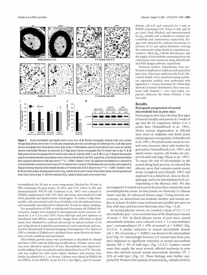
ResultsRetrograde progression of axonalmicrotubule loss in pmn miceHomozygous pmn mice develop first signsof muscle atrophy and paresis at 2 weeks ofage and die by respiratory failure 4 to 5weeks later (Schmalbruch et al., 1991).Motor neuron degeneration in affectedmice starts at endplates and distal axonsand progresses retrogradely (Schmalbruchet al., 1991). Proximal motor axons in ven-tral roots, however, show only modest de-generation (Schmalbruch et al., 1991), andspinal motor neuron cell bodies are pre-served until end stage (Haase et al., 1997).To assess the role of microtubules in theaxonal dying back process, we selected thephrenic nerve that contains mainly motoraxons (Langford and Schmidt, 1983) andanalyzed it at a distal level, close to the di-aphragm, and at an intermediate level cor-responding to the thoracic inlet. We also
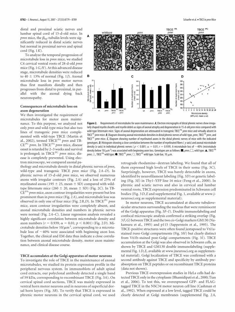
investigated C4 ventral root nerves because they contain the mostproximal phrenic axons. As time points, we chose day 15 (diseaseonset) and day 28 (advanced disease stage). Using electron mi-croscopy, we determined microtubule number and axonal sur-face in at least 10 entire cross-sectional axon profiles per nerve infour wild-type and four pmn littermate mice (Fig. 1A,B).
In normal phrenic nerves, we counted a mean of 15.2 � 2.3microtubules/�m 2 cross-sectional area of the distal axon (meansof mean � SD). In distal phrenic nerves of pmn mice, axonalmicrotubule densities were reduced to 6.0 � 1.6 microtubules/�m 2, corresponding to 39 � 10% of normal ( p � 0.0001) (Fig.1C,D, I). A similar reduction in axonal microtubule density(44 � 9% of normal; p � 0.0001) was found at the intermediatelevel (Fig. 1I). Interestingly however, C4 ventral roots from pmnmice displayed no significant reduction in axonal microtubuledensity (89 � 5% of wild type) (Fig. 1E,F,I). Lumbar ventralroots that supply the most severely affected territories in pmnmice also showed normal axonal microtubule densities (97 �22% of wild type) (Fig. 1 I). These findings were further sup-ported by Western blot analysis of neuronal �III-tubulin levels in
Figure 1. Axonal microtubules and tubulin levels in pmn mice. A, B, Electron micrographs showing entire cross sectionsthrough distal phrenic nerves from 15-d-old early symptomatic pmn mice and wild-type (wt) littermate mice. C, D, High-powerelectron micrographs from distal phrenic nerve axons at day 15. Microtubules (arrows) in pmn phrenic nerve axons are rarefied,whereas intermediate filaments are preserved. E, F, High-power electron micrographs from C4 ventral roots at day 15. G, H,High-power electron micrographs from C4 ventral roots at day 28. Scale bars: A, B, 1 �m; C–H, 0.5 �m. I, Diagram showing thataxonal microtubule densities in pmn phrenic nerves mice are reduced by 61 and 56%, respectively, at the distal and intermediatelevel compared with those in wild-type nerves (***p � 0.0001, Student’s t test). No significant microtubule loss is observed incervical and lumbar ventral root motor axons of 15-d-old pmn mice. A total of 378 individual axon cross sections were analyzed. J,Diagram showing reduction of microtubule densities in C4 ventral roots of 28-d-old pmn mice (***p � 0.0001, Student’s t test).K, Western blot analysis showing protein levels of �III-tubulin and �-actin in lysates from lumbar spinal cord and proximal anddistal sciatic nerves at day 15. Note the reduction of �III-tubulin in distal sciatic nerves of pmn mice.
Schaefer et al. • TBCE in pmn Mice J. Neurosci., August 15, 2007 • 27(33):8779 – 8789 • 8781
distal and proximal sciatic nerves andlumbar spinal cord of 15-d-old mice. Inpmn mice, the �III-tubulin levels were sig-nificantly reduced in distal sciatic nervesbut normal in proximal nerves and spinalcord (Fig. 1K).
To analyze the temporal progression ofmicrotubule loss in pmn mice, we studiedC4 cervical ventral roots of 28-d-old pmnmice (Fig. 1G,H). At this advanced diseasestage, microtubule densities were reducedto 40 � 13% of normal (Fig. 1 J). Axonalmicrotubule loss in pmn motor nervesthus first manifests distally and thenprogresses from distal to proximal, in par-allel with the axonal dying backneuronopathy.
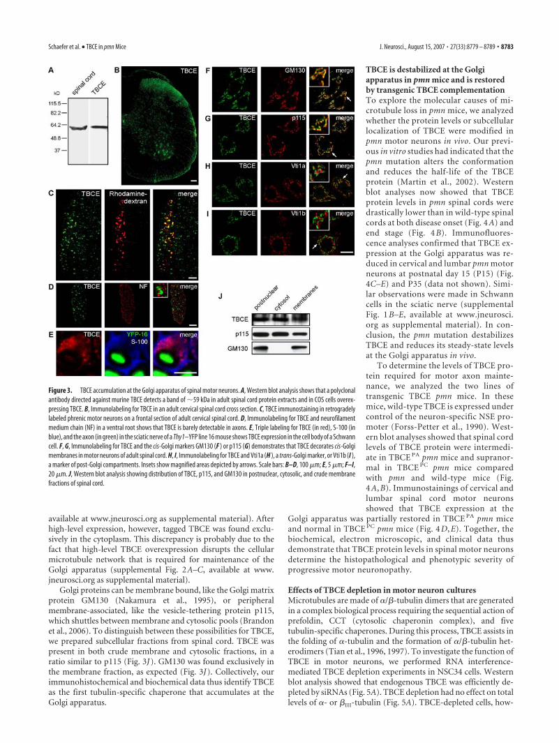
Consequences of microtubule loss onaxon degenerationWe then investigated the requirement ofmicrotubules for motor axon mainte-nance. To this purpose, we analyzed notonly pmn and wild-type mice but also twolines of transgenic pmn mice comple-mented with wild-type TBCE (Martin etal., 2002), termed TBCE PA pmn and TB-CE PC pmn. In TBCE PA pmn mice, diseaseonset is retarded by 2–3 weeks and survivalis prolonged; in TBCE PC pmn mice, dis-ease is completely prevented. Using elec-tron microscopy, we compared axonal pa-thology and microtubule density in distal phrenic nerves of pmn,wild-type and transgenic TBCE pmn mice (Fig. 2A–D). Inphrenic nerves of 15-d-old pmn mice, we observed numerousaxons with irregular contours (Fig. 2A) and a loss of 25% ofmyelinated axons (195 � 25, mean � SD) compared with wild-type littermate mice (260 � 20, mean � SD) (Fig. 2C). In TB-CE PA pmn mice, axon contour irregularities were present but lessprominent than in pmn mice (Fig. 2A), and microtubule loss wasobserved in only one of four mice (Fig. 2B,D). In TBCE PC pmnmice, axon contour irregularities were completely absent, andaxonal microtubule densities and numbers in phrenic nerveswere normal (Fig. 2A–C). Linear regression analysis revealed ahighly significant correlation between microtubule density andaxon numbers (r � 0.939; � � 0.01; p � 0.001) (Fig. 2D). Mi-crotubule densities below 10/�m 2, corresponding to a microtu-bule loss of �40% were associated with beginning axon loss.Together, the clinical and EM data thus indicate a close correla-tion between axonal microtubule density, motor axon mainte-nance, and clinical disease course.
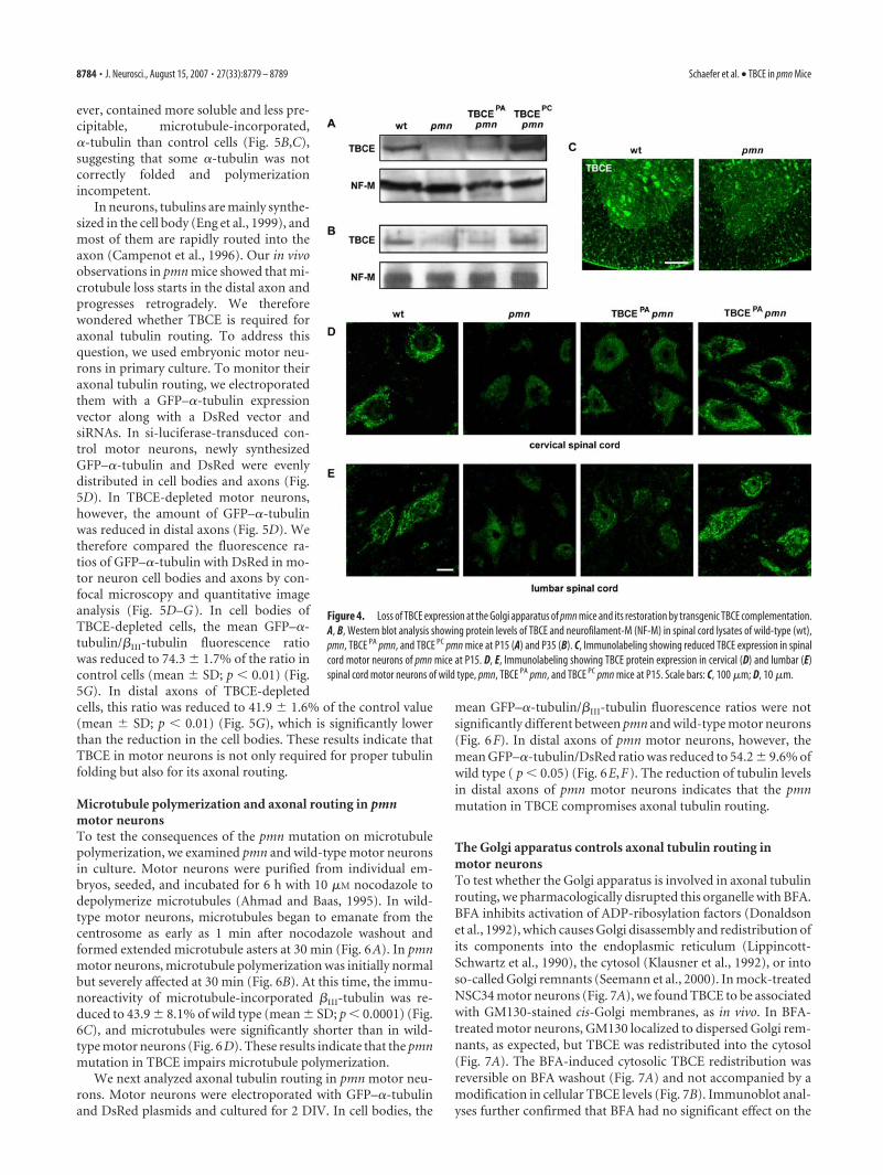
TBCE accumulates at the Golgi apparatus of motor neuronsTo investigate the role of TBCE in the maintenance of axonalmicrotubules, we studied its protein expression profile in theperipheral nervous system. In immunoblots of adult spinalcord extracts, our polyclonal antibody detected a single bandof 59 kDa, corresponding to recombinant TBCE (Fig. 3A). Oncervical spinal cord sections, TBCE was mainly expressed inventral horn motor neurons and in neurons of superficial dor-sal horn layers (Fig. 3B). To verify that TBCE is expressed inphrenic motor neurons in the cervical spinal cord, we used
retrograde rhodamine– dextran labeling. We found that all ofthem expressed high levels of TBCE in their soma (Fig. 3C).Surprisingly, however, TBCE was barely detectable in axons,identified by neurofilament labeling (Fig. 3D) or genetic label-ing (Fig. 3E) in Thy1–YFP line 16 mice (Feng et al., 2000). Inphrenic and sciatic nerves and also in cervical and lumbarventral roots, TBCE expression predominated in Schwann cellbodies (Fig. 3 D, E and supplemental Fig. 1, available at www.j-neurosci.org as supplemental material).
In motor neurons, TBCE accumulated at discrete tubulove-sicular structures surrounding the nucleus that were reminiscentof the Golgi apparatus (Fig. 3F–I). Double immunolabeling andconfocal microscopic analysis confirmed a striking overlap (Fig.3F,G) between TBCE and the two cis-Golgi markers GM130 (Na-kamura et al., 1995) and p115 (Sapperstein et al., 1995). TheTBCE-positive structures were often found juxtaposed to Vti1a-stained trans-Golgi compartments (Fig. 3H) but clearly distinctfrom Vti1b-stained post-Golgi compartments (Fig. 3I). TBCEaccumulation at the Golgi was also observed in Schwann cells, asshown by TBCE and GM130 double immunolabeling (supple-mental Fig. 1D,E, available at www.jneurosci.org as supplemen-tal material). Golgi localization of TBCE was confirmed with asecond antibody against TBCE and specificity by antibody pre-absorption on TBCE peptides or on recombinant TBCE proteins(data not shown).
Previous TBCE overexpression studies in HeLa cells had de-tected TBCE only in the cytoplasm (Bhamidipati et al., 2000; Tianet al., 2006). To test this, we overexpressed GFP- and FLAG-tagged TBCE in the NSC34 motor neuron cell line (Cashman etal., 1992). When expressed at a low level, tagged TBCE could beclearly detected at Golgi membranes (supplemental Fig. 2A,
Figure 2. Requirements of microtubules for axon maintenance. A, Electron micrographs of distal phrenic nerves show irregu-larly shaped myelin sheaths and myelin debris as signs of axonal atrophy and degeneration in 15-d-old pmn mice compared withwild-type littermate mice. Signs of axonal degeneration are attenuated in transgenic TBCE PA pmn mice and virtually absent inTBCE PC pmn mice. B, Diagram showing axonal microtubule densities in distal phrenic nerves of wild-type, pmn, TBCE PA pmn, andTBCE PC pmn mice. C, Diagram showing number of myelinated axons in the distal phrenic nerves of mice with the indicatedgenotypes. D, Histogram showing a close correlation between the number of myelinated fibers ( y-axis) and axonal microtubuledensity (x-axis) in individual phrenic nerves ( p � 0.001; � � 0.01; r � 0.939). A microtubule loss of �40% (microtubuledensity below 10/�m 2) was associated with beginning axon loss. Genotypes are as follows: f, pmn; �, wild type; Œ, TBCE PA
pmn; ‚, TBCE PA wild type; F, TBCE PC pmn; E, TBCE PC wild type. Scale bar, 10 �m.
8782 • J. Neurosci., August 15, 2007 • 27(33):8779 – 8789 Schaefer et al. • TBCE in pmn Mice
available at www.jneurosci.org as supplemental material). Afterhigh-level expression, however, tagged TBCE was found exclu-sively in the cytoplasm. This discrepancy is probably due to thefact that high-level TBCE overexpression disrupts the cellularmicrotubule network that is required for maintenance of theGolgi apparatus (supplemental Fig. 2A–C, available at www.jneurosci.org as supplemental material).
Golgi proteins can be membrane bound, like the Golgi matrixprotein GM130 (Nakamura et al., 1995), or peripheralmembrane-associated, like the vesicle-tethering protein p115,which shuttles between membrane and cytosolic pools (Brandonet al., 2006). To distinguish between these possibilities for TBCE,we prepared subcellular fractions from spinal cord. TBCE waspresent in both crude membrane and cytosolic fractions, in aratio similar to p115 (Fig. 3J). GM130 was found exclusively inthe membrane fraction, as expected (Fig. 3J). Collectively, ourimmunohistochemical and biochemical data thus identify TBCEas the first tubulin-specific chaperone that accumulates at theGolgi apparatus.
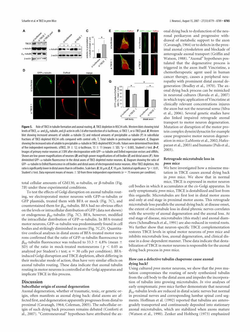
TBCE is destabilized at the Golgiapparatus in pmn mice and is restoredby transgenic TBCE complementationTo explore the molecular causes of mi-crotubule loss in pmn mice, we analyzedwhether the protein levels or subcellularlocalization of TBCE were modified inpmn motor neurons in vivo. Our previ-ous in vitro studies had indicated that thepmn mutation alters the conformationand reduces the half-life of the TBCEprotein (Martin et al., 2002). Westernblot analyses now showed that TBCEprotein levels in pmn spinal cords weredrastically lower than in wild-type spinalcords at both disease onset (Fig. 4 A) andend stage (Fig. 4 B). Immunofluores-cence analyses confirmed that TBCE ex-pression at the Golgi apparatus was re-duced in cervical and lumbar pmn motorneurons at postnatal day 15 (P15) (Fig.4C–E) and P35 (data not shown). Simi-lar observations were made in Schwanncells in the sciatic nerve (supplementalFig. 1 B–E, available at www.jneurosci.org as supplemental material). In con-clusion, the pmn mutation destabilizesTBCE and reduces its steady-state levelsat the Golgi apparatus in vivo.
To determine the levels of TBCE pro-tein required for motor axon mainte-nance, we analyzed the two lines oftransgenic TBCE pmn mice. In thesemice, wild-type TBCE is expressed undercontrol of the neuron-specific NSE pro-moter (Forss-Petter et al., 1990). West-ern blot analyses showed that spinal cordlevels of TBCE protein were intermedi-ate in TBCE PA pmn mice and supranor-mal in TBCE PC pmn mice comparedwith pmn and wild-type mice (Fig.4 A, B). Immunostainings of cervical andlumbar spinal cord motor neuronsshowed that TBCE expression at the
Golgi apparatus was partially restored in TBCE PA pmn miceand normal in TBCE PC pmn mice (Fig. 4 D, E). Together, thebiochemical, electron microscopic, and clinical data thusdemonstrate that TBCE protein levels in spinal motor neuronsdetermine the histopathological and phenotypic severity ofprogressive motor neuronopathy.
Effects of TBCE depletion in motor neuron culturesMicrotubules are made of �/�-tubulin dimers that are generatedin a complex biological process requiring the sequential action ofprefoldin, CCT (cytosolic chaperonin complex), and fivetubulin-specific chaperones. During this process, TBCE assists inthe folding of �-tubulin and the formation of �/�-tubulin het-erodimers (Tian et al., 1996, 1997). To investigate the function ofTBCE in motor neurons, we performed RNA interference-mediated TBCE depletion experiments in NSC34 cells. Westernblot analysis showed that endogenous TBCE was efficiently de-pleted by siRNAs (Fig. 5A). TBCE depletion had no effect on totallevels of �- or �III-tubulin (Fig. 5A). TBCE-depleted cells, how-
Figure 3. TBCE accumulation at the Golgi apparatus of spinal motor neurons. A, Western blot analysis shows that a polyclonalantibody directed against murine TBCE detects a band of �59 kDa in adult spinal cord protein extracts and in COS cells overex-pressing TBCE. B, Immunolabeling for TBCE in an adult cervical spinal cord cross section. C, TBCE immunostaining in retrogradelylabeled phrenic motor neurons on a frontal section of adult cervical spinal cord. D, Immunolabeling for TBCE and neurofilamentmedium chain (NF) in a ventral root shows that TBCE is barely detectable in axons. E, Triple labeling for TBCE (in red), S-100 (inblue), and the axon (in green) in the sciatic nerve of a Thy1–YFP line 16 mouse shows TBCE expression in the cell body of a Schwanncell. F, G, Immunolabeling for TBCE and the cis-Golgi markers GM130 (F ) or p115 (G) demonstrates that TBCE decorates cis-Golgimembranes in motor neurons of adult spinal cord. H, I, Immunolabeling for TBCE and Vti1a (H ), a trans-Golgi marker, or Vti1b (I ),a marker of post-Golgi compartments. Insets show magnified areas depicted by arrows. Scale bars: B–D, 100 �m; E, 5 �m; F–I,20 �m. J, Western blot analysis showing distribution of TBCE, p115, and GM130 in postnuclear, cytosolic, and crude membranefractions of spinal cord.
Schaefer et al. • TBCE in pmn Mice J. Neurosci., August 15, 2007 • 27(33):8779 – 8789 • 8783
ever, contained more soluble and less pre-cipitable, microtubule-incorporated,�-tubulin than control cells (Fig. 5B,C),suggesting that some �-tubulin was notcorrectly folded and polymerizationincompetent.
In neurons, tubulins are mainly synthe-sized in the cell body (Eng et al., 1999), andmost of them are rapidly routed into theaxon (Campenot et al., 1996). Our in vivoobservations in pmn mice showed that mi-crotubule loss starts in the distal axon andprogresses retrogradely. We thereforewondered whether TBCE is required foraxonal tubulin routing. To address thisquestion, we used embryonic motor neu-rons in primary culture. To monitor theiraxonal tubulin routing, we electroporatedthem with a GFP–�-tubulin expressionvector along with a DsRed vector andsiRNAs. In si-luciferase-transduced con-trol motor neurons, newly synthesizedGFP–�-tubulin and DsRed were evenlydistributed in cell bodies and axons (Fig.5D). In TBCE-depleted motor neurons,however, the amount of GFP–�-tubulinwas reduced in distal axons (Fig. 5D). Wetherefore compared the fluorescence ra-tios of GFP–�-tubulin with DsRed in mo-tor neuron cell bodies and axons by con-focal microscopy and quantitative imageanalysis (Fig. 5D–G). In cell bodies ofTBCE-depleted cells, the mean GFP–�-tubulin/�III-tubulin fluorescence ratiowas reduced to 74.3 � 1.7% of the ratio incontrol cells (mean � SD; p � 0.01) (Fig.5G). In distal axons of TBCE-depletedcells, this ratio was reduced to 41.9 � 1.6% of the control value(mean � SD; p � 0.01) (Fig. 5G), which is significantly lowerthan the reduction in the cell bodies. These results indicate thatTBCE in motor neurons is not only required for proper tubulinfolding but also for its axonal routing.
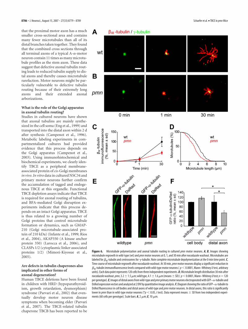
Microtubule polymerization and axonal routing in pmnmotor neuronsTo test the consequences of the pmn mutation on microtubulepolymerization, we examined pmn and wild-type motor neuronsin culture. Motor neurons were purified from individual em-bryos, seeded, and incubated for 6 h with 10 �M nocodazole todepolymerize microtubules (Ahmad and Baas, 1995). In wild-type motor neurons, microtubules began to emanate from thecentrosome as early as 1 min after nocodazole washout andformed extended microtubule asters at 30 min (Fig. 6A). In pmnmotor neurons, microtubule polymerization was initially normalbut severely affected at 30 min (Fig. 6B). At this time, the immu-noreactivity of microtubule-incorporated �III-tubulin was re-duced to 43.9 � 8.1% of wild type (mean � SD; p � 0.0001) (Fig.6C), and microtubules were significantly shorter than in wild-type motor neurons (Fig. 6D). These results indicate that the pmnmutation in TBCE impairs microtubule polymerization.
We next analyzed axonal tubulin routing in pmn motor neu-rons. Motor neurons were electroporated with GFP–�-tubulinand DsRed plasmids and cultured for 2 DIV. In cell bodies, the
mean GFP–�-tubulin/�III-tubulin fluorescence ratios were notsignificantly different between pmn and wild-type motor neurons(Fig. 6F). In distal axons of pmn motor neurons, however, themean GFP–�-tubulin/DsRed ratio was reduced to 54.2 � 9.6% ofwild type ( p � 0.05) (Fig. 6E,F). The reduction of tubulin levelsin distal axons of pmn motor neurons indicates that the pmnmutation in TBCE compromises axonal tubulin routing.
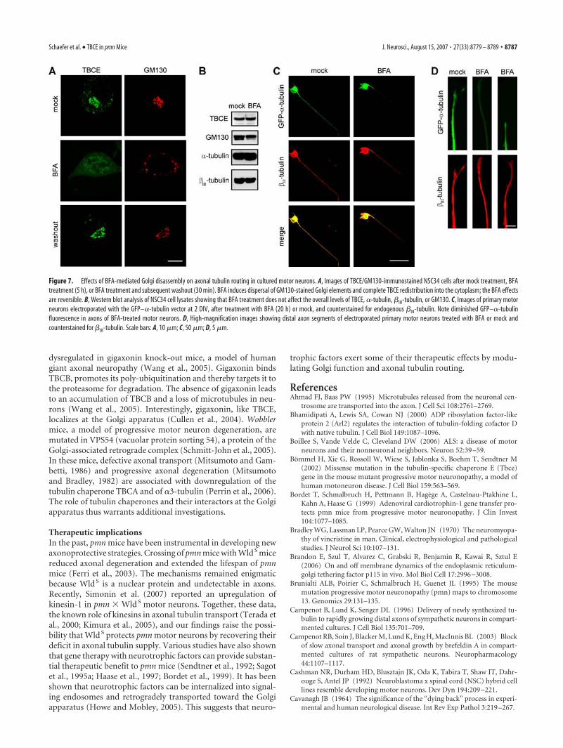
The Golgi apparatus controls axonal tubulin routing inmotor neuronsTo test whether the Golgi apparatus is involved in axonal tubulinrouting, we pharmacologically disrupted this organelle with BFA.BFA inhibits activation of ADP-ribosylation factors (Donaldsonet al., 1992), which causes Golgi disassembly and redistribution ofits components into the endoplasmic reticulum (Lippincott-Schwartz et al., 1990), the cytosol (Klausner et al., 1992), or intoso-called Golgi remnants (Seemann et al., 2000). In mock-treatedNSC34 motor neurons (Fig. 7A), we found TBCE to be associatedwith GM130-stained cis-Golgi membranes, as in vivo. In BFA-treated motor neurons, GM130 localized to dispersed Golgi rem-nants, as expected, but TBCE was redistributed into the cytosol(Fig. 7A). The BFA-induced cytosolic TBCE redistribution wasreversible on BFA washout (Fig. 7A) and not accompanied by amodification in cellular TBCE levels (Fig. 7B). Immunoblot anal-yses further confirmed that BFA had no significant effect on the
Figure 4. Loss of TBCE expression at the Golgi apparatus of pmn mice and its restoration by transgenic TBCE complementation.A, B, Western blot analysis showing protein levels of TBCE and neurofilament-M (NF-M) in spinal cord lysates of wild-type (wt),pmn, TBCE PA pmn, and TBCE PC pmn mice at P15 (A) and P35 (B). C, Immunolabeling showing reduced TBCE expression in spinalcord motor neurons of pmn mice at P15. D, E, Immunolabeling showing TBCE protein expression in cervical (D) and lumbar (E)spinal cord motor neurons of wild type, pmn, TBCE PA pmn, and TBCE PC pmn mice at P15. Scale bars: C, 100 �m; D, 10 �m.
8784 • J. Neurosci., August 15, 2007 • 27(33):8779 – 8789 Schaefer et al. • TBCE in pmn Mice
total cellular amounts of GM130, �-tubulin, or �-tubulin (Fig.7B) under these experimental conditions.
To test the effects of Golgi disruption on axonal tubulin rout-ing, we electroporated motor neurons with GFP–�-tubulin orGFP plasmids, treated them with BFA or mock (Fig. 7C), andcounterstained them for �III-tubulin. BFA had no obvious effecton the levels or intracellular distribution of GFP (data not shown)or endogenous �III-tubulin (Fig. 7C). BFA, however, modifiedthe intracellular distribution of GFP–�-tubulin. In BFA-treatedmotor neurons, GFP–�-tubulin was predominantly found in cellbodies and strikingly diminished in axons (Fig. 7C,D). Quantita-tive confocal analyses in distal axons of BFA-treated motor neu-rons confirmed that the ratio of GFP–�-tubulin fluorescence to�III-tubulin fluorescence was reduced to 33.3 � 4.8% (mean �SD) of the ratio in mock-treated motoneurons ( p � 0.05 asanalyzed per Student’s t test; n � 30 cells per condition). BFA-induced Golgi disruption and TBCE depletion, albeit differing intheir molecular mode of action, thus have very similar effects onaxonal tubulin routing. These data indicate that axonal tubulinrouting in motor neurons is controlled at the Golgi apparatus andimplicate TBCE in this process.
DiscussionSubcellular origin of axonal degenerationAxonal degeneration, whether of traumatic, toxic, or genetic or-igin, often manifests as axonal dying back: distal axons are af-fected first, and degeneration apparently progresses from distal toproximal (Cavanagh, 1964; Coleman, 2005). The subcellular or-igin of such dying back processes remains debated (Conforti etal., 2007). “Centroneuronal” hypotheses have attributed the ax-
onal dying back to dysfunction of the neu-ronal perikaryon and progressive with-drawal of metabolic support to the axon(Cavanagh, 1964) or to defects in the prox-imal axonal cytoskeleton and blockade ofanterograde axonal transport (Griffin andWatson, 1988). “Axonal” hypotheses pos-tulated that the degenerative process istriggered in the axon itself: Vincristine, achemotherapeutic agent used in humancancer therapy, causes a peripheral neu-ropathy with prominent distal axonal de-generation (Bradley et al., 1970). The ax-onal dying back process can be mimickedin neuronal cultures (Ravula et al., 2007)in which topic application of Vincristine atclinically relevant concentrations injuresthe axon but not the neuronal soma (Silvaet al., 2006). Several genetic studies havealso linked impaired retrograde axonaltransport to motor neuron degeneration.Mutation or disruption of the motor pro-tein complex dynein/dynactin for examplecause progressive motor neuron degener-ation in mice (LaMonte et al., 2002; Hafez-parast et al., 2003) and humans (Puls et al.,2003).
Retrograde microtubule loss inpmn miceWe here investigated how a missense mu-tation in TBCE causes axonal dying backin pmn mice. We show that in normalmice, TBCE is expressed in motor neuron
cell bodies in which it accumulates at the cis-Golgi apparatus. Inearly symptomatic pmn mice, TBCE is destabilized and lost fromthis organelle. Microtubules are first lost in distal motor axonsand only at end stage in proximal motor axons. This retrogrademicrotubule loss parallels the axonal dying back: at disease onset,the extent of microtubule loss in distal phrenic nerves correlateswith the severity of axonal degeneration and the axonal loss. Atend stage of disease, microtubules (this study) and axonal diam-eters (Schmalbruch et al., 1991) are also reduced in ventral roots.We further show that neuron-specific TBCE complementationrestores TBCE levels in spinal motor neurons of pmn mice andinhibits microtubule loss, axonal degeneration, and clinical dis-ease in a dose-dependent manner. These data indicate that desta-bilization of TBCE in motor neurons is responsible for the axonaldying back process in pmn mice.
How can a defective tubulin chaperone cause axonaldying back?Using cultured pmn motor neurons, we show that the pmn mu-tation compromises the routing of newly synthesized tubulinfrom the cell body to the distal axon and impedes the incorpora-tion of tubulin into growing microtubules. In vivo analyses ofearly symptomatic pmn mice further demonstrate that neuronal�III-tubulin levels are reduced in distal sciatic nerves but normalin proximal nerves and corresponding lumbar spinal cord seg-ments. Hoffman et al. (1992) reported that tubulins are antero-gradely transported and continuously deposited into stationaryaxonal microtubules, which are stabilized when axons mature(Watson et al., 1990). Zenker and Hohberg (1973) emphasized
Figure 5. Role of TBCE in tubulin formation and axonal routing. A, TBCE depletion in NSC34 cells. Western blots showing totallevels of TBCE, �- and �III-tubulin, and �-actin in cells 3 d after transfection of si-luciferase, si-TBCE 1, or si-TBCE pool. B, Westernblot showing increased amounts of soluble �-tubulin (S) and reduced amounts of precipitable �-tubulin (P) in subcellularfractions of TBCE-depleted NSC34 cells compared with control cells. T, Total tubulin in postnuclear supernatant. C, Diagramshowing the increased ratio of soluble to precipitable �-tubulin in TBCE-depleted NSC34 cells. Values were determined from blotsof five independent experiments. siTBCE, 39 � 12; si-luciferase, 13 � 11 (means � SD). *p � 0.005, Student’s t test. D–F,Images of primary motor neurons at 3 DIV after electroporation with GFP–�-tubulin and DsRed expression vectors and siRNAs.Shown are low-power magnifications of neurons (D) and high-power magnifications of cell bodies (E) and distal axons (F ). Notediminished GFP–�-tubulin fluorescence in the distal axons of TBCE-depleted motor neurons. G, Diagram showing the ratio ofGFP–�-tubulin to DsRed fluorescence in cell bodies and distal axons of electroporated motor neurons. After TBCE depletion, thisratio is significantly lower in distal axons than in cell bodies. Scale bars: D, 50 �m; E, F, 10 �m. Statistical significance: *p � 0.01,Student’s t test. Data represent means of means � SD from three independent experiments (n � 75 neurons per condition).
Schaefer et al. • TBCE in pmn Mice J. Neurosci., August 15, 2007 • 27(33):8779 – 8789 • 8785
that the proximal motor axon has a muchsmaller cross-sectional area and containsmany fewer microtubules than all of itsdistal branches taken together. They foundthat the combined cross sections throughall terminal axons of a typical A-�-motorneuron contain 11 times as many microtu-bule profiles as the stem axon. These datasuggest that defective axonal tubulin rout-ing leads to reduced tubulin supply to dis-tal axons and thereby causes microtubulerarefaction. Motor neurons might be par-ticularly vulnerable to defective tubulinrouting because of their extremely longaxons and their extended axonalarborizations.
What is the role of the Golgi apparatusin axonal tubulin routing?Studies in cultured neurons have shownthat axonal tubulins are mainly synthe-sized in the cell soma (Eng et al., 1999) andtransported into the distal axon within 2 dafter synthesis (Campenot et al., 1996).Metabolic labeling experiments in com-partmentalized cultures had providedevidence that this process depends onthe Golgi apparatus (Campenot et al.,2003). Using immunohistochemical andbiochemical experiments, we clearly iden-tify TBCE as a peripheral membrane-associated protein of cis-Golgi membranesin vivo. In vitro data in cultured NSC34 andprimary motor neurons further confirmthe accumulation of tagged and endoge-nous TBCE at this organelle. FunctionalTBCE depletion assays indicate that TBCEis required for axonal routing of tubulins,and BFA-mediated Golgi disruption ex-periments indicate that this process de-pends on an intact Golgi apparatus. TBCEis thus related to a growing number ofGolgi proteins that control microtubuleformation or dynamics, such as GMAP-210 (Golgi microtubule-associated pro-tein of 210 kDa) (Infante et al., 1999; Rioset al., 2004), AKAP350 (A kinase anchorprotein 350) (Larocca et al., 2006), andCLASPs 1/2 (cytoplasmic linker associatedproteins 1/2) (Mimori-Kiyosue et al.,2005).
Are defects in tubulin chaperones alsoimplicated in other forms ofaxonal degeneration?Human TBCE deletions have been foundin children with HRD (hypoparathyroid-ism, growth retardation, dysmorphism)syndrome (Parvari et al., 2002) that even-tually develop motor neuron diseasesymptoms when becoming elder (Parvariet al., 2007). The TBCE-related tubulinchaperone TBCB has been reported to be
Figure 6. Microtubule polymerization and axonal tubulin routing in cultured pmn motor neurons. A, B, Images showingmicrotubule regrowth in wild-type (wt) and pmn motor neurons at 0, 1, and 30 min after nocodazole washout. Microtubules arelabeled for �III-tubulin and centrosomes for �-tubulin. Note complete microtubule depolymerization at the 0 min time point. C,Time course of microtubule regrowth after nocodazole washout. At 30 min, pmn motor neurons display a significant reduction in�III-tubulin immunofluorescence levels compared with wild-type motor neurons ( p � 0.0001, Mann–Whitney U test, arbitraryunits). Each data point represents 120 cells from three independent experiments. D, Microtubule length distribution 30 min afternocodazole washout. pmn, 3.2�1.5 �m; wild type, 4.1�1.6 �m (means�SD) ( p�0.0001, Mann–Whitney U test; n�120per genotype). E, Images of distal axons from wild-type and pmn primary motor neurons electroporated with GFP–�-tubulin andDsRed expression vectors and analyzed at 2 DIV by quantitative image analysis. F, Diagram showing the ratio of GFP–�-tubulin toDsRed fluorescence in cell bodies and distal axons of wild-type and pmn motor neurons. In distal axons, this ratio is significantlylower in pmn than in wild-type motor neurons (*p � 0.05, t test). Data represent means � SD from two independent experi-ments (60 cells per genotype). Scale bars: A, 2 �m; E, 10 �m.
8786 • J. Neurosci., August 15, 2007 • 27(33):8779 – 8789 Schaefer et al. • TBCE in pmn Mice
dysregulated in gigaxonin knock-out mice, a model of humangiant axonal neuropathy (Wang et al., 2005). Gigaxonin bindsTBCB, promotes its poly-ubiquitination and thereby targets it tothe proteasome for degradation. The absence of gigaxonin leadsto an accumulation of TBCB and a loss of microtubules in neu-rons (Wang et al., 2005). Interestingly, gigaxonin, like TBCE,localizes at the Golgi apparatus (Cullen et al., 2004). Wobblermice, a model of progressive motor neuron degeneration, aremutated in VPS54 (vacuolar protein sorting 54), a protein of theGolgi-associated retrograde complex (Schmitt-John et al., 2005).In these mice, defective axonal transport (Mitsumoto and Gam-betti, 1986) and progressive axonal degeneration (Mitsumotoand Bradley, 1982) are associated with downregulation of thetubulin chaperone TBCA and of �3-tubulin (Perrin et al., 2006).The role of tubulin chaperones and their interactors at the Golgiapparatus thus warrants additional investigations.
Therapeutic implicationsIn the past, pmn mice have been instrumental in developing newaxonoprotective strategies. Crossing of pmn mice with Wld S micereduced axonal degeneration and extended the lifespan of pmnmice (Ferri et al., 2003). The mechanisms remained enigmaticbecause Wld S is a nuclear protein and undetectable in axons.Recently, Simonin et al. (2007) reported an upregulation ofkinesin-1 in pmn � Wld S motor neurons. Together, these data,the known role of kinesins in axonal tubulin transport (Terada etal., 2000; Kimura et al., 2005), and our findings raise the possi-bility that Wld S protects pmn motor neurons by recovering theirdeficit in axonal tubulin supply. Various studies have also shownthat gene therapy with neurotrophic factors can provide substan-tial therapeutic benefit to pmn mice (Sendtner et al., 1992; Sagotet al., 1995a; Haase et al., 1997; Bordet et al., 1999). It has beenshown that neurotrophic factors can be internalized into signal-ing endosomes and retrogradely transported toward the Golgiapparatus (Howe and Mobley, 2005). This suggests that neuro-
trophic factors exert some of their therapeutic effects by modu-lating Golgi function and axonal tubulin routing.
ReferencesAhmad FJ, Baas PW (1995) Microtubules released from the neuronal cen-
trosome are transported into the axon. J Cell Sci 108:2761–2769.Bhamidipati A, Lewis SA, Cowan NJ (2000) ADP ribosylation factor-like
protein 2 (Arl2) regulates the interaction of tubulin-folding cofactor Dwith native tubulin. J Cell Biol 149:1087–1096.
Boillee S, Vande Velde C, Cleveland DW (2006) ALS: a disease of motorneurons and their nonneuronal neighbors. Neuron 52:39 –59.
Bommel H, Xie G, Rossoll W, Wiese S, Jablonka S, Boehm T, Sendtner M(2002) Missense mutation in the tubulin-specific chaperone E (Tbce)gene in the mouse mutant progressive motor neuronopathy, a model ofhuman motoneuron disease. J Cell Biol 159:563–569.
Bordet T, Schmalbruch H, Pettmann B, Hagege A, Castelnau-Ptakhine L,Kahn A, Haase G (1999) Adenoviral cardiotrophin-1 gene transfer pro-tects pmn mice from progressive motor neuronopathy. J Clin Invest104:1077–1085.
Bradley WG, Lassman LP, Pearce GW, Walton JN (1970) The neuromyopa-thy of vincristine in man. Clinical, electrophysiological and pathologicalstudies. J Neurol Sci 10:107–131.
Brandon E, Szul T, Alvarez C, Grabski R, Benjamin R, Kawai R, Sztul E(2006) On and off membrane dynamics of the endoplasmic reticulum-golgi tethering factor p115 in vivo. Mol Biol Cell 17:2996 –3008.
Brunialti ALB, Poirier C, Schmalbruch H, Guenet JL (1995) The mousemutation progressive motor neuronopathy (pmn) maps to chromosome13. Genomics 29:131–135.
Campenot B, Lund K, Senger DL (1996) Delivery of newly synthesized tu-bulin to rapidly growing distal axons of sympathetic neurons in compart-mented cultures. J Cell Biol 135:701–709.
Campenot RB, Soin J, Blacker M, Lund K, Eng H, MacInnis BL (2003) Blockof slow axonal transport and axonal growth by brefeldin A in compart-mented cultures of rat sympathetic neurons. Neuropharmacology44:1107–1117.
Cashman NR, Durham HD, Blusztajn JK, Oda K, Tabira T, Shaw IT, Dahr-ouge S, Antel JP (1992) Neuroblastoma x spinal cord (NSC) hybrid celllines resemble developing motor neurons. Dev Dyn 194:209 –221.
Cavanagh JB (1964) The significance of the “dying back” process in experi-mental and human neurological disease. Int Rev Exp Pathol 3:219 –267.
Figure 7. Effects of BFA-mediated Golgi disassembly on axonal tubulin routing in cultured motor neurons. A, Images of TBCE/GM130-immunostained NSC34 cells after mock treatment, BFAtreatment (5 h), or BFA treatment and subsequent washout (30 min). BFA induces dispersal of GM130-stained Golgi elements and complete TBCE redistribution into the cytoplasm; the BFA effectsare reversible. B, Western blot analysis of NSC34 cell lysates showing that BFA treatment does not affect the overall levels of TBCE, �-tubulin, �III-tubulin, or GM130. C, Images of primary motorneurons electroporated with the GFP–�-tubulin vector at 2 DIV, after treatment with BFA (20 h) or mock, and counterstained for endogenous �III-tubulin. Note diminished GFP–�-tubulinfluorescence in axons of BFA-treated motor neurons. D, High-magnification images showing distal axon segments of electroporated primary motor neurons treated with BFA or mock andcounterstained for �III-tubulin. Scale bars: A, 10 �m; C, 50 �m; D, 5 �m.
Schaefer et al. • TBCE in pmn Mice J. Neurosci., August 15, 2007 • 27(33):8779 – 8789 • 8787
Cifuentes-Diaz C, Nicole S, Velasco ME, Borra-Cebrian C, Panozzo C, Fru-gier T, Millet G, Roblot N, Joshi V, Melki J (2002) Neurofilament accu-mulation at the motor endplate and lack of axonal sprouting in a spinalmuscular atrophy mouse model. Hum Mol Genet 11:1439 –1447.
Coleman M (2005) Axon degeneration mechanisms: commonality amid di-versity. Nat Rev Neurosci 6:889 – 898.
Conforti L, Adalbert R, Coleman MP (2007) Neuronal death: where doesthe end begin? Trends Neurosci 30:159 –166.
Cullen VC, Brownlees J, Banner S, Anderton BH, Leigh PN, Shaw CE, MillerCC (2004) Gigaxonin is associated with the Golgi and dimerises via itsBTB domain. NeuroReport 15:873– 876.
Devon RS, Orban PC, Gerrow K, Barbieri MA, Schwab C, Cao LP, Helm JR,Bissada N, Cruz-Aquado R, Davidson TL, Witmer J, Metzler M, Lam CK,Tetzlaff W, Simpson EM, McCaffery JM, El-Husseini AE, Leavitt BR,Hayden MR (2006) Als2-deficient mice exhibit disturbances in endosometrafficking associated with motor behavioral abnormalities. Proc NatlAcad Sci USA 103:9595–9600.
Donaldson JG, Finazzi D, Klausner RD (1992) Brefeldin A inhibits Golgimembrane-catalysed exchange of guanine nucleotide onto ARF protein.Nature 360:350 –352.
Eng H, Lund K, Campenot RB (1999) Synthesis of �-tubulin, actin, andother proteins in axons of sympathetic neurons in compartmented cul-tures. J Neurosci 19:1–9.
Feng G, Mellor RH, Bernstein M, Keller-Peck C, Nguyen QT, Wallace M,Nerbonne JM, Lichtman JW, Sanes JR (2000) Imaging neuronal subsetsin transgenic mice expressing multiple spectral variants of GFP. Neuron28:41–51.
Ferri A, Sanes JR, Coleman MP, Cunningham JM, Kato AC (2003) Inhibit-ing axon degeneration and synapse loss attenuates apoptosis and diseaseprogression in a mouse model of motoneuron disease. Curr Biol13:669 – 673.
Fischer LR, Culver DG, Tennant P, Davis AA, Wang M, Castellano-SanchezA, Khan J, Polak MA, Glass JD (2004) Amyotrophic lateral sclerosis is adistal axonopathy: evidence in mice and man. Exp Neurol 185:232–240.
Forss-Petter S, Danielson PE, Catsicas S, Battenberg E, Price J, Nerenberg M,Sutcliffe JG (1990) Transgenic mice expressing beta-galactosidase inmature neurons under neuron-specific enolase promoter control. Neu-ron 5:187–197.
Frey D, Schneider C, Xu L, Borg J, Spooren W, Caroni P (2000) Early andselective loss of neuromuscular synapse subtypes with low sproutingcompetence in motoneuron diseases. J Neurosci 20:2534 –2542.
Griffin JW, Watson DF (1988) Axonal transport in neurological disease.Ann Neurol 23:3–13.
Haase G, Kennel P, Pettmann B, Vigne E, Akli S, Revah F, Schmalbruch H,Kahn A (1997) Gene therapy of a murine motor neuron disease usingadenoviral vectors for neurotrophic factors. Nat Med 3:429 – 436.
Hafezparast M, Klocke R, Ruhrberg C, Marquardt A, Ahmad-Annuar A,Bowen S, Lalli G, Witherden AS, Hummerich H, Nicholson S, Morgan PJ,Oozageer R, Priestley JV, Averill S, King VR, Ball S, Peters J, Toda T,Yamamoto A, Hiraoka Y, et al. (2003) Mutations in dynein link motorneuron degeneration to defects in retrograde transport. Science300:808 – 812.
Hoffman PN, Lopata MA, Watson DF, Luduena RF (1992) Axonal trans-port of class II and III beta-tubulin: evidence that the slow componentwave represents the movement of only a small fraction of the tubulin inmature motor axons. J Cell Biol 119:595– 604.
Howe CL, Mobley WC (2005) Long-distance retrograde neurotrophic sig-naling. Curr Opin Neurobiol 15:40 – 48.
Infante C, Ramos-Morales F, Fedriani C, Bornens M, Rios RM (1999)GMAP-210, a cis-Golgi network-associated protein, is a minus endmicrotubule-binding protein. J Cell Biol 145:83–98.
Jacquier A, Buhler E, Schafer MK, Bohl D, Blanchard S, Beclin C, Haase G(2006) Alsin/Rac1 signaling controls survival and growth of spinal mo-toneurons. Ann Neurol 60:105–117.
Kennel PF, Finiels F, Revah F, Mallet J (1996a) Neuromuscular functionimpairment is not caused by motor neurone loss in FALS mice: an elec-tromyographic study. NeuroReport 7:1427–1431.
Kennel PF, Fonteneau P, Martin E, Schmidt JM, Azzouz M, Borg J, Guenet JL,Schmalbruch H, Warter JM, Poindron P (1996b) Electromyographicaland motor performance studies in the pmn mouse model of neurodegen-erative disease. Neurobiol Disease 3:137–147.
Kimura T, Watanabe H, Iwamatsu A, Kaibuchi K (2005) Tubulin and
CRMP-2 complex is transported via Kinesin-1. J Neurochem93:1371–1382.
Klausner RD, Donaldson JG, Lippincott-Schwartz J (1992) Brefeldin A: in-sights into the control of membrane traffic and organelle structure. J CellBiol 116:1071–1080.
LaMonte BH, Wallace KE, Holloway BA, Shelly SS, Ascano J, Tokito M, VanWinkle T, Howland DS, Holzbaur EL (2002) Disruption of dynein/dy-nactin inhibits axonal transport in motor neurons causing late-onset pro-gressive degeneration. Neuron 34:715–727.
Langford LA, Schmidt RF (1983) An electron microscopic analysis of the leftphrenic nerve in the rat. Anat Rec 205:207–213.
Larocca MC, Jin M, Goldenring JR (2006) AKAP350 modulates microtu-bule dynamics. Eur J Cell Biol 85:611– 619.
Lippincott-Schwartz J, Donaldson JG, Schweizer A, Berger EG, Hauri HP,Yuan LC, Klausner RD (1990) Microtubule-dependent retrogradetransport of proteins into the ER in the presence of brefeldin A suggests anER recycling pathway. Cell 60:821– 836.
Martin N, Jaubert J, Gounon P, Salido E, Haase G, Szatanik M, Guenet JL(2002) A missense mutation in Tbce causes progressive motor neu-ronopathy in mice. Nat Genet 32:443– 447.
Mimori-Kiyosue Y, Grigoriev I, Lansbergen G, Sasaki H, Matsui C, Severin F,Galjart N, Grosveld F, Vorobjev I, Tsukita S, Akhmanova A (2005)CLASP1 and CLASP2 bind to EB1 and regulate microtubule plus-enddynamics at the cell cortex. J Cell Biol 168:141–153.
Mitsumoto H, Bradley WG (1982) Murine motor neuron disease (the wob-bler mouse): degeneration and regeneration of the lower motor neuron.Brain 105:811– 834.
Mitsumoto H, Gambetti P (1986) Impaired slow axonal transport in wob-bler mouse motor neuron disease. Ann Neurol 19:36 – 43.
Nakamura N, Rabouille C, Watson R, Nilsson T, Hui N, Slusarewicz P, KreisTE, Warren G (1995) Characterization of a cis-Golgi matrix protein,GM130. J Cell Biol 131:1715–1726.
Parvari R, Hershkovitz E, Grossman N, Gorodischer R, Loeys B, Zecic A,Mortier G, Gregory S, Sharony R, Kambouris M, Sakati N, Meyer BF, AlAqeel AI, Al Humaidan AK, Al Zanhrani F, Al Swaid A, Al Othman J, DiazGA, Weiner R, Khan KT, Gordon R, Gelb BD (2002) Mutation of TBCEcauses hypoparathyroidism-retardation-dysmorphism and autosomalrecessive Kenny-Caffey syndrome. Nat Genet 32:448 – 452.
Parvari R, Diaz GA, Hershkovitz E (2007) Parathyroid development and therole of tubulin chaperone E. Horm Res 67:12–21.
Pasinelli P, Brown RH (2006) Molecular biology of amyotrophic lateralsclerosis: insights from genetics. Nat Rev Neurosci 7:710 –723.
Perrin FE, Boisset G, Lathuiliere A, Kato AC (2006) Cell death pathwaysdiffer in several mouse models with motoneurone disease: analysis of puremotoneurone populations at a presymptomatic age. J Neurochem98:1959 –1972.
Puls I, Jonnakuty C, LaMonte BH, Holzbaur EL, Tokito M, Mann E, FloeterMK, Bidus K, Drayna D, Oh SJ, Brown Jr RH, Ludlow CL, Fischbeck KH(2003) Mutant dynactin in motor neuron disease. Nat Genet33:455– 456.
Pun S, Santos AF, Saxena S, Xu L, Caroni P (2006) Selective vulnerabilityand pruning of phasic motoneuron axons in motoneuron disease allevi-ated by CNTF. Nat Neurosci 9:408 – 419.
Raoul C, Estevez AG, Nishimune H, Cleveland DW, deLapeyriere O, Hender-son CE, Haase G, Pettmann B (2002) Motoneuron death triggered by aspecific pathway downstream of Fas. Potentiation by ALS-linked SOD1mutations. Neuron 35:1067–1083.
Ravula SK, Wang MS, McClain MA, Asress SA, Frazier B, Glass JD (2007)Spatiotemporal localization of injury potentials in DRG neurons duringvincristine-induced axonal degeneration. Neurosci Lett 415:34 –39.
Richmond FJ, Gladdy R, Creasy JL, Kitamura S, Smits E, Thomson DB(1994) Efficacy of seven retrograde tracers, compared in multiple-labelling studies of feline motoneurones. J Neurosci Methods 53:35– 46.
Rios RM, Sanchis A, Tassin AM, Fedriani C, Bornens M (2004) GMAP-210recruits gamma-tubulin complexes to cis-Golgi membranes and is re-quired for Golgi ribbon formation. Cell 118:323–335.
Sagot Y, Tan SA, Baetge E, Schmalbruch H, Kato AC, Aebischer P (1995a)Polymer encapsulated cell lines genetically engineered to release ciliaryneurotrophic factor can slow down progressive motor neuronopathy inthe mouse. Eur J Neurosci 7:1313–1322.
Sagot Y, Duboisdauphin M, Tan SA, Debilbao F, Aebischer P, Martinou JC,Kato AC (1995b) Bcl-2 overexpression prevents motoneuron cell body
8788 • J. Neurosci., August 15, 2007 • 27(33):8779 – 8789 Schaefer et al. • TBCE in pmn Mice
loss but not axonal degeneration in a mouse model of a neurodegenera-tive disease. J Neurosci 15:7727–7733.
Sapperstein SK, Walter DM, Grosvenor AR, Heuser JE, Waters MG (1995)p115 is a general vesicular transport factor related to the yeast endoplas-mic reticulum to Golgi transport factor Uso1p. Proc Natl Acad Sci USA92:522–526.
Schmalbruch H, Jensen HS, Bjaerg M, Kamieniecka Z, Kurland L (1991) Anew mouse mutant with progressive motor neuronopathy. J NeuropatholExp Neurol 50:192–204.
Schmitt-John T, Drepper C, Mussmann A, Hahn P, Kuhlmann M, Thiel C,Hafner M, Lengeling A, Heimann P, Jones JM, Meisler MH, Jockusch H(2005) Mutation of Vps54 causes motor neuron disease and defectivespermiogenesis in the wobbler mouse. Nat Genet 37:1213–1215.
Seemann J, Jokitalo E, Pypaert M, Warren G (2000) Matrix proteins cangenerate the higher order architecture of the Golgi apparatus. Nature407:1022–1026.
Sendtner M, Schmalbruch H, Stockli KA, Carroll P, Kreutzberg GW,Thoenen H (1992) Ciliary neurotrophic factor prevents degeneration ofmotor neurons in mouse mutant progressive motor neuronopathy. Na-ture 358:502–504.
Silva A, Wang Q, Wang M, Ravula SK, Glass JD (2006) Evidence for directaxonal toxicity in vincristine neuropathy. J Peripher Nerv Syst11:211–216.
Simonin Y, Perrin FE, Kato AC (2007) Axonal involvement in the Wldsneuroprotective effect: analysis of pure motoneurons in a mouse model
protected from motor neuron disease at a pre-symptomatic age. J Neuro-chem 101:530 –542.
Terada S, Kinjo M, Hirokawa N (2000) Oligomeric tubulin in large trans-porting complex is transported via kinesin in squid giant axons. Cell103:141–155.
Tian G, Huang Y, Rommelaere H, Vandekerckhove J, Ampe C, Cowan NJ(1996) Pathway leading to correctly folded beta-tubulin. Cell86:287–296.
Tian G, Lewis SA, Feierbach B, Stearns T, Rommelaere H, Ampe C, Cowan NJ(1997) Tubulin subunits exist in an activated conformational state gen-erated and maintained by protein cofactors. J Cell Biol 138:821– 832.
Tian G, Huang MC, Parvari R, Diaz GA, Cowan NJ (2006) Cryptic out-of-frame translational initiation of TBCE rescues tubulin formation in com-pound heterozygous HRD. Proc Natl Acad Sci USA 103:13491–13496.
Wang W, Ding J, Allen E, Zhu P, Zhang L, Vogel H, Yang Y (2005)Gigaxonin interacts with tubulin folding cofactor B and controls itsdegradation through the ubiquitin-proteasome pathway. Curr Biol15:2050 –2055.
Watson DF, Hoffman PN, Griffin JW (1990) The cold stability of microtu-bules increases during axonal maturation. J Neurosci 10:3344 –3352.
Yamanaka K, Miller TM, McAlonis-Downes M, Chun SJ, Cleveland DW(2006) Progressive spinal axonal degeneration and slowness in ALS2-deficient mice. Ann Neurol 60:95–104.
Zenker W, Hohberg E (1973) A-alpha-nerve-fiber: number of neurotubulesin the stem fibre and in the terminal branches. J Neurocytol 2:143–148.
Schaefer et al. • TBCE in pmn Mice J. Neurosci., August 15, 2007 • 27(33):8779 – 8789 • 8789