Embed Size (px)
Citation preview

PRAB
ROGBSTR
GRAMRACT
M AT BO
AND OOK
K
YBMRS 2014 1
Dear YBMRS Participant,
Welcome to the 13th edition of the Young Belgian Magnetic Resonance Scientist symposium series! We thank you for joining us and hope you will enjoy the conference, as well as your stay here in the heart of the most beautiful Belgian forest. The organizers have strived to present an attractive program, including two educational sessions, two invited lectures and a selection of short oral communications on a wide variety of topics. As always, the goal has been to promote the work of young scientists working in any field related to magnetic resonance, and to create opportunities for interesting discussions and contacts. General practical information All scientific activities are organized around the “Pierre le Grand” conference room, which can be found to the right from the main in the lobby at the level ‐1. All participating sponsors are located in the meeting area of the Sol Cress, where coffee breaks will be served during the poster sessions and in between the morning sessions. A wardrobe is available, however we urge you not to leave any valuables as we cannot guarantee it will be guarded at all times. Should you have any question during your stay, please address yourself to the conference desk located in the meeting area. During sessions
• Please switch off your mobile phone or put it in silent mode. • Do not take pictures of slides or posters, but rather approach the author during the
meeting and request a reprint or additional information. • Have the courtesy not to use your laptop in the conference room during talks, but
rather use the main lobby, foyer or bar. Poster sessions All posters are to be set up in the “Wellington” and “Sources de la Reine” rooms. The posters panels are numbered. Your poster number is available from the abstract book or on the list provided in the poster room. Participants presenting a poster are kindly requested to set up their poster no later than the lunch Break on Monday, and to remove them after the coffee break on Tuesday afternoon. Transparent adhesive tape is available in the poster room to fix your poster to the panels. Please do not use staples or pushpins in any circumstances. Rooms If you have not done so, please register for your room at the main desk in the lobby of the Sol Cress. Check out time: Please note that all rooms should be vacated no later than 10:00 a.m. on Tuesday morning.
13TH EDITION
YBMRS 2014 2
A closed luggage room, next to the poster room, will be available on Tuesday for safekeeping of your luggage. Should you have any question or request concerning your accommodation, please contact the Soll Cress staff. Breakfast, lunch and dinner Breakfast and lunch will be served in the Restaurant. Please follow the indications from the main lobby. Breakfast is served from 07:30 until 09:00 on Monday and Tuesday morning. Lunch starts immediately after the morning session and consists of a buffet with beverages on Monday and of a hot meal on Tuesday. The main conference dinner will start at 20:30 on Monday evening, with a choice between a vegetarian or a non‐vegetarian menu, as indicated during your registration. If you have any additional dietary requirements, please contact the Sol Cress staff. Internet access Free wireless internet access is available in the entire domain. YBMRS party A party will be organized after dinner on Monday evening, starting around 10 p.m. and featuring the House DJ. At the request of the Sol Cress, and as a courtesy to the other guests, please note that the party is set to close at 01:00. Please note that the bar remains available to provide more tranquil surrounding. If you have further questions, please contact the conference desk in the foyer. Yours sincerely, The OrganizingCommittee
• Luce Vander Elst, Université de Mons • Christian Damblon, Université de Liège • Céline Henoumont, Université de Mons • Uwe Himmelreich, Katholieke Universiteit Leuven • Yves Gossuin, Université de Mons • Q.L. Vuong, Université de Mons • J. Martins, Universiteit Gent
The Scientific Committee
• Carmela Aprile, Facultés Universitaires Notre-Dame de la Paix, Namur • Christian Damblon, Université de Liège • Bernard Gallez, Université Catholique de Louvain • Uwe Himmelreich, Katholieke Universiteit Leuven • Sabine Van Doorslaer, Universiteit Antwerpen • Luce Vander Elst, Université de Mons • Alex Volkov, Vrije Universiteit Brussel • Michel Luhmer, Université Libre de Bruxelles • Davy Sinnaeve, Universiteit Gent • Kristin Bartik, Université Libre de Bruxelles • Nico Van Nuland, Vrije Universiteit Brussel

YBMRS 2
Tfo
2014
The orgaollowing
anizing g institu
commitutions, s
ki
ttee grasocietieind sup
atefullyes and cpport
acknowcompan
wledgesnies for
3
s the their

YBMRS 2014‐PROGRAM 4
Symposium Program Monday, 24th November 9:00 Registration, Welcome Coffee and Poster Setup 10:15 Welcome 10:20 Educational Session 1 Rudi WILLEM (VUB) High Resolution Magic Angle Spinning (hr‐MAS) NMR: investigation of solid‐liquid
interfaces in materials 11:20 Break 11:35 Educational Session 2 Claudio Luchinat (University of Florence) Paramagnetic NMR: a versatile tool in structural biology (Part 1) 13:00 Lunch 14:00 Symposium Opening Plenary Session 1 14:10 Claudio Luchinat (University of Florence) Paramagnetic NMR: a versatile tool in structural biology (Part 2) 15:20 J. De Roo (Universiteit Gent) “The surface chemistry of metal oxide nanocrystals; a
solution NMR study” 15:40 D. Henrard (Université de Mons) “Characterization of iron oxide nanoparticles by NMR
relaxometry and magnetometry: effects of size distribution and agglomeration” 16:00 M. Harris (KU Leuven) “Dysprosium and terbium magnetofluorescent micellar
Complexes as potential bimodal agents for magnetic resonance and optical imaging” 16:20 Poster session 1 – Coffee break Plenary session 2 17:40 E. Brunetti (Université Libre de Bruxelles) “NMR Study of the Recognition Properties of a
Calix[6]aza‐cryptand Incorporated in DPC Micelles” 18:00 M. Dentamaro (Université de Mons) “Targeting of Apoptotic Cells by a New Bimodal
Probe Based on AGuIX® Nanoparticles” 18:20 E. Louis( Hasselt University) “Validation of 1H‐NMR‐based metabolomics as a tool to
detect lung cancer in human blood plasma” 19:30 Reception 20:30 Dinner and YBMRS Party

YBMRS 2014‐PROGRAM 5
Tuesday, 25th November 07:30 Breakfast Plenary session 3 09:00 D. Buyst (Universiteit Gent) “Identification of a pKa‐regulating motif stabilizing
Imidazole modified double stranded DNA” 09:20 R. Lavendomme (Université Libre de Bruxelles) “Characterization of calixarene
derivatives by liquid‐state NMR spectroscopy: challenges and solutions” 09:40 M. Tassi (Hasselt University) “Characterization of phosphonic acid grafted titanium
dioxide surfaces by 31P NMR and ATR‐FTIR” 10:00 J. Kusakovskij (Universiteit Gent) “Study of transient radicals created immediately
after room temperature X‐ray irradiation in single crystal sucrose” 10:20 Gérald REMAUD (Université de Nantes) Isotopic NMR and analysis of food : 2H and 13C NMR 11:20 Poster session 2‐ Coffee Break 13:00 Lunch Plenary session 4 14:00 Klaas Nicolaij (Eindhoven University of Technology) MR techniques for guiding cancer therapy 15:00 B. Cuypers (University of Antwerp) “An investigation of Antarctic fish cytoglobins using
EPR and optical spectroscopy” 15:20 R. Garcia Ribeiro (KU Leuven) “Synthesis and characterization of functionalized
magnetoliposomes for Magnetic Resonance Imaging and theranostics applications” 15:40 N. Sauwen (KU Leuven) “Hierarchical non‐negative matrix factorization for brain tumor
characterization using multi‐parametric MRI” 16:00 Coffee break 16:20 A. De Rache (Université de Bordeaux) “G‐quadruplex in the HIV promoter region: UV‐
spectroscopy and NMR insight” 16:40 A. Kretschmer (Dow Corning) “Application of NMR Spectroscopy in the Development of
Silicone Materials” 17:00 Awards/ Closure

YBMRS 2014‐ LIST OF POSTERS 6
LIST OF POSTERS P1 F. Chain, R. Ribić, D. Sinnaeve, J. C. Martins, S. Tomić, K. Fehér
Targeted delivery of peptidoglycan immunomodulators using liposomal carriers: NMR study of the lipid encapsulation
P2 M. De Vleeschouwer, N. Matthijs, D. Sinnaeve, T. Coenye, J.C. Martins, A. Madder First insights in the structure-function relationship of a natural cyclic lipodepsipeptide by synthetic modification and NMR investigation
P3 Matthias Ceulemans, Wim De Borggraeve, Luce Vander Elst, Sophie Laurent, Robert N. Muller and Tatjana N. Parac-Vogt Synthesis of bimodal MRI contrast agents based on metallostar complexes
P4 G. Circelli , L. Vander Elst , R. N. Muller , S. Laurent Design of contrast agent for neurodegenerative diseases diagnosis
P5 C. Desmet, A. Lafosse, S. Vériter, Ph. Levêque, D. Dufrane and B. Gallez
Development of EPR oximetry in diabetic wound healing models
P6 S. Delangre, Q.L. Vuong, C. Po, B. Gallez and Y. Gossuin Theoretical and Experimental study of the Off-Resonant Saturation, an MRI Sequence for Positive Contrast With Superparamagnetic Particles
P7 N. Geudens, K. Fehér, M. De Vleeschouwer,J-M Crowet, M.N. Nasir, A. Madder, L. Lins, J.C. Martins and D. Sinnaeve
Membrane interactions of natural cyclic lipodepsipeptides
P8 L. Fusaro, M. Luhmer, G. Casella and A. Bagno 17O NMR Study of Diamagnetic and Paramagnetic Lanthanide(III)− DOTA Complexes in Aqueous Solution
P9 L. Capette, S. Laurent, L. Granato, R. N. Muller and L. Vander Elst Design and characterization of a dendrimeric contrast agent dedicated to the imaging of the nervous central system
P10 S. Gillet, M. Aguedo, C. Blecker, N. Jacquet, A. Richel
Use of 13C-NMR in structural elucidation of polysaccharides: case of locust bean gum.
P11 A. Hannecart, L. Vander Elst, R. N. Muller, S. Laurent Conception of superparamagnetic polymersomes for potential drug delivery and magnetic resonance imaging applications

YBMRS 2014‐ LIST OF POSTERS 7
P12 J. Kay, D. C. Thorn, C. Pain, N. Scarafone, C. Huynen, S. Preumont, A. Corazza, C.
Damblon, and M. Dumoulin
Investigation of the effects due to the insertion of a polyQ tract increasingly long on the structure and dynamic of BlaP using NMR spectroscopy
P13 V. Marchand, J. Magat, G. De Preter,P. Sonveaux, B. Jordan, B. Gallez. Evaluation of extracellular pH andenergetic status response tohyperglycemia and MIBG treatment on oxidative versus glycolytic tumor phenotype: An in vivo 31P MRS study
P14 G. Maniet, N. Jacquet, S. Gillet, A. Richel Impact of steam explosion treatment on chemical configuration of Festuca L. lignin : structural elucidation using NMR spectroscopy
P15 C. Henoumont, S. Laurent, R.N. Muller, L. Vander Elst DOSY in HR-MAS : a tool to be used with caution.
P16 U. B. le Paige, P. S. Mercuri, A. I. Karsisiotis, J-D. Docquier, M. Galleni, C. Damblon Metalloβ-lactamase Cau-1 : Interaction with Adenosine Triphosphate and Metal-binding studies
P17 R. Michez, L. Fusaro, M. Luhmer, Th.Doneux and C. Buess-Herman Electrochemical degradation of imidazolium based ionic liquids studied by NMR spectroscopy
P18 Maité Callewaert, Cyril Cadiou, Valérie G. Roullin, Elodie Millart, M.C. Andry, Christophe Portefaix, Michael Molinari, Françoise Chuburu, Robert. N. Muller, Sophie Laurent, Céline Henoumont, Luce Vander Elst A nanohydrogel approach to boost the relaxivity of conventional MRI Gd contrast agents
P19 M.-A. Neveu, V. Bol, A. Bol, V. Grégoire and B. Gallez
Impact of oxygenation status on 18F-FDG uptake inside solid tumors
P20 D. Sinnaeve Simultaneous solvent and J-modulation suppression in PFGSTE diffusion measurements
P21 I. Nevjestić, H. Depauw, K. Leus, V. Kalendra, I. Caretti, G. Jeschke, S. Van Doorslaer, F. Callens, P. Van Der Voort, H. Vrielinck Confirming vanadium dopant incorporation in an Al-Metal-Organic Framework

YBMRS 2014‐ LIST OF POSTERS 8
MIL-53 by EPR and ENDOR spectroscopy
P22 S. Montante, L. Vander Elst, R. N. Muller, S. Laurent Nanodiamond Particles for Medical Imaging: Chemical Oxidative Treatment and Analysis of the Surface
P23 D. Sinnaeve, K. Van Hecke, I. Van Driessche and P. Lommens Analysis of the multimerization of Cu2+ complexes in aqueous solution by1H NMR and Evans’ method
P24 L. Van Lokeren, C. Stassen, G. Desmet, K. Broeckhoven, S. Eeltink Investigation of Total Pore-Blocking Conditions in Polymer Monolithic Columns
P25 C. Wauters, A. Tatton, T. Defize, P. Lecompte, C. Damblon Solid-state NMR characterisation of a shape memory material
P26 D. Stanicki , S. Boutry, L. Vander Elst, R. N. Muller , S. Laurent Polysiloxane coated nanoparticles, an innovative platform for bimodal molecular imaging
P27 N. Álvarez, G. Alejandro, J. Gómez, E. Goovaerts and A. Butera Relaxation dynamics in ferromagnetic resonance for chemically disordered FePt thin films
P28 A.Weerasekera, T. Dresslaers, D M Sima, U.Himmelreich Non-invasive assessment of the onset and progression of amyotrophic lateral sclerosis in transgenic animal models using magnetic resonance spectroscopy.
P29 M. Retout, Th. Doneux, G. Bruylants Development of a colorimetric biosensor specific to the oncoprotein Mdm2
P30 A. S. Tatton, R. Wechselberger, H. Nova De Armas and C. Damblon
Using solid-state NMR to characterise pharmaceutical polymorphs

YBMRS 2014‐ INVITED LECTURES 9
INVITED LECTURES

YBMRS 2014‐ INVITED LECTURES 10
High Resolution Magic Angle Spinning (hr-MAS) NMR: investigation of solid-liquid interfaces in materials
Rudolph WILLEM
High Resolution NMR Centre, Department of Materials and Chemistry, Vrije Universiteit Brussel, Pleinlaan 2, 1050 Brussel (Belgium)
High resolution Magic Angle Spinning (hr-MAS) NMR, applied to proton, carbon-13 and other nuclei, enables in situ analysis at solid-liquid interfaces of chemical functionalities grafted onto insoluble solid supports and dipped in a suitable solvent.1 Swelling of the solid material by the solvent ensures fast isotropic rotational and/or conformational mobility of the graft, resulting in natural cancellation of dipolar and chemical shift anisotropy interactions for the graft but not for the solid support. Hence, upon application of usual liquid NMR techniques, 1D or 2D spectra of only the mobile graft are obtained within standard chemical shift ranges of liquids, resonances from the solid support being smeared out into the noise of the spectral baseline. Magic Angle Spinning at moderate frequencies (2 – 4 kHz) cancels residual line broadening due exclusively to magnetic susceptibility heterogeneity at the interface, while application of diffusion filters edits resonances from grafts, those of translationally mobile species being suppressed. These techniques are illustrated on synthesis and activity monitoring of grafted organotin catalysts used, in particular, in ring opening polymerization of ε-caprolactone.2 The power of the technique is also emphasized by the ability to observe unusual 1J(1H-14N) and 1J(2H-14N) scalar couplings in 1H and 2H hr-MAS NMR spectra of Double Network (DN) hydrogels composed of poly(2-acrylamido-2-methyl-1-propanesulfonic acid) (PAMPS) and poly(acrylamide) (PAAm) cross-linked by N,N’-methylene bis(acrylamide) (MBAA).3 This enabled not only a complete identification of the chemical structure and morphology of such materials, but also insight into the origin of the exceptional mechanical strength and stress resistance of such materials in comparison with single network gels.3
MBAA PAAm PAMPS
1. Martins, J. C.; Mercier, F. A. G.; Vandervelden, A.; Biesemans, M.; Wieruszeski, J.-M.; Humpfer E.;
Willem R.; Lippens, G. Chem. Eur. J. 2002, 8, 3431−3441.
2. Poelmans, K.; Pinoie, V.; Verbruggen, I.; Biesemans, M.; Deshayes, G.; Duquesne, E.; Delcourt, C.; Degée, P.; Miltner, H. E.; Dubois, P.; Willem, R. Organometallics 2008, 27, 1841-1849.
3. Shestakova, P.; Vassileva, E.; Willem, R. Chem. Eur. J. 2011, 17, 14867−14877.
NH
NH
O O
NH2O
n
NH
S
O
O
OHO
CH3
CH3
n

YBMRS 2014‐ INVITED LECTURES 11
Paramagnetic NMR: a versatile tool in structural biology
Claudio Luchinat
CERM and Department of Chemistry, University of Florence, via Sacconi 6, 50019 Sesto Fiorentino, Italy
Paramagnetic centers such as metal ions have long been known to strongly affect NMR parameters and sometimes even prevent signal detection. For this reason, in structural biology studies in solution, metal ions are often avoided. Indeed, the presence of a paramagnetic center in e.g. a protein increases the linewidths of the protein nuclei and therefore makes it more difficult to detect signals and collect the NMR-based restraints necessary to solve the three-dimensional structure. At the same time, however, paramagnetism-induced relaxation, contact and pseudocontact shifts, residual dipolar couplings and cross-correlation effects provide novel restraints that can compensate for the losses in diamagnetic restraints. In addition, being of a different nature, paramagnetism-based restraints can provide information that cannot be possibly obtained otherwise. The purpose of my lectures is to provide the basic information necessary to plan the experiments in such a way as to minimize the adverse effects and maximize the extra structural information. To do so, the various paramagnetic effects and their physical principles will be reviewed, and their exploitation described with examples from the author’s own experience. Then, examples from recent work carried out at CERM, both in solution and in the solid state, will be presented. It has to be kept in mind that about 1/3-1/4 of all proteins encoded in the genomes are metalloproteins, a non-negligible fraction of which contain paramagnetic metals. So, sometimes dealing with metals cannot be avoided. It should be also appreciated that, even in the absence of paramagnetic line broadening, i.e. when the metal is diamagnetic, the very presence of the metal prevents the obtainment of NOE restraints: a metal ion surrounded by liganded protein side chains constitutes a “black hole” across which NOEs are hardly measured. Furthermore, with a few exceptions, metal-protein restraints are not available, so that the metal coordination cage is often ill-determined. In this case, substitution of the diamagnetic metal with a paramagnetic one may provide a dramatic improvement in i) defining the protein ligands and ii) defining the metal coordinates in the structure. The first information can be provided by contact shifts, as they depend on the presence of metal-donor atom coordination bonds, while the second is provided by pseudocontact shifts. Finally, contact shifts can provide information on dihedral angles, in much the same way as 3J coupling measurements do in diamagnetic systems. Another instance where paramagnetic relaxation enhancement may be beneficial is in the study of relatively weak protein-protein and protein-small molecules interaction, where the strong relaxation enhancement permits the detection and the partial characterization of the interaction even in the presence of high molar ratios between unbound and bound forms. One of the most promising exploitations of paramagnetism in metalloproteins is based on the combination of the various pieces of information derived from the anisotropic magnetic susceptibility tensor (pseudocontact shifts and residual dipolar couplings) to learn about the relative degrees of freedom of one protein domain with respect to another. Broadly speaking, NMR is in principle able to provide information on unstructured or partially structured protein systems, thereby complementing other structural techniques. More and more efforts are

YBMRS 2014‐ INVITED LECTURES 12
dedicated to understand the behavior of unfolded proteins that may be natively lacking tertiary structure. In parallel, there is a continuing interest in understanding the dynamics of proteins that perform their function by changing their structure. The presence of a paramagnetic metal ion helps acquiring information on, e.g., global order parameters of one domain with respect to another, or on the relative population of different conformers. Finally, paramagnetic effects have been recently shown to be a very promising tool for the determination of protein structures by solid state NMR. Indeed, Curie relaxation, often the major source of paramagnetic line broadening, is absent in the solid state. Pseudocontact shifts are measured as easily as in solution, while distance restraints of NOE type are much less readily obtained. As a consequence, the relative importance of pseudocontact shifts as structural restraints is higher in the solid state than it is in solution. Furthermore, when the magnetic susceptibility tensor is strongly anisotropic, the pseudocontact shifts in microcrystalline materials may reach out neighboring molecules in the crystal, permitting the obtainment of information on the reciprocal disposition of the molecules, in a sort of “NMR crystallography”.

YBMRS 2014‐ INVITED LECTURES 13
Isotopic NMR and analysis of food : 2H and 13C NMR
Gérald S. Remaud
EBSI Team, CEISAM UMR-CNRS 6230, University of Nantes, France. The traceability of a given product may be defined as the “ability to trace the history, application or location of manufactured or distributed products” [1]. Industry today faces major problems such as product substitution or copy and adulterations. Techniques employing stable isotope analyses [2], using isotope ratio mass spectrometry (IRMS), have found increasing popularity in forensic science [3]. However, it can only determine the global isotope content of a given element, leading to the loss of much valuable data. The development of isotopic NMR spectrometry at natural abundance enables the quantification each isotopomer constituting a given molecule for a given element. Measurement of 2H/1H ratios by NMR is a well-established technique for food authentication and is used for the official control of wine, spirits, fruit juices and flavors [4]. The possibility of measuring site-specific 13C/12C ratios directly using 13C NMR has been established more recently [5]. The main difficulty of isotopic 13C NMR is meeting the requirement for a high level of precision: better than 1‰! Advanced protocols have been developed that overcome several obstacles and allow its successful application to a number of fields [6, 7, 8]. Recent technological developments have made further improvements, specifically by the exploitation of polarization transfer techniques, in which the abundance of the 1H atom is exploited to enhance sensitivity [9]. The relative 13C distribution within the molecule is characteristic and sufficient for establishing an isotope fingerprint. The representation of the data can then be considered analogous to that carried out in ‘metabolomic’ (or any ‘omic’ approaches): comparison of data built up from a multiplicity of variables and collected from several samples using the same protocol for which the precision has been established. Thus the isotope profile could be considered as an ‘isotopomic’ study. References
[1] ISO9000:2000, European standard, point 3.5.4, Committee for Standardisation, Brussels, Belgium. [2] Zhao Y, Zhang B, Chen G, Chen A, Yang S, Ye Z. Food Chem. 2014, 145, 300-305. [3] Gentile, N., Besson, L., Pazos, D., Delémont, O., Esseiva, P., Forensic Sci. Int. 2011, 212 260-271. [4] Martin, G.J., Martin, M.L., Remaud, G., SNIF-NMR-Part 3: From mechanistic affiliation to origin inference, in: G.A. Webb (Ed.), Modern Magnetic Resonance, Springer (2006) 1647-1658. [5] Caytan, E., Botosoa, E.P., Silvestre, V., Robins, R.J., Akoka, S., Remaud, G.S., Anal. Chem. 2007, 79, 8266-8269. [6] Silvestre, V., Maroga Mboula, V., Jouitteau, C., Akoka, S., Robins, R.J., Remaud, G.S., J. Pharm. Biomed. Anal. 2009, 50, 336-341. [7] Thomas F, Randet C, Gilbert A, Silvestre V, Jamin E, Akoka S, et al. J. Agric. Food Chem. 2010, 58, 11580-5. [8] Chaintreau A, Fieber W, Sommer H, Gilbert A, Yamada K, Yoshida N, et al. Anal. Chim. Acta. 2013, 788, 108-113. [9] Remaud, G.S., Bussy, U., Lees, M., Thomas, F., Desmurs, J.R., Jamin, E., Silvestre, V., Akoka, S., Eur. J. Pharm. Sci. 2013, 48, 464–473.

YBMRS 2014‐ INVITED LECTURES 14
MR techniques for guiding cancer therapy
Prof. Klaas Nicolay
Biomedical NMR Group Department of Biomedical Engineering
Eindhoven University of Technology, Eindhoven, The Netherlands
Traditionally, MR imaging has a lot to offer to the diagnostics of a wide variety of diseases. In recent years, MRI is also intensely explored for its utility to steer therapeutic interventions, particularly in the setting of cancer therapy. Real-time MRI guidance is being exploited for thermal interventions (like High-Intensity Focused Ultrasound), photodynamic therapy and radiotherapy. This presentation will describe how MRI is being used to steer and monitor tumor treatment and also highlight the use of MRI for the early prediction of the efficacy of the therapy.
YBMRS 2014‐ORAL COMMUNICATIONS 15
ORAL COMMUNICATIONS

Th
J. De
1 N2 S3 P
Ceramiccatalysiprocesschemistaddress
surface oleylamand theacid/basWe demand thepropertywas shocarboxy
Sligand eNC surtitration demonsNCs dumonitorcharacte
In csulfides
1. D
2. D
he surface
Roo1,2,3, F.
NMR and StrSCRIPTS, VaPCN, Vakgro
c nanocryss, gas sen
sed in ordetry of the Ns this due toHfO2 NCs aof the obta
mine to allowe constituense processe
monstrate the more widey of oxygenown with thylic acid on
Sch
Subsequenexchange rerface. We e
experimenstrate that aue to the red in situ erized the fconclusion, s. This prope
De Roo, J.; De
De Roo, J.; Van
136, 9650-9657
e chemistr
Van den B
ructure Analyakgroep Anooep Anorgani
tals (NCs) nsing, LED’r to be use
NCs is thereo the extensare solvotheained chargew solubility t particles aes during thhat there is ely studied n allows fore help of ea metal oxid
heme 1: Surfac
ntly, we shoeactions, wexamined tnts which acid catalyzsurface attthe ester
final compometal oxideerty allows
Keukeleere, K.;
n den Broeck, F
7.
ry of meta
Broeck1, K. D
ysis Unit, Vakorganische enische en Fys
are of gen’s, and othed in a speefore of spesive T2 relaxermally syne stabilizedin nonpolarare obtainehe surface ma crucial dichalcogeni
r protons toxchange exde NC is th
ce reaction from
ow the prachich were che exchangwere follow
zed organictached prorification ofund with HMe NCs hold unexpected
; Feys, J.; Lomm
.; De Keukeleer
al oxide na
De Keukele
Martins1
kgroep Organ Fysische Csische Chem
neral interesher areas. ecific applicaecial importaxation of bothesized in NCs can sr solvents.1d. We presmodificationifference in de NCs, su
o be accomxperiments us a dissoc
m charge stabi
ctical implicaconsidered ge of X-typwed by 1Dc reactions otons. As af ethanol aMBC, HSQCprotons on
d catalytic a
mens, P.; Hens,
re, K.; Martins, J
YBMR
anocrystal
eere2, I. Van1
nische ChemChemie, Univ
mie, Universite
st becauseHowever, tation. Gainance and N
ound ligands benzyl alco
subsequentlDuring this
ent here a n, using 1D
surface chuch as PbS
mmodated awith deuter
ciative proce
ilized to sterica
ations of thimpossible,
pe ligands D proton acould proc
an exampleand oleic aC and COSthe surface
activity and
, Z.; Van Driess
J. C.; Van Dries
S 2014‐ORA
ls; a solut
n Driessche
mie, Universiversiteit Geneit Gent
e of their pothe formeding knowle
NMR provids. ohol with mly be modifis process, cdetailed stuproton, NOemistry betw
S and CdSeat the surfarated acid. ess, see sch
ally stabilized p
hese finding, do take plfor L-type
and DOSY ceed in the e of such acid with
SY. e, in contrasligand exch
che, I. J. Nanop
ssche, I.; Hens,
L COMMUNI
tion NMR s
2, Z. Hens3
iteit Gent t
otential app particles dge about es us with
microwave hied with fattclusters areudy of the f
OESY and Dween meta
e.2 The elecce of HfO2 The bindingheme 1.
particles
gs. First, welace at the ligands by NMR. Sepresence catalytic p1D proton
st to metal shange to tak
part. Res. 2013,
Z. J. Am. Chem
ICATIONS 16
study
and J. C.
plications inneed to bethe surfacethe tools to
heating. Thety acids ande broken upfundamentaDOSY NMRl oxide NCsctronegativeNCs which
g event of a
e show thametal oxideperforming
econdly, weof the HfO2
process, weNMR and
selenides oke place.
, 15, 1778.
m. Soc. 2014,
6
n e e o
e d p al R. s e h a
t e g e 2 e d
r

YBMRS 2014‐ORAL COMMUNICATIONS 17
Characterization of iron oxide nanoparticles by NMR relaxometry and magnetometry: effects of size distribution and agglomeration.
D. Henrard1, Q. L. Vuong1, S. Delangre1, X. Valentini2, D. Nonclercq2, Y. Gossuin1
1 Biomedical Physics Department, University of Mons, Belgium 2 Department of Histology, University of Mons, Belgium Iron oxide nanoparticles (NP) are of great interest in nanomedicine. They are used in Magnetic Resonance Imaging (MRI) as negative contrast agent, in tumor targeting, in drug delivery and in magnetic hyperthermia. With suited sequences they could even generate positive contrast. In this work, we explore the effects of size distribution, magnetization and agglomeration of different-sized magnetite NP (Fe3O4) on NMR relaxation and magnetometry experiments. Indeed, these parameters play a key role in their efficiency as MRI contrast agents. First, using a Vibrating Sample Magnetometer (VSM), Zero-Field-Cooling (ZFC) measurements are carried out. The particle size distribution can be obtained by fitting the obtained curves with the standard ZFC theory[1]: each NP is considered to be either blocked or superparamagnetic, depending on its size and on the temperature. Then, nuclear magnetic relaxation dispersion (NMRD) profiles were recorded at 37°C using a Fast Field Cycling (FFC) relaxometer. Fitting the data with the RMG model[2] allows to get the average NP radius and the saturation magnetization. NMRD profiles are also more sensitive to the agglomeration of the NP. Finally, direct measurements of the size distribution and of the NP agglomeration were carried out by transmission electron microscopy (TEM). The comparison of these three sets of results allows to finely characterize iron oxide NP in aqueous solutions since the different techniques are sensitive to different factors. For example, we obtained larger NP radii and lower saturation magnetizations from the NMRD profiles than those obtained from magnetometric experiments. This is consistent since magnetometry is sensitive to the iron oxide monocrystals (even if clustered in a larger structure), while relaxometry will consider a cluster as a global, less magnetized particle. NP radii measured by TEM are very similar to those obtained from magnetometric experiments which is consistent too. On the other side, distribution widths are larger in magnetometric experiments than in TEM. In a later work, biological samples containing iron oxide NP will be studied in order to optimize protocols of iron quantification by magnetometry. [1] Lévy M., Gazeau F., Bacri J-C., Wilhelm C. and Devaud M., Physical Review B 2011, 84, 075480. [2] Roch A., Gillis P., Ouakssim A. and Muller R.N., J. Magn. Magn. Mater 1999, 201, 77-79.
YBMRS 2014‐ORAL COMMUNICATIONS 18
Dysprosium and terbium magnetofluorescent micellar complexes as potential bimodal agents for magnetic resonance and optical imaging
M. Harris1, T. N. Parac-Vogt1, S. Laurent2, L. Vander Elst2, R. N. Muller2
1 Laboratory of Bioinorganic Chemistry, KU Leuven. 2 Department of General Organic and Biomedical Chemistry, University of Mons.
Most of the contrast agents currently used in magnetic resonance imaging (MRI), are based on complexes of gadolinium(III) with diethylenetriaminepentaacetic acid (DTPA) or 1,4,7,10-tetraazacyclododecane-1,4,7,10-tetraacetic acid (DOTA), which cause shortening of the longitudinal relaxation time (T1) of water protons resulting in a positive contrast. Alternatively, iron oxide particles can be used to accelerate the transverse relaxation time (T2) resulting in a negative contrast. The high spatial resolution of the MRI technique unfortunately suffers from low sensitivity and a decrease in relaxation efficiency of current contrast agents with increasing magnetic field strengths. Decreasing the molecular tumbling rate has been identified as a method to increase their performance.1-4 Recently, contrast agents combining paramagnetic and luminescent properties have been investigated for the purpose of enhancing imaging performance because optical imaging has a high sensitivity due to the low detection limit together with the good resolution of MR imaging.1,2,5 The use of lanthanide systems circumvents restrictions of bio-conjugates such as short luminescence lifetimes, small Stokes shifts, and photobleaching. Our research group has recently published a review of the topic in Chemical Society Reviews.5
In our most recent work, two diethylene triamine pentaacetic acid (DTPA) and four 1,4,7,10-tetraazacyclododecane-1,4,7,10-tetraacetic acid (DOTA) bisamide derivatives functionalized with amphiphilic p-dodecylaniline and p-tetradecylaniline. For the DOTA derivatives this was in a differing cis- and trans-orientation. The DOTA derivatives were coordinated to dysprosium (III) and both DTPA and DOTA were coordinated to terbium (III). The complexes were assembled into mono-disperse micelles of approx. 10 nm. For the first time terbium (III) has been evaluated as a single lanthanide negative bimodal contrast agent for MRI, dysprosium (III) DOTA complexes are compared with similar DTPA complexes1 and the magnetic and optical properties of the complexes were examined in detail. The complexes show characteristic DyIII and TbIII emission. The transverse relaxivity r2 per DyIII and TbIII ion at 500 MHz and 310 K reaches maximum values around 20 s-1 mM-1 and 15 s-1 mM-1 respectively. The efficient T2 relaxation especially at high magnetic field strengths is sustained by the high magnetic moment of the lanthanide ions.
1 E. Debroye, S. Laurent, L. Vander Elst, R. N. Muller, and T. N. Parac‐Vogt, Chem. Eur. J. 19, 16019‐16028 (2013).
2 E. Debroye, S. V. Eliseeva, S. Laurent, L. Vander Elst, R. N. Muller, and T. N. Parac‐Vogt. Dalton Transactions 43, 3589‐3600 (2014).
3 G. Dehaen, S. V. Eliseeva, K. Kimpe, S. Laurent, L. Vander Elst, R. N. Muller, W. Dehaen, K. Binnemans, T. N. Parac‐Vogt. Chem. Eur. J. 18, 293‐302 (2012).
4 G. Dehaen, S. V. Eliseeva, P. Verwilst, S. Laurent, L. Vander Elst, R. N. Muller, W. Deborggraeve, K. Binnemans, T. N. Parac‐Vogt. Inorg. Chem. 51, 8775‐8783 (2012).
5 E. Debroye, and T. N. Parac‐Vogt. Chem. Soc Rev, doi:10.1039/c4cs00201f (2014).

YBMRS 2014‐ORAL COMMUNICATIONS 19
NMR Study of the Recognition Properties of a Calix[6]aza‐cryptand Incorporated in DPC Micelles
Emilio Brunettia,b, Ivan Jabinb and Kristin Bartika
a Engineering of Molecular NanoSystems, Ecole Polytechnique de Bruxelles b Laboratoire de Chimie Organique, Faculté des Sciences
Université Libre de Bruxelles (ULB)
contact: [email protected]
Water is a unique solvent and the design of selective artificial hosts that can efficiently work in an aqueous medium is a challenging task.1 The solubilisation of organo‐soluble receptors in micelles is an elegant and very simple strategy to obtain water compatible nanosized supramolecular recognition devices which can be prepared via a straightforward self‐assembly process.2 It was notably shown that calix[6]tren complex 1.Zn2+ (Figure) can efficiently and selectively bind neutral guests in CDCl3.3,4 In order to study this complex in water, 1.Zn2+ was incorporated into dodecylphoscholine micelles (DPC) and intensive NMR binding studies were undertaken.
DPC
NH
MeO OMeO
2+NNH HN
O OMe O
Zn
1.Zn2+
O PO
O N+ CH3
H3C
CH3
O
1 Oshovsky, G. V.; Reinhoudt, D. N.; Verboom, W. Angew. Chem. Int. Ed. 2007, 46, 2366‐2393. 2 Cametti M., Dalla Cort A. and Bartik K., Chem. Phys. Chem. 2008, 9, 2168‐2171. 3 U. Darbost, X. Zeng, X.; M.‐N. Rager, M. Giorgi, I. Jabin, O. Reinaud, Eur. J. Inorg. Chem. 2004, 4371–4374. 4 U. Darbost, M.‐N. Rager, S. Petit, I. Jabin, O. Reinaud, J. Am. Chem. Soc. 2005, 127, 8517–8525.

YBMRS 2014‐ORAL COMMUNICATIONS 20
Targeting of Apoptotic Cells by a New Bimodal Probe Based on AGuIX®
Nanoparticles
Mario Dentamaro1,2, Sophie Laurent1,3, François Lux2, Luce Vander Elst1,3, Olivier Tillement2, Robert Muller1,3
1University of Mons, Department of de General, Organic and Biomedical chemistry, Laboratory of NMR and Molecular Imaging.
2University Claude Bernard-Lyon 1, Institut Lumière Matière, FENNEC Team, UMR CNRS-5305. 3CMMI - Center for Microscopy and Molecular Imaging, Gosselies.
AGuIX® nanoparticles are small and rigid platforms of polysiloxane recently developed and used in various applications of medical imaging. These nanoparticles are formed after the dissolution in aqueous solution, of gadolinium oxide nanoparticles with a core/shell structure of polysiloxane. The coating contains several amine functions used for the fixation of DOTA ligands on nanoparticle surface. The dissolution of Gd2O3 is followed by the complexation of the Gd3+ ions by approximately 70 percent of DOTA ligands [1]. These paramagnetic platforms have a diameter less than 5 nm and a low transmetalation [2]. Different studies have moreover been achieved in the biomedical domain, showing that they allow to combine multimodal and theragnostic properties. A passive tumoral targeting has already been observed by EPR effect (Enhanced Permeation Effect). Otherwise, their small size allows a quick elimination by the kidney [3].
AGuIX® nanoparticle
Previously phage display studies showed that TLVSSL peptide has a high affinity for phosphatidylserine, a phospholipid overexpressed on membrane of apoptotic cells. Apoptosis is a natural process of cell death [4]. The targeting of apoptotic cells is interesting in following the efficiency of an antitumoral therapy and for diagnosis of diseases related to this process. This peptide has been fixed on nanoparticles AGuIX® by activation with EDC of carboxylic functions available on nanoparticle surface. Furthermore, previous addition of an optical dye allows their applications in optical imaging. Different techniques such as PCS, fluorescence spectroscopy, TGA, HPLC and relaxometry were used to characterize this platform. Relaxometric studies by NMRD profiles were mostly used to confirm the increase of the rotational correlation time after linking of the peptide and to study the time stability of the platform. The biological efficiency of this novel bimodal agent to target apoptotic cells was evaluated by fluorescence microscopy on lymphablastic human T cell line. In-vitro cell apoptosis was chemically induced by incubation with campthothecin.
These characterizations and biological tests confirm the substituent linking and the efficient targeting of apoptotic cells. Further applications will be achieved in in-vivo systems.
References [1] F. Lux et Al., Angew. Chem. Int. Ed. 2011, 50, 1-6. [2] A. Mignot et Al., Chem. Eur. J., 2013, 19, 6122-6136. [3] G. Le Duc et Al., ACS Nano, 2011, 5, 9566-9574. [4] C. Laumonier, thesis, 2005, Université de Mons-Hainaut, « Imagerie moléculaire : recherche de vecteurs peptidiques spécifiques de l'apoptose par la méthode du phage display ».

YBMRS 2014‐ORAL COMMUNICATIONS 21
Validation of 1H-NMR-based metabolomics as a tool to detect lung cancer in human blood plasma
E. Louis1, M. Thomeer1,2, L. Mesotten1,3, K. Vanhove1,4, K. Vandeurzen5, A. Sadowska6, G. Reekmans7 and P. Adriaensens7
1Faculty of Medicine and Life Sciences, Hasselt University, Diepenbeek, Belgium 2Department of Respiratory Medicine, Ziekenhuis Oost-Limburg, Genk, Belgium 3Department of Nuclear Medicine, Ziekenhuis Oost-Limburg, Genk, Belgium 4Department of Respiratory Medicine, Algemeen Ziekenhuis Vesalius, Tongeren, Belgium 5Department of Respiratory Medicine, Mariaziekenhuis Noord-Limburg, Overpelt, Belgium 6Department of Respiratory Medicine, Ziekenhuis Maas en Kempen, Maaseik, Belgium 7Institute for Materials Research, Hasselt University, Diepenbeek, Belgium
Background. Until today no effective method permits the early detection of lung cancer. Evidence has shown that disturbances in biochemical pathways which occur during the development of cancer provoke changes in the metabolic phenotype. Recently, our research group has constructed a statistical classifier by means of multivariate orthogonal partial least squares-discriminant analysis (OPLS-DA). This classifier (constructed with 110 spectral integration regions as variables) allows to discriminate between 209 lung cancer patients (70% male, 30% female, age: 68 ± 10, BMI: 25.8 ± 4.6) and 199 controls (52% male, 48% female, age: 67 + 11, BMI: 28.2 ± 5.1) with a sensitivity of 81%, a specificity of 92%, and an area under the curve (AUC) of 0.86. When only the 28 most discriminating variables (VIP value > 0.8) were selected to construct a classifier (i.e. regions representing glucose, lactate, myo-inositol, β-hydroxybutyrate, threonine, citrate and lipids) a sensitivity of 72%, a specificity of 88% and an AUC of 0.80 is achieved. The present study aims to examine the predictive accuracy of these classifiers in an independent cohort of 50 lung cancer patients (58% male, 42% female, age: 67 ± 9, BMI: 25.7 ± 4.2) and 64 controls (44% male, 56% female, age: 70 ± 10, BMI: 28.3 ± 6.1). Methods. The classification of this independent cohort is accomplished by means of the classifiers described above. The predictive accuracy of these classifiers is further evaluated by means of a receiver operating characteristic (ROC) curve, using the independent cohort as a hold-out dataset. Results. By using the classifier constructed with all variables, 86% of the lung cancer patients and 72% of the controls are correctly classified, with an AUC of 0.93. When the classifier constructed with the 28 most discriminating variables is used, a sensitivity of 90%, a specificity of 83% and an AUC of 0.86 is achieved. Conclusions. Both statistical classifiers show a good predictive accuracy. Further experiments are ongoing to investigate whether the constructed classifiers have potential as valid screening tool.
YBMRS 2014‐ORAL COMMUNICATIONS 22
Identification of a pKa-regulating motif stabilizing imidazole modified double stranded DNA
D. Buyst1, V. Gheerardijn2, K. Fehér1, B. Van Gasse1, J. Van Den Begin2,
J.C. Martins1 and A. Madder2
1 NMR and Structure Analysis Unit, Dept. of Organic and Macromolecular Chemistry, Ghent University 2 Organic and Biomimentic Chemistry Research Group, Dept. of Organic and Macromolecular Chemistry, Ghent University
Inspired by nature, the de novo design of artificial enzymes using physico-chemical principles intuition and computational methods is rapidly coming of age (1). A major design problem is the requirement to precisely engineer the position of the various functionalities required for catalysis in a productive arrangement capable of providing the basic reactivity. An alternative approach has been to simplify the design by grafting onto a simpler ‘template’ structure, which is selected to ensure the formation of a sufficiently rigid unit bearing the catalytic core (2,3). Using a 14mer DNA duplex as a rigid scaffold for the precise and predictable positioning of catalytic functionalities, our systems of interest can be classified as first generation hydrolase-like DNAzymes equipped with one histidine mimicking functionality based on a modified thymine nucleotide building block (TIm). Depending on the position of this peptide-like functionality, a significant increase in stability with respect to the non-modified wild type duplex due to the contribution of a single modification has been observed using UV melting experiments. In addition, an increase in pKaH of the imidazole functionality depending on its position inside the DNA framework has been demonstrated. Most notably this is the case in the T8
ImH+ system, where both a significant increase in stability and pKaH-value is perceived. Following complete 1H NMR assignments of all modified systems, an initial view on the exact position and interactions of the imidazole moiety with the duplex is achieved using chemical shift difference and nOe-contact mapping. In a second stage, GPU-accelerated unrestrained molecular dynamics trajectories in the AMBER FF12SB force field with explicit water have been used to obtain an atomic view on the systems at hand. The overall quality and validity of the simulations was assessed by extracting relevant parameters (e.g. sampling of the α/γ conformational space (4)) for each system. Subsequently, distances between the imidazole and duplex hydrogen atoms were monitored during the trajectory and confronted with the available nOe data. Using this integrated methodology, a new pKaH-regulating DNA motif has been identified in T8
ImH+ and subsequently validated in other duplexes. When integrated into a DNA sequence, this generic motif enables a specific interaction and pKaH-regulation of the imidazole functionality within the major groove. Simultaneous introduction with non-interacting tethered imidazoles should allow specific tuning of relative pKaH in multiple modified systems. 1. Nanda, V. and Koder, R.L., Nat Chem, 2010, 2, 15-24. 2. Roelfes, G. and Feringa, B.L., Angewandte Chemie, 2005, 44, 3230-3232. 3. Albada, H.B. and Liskamp, R.M.J., J. Comb. Chem. 2008, 10, 814-824. 4. Varnai, P., Djuranovic, D., Lavery, R. and Hartmann, B. Nucleic Acids Res., 2002, 30, 5398-5406.

Cha
b Lab Calixaremoleculselectiv Calixareunits thatropisocharactenightmawell asbroaden These istrategycharacteillustrate
Figure
1.
2.
aracterizat
boratoire de
enes are plar receptore, functiona
enes exhibihrough the omers or cerization of
are at first s on the exned 1H NMR
issues will y used for erization ofed.
e 1: Genera
Calixarenes
Tailored Fun
Luhmer, M.;
tion of cal
Ra LaboratoireRésonance
polyphenolicrs.1 We arealization of s
it high confmacrocycl
conformersf calixarenesight. IndeexperimentaR signals, le
be overviesignal ass
f some calix
al structure o
in Action (Ed
nctionalizatio
Jabin, I. J. O
lixarene dchalle
. Lavendome de Chimie Magnétique
c macrocyce currently such compo
formationale annulus , and/or a
e derivativeed, dependil conditionseading to po
ewed, hints signment wxarene der
of p-tBu-cal
ds.: L. Mand
on of Polyphe
Org. Chem. 2
erivativesnges and
mmea,b, I. JaOrganique, Nucléaire H
cles used developingounds.2
flexibility o(Figure 1)
as a cos by solutiong on the ns, calixarenoorly resolv
for obtainiwill be presivatives tha
lix[n]arenes
olini, R. Ung
enol-Based M
2014, 79, 656
YBMR
s by liquidsolutions
abina, M. LuUniversité lib
Haute Résolut
in host-gu a new stra
originating . Due to onsequenceon-state NMnature and ne derivativ
ved overall s
ng suitablesented and at were syn
s and origin
aro), Imperia
Molecular Pla
63-6570.
S 2014‐ORA
-state NMs
hmerb bre de Bruxetion, Univers
est chemisategy for th
from the ro the coe
e of inMR spectronumber of ves may yspectra.
e NMR speadditional
nthesized in
of their con
al College Pr
atforms, Lave
L COMMUNI
R spectro
elles. sité libre de B
stry for thehe tailored,
otation of thexistence herent ch
oscopy maysubstituent
yield numer
ectra will betopics rela
n our labora
nformationa
ress, London
endomme, R
ICATIONS 23
oscopy:
Bruxelles.
e design o and highly
he phenolicof severahirality, the
y look like at groups, asrous and/o
e given, theated to theatory will be
al flexibility.
n, 2000.
R.; Leroy, A.;
3
f y
c al e a s r
e e e

YBMRS 2014‐ORAL COMMUNICATIONS 24
Characterization of phosphonic acid grafted titanium dioxide surfaces by 31P NMR and ATR-FTIR
M. Tassi, G. Reekmans, M. Vanhamel, R. Carleer, P. Adriaensens.
Hasselt University, IMO-TANC (Applied and Analytical Chemistry), Agoralaan Building D, B-3590, Belgium
The reaction between titanium dioxide (TiO2) and aromatic or alkyl phosphonic acids results in the formation of a hydrophobic layer on the transition metal oxide surface1. The phosphonic acids can be physically or chemically adsorbed to the surface. The chemical adsorption is characterized by the presence of covalent bonds between the titanium and the phosphonic group2. The mode of these bonds is strongly dependent on the reaction conditions and has been investigated using ATR-FTIR and 31P CP-MAS SS-NMR3. The binding mode is important since it is correlated to the stability of the surface layer4. Generally it is possible to distinguish three different binding modes: monodentate, bidentate and tridentate5. The detailed results coming out of an in depth investigation of several reaction conditions provide information on the correlation between the reaction temperature and reaction mechanism. We demonstrated that the formation of bidentate structures is possible at room temperature via heterocondensation reactions, while the formation of the tridentate structure at the TiO2 surface starts at 45°C and increases as a function of the temperature. At 150°C, both the aromatic and alkyl phosphonic acids are bonded prevalently via the tridentate binding mode. In order to explain the formation of tridentate structures, a reaction mechanism is hypothesized in which a nucleophilic attack of water at the phosphorus atom of a bidentate structure takes place leading to the formation of the third P-O-Ti bond. A qualitative monitoring of the tridentate structure formation is possible by ATR-FTIR due to the presence of two dedicated absorptions (1023 cm-1, 1080 cm-1) and a typical resonance signal in the solid-state (SS) 31P CP-MAS NMR spectrum (-8 ppm for aromatic phosphonic acids and 4 ppm for alkyl phosphonic acids). In addition to this qualitative investigation, we also developed a measuring protocol to absolutely quantify the reaction between TiO2 and phenylphosphonic acid by SS-31P-MAS NMR.
1- J. Schwartz et al., Materials Science and Engineering C, 23, 395-400, 2003. 2- B.M. Silverman, K.A. Wieghaus, J. Schwartz, Langmuir, 21, 225-228, 2005. 3- P.H. Mutin, G. Guerrero, A. Vioux, J. Mater. Chem., 15, 3761-3778, 2005. 4- G. Guerrero, P.H. Mutin, A. Vioux, Chem. Mater., 13, 4367-4373, 2001. 5- Fabio Augusto, Leandro W Hantao, Trends in Analytical Chemistry, 43, 14-23, 2013
S
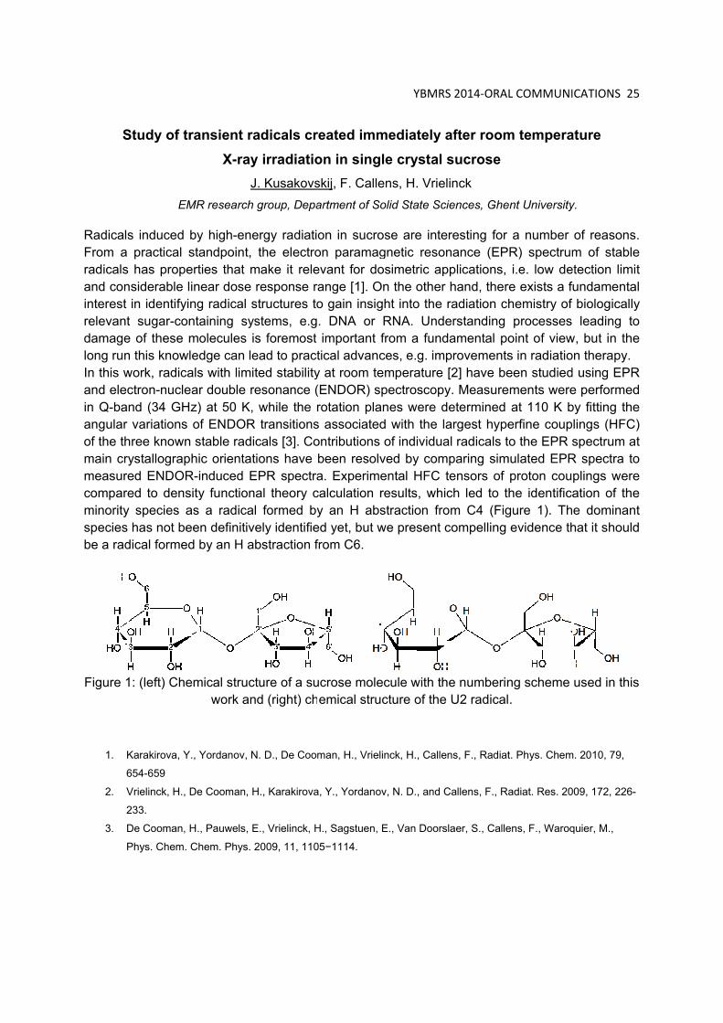
RadicalFrom aradicalsand coninterest relevantdamagelong runIn this wand elein Q-baangularof the thmain crmeasurcomparminorityspeciesbe a rad
Figure 1
1. K
6
2. V
2
3.
Study of tr
EM
s induced b practical s
s has propensiderable l
in identifyint sugar-cone of these mn this knowlwork, radicactron-nucle
and (34 GHzr variations hree knownrystallograpred ENDORred to densy species as has not bedical formed
1: (left) Che
Karakirova, Y
654-659
Vrielinck, H., D
233.
De Cooman, H
Phys. Chem. C
ransient raX-ray
JMR research
by high-enestandpoint, erties that minear dose ng radical sntaining symolecules iedge can le
als with limiar double rz) at 50 K, of ENDOR stable radihic orientat
R-induced Esity functionas a radicaeen definitivd by an H a
emical strucwork an
., Yordanov, N
De Cooman, H
H., Pauwels, E
Chem. Phys.
adicals cry irradiatioJ. Kusakovsgroup, Depa
ergy radiatithe electro
make it releresponse ra
structures tostems, e.gs foremost ead to practted stability
resonance (while the r
R transitionscals [3]. Cotions have EPR spectranal theory cl formed by
vely identifiebstraction f
cture of a sund (right) ch
N. D., De Coom
H., Karakirova
E., Vrielinck, H
2009, 11, 110
reated immon in singskij, F. Calleartment of So
on in sucroon paramagevant for doange [1]. Oo gain insig. DNA or important f
tical advancy at room te(ENDOR) srotation plas associatedontributions been resolva. Experime
calculation ry an H abs
ed yet, but wfrom C6.
ucrose moleemical stru
man, H., Vriel
a, Y., Yordanov
H., Sagstuen,
05−1114.
YBMR
mediately le crystal ens, H. Vrieolid State Sc
ose are integnetic resoosimetric ap
On the otherght into the RNA. Undfrom a fundces, e.g. imemperature pectroscopy
anes were dd with the lof individua
ved by comental HFC results, whistraction frowe present c
ecule with thcture of the
inck, H., Calle
v, N. D., and C
E., Van Doors
S 2014‐ORA
after roomsucrose
elinck
ciences, Ghe
eresting for nance (EP
pplications, r hand, therradiation cherstanding damental poprovements[2] have bey. Measuredeterminedargest hypeal radicals t
mparing simtensors of ich led to tom C4 (Figcompelling
he numberine U2 radical
ens, F., Radiat
Callens, F., Ra
slaer, S., Calle
L COMMUNI
m tempera
ent University
a number R) spectrumi.e. low de
re exists a fhemistry of processes
oint of views in radiatioeen studiedments wereat 110 K b
erfine coupo the EPR
mulated EPRproton couhe identificgure 1). Thevidence th
ng scheme .
t. Phys. Chem
adiat. Res. 20
ens, F., Waroq
ICATIONS 25
ature
y.
of reasonsm of stable
etection limifundamentaf biologically leading to
w, but in theon therapy.d using EPRe performedby fitting theplings (HFCspectrum a
R spectra toplings wereation of the
he dominanhat it should
used in this
m. 2010, 79,
009, 172, 226-
quier, M.,
5
. e t
al y o e
R d e ) t
o e e t
d
s
-

YBMRS 2014‐ORAL COMMUNICATIONS 26
An investigation of Antarctic fish cytoglobins using EPR and optical spectroscopy
B. Cuypers1, S. Vermeylen2, A. De Schutter1, V. Rahemi3, C. Verde4, K. De Wael3,
S. Dewilde2 and S. Van Doorslaer1
1 BIMEF Laboratory, Department of Physics, University of Antwerp 2 PPES Laboratory, Department of Biomedical Sciences, University of Antwerp 3 AXES Laboratory, Department of Chemistry, University of Antwerp 4Institute of Protein Biochemistry, Consiglio Nazionale delle Ricerche, Italy
Up to now, five different globins have been identified in humans: hemoglobin, myoglobin, neuroglobin, cytoglobin and androglobin. Both neuroglobin and cytoglobin are hexacoordinated globins (HisE7-Fe-HisF8), which means that the heme iron has no free binding site in comparison with hemoglobin and myoglobin. Cytoglobin occurs in different tissues in a rather low concentration. In this work, two Antarctic fish cytoglobins (Dissostichus mawsoni and Chaenocephalus aceratus) are investigated using different spectroscopic techniques: optical absorption spectroscopy, electron paramagnetic resonance (EPR) and resonance Raman spectroscopy (RRS). Both Antarctic fish lack myoglobin and C.ace also lacks hemoglobin, which makes their corresponding cytoglobins interesting candidates to investigate and compare with their human variant.1 Unlike in human neuroglobin, where the presence of an intramolecular disulfide bond is found to modulate the gas binding affinity through a change in the heme-pocket structure, the formation of a disulfide bridge in human cytoglobin has only a negligible effect.2 The Antarctic fish and human cytoglobins have cysteines at different positions. By comparing the principal g-values of the ferric cytoglobins with those of their Cys Ser mutants, the effect of possible intra- and/or intermolecular disulfide bridges on the heme-pocket structure can be determined. Combination of all the data from the above mentioned spectroscopic techniques reveals slight differences in the heme-pocket structure of human and Antarctic fish cytoglobins. The most pronounced difference is found in the stabilization of the CO-ligand.
1. E. Vinck, S. Van Doorslaer, S. Dewilde and L. Moens, J. Am. Chem. Soc., 2004, 126, 4516-4517.
2. D. Hamdane, L. Kiger, S. Dewilde, B. Green, et al., J. Biol. Chem., 2003, 278, 51713-51721.
His E7
His F8

YBMRS 2014‐ORAL COMMUNICATIONS 27
Synthesis and characterization of functionalized magnetoliposomes for Magnetic Resonance Imaging and theranostics applications
R. Garcia Ribeiro1, A. Ketkar-Atre1, S. Soenen1, M. De Cuyper3, U. Himmelreich1 1 Biomedical MRI Unit/MoSaic, Department of Imaging and Pathology, KU Leuven; 2 Lab of BioNanoColloids, Interdisciplinary Research Center, KU Leuven, KULAK.
Magnetoliposomes (MLs), which were earlier developed in our lab, consist of nanosized, magnetisable iron oxide cores (magnetite, Fe3O4) which are individually enveloped by a bilayer of phospholipid molecules. In the past, it was already shown that these structures are biocompatible imaging agents, resulting in a highly efficient labeling of cells without evoking toxic effects and with a powerful imaging signal that remains stable over a long time period (1). A unique feature of MLs is that the coating can be easily modified, for instance by inserting fluorescent or radioactive labels and/or targeting molecules into the lipid bilayer. Thus, these highly sophisticated MLs offer exciting possibilities for multimodal molecular imaging and drug delivery. Till now, in-house synthetized MLs were functionalized and characterized by Transmission Electron Microscopy (TEM) and Dynamic Light Scattering (DLS). The results were satisfactory in both techniques, with TEM defining the size (30-35 nm) and DLS the hydrodynamic diameter (45 nm) of the MLs, and with the morphology of the phospholipidic bilayers very discernible in the TEM images. These MLs were further employed for cell labeling and longitudinal in vivo imaging of prelabeled INS-1E cells and Pancreatic Islets (PIs). To test the MR detectability of cells labeled under different conditions, agarose phantoms were prepared and MR imaging was performed. Phantoms were scanned using a 3D T2*-weighted gradient echo sequence with a Fast Low Angle Shot sequence (FLASH, TR = 200 ms, TE = 15 ms) resulting in an isotropic resolution of 234 µm3. First proof-of-principle experiments were also successfully conducted to transplant the prelabeled cells and PIs in the kidney capsule and portal vein of C57Bl6 adult mice. Animals were scanned on the day of the islet engraftment and until 14 days post islet transplantation. A respiration-gated FLASH sequence (TE= 2.3ms, TR= 202.56ms, six slices with a thickness of 1mm and an in-plane resolution of 136µm2) was used to determine the decrease in the signal intensity due to labeled islets at the site of transplantation. All MR measurements were performed using a 9.4 T Bruker Biospec small animal MR scanner (Bruker Biospin, Ettlingen, Germany). The results obtained here show that MLs have highly sensitive T2/T2* MR contrast and can be successfully used for prelabeling or cells/ islets and subsequent in vivo imaging. MLs express lower level of toxic effects compared to other iron oxide particles (2) and functionalized MLs using cell recognizing ligands such as peptides (e.g. cRGD peptide), small molecules (e.g. lactose moieties) and vitamins (e.g vitamin A) were already validate by our group for the in vitro and in vivo visualization of hepatocytes and hepatic setellate cells, respectively (3). The later were used for the early detection of liver fibrosis. The focus of our future work is on in vivo targeting of beta cells for diabetes therapy using molecules that target receptors of beta cells, including the GPR-40 receptor using a thio-derivative of TAK-875, a potent GPR-40 agonist.
1. 1. De Cuyper, M. et al (2010) Cationic Magnetoliposomes. Liposomes, Methd.Mol.Bio.(605), chapter 6. 2. 2. Soenen, S et al (2011) Magnetoliposomes as MRI contrast agents. Wiley interdisciplinary revies.
Nanomedicine and nanobiotechnology, 3, 197-211. 3. 3. Ketkar-Atre,A (2013) In vivo hepatocyte MR imaging using lactose functionalized magnetoliposomes.
Biomaterials (In press). The research leading to these results has received funding from the People Programme (Marie Curie Actions) of the European Union's Seventh Framework Programme FP7/2007-2013/ under REA grant agreement n° 289932.

YBMRS 2014‐ORAL COMMUNICATIONS 28
Hierarchical non-negative matrix factorization for brain tumor characterization using multi-parametric MRI
N. Sauwen1,2, D.M. Sima1,2, S. Van Cauter3, J. Veraart4, A. Leemans5, F. Maes6, U. Himmelreich7
and S. Van Huffel1,2
1 Department of Electrical Engineering (ESAT), STADIUS Center for Dynamical Systems, Signal Processing and Data Analytics, KU Leuven 2 iMinds Medical IT 3 Department of Radiology, University Hospitals of Leuven 4 Center for Biomedical Imaging, Department of Radiology, New York University Langone Medical Center 5 Image Sciences Institute, University Medical Center Utrecht, Utrecht University 6 Department of Electrical Engineering (ESAT), Center for Processing Speech and Images, KU Leuven 7 Biomedical MRI/MoSAIC, Department of Imaging and Pathology, KU Leuven
Advanced MR modalities such as MRSI, PWI (perfusion-weighted imaging) and DWI (diffusion-weighted imaging) have shown their added value to characterize brain tumors in a non-invasive way, detect full tumor extent and assess early success of therapy. The combination of MR modalities in a multi-parametric MRI (MP-MRI) approach provides us with complementary information and allows us to answer clinical questions more specifically and unambiguously [1]. Tissue characterization within gliomas is challenging due to the co-existence of several intra-tumoral tissue types within the same region and the high spatial heterogeneity in high-grade gliomas. Previous advanced MR studies have often neglected this aspect of tissue complexity. An accurate and reproducible method for brain tumor characterization and the detection of the relevant tumor substructures could be of great added value for the diagnosis, treatment planning and follow-up of individual patients. This study presents a hierarchical non-negative matrix factorization (hNMF) technique, providing a voxelwise tissue characterization and incorporating the concept of tissue mixtures. The hNMF algorithm was originally developed to process MRSI data only [2], but has been modified to cope with an extended set of MP-MRI data. Tissue-specific patterns are obtained and the spatial distribution of each tissue type is visualized by means of abundance maps. The hNMF algorithm is applied to the MP-MRI data of 13 non-necrotic glioma patients and 11 patients with glioblastoma multiforme. Dice-scores were calculated by converting the abundance maps into a tissue segmentation and comparing it to the manual segmentation by a radiologist. Correlation coefficients were calculated between the pathologic tissue sources and the average feature vector within the corresponding tissue region. For the non-necrotic patients, an average dice-score of 88% and an average correlation coefficient of 0.97 were found for the tumor region. For the GBM patients, average dice-scores of 76%, 69% and 84% were obtained for active tumor, necrosis and the whole tumor region. The average correlation coefficients were 0.90 for active tumor and 0.96 for necrosis. hNMF can be applied on a patient-by-patient basis, it does not require large training datasets nor data normalization and it provides a more refined tissue characterization compared to black-and-white segmentation techniques.
1. Van Cauter S, De Keyzer F, Sima DM, et al. Neuro‐Oncol. 2014;16:1010–1021.
2. Li Y, Sima DM, Van Cauter S, Himmelreich U, et al. Proc BIOSIGNALS 2012. Algarve, Portugal;
212–217.

YBMRS 2014‐ORAL COMMUNICATIONS 29
G-quadruplex in the HIV promoter region: UV-spectroscopy and NMR insight
A. De Rache,1,2 S. Amrane,1,2 A. Bedrat,1,2 M.L. Andréola,1,3 J.L. Mergny 1,2
1 Univ. Bordeaux, F-33000 Bordeaux, France; 2 INSERM U869, IECB, ARNA laboratory, 2 Rue Robert Escarpit, F-33600 Pessac, France;
3 CNRS UMR 5234, 146 Rue Leo Saignat, 33076 Bordeaux, France [email protected]
G-quadruplexes are nucleic acid structures formed by stacking of a minimum of two G-quartets. Each G-quartet is composed of four guanines interacting through Hoogsteen H-bonds. Several G-quadruplex forming aptamers (nucleic acid sequences selected in vitro for their affinity towards a given target) are able to interact with key proteins in the HIV replication cycle like the integrase, the reverse transcriptase, the gp120, etc. They inhibit the HIV infectivity at various steps of the viral cycle. We have demonstrated that the sequence HIV_PRO1, located in the promoter of the virus, is prone to adopt a G-quadruplex folding in vitro and particularly conserved among the HIV variants.[1] In the present work we investigate the biophysical characteristics of a second sequence, HIV_PRO2, located in the viral promoter region. The formation of a G-quadruplex structure is attested by UV spectroscopy and by the presence of twelve imino protons in the 1H NMR spectrum. Indications related to the topology (i.e. the organization of the G-quartet and base pairs) of the G-quadruplex were obtained by circular dichroism and NMR (including H2O to D2O exchange, HMBC and NOESY experiments). Single base 13C and 15N labelling are used to assess key assignments.
1. S. Amrane, A. Kerkour, A. Bedrat, B. Vialet, M. L. Andreola, J. L. Mergny, J. Am. Chem. Soc.
2014, 136, 5249.

YBMRS 2014‐ORAL COMMUNICATIONS 30
Application of NMR Spectroscopy in the Development of Silicone Materials
A.Kretschmer
Dow Corning Belgium, Analytical Sciences
Silicones bringing inorganic (Si-O-Si bonds) with organic chemistry (Si-C bonds) together, show very special properties like thermal and chemical stability, resistance to light, low surface tension and others. Beside well known poly (dimethyl siloxanes), the silicone backbone can be linked to other organic groups like phenyl, polyether and others; the silicone can be branched by incorporation of tri- or tetra-functional silanes. The silicone materials, their formation, their reactions like hydrosilylation or condensation, can be monitored by mainly 29Si NMR spectroscopy, but also 1H and 13C NMR methods are of interest. The NMR characterization of silicones, their theoretical and practical aspects, were developed in detail in the 1970ies and 1980ies by Marsmann, Radeglia, Engelhardt, Jancke, Harris, Schraml and others [1-5]. The chemical shift range in 29Si NMR allows to distinguish between different functionalities (M,D,T and Q units), neighborhoods; quantification allows to determine molecular weights, branching levels, average structures [6]. In addition to classic solution state NMR, a few examples of characterizing emulsions and solids will be presented. Solid applications do not only reflect structures, but also dynamic aspects. The relative low glass transition temperature of poly (dimethyl siloxane) with its high mobility at room temperature makes investigations of copolymers with rather rigid components like poly amides very interesting for 1H solid state applications [7]. References
1 H.Marsmann,Makromolek.Chemie,1972,162,255‐267 2 R.Radeglia, Z.Phys.Chemie., 1975, 256, 453‐464 3 R.K.Harris,B.J.Kimber, J.Magn.Reson., 1975,17,174‐188 4 G.Engelhardt,H.Jancke,E.Lipmaa,A.Samoson,J.Organomet.Chem.,1981,210,295‐301 5 J.Schraml,Progress in NNMR Spectroscopy, 1990,22,289‐348 6 A.L.Smith (Ed.),The Analytical Chemistry of Silicones, John Wiley & Sons, 1991,347‐419 7 A.Kretschmer,R.Drake,M.Neidhoefer,M.Wilhelm,Sol.StateNucl.Magn.Reson., 2002,22,204‐
217

YBMRS 2014‐ POSTERS 31
POSTERS
Ta
Peptidodocumereceptoas adjuvIn orderdevelopcarriers
We havinteractthe PGNintensityspectrathe adautilised the bilayand actsis expos[1] S[2] O
argeted del
F. Ch aDepartm
bDepart
oglycan (PGented immrs. Such imvants in vacr to selectivped actively.
ve investigaion betweeN derivativey of the nO. We have
amantly groSTD experyer. We havs as an ancsed on the s
Szilagyi L. PristO’Hagan DT, D
ivery of peN
aina, R. Rib
ment of Orgatment of Che
GN) as weunomodula
mmunomoduccines or in ely modulaty targeted
ated the en them on es are incor
Oe as well adeterminedup attachmriments for ve found thchor of the Psurface. tovsek P. Min
De Gregorio E
eptidoglycaNMR study
bićb, D. Sinn
anic and Macemistry, Facu
*Kr
ell as fragating propeulators are aiding cancte a certaindelivery o
ncapsulatioa molecula
rporated in as by the obd the encap
ment had a the charact
hat the adamPGN deriva
niRev.Med.Ch. Drug Discov
an immunoof the lipid
naevea, J. C
cromolecularulty of Scienc
risztina.Fehe
gments anderties[1] exe
urgently necer therapie subset of i
of PGN-bas
on of targear level usinthe studied
bservation opsulation eflarge influeterisation omantyl grouative cargo,
hem. 2007, 7. ery Today, 20
omodulatord encapsul
C. Martinsa, S
r Chemistry, ce, University
d substancerted by ieeded in mes. mmune cel
sed immun
ting compong NMR spd lipid bilayeof slower trfficiency anence on thef the orient
up does penwhile the h
p.861. 009, 14, Nr 11/
YB
rs using lipation
S. Tomićb, K
University oy of Zagreb,
ces derivedinteraction any medica
ls using PGomodulants
ounds into pectroscopyers by the canslational
nd have foue entrapmetation of thenetrate the lydrophilic p
/12, 541-551.
BMRS 2014‐ P
posomal ca
K. Fehéra*
of Ghent Zagreb, Cro
d from it, with inna
al interventi
GN fragmens using lip
lipid bilayey. We have change of thdiffusion in
und that thent efficiency
e studied delipid core ofpeptidoglyca
POSTERS 32
arriers:
oatia
have wellte immuneions[2], such
nts, we haveosomes as
ers and theshown tha
he sign andn PFG-NMRe chirality oy. We haveerivatives inf the bilayean fragmen
2
-e h
e s
e t d R f e n r t

YBMRS 2014‐ POSTERS 33
First insights in the structure-function relationship of a natural cyclic lipodepsipeptide by synthetic modification and NMR investigation
M. De Vleeschouwer1,2, N. Matthijs3, D. Sinnaeve1, T. Coenye3, J.C. Martins1, A. Madder2
1 NMR and Structure Analysis Unit, Ghent University 2 Organic and Biomimetic Chemistry Research Group, Ghent University 3 Laboratory of Pharmaceutical Microbiology, Ghent University
Pseudodesmin A (1) is a secondary metabolite produced by Pseudomonas bacteria, belonging
to the viscosin group of cyclic lipodepsipeptides. It displays moderate antibiotic activity, including
against MRSA and vancomycin-resistant Enterococcus. Extensive NMR studies revealed that
individual molecules self-assemble into well-defined supramolecular structures in non-polar
solvents.[1,2]
Our goal is to investigate in detail the molecular structure of the self-assembly and its role in
biological activity, which involves membrane interaction. For this, a rapid, efficient solid-phase
synthesis strategy for pseudodesmin A was developed.[3] The newly developed route allows the
straightforward production of analogues for structure-activity relationship studies, including an
Ala-scan and modifications to the fatty acid moiety. Using NMR diffusion measurements, the
modulation of the self-assembly could be monitored, revealing fundamental intermolecular
contacts. Additionally, the enantiomer of pseudodesmin A was produced, revealing identical
biological activity, for the first time demonstrating that no chiral interactions mediate these
compounds’ mode of action.
1. Sinnaeve D., Hendrickx P.M.S., Van hemel J. et al. Chem. Eur. J. 2009, 15, 12653-12662.
2. Sinnaeve D., Delsuc M.A., Martins J.C. and Kieffer B. Chem. Sci. 2012, 3, 1284-1292
3. De Vleeschouwer M., Sinnaeve D., Van den Begin J. et al. Chem. Eur. J., 2014, 20, 7766-7775
αL‐helix
loop
L‐Leu1HDA
L‐Ile9
D‐Ser8L‐Leu7
D‐Ser6

YBMRS 2014‐ POSTERS 34
Synthesis of bimodal MRI contrast agents based on metallostar complexes
Matthias Ceulemans1, Wim De Borggraeve1, Luce Vander Elst², Sophie Laurent², Robert N. Muller² and Tatjana N. Parac-Vogt1
1 Department of Chemistry, KU Leuven. 2 Department of General, Organic and Biomedical Chemistry, University of Mons.
During the last decades, Magnetic Resonance Imaging became a well established medical imaging technique. Most contrast agents used today are complexes of Gd3+ with diethylenetriaminepentaacetic acid ([Gd(DTPA)]2-), 1,4,7,10-tetraazacyclododecane-1,4,7,10-tetraacetic acid (Gd(DOTA)]-) or structural analogues. The paramagnetic gadolinium(III) with its seven unpaired electrons and sphere symmetric ground state (8S7/2) is an ideal metal for this application. The relaxivity of the molecule is determined by several parameters such as the number of inner-sphere water molecules, the residence time of the coordinated water molecules and the rotational correlation time of the contrast agent. Multimodal contrast agents are gaining in demand as combination of different imaging techniques allows analysis in more exquisite details. Compounds being both luminescent and paramagnetic hold potential as bimodal contrast agents for MRI and optical imaging. Luminescence is known to have a high sensitivity, while MRI shows a much better spacial resolution and deeper penetration depth. In this work we present a ligand consisting of a parasubstituted pyridine-2,6-dicarboxylate derivative of DTPA. In the envisioned complex, water molecules are able to approach the paramagnetic entity in order to achieve efficient relaxation enhancement, while water is excluded from the first coordination sphere of central lanthanide(III) resulting in a bright emissive compound. Furthermore, the sensitivity difference between optical and MRI is encountered by assembling three paramagnetic components around one luminescent metal ion.
1 Debroye, E., eulemans, M., Vander Elst, L., Laurent, S., Muller, R., Parac-Vogt, T. (2014). Controlled Synthesis of a Novel Heteropolymetallic Complex with Selectively Incorporated Lanthanide(III) Ions. Inorganic Chemistry, 53 (3), 1257-1259.
Figure 1. Structure of the metallostar complex

YBMRS 2014‐ POSTERS 35
DESIGN OF CONTRAST AGENT FOR NEURODEGENERATIVE DISEASES DIAGNOSIS
G. Circelli 1, L. Vander Elst 1,2, R. N. Muller 1,2, S. Laurent 1,2
1 NMR and Molecular Imaging Laboratory, University of Mons; 2 Center for Microscopy and Molecular Imaging, Gosselies. In the last decades, molecular imaging technologies began to attract increased interest for the targeting of diseases. In particular, Alzheimer disease diagnosis is nowadays investigated through the design of tailor-made contrast agents able to cross over the blood-brain barrier (BBB). Conventional contrast agents are composed of three parts: contrast agent, spacer and vector. Over the last three decades, gadolinium complex of DOTA macrocycle has exhibited enhanced stability properties as compared to other first generation contrast agents. In addition, the versatility of the complexes confers to DOTA-based contrast agents the ability to be used in a large range of imaging modalities, including magnetic resonance (MR), positron emission tomography (PET), single photon emission computed tomography (SPECT) and fluorescence imaging.1 Variations on the arms of the DOTA macrocycle appears as the way towards the generation of highly stable responsive and selective probes.2 In this context, the specific targeting of diseases has been drastically enhanced by the incorporation of peptides as vectors. In the present study, the design of L-DOPA functionalized Gd-DOTA complex as a bluilding block for further modifications is under consideration. The synthetic strategy involves the following steps : (i) the selective three-arms protection of cyclene by tert-butyl acetate moeities; (ii) the incorporation of a fourth arm exhibiting a primary amine moeity; (iii) amidation reaction on the fourth arm with Boc-L-DOPA; (iv) BOC deprotection; (v) Gd3+ complexation by the modified DOTA macrocycle. For instance, the two first steps were successfully achieved. Both NMR and ESI-MS characterization techniques were used to follow the progress of the reactions. The next steps are under progress. 1. G. J. Stasiuk, N.J. Long, Chem. Commun., 49, 2732 (2013). 2. C.F.G.C. Geraldes, S. Laurent, Contrast Media & Molecular Imaging, 4(1), 1 (2009)

YBMRS 2014‐ POSTERS 36
Development of EPR oximetry in diabetic wound healing models
C. Desmet1, A. Lafosse2, 3, S. Vériter2, Ph. Levêque1, D. Dufrane2 and B. Gallez1 1 Biomedical Magnetic Resonance Research group, Louvain Drug Research Institute, Université Catholique de Louvain, Brussels. 2 Endocrine Cell Therapy Unit, Center of Tissue/Cell Therapy, Cliniques Universitaires Saint-Luc, Université Catholique de Louvain, Brussels. 3 Plastic and Reconstructive Surgery Unit, Cliniques Universitaires Saint-Luc, Université Catholique de Louvain, Brussels.
Introduction: One major complication of diabetes is the development of skin ulcers with impaired healing. Indeed, 15 % of diabetic patients will suffer from lower extremity ulcers during their life and 7 to 20 % of these patients with foot ulcers will require amputation [1]. A main cause of the nonhealing of foot ulcers is tissue ischemia [2] as it is known that oxygen is a key parameter in wound healing. Nevertheless, the variation of the pO2 in the wound remains uncertain as there still lacks non invasive methods for absolute and repeated pO2 measurements in situ. EPR oximetry is a technique that allows repeated measurements of the absolute tissue pO2. The technique is based on the measurement of the linewidth of the EPR signal recorded with a biocompatible oxygen sensor like LiPc implanted in the tissue. Aim: To investigate whether EPR oximetry with LiPc is a possible tool to follow the pO2 in the wound in diabetic wound healing. Materials and methods: Two diabetes models were used: chemically-induced type I diabetes (with streptozotocin) and genetically-induced type II diabetes (db/db mice). Two types of wounds were tested: a 30 x 8 mm pedicled flap and a 6 mm diameter excisional wound. LiPc crystals were inserted in different positions inside the wound and the EPR signal recorded repeatedly during the wound healing process. Results: In the flap model, the pO2 decreased the first day after wounding due to the flap surgery. Then, the pO2 increased during the healing process. Interestingly, this increase was more rapid for non diabetic mice than for diabetic mice (fig. 1). For the excisional wound model, a variation of the pO2 was observed in the wound periphery during the healing process but it did not differ from what was observed in the control non wounded tissue. In the center of the excisional wound, the pO2 was elevated the first days after the wounding without any difference between the diabetic and non diabetic state (fig. 2).
Fig. 1: pO2 measurements during the healing process obtained with LiPc crystals implanted in the flap in control db/+ and db/db mice.
Fig. 2: pO2 measured in the wound during the healing process in non diabetic mice and db/db mice.
Conclusion: EPR oximetry using LiPc is a suitable methodology for monitoring the variation of the pO2 over time in wounds. The flap model containing LiPc in db/db mice is an adequate wound model to study the tissue pO2 during the wound healing process in diabetic animals.
1. Frykberg RG, Zgonis T, Armstrong DG, Driver VR, Giurini JM, Kravitz SR, Landsman AS, Lavery LA, Moore JC, Schubert JM, et al. The Journal of Foot and Ankle Surgery. 2006, 45, S1-S66.
2. Schreml S, Szeimies RM, Prantl L, Karrer S, Landthaler M & Babilas P. The British Journal of Dermatology 2010, 163, 257-268.
0
5
10
15
20
25
30
‐2 0 2 4 6 8 10 12 14
pO2
(mm
Hg)
Time (days)
pO2 evolution in flap
db/+
db/db
WOUND
0
10
20
30
40
50
60
0 10 20 30
pO2
(mm
Hg)
Time (days)
pO2 evolution in excisional wound
non diabetic mice
db/db mice

YBMRS 2014‐ POSTERS 37
Theoretical and Experimental study of the Off-Resonant Saturation, an MRI Sequence for Positive Contrast With Superparamagnetic Particles
S. Delangre1, Q.L. Vuong1, C. Po2, B. Gallez2 and Y. Gossuin1 1 Biomedical Physics Department, UMONS 2 Biomedical Magnetic Resonance Unit (REMA), UCL
Magnetic Resonance Imaging (MRI) is a powerful non invasive medical imaging technique that provides images with excellent resolution and intrinsic contrast. However, MRI sometimes necessitates contrast agents to highlight regions of interest such as tumors. The Superparamagnetic Iron Oxide Nanoparticles (SPM particles)1 are one kind of contrast agent: these biocompatible particles appear as hypointense spots in MRI images obtained with conventional imaging sequences due to their high r2/r1 ratio – and thus, belong to the negative contrast agent category.
Unfortunately, negative contrast can be difficult to interpret because it can also be produced by many sources which are independent of the SPM nanoparticles – such as air bubbles or tissue interfaces. To overcome this problem, new imaging sequences producing positive contrast with SPM nanoparticles were developed during the last few years. One of them is the Off-Resonance Saturation (ORS) technique2. The idea behind this sequence is to saturate the signal near the SPM particles and to subtract the resulting image from a classical image obtained without saturation. This computation yields an image with a positive contrast near the SPM nanoparticles.
Different studies have experimentally shown that ORS is able to generate positive contrast both in vitro and in vivo2,3. However, a complete theoretical study of the ORS sequence is still missing, which makes impossible the optimization of the ORS sequence. For this reason, this work proposes a theoretical study of the ORS sequence, validated by numerical simulations and verified by experiments on agarose gel phantom on a 11.7 T MRI scanner system. An analytical expression describing the contrast dependence on the sequence parameters and the SPM particles properties was developed. This expression provides a fundamental comprehension of the mechanisms leading to the ORS contrast and allows an optimization of the ORS sequence. Therefore, the theoretical model can be useful for future in-vivo applications.
The influences of the SPM particles relaxivities and concentration, the echo time and the saturation pulse parameters on the contrast were investigated. The best contrast was achieved for SPM particles possessing the lowest transverse relaxivity, for specific values of particles concentration predicted by the theory and for the lowest echo times. 1. Wang, Y. X., Hussain, S. M. & Krestin, G. P. Superparamagnetic iron oxide contrast agents: physicochemical
characteristics and applications in MR imaging. Eur. Radiol. 11, 2319–2331 (2001). 2. Zurkiya, O. & Hu, X. Off-resonance saturation as a means of generating contrast with superparamagnetic
nanoparticles. Magn. Reson. Med. 56, 726–732 (2006). 3. Khemtong, C. et al. In vivo Off-Resonance Saturation Magnetic Resonance Imaging of v 3-Targeted
Superparamagnetic Nanoparticles. Cancer Res. 69, 1651–1658 (2009).
YBMRS 2014‐ POSTERS 38
Membrane interactions of natural cyclic lipodepsipeptides N. Geudens1, K. Fehér1, M. De Vleeschouwer1,2, J-M Crowet3, M.N. Nasir3,
A. Madder2, L. Lins3, J.C. Martins1 and D. Sinnaeve1 1 Ghent University, NMR and Structural Analysis Unit, 2 Ghent University, Organic and Biomimetic
Chemistry,3 University of Liege, Gembloux, Unité de Chimie Biologique Industrielle
Cyclic lipodepsipeptides (CLPs) are non-ribosomal peptides produced by bacteria, mainly Pseudomonas and Bacillus spp. They consist of a short sequence of both D- and L-amino acids that forms a cyclic structure by means of an ester bond between the C-terminus and a side-chain alcohol. They possess antimicrobial activity against clinically relevant Gram-positive bacteria and certain agricultural relevant plant pathogens.
In the past, efforts have been made in analysing the conformations and self-assembling properties of a collection of CLPs known as the viscosin group. [1-4] It is hypothesized that the antimicrobial activity of CLPs can be related to their ability to interact with cell membranes. We apply a multidisciplinary approach to investigate the CLP’s working mechanism, focussing either on the peptides themselves or the impact on the lipid bilayer.
NMR spectroscopy has emerged as a powerful tool for the structure determination of peptides in the presence of model membranes. In liquid-state NMR, it is important that the model membrane is sufficiently small to obtain good spectral resolution. This requirement imposes a restriction on the types of model membranes that are suitable. In this respect, isotropic bicelles have been developed as model membranes for NMR, combining the advantageous NMR properties of micelles with the characteristics of lipid bilayers. [5]
Complementary biophysical experiments are performed, including fluorescence spectroscopy and circular dichroism. Additionally, preliminary Molecular Dynamics simulations in the presence of bilayers have been performed to better understand the molecular mechanism of CLPs at an atomic level.
1. Sinnaeve, D., P. M. Hendrickx, et al. Chemistry - A European Journal, 2009 15(46): 12653-12662. 2. Sinnaeve, D., M.-A. Delsuc, et al. Chemical Science , 2012 3: 1284-1292. 3. De Vleeschouwer, M., D. Sinnaeve, et al. Chemistry - A European Journal, 2014 20(25): 7766-7775 4. Geudens N., De Vleeschouwer M., et al, ChemBioChem, in press, DOI: 10.1002/cbic.201402389 5. Durr, U. H., M. Gildenberg, et al. Chem Rev, 2012 112(11): 6054-6074.

YBMRS 2014‐ POSTERS 39
17O NMR Study of Diamagnetic and Paramagnetic Lanthanide(III)− DOTA Complexes in Aqueous Solution
L. Fusaro1,2, M. Luhmer2, G. Casella3 and A. Bagno4 1 Laboratoire de RMN, University of Namur, Belgium 2 Laboratoire de RMN Haute Reśolution, Université Libre de Bruxelles, Belgium 3 Dipartimento di Scienze della Terra e del Mare (DiSTeM), Università di Palermo, Italy. 4 Dipartimento di Scienze Chimiche, Università di Padova, Italy.
The complexes between the 17O-enriched polyaminocarboxylate DOTA ligand and the whole series of stable lanthanide-(III) metal ions were studied in aqueous solution by 17O NMR: I) The complex between DOTA and praseodymium(III) (Pr3+) was studied in aqueous solution by variable-temperature 17O NMR. pH effects as well as the influence of metal ions free in solution were investigated. The 17O NMR signals of both the nonchelating (O1) and chelating (O2) oxygen atoms could be detected. At low temperature, the signals of both the square antiprismatic (SAP) and twisted square antiprismatic (TSAP) conformational isomers were also observed. At high temperature, the spectra exhibit signal broadening that reveals the interchange of the O1 and O2 oxygen atoms of the carboxylate groups. The linewidths measured for O1 were deconvolved into contributions from quadrupole relaxation and chemical exchange, allowing the corresponding activation barriers to be determined.1 II) For all of the paramagnetic systems, except Gd3+, the 17O NMR signals of both O1 and O2 could be detected, and for some of them, the signals of both the SAP and TSAP (TSAP′) conformational isomers were also observed. Line width data analysis reveals that signal broadening is not dominated by paramagnetic relaxation enhancement, as it was believed to be. The data indicate that quadrupole relaxation and, for some complexes, chemical exchange between the SAP and TSAP isomers are the major contributions to the 17O NMR line width at 25 °C. Besides, the Fermi contact and pseudocontact contributions to the observed lanthanide-induced shifts could be extracted.2 III) The 17O NMR spectrum of the non-coordinated carboxyl oxygen in the Gd(III) DOTA complex has been observed experimentally. Its line width is essentially unaffected by paramagnetic relaxation due to Gd, and due only to the quadrupole pathway. The results are supported by the relevant parameters (hyperfine and quadrupole coupling constant) calculated by relativistic DFT methods. This finding opens new avenues for investigating the structure and reactivity of paramagnetic Gd(III) complexes used as contrast agents in MRI.3 1. Fusaro L. and Luhmer. M., DaltonTrans. 2014, 43, 967–972. 2. Fusaro L. and Luhmer. M., Inorg. Chem. 2014, 53, 8717-8722. 3. Fusaro L., Casella G. and A. Bagno. Chemistry 2014, (in press).

Des
1
2
DendrimsphericaapplicatThe aimseveral Indeed, efficiencgeneratuptake aThe zedifferenreagentmagnetThe maand fullyIn the ncomplexcontrast
sign and c
L. Cape1 NMR and M2 Center for M
mers are a cal structuretions. m of this w
paramagn previous scy in the mation dendrimand transporo, the first synthesis ts. Finally, ic resonanc
acrocycle wy characternear future,x and also t agent able
Fig.1 : D
characterizima
ette1, S. LauMolecular ImMicroscopy a
class of pare. Their p
work is theretic centertudies showagnetic fieldmer and to gort. st and the
parametersall generat
ce (NMR) spwhich is a D
ized. we plan tothe biocyti
e to undergo
Design of (a
zation of aaging of th
urent1,2, L. Gaging Laboraand Molecula
rticular polyproperties
refore the drs with a rwed that theds of medicgraft it with
second ges such as ttions were pectroscopy
DOTA deriv
o graft on tn or the who uptake an
a) second ge
a dendrimhe nervous
Granato1, Ratory, Univerar Imaging, C
ymer with a make them
developmenreduced moe reduction al imaging. a paramag
enerations the solvents
characterizy (Fig. 1).
vative (a we
the second heat germ nd transport
eneration d
eric contrs central s
R. N. Muller1
rsity of MonsCharleroi, Be
large numbm ideal fo
nt of a maobility, ableof the mobOur object
gnetic chela
of dendrims, the tempezed by ma
ell-known c
generationagglutinin (t by neurons
endrimer a
YB
rast agent system
,2 and L. Vas elgium.
ber of functor a wide
cromoleculae to be traility of a Gdive is thus t
ates and a v
mer were oerature, the
ass spectro
ontrast age
n dendrimer(WGA) in os.
nd (b) DOT
BMRS 2014‐ P
dedicated
ander Elst1,2
tions and a range of
ar contrast ansported bd-complex into synthesizvector for th
obtained bye reaction timetry and
ent) was als
r the Gd paorder to obt
TA derivative
POSTERS 40
d to the
2
rigid quasibiomedica
agent withby neuronsncreases itsze a secondhe neurona
y optimizingime and theby nuclea
so obtained
aramagnetictain an MR
e
0
-al
h s. s d al
g e r
d
c RI

YBMRS 2014‐ POSTERS 41
Use of 13C‐NMR in structural elucidation of polysaccharides: case of locust bean gum. S. Gillet1, M. Aguedo1, C. Blecker2, N. Jacquet1, A. Richel1
1Unit of Biological and Industrial Chemistry (CBI) ‐ Gembloux Agro‐Bio Tech ‐ University of Liege 2Food science and formulation department (SAF) ‐ Gembloux Agro‐Bio Tech ‐ University of Liege Locust bean gum (LBG) galactomannans are polysaccharides consisting of a β‐(1→4) D‐mannopyranosyl backbone substituted to varying degrees in α‐(1→6) with single D‐galactopyranosyl residues. This basic structure is the same for all galactomannans (Fig. 1). However, when locust bean gum is extracted at different temperatures, the generated fractions exhibit different properties in aqueous solution (viscosity, viscoelasticity, gel formation, thermohydrolysis resistance, etc.). This means that there are differences within the fine structure of the polymers (although the basic structure is the same). Analysis of [13C]‐NMR spectra of galactomannans, in combination with other techniques, can provide capital information about fine structural elucidation of the polymers. The method specifies the distribution of lateral galactosyls along the main chain of mannans. Two fractions extracted from locust bean gum at 25 and 80 °C (respectively GM25 and GM80) were comparatively studied by [13C]‐NMR. Mannosyls/Galactosyls (M/G) ratios can be determined by considering the intensities of C‐1 mannose and galactose signals in [13C]‐NMR spectra. This method provides results relatively close to those obtained by GC‐MS analysis. Spectra also showed that resonance from C4 of D‐mannose residues were split, in evident dependence upon the nearest‐neighbor probabilities (“diad frequencies”) of D‐galactosyl groups along the mannan chains (Fig. 1). Diad frequencies were obtained by integrating C4(Man) peak areas. F11, F21/F12 and F22 gave respectively the di‐, mono‐ or non‐substituted mannose pairs proportions. High percentages of F11 and F22 therefore indicate a more non‐homogeneous distribution of lateral galactosyls along the polysaccharide backbone as observed for GM80. The percentages of total lateral substituents obtained by C4(Man) peak analysis [F11 + (F21 or F12)/2] were fairly well correlated with M/G ratios. Splitting of the C‐6 substituted D‐mannose resonance provides, therefore the basis for determining the next‐nearest‐neighbor probabilities (triad frequencies) (Fig. 1). However, the spectrum is often not sufficiently resolved to accurately quantify and interpret the results.
Fig. 1 : [13C]‐NMR spectrum of GM80 and superimposed mannosyl C4 signals of different galactomannans.

YBMRS 2014‐ POSTERS 42
Conception of superparamagnetic polymersomes for potential drug delivery and magnetic resonance imaging applications
A. Hannecart1, L. Vander Elst1,2, R. N. Muller1, 2, S. Laurent1,2
1 Department of General, Organic and Biomedical Chemistry, NMR and Molecular Imaging Laboratory, University of Mons 2 Center for Microscopy and Molecular Imaging, Gosselies
Ultra-small superparamagnetic iron oxide nanoparticles (USPIO) have been extensively investigated for biomedical applications due to their numerous advantages like their high magnetization, their biodegradability and their biocompatibility. Thanks to their high magnetic moment, USPIO lead to a Magnetic Resonance Imaging (MRI) contrast enhancement by accelerating the relaxation of water protons. Besides their use as MR imaging contrast agents, USPIO can also create an increase in temperature by applying an oscillating magnetic field (magnetic hyperthermia). The objective of this study is to develop nanoscale drug delivery systems encapsulating iron oxide nanoparticles. Among the different delivery systems, polymer vesicles called polymersomes have emerged as promising nanocarriers thanks to their robustness and their ability to load hydrophilic as well as hydrophobic compounds [1]. Firstly, commercial copolymers of poly(ethylene oxide) (PEO) and poly(caprolactone) (PCL) were used to prepare polymersomes. Then iron oxide nanoparticles were synthesized by a coprecipitation method and stabilized by an organosilane (3-triethoxysilylpropyl succinic anhydride (TEPSA)). Transverse (r2) and longitudinal (r1) relaxivities of the USPIO-TEPSA (describing their efficiency as MRI contrast agent) were measured at 20 and at 60 MHz. A nuclear magnetic relaxation dispersion (NMRD) profile reporting the longitudinal relaxivity over a magnetic field range extending from 0.15 MHz to 60 MHz was recorded on these USPIO-TEPSA. The saturation magnetization (Ms) and the size of superparamagnetic crystals (RNMR) were determined by fitting this NMRD curve by a theoretical model [2]. The next step in further studies will be the encapsulation of iron oxide nanoparticles into the polymersomes which could be used to release a drug in a controlled manner due to an increase of the fluidity of the semicrystalline membrane by heat produced by magnetic nanoparticles. The attachment of an active targeting group such as an RGD-containing peptide to the polymersome surface is also considered.
1. D. H. Levine, P. P. Ghoroghchian, J. Freudenberg, G. Zhang, M. J. Therien, M. I. Greene, D. A.
Hammer, and R. Murali. Methods, 2008, 46, 25-32.
2. A. Roch, R. N. Muller and P. Gillis. J. Chem. Phys., 1999, 110, 5403-5411.

YBMRS 2014‐ POSTERS 43
Investigation of the effects due to the insertion of a polyQ tract increasingly long on the structure and dynamic of BlaP using NMR spectroscopy
J. Kay1, D. C. Thorn1, C. Pain1, N. Scarafone1, C. Huynen1, S. Preumont1, A. Corazza2, C. Damblon3, and M. Dumoulin1
1 Centre for Protein Engineering, University of Liège, Liège 2
Dipartimento di Scienze Mediche e Biologiche, Università di Udine, I-33100 Udine, Italy 3 Institute of Chemistry, University of Liège, Liège
Polyglutamine (polyQ) diseases, which include the well-known Huntington’s disease, arise from an anomalous expansion of a polyQ tract within proteins specific to each disease1. When the length of this polyQ tract is higher than a characteristic threshold (~35-45Q), it becomes pathogenic by triggering the aggregation of disease-associated proteins into amyloid fibrils which are deposited in neurons as nuclear inclusions and lead to the death2. Although the polyQ repeat was thought to be the only determinant factor that mediates the aggregation, recent studies have demonstrated that non-polyQ regions of these proteins play also a significant role, either preventive or facilitative, into the aggregation process3-4. In order to better understand the complex interplay between the ability of the polyQ tract to mediate the aggregation and the modulating effect of non-polyQ regions, we engineered in our lab chimeric proteins by inserting at two positions (i.e. 197 and 216) a polyQ tract of various lengths (i.e. 23Q, 30Q, 55Q and 79Q) into the β-lactamase BlaP from Bacillus licheniformis 749/C5. Both sets of BlaP chimeras recapitulate the aggregation behaviour of disease-associated proteins: indeed there is a repeat threshold length above which the chimeras readily assemble into amyloid fibrils and the rate of assembly increases with polyQ length. While having little impact on the overall structure, the location of the polyQ tract affects the tertiary structure, enzymatic activity, unfolding cooperativity and stability of the chimeric proteins, and more importantly, it significantly affects their aggregation propensity. Indeed, the 216 chimeras exhibit a significantly higher fibril-forming propensity than their 197 counterparts6. The aim of my work was to use NMR to investigate, at the molecular level, the effects of polyQ insertions on the structure and the dynamics of BlaP, in order to better understand the differences in the aggregating properties of the two sets of chimeras. First, I assign 98.5% of the backbone 1H-15N correlations in the 2D-HSQC spectrum of BlaP216Q0 using a series of triple resonance NMR experiments. Then, the regions of BlaP affected by the insertion of the dipeptide PG at position 197 and 216 were identified by comparing their respective [1H-15N] 2D-HSQC spectrum with that of the wild-type been introduced within the gene of BlaP, at position 197 or 216, to allow poly(CAG) sequence insertion. Changes in the chemical shift of each residue due to the PG insertion were determined. In both cases, the perturbations are essentially located around the insertion site (i.e. helix H8 for BlaP197Q0 and helix H9 for BlaP216Q0); they are, however, more significant when the dipeptide is inserted at position 216 than at position 197. Perturbations also occur in the three left-located helixes (i.e. H3, H4 and H9) that seem to laterally propagate the perturbation because of side chains interactions. In order to investigate eventual changes in the dynamic of BlaP upon the insertion of the dipeptide PG, real time hydrogen/deuterium exchange experiments were carried out. The preliminary analysis indicates that the dynamics of BlaP is more perturbed by the insertion at position 216 than at position 197; this result is in agreement with the lower thermodynamic stability of this chimera. Finally, the perturbations due to the presence of a polyQ tract increasingly long were also investigated. Globally, polyQ insertion at position 197 induces perturbations essentially located in the α-domain of BlaP while the perturbations due to insertion at position 216 are located in both domains. For both sets of chimera, the longer the polyQ, the farther are the perturbations on the surface of BlaP. These differences may explain the different aggregation propensity of the two sets of chimeras.
1. Hands, S. L.; Wyttenbach, A. Acta Neuropathol 2010, 120, 419. 2: Orr, H. T.; Zoghbi, H. Y. Annu Rev Neurosci 2007, 30, 575.
2. Robertson, A. L.; Bottomley, S. P. Current medicinal chemistry 2010, 17, 3058. 4: Wetzel, R. Journal of molecular biology 2012, 421, 466.
3. Scarafone, N.; Pain, C.; Fratamico, A.; Gaspard, G.; Yilmaz, N.; Filee, P.; Galleni, M.; Matagne, A.; Dumoulin, M. PloS one 2012, 7, e31253.
4. Thorn, D.; Pain, C.; Huynen, C.; Preumont, S.; Duez, C.; Brisotto, G.; Matagne, A.; Dumoulin, M.; Scarafone, N. In preparation.

YBMRS 2014‐ POSTERS 44
Evaluation of extracellular pH and energetic status response to hyperglycemia and MIBG treatment on oxidative versus glycolytic tumor phenotype:
An in vivo 31P MRS study
V. Marchand1, J. Magat1, G. De Preter1, P. Sonveaux2, B. Jordan1, B. Gallez1.
1. Louvain Drug Research Institute (LDRI), Biomedical Magnetic Resonance Section (REMA), Université catholique de
Louvain(UCL), Brussels 1200, Belgium.
2. Institut de Recherche Expérimentale et Clinique (IREC), Pole of Pharmacology (FATH), Université catholique de Louvain (UCL),
Brussels 1200, Belgium
Context: While normal cells generate ATP and biosynthetic precursors through a combination of glycolytic
and oxidative metabolism, tumor cells reprogram their metabolism depending on their microenvironment to
support a rapid, invasive and metastatic growth. Indeed, it has been shown that solid tumors show an
increased glycolytic metabolism involved in tumor aggressiveness.
There are two types of glycolytic phenotype: some tumor cells switch from oxidative phosphorylation to
glycolysis under hypoxic conditions (Pasteur effect, anaerobic glycolysis), and some cells remain glycolytic
even when oxygen availability is restored (Warburg effect, aerobic glycolysis) .This Warburg phenotype is
a hallmark of aggressive cancers. So the evaluation of metabolic pathways in solid tumors and their
interaction is very important for the understanding of mechanisms of transformation, malignant tumor
resistance to cancer chemotherapy and developing new treatments
Purpose: Our project aims to establish an in vivo non-invasive dynamic analysis of tumor metabolism by 31P magnetic resonance spectroscopy. We study the effect of mitochondrial inhibition by hyperglycemia
and MIBG on the pHe and energetic status. We assume that the response of these metabolic biomarkers
would discriminate an oxidative metabolism (Pasteur Effect) of a glycolytic metabolism (Warburg Effect).
Methods and results: The NMR spectroscopy is performed with 11.7 T Bruker Biospec system and a
1H/31P Transceiver Surface Coil with a single pulse sequence. The pHe measurement is based on
chemical shift of 31P exogenous probe (3-APP) and energetic status is measured via the ratio of NTP/total
phosphate. The choice of tumor models is based on in vitro phenotypic characterization performed
beforehand in the laboratory. (De Preter et al.) The oxidative phenotype is represented by the TLT and
SiHa models and glycolytic phenotype by MDA-MB-231 model. The treatment with hyperglycemia caused
a significant acidification in both oxidative models but not in MDA-MB-23. No significant differences were
observed in all three models for the energetic status response. The combination of hyperglycemia with
MIBG induced an acidification in each model and decreased energetic status for SiHa and MDA-MB-231.
Moreover, there are no significant differences in pH variations induced between the three lines.
Conclusion: We observe a different pHe response to hyperglycemia alone (Crabtree Effect) between
oxidative and glycolytic metabolism

YBMRS 2014‐ POSTERS 45
Impact of steam explosion treatment on chemical configuration of Festuca L. lignin : structural elucidation using NMR spectroscopy
G. Maniet1, N. Jacquet1, S. Gillet1, A. Richel1
1 Biological and Industrial Chemistry, ULg-GxABT
In the economic and energy context of our society, it is universally recognized that alternatives to petrochemicals products must be found. To overcome this problem, renewable lignocellulosic biomass could be used to produce high value products. To achieve this objective, pretreatment processes are required to allow the breakdown of lignocellulosic structure and increase accessibility of the material. In this way, steam explosion is a thermo-mechano-chemical pretreatment which allows the opening of lignocellulosic material structural components and includes modifications of the physical properties of the material, hydrolysis of hemicellulosic components and modification of the chemical structure of lignin [1]. This study is focused on the impact of various steam explosion treatments on the chemical configuration of Festuca L. lignin. NMR-2D analyses perform on the Festuca L. pretreated samples show a slight increase of the β-β link with the treatment intensities, which traduce a partial re-polymerization of the lignin structure. In parallel, HPSEC analyses show modifications in the molecular weight of the lignin obtained after the steam explosion treatment.
1) Jacquet, N., Quievy, N., Vanderghem, C., Janas, S., Blecker, C., Wathelet, B., Devaux, J. and
Paquot, M. Polymer Degradation ans Stability. 2011, 96, 1582-1588.

YBMRS 2014‐ POSTERS 46
DOSY in HR-MAS : a tool to be used with caution.
C. Henoumont1, S. Laurent1,2, R.N. Muller1,2, L. Vander Elst1,2
1 Department of General, Organic and Biomedical Chemistry, NMR and Molecular Imaging Laboratory, University of Mons. 2 Center for Microscopy and Molecular Imaging, Gosselies.
Our laboratory is developping since several years iron oxide nanoparticles for molecular
imaging by MRI. Molecular imaging aims to visualize at an early stage molecular entities which
are expressed or overexpressed in pathological conditions. To achieve this, iron oxide
nanoparticles are vectorized in order to recognize specifically these molecular targets. The
vectorization is performed by grafting at the nanoparticle surface small peptides selected in our
laboratory by the phage display technique.
Before testing these nanoparticles in vitro and in vivo, it is important to fully characterize
them, and particularly it is important to verify the covalent grafting of the small vectors on the
nanoparticle surface. A method of choice to achieve it could be NMR, and particularly DOSY. A
significant decrease of the diffusion coefficient of the small organic molecules should indeed be
observed when they are grafted at the nanoparticle surface.
Iron oxide nanoparticles being superparamagnetic, HR-MAS spectroscopy is needed to
obtain 1D and 2D spectra of grafted organic molecules, as it has been shown in an article of
Polito et al.1 Nevertheless, we will show here that the recording of DOSY spectra in HR-MAS is
not so easy and requires to take some precautions, as it was already demonstrated in the
literature2. We will also show that it becomes impossible to obtain accurate diffusion coefficients
on iron oxide nanoparticles grafted with a test molecule, polyethylene glycol (PEG).
1. Polito, L.; Colombo, M.; Monti, D.; Melato, S.; Caneva, E.; Prosperi, D. J. Am. Chem. Soc. 2008, 130,
12712-12724.
2. Viel, S.; Ziarelli, F.; Pagès, G.; Carrara, C.; Caldarelli, S. J. Magn. Reson. 2008, 190, 113-123.

YBMRS 2014‐ POSTERS 47
Metallo β-lactamase Cau-1 : Interaction with Adenosine Triphosphate and Metal-binding studies
U. B. le Paige1,2, P. S. Mercuri2, A. I. Karsisiotis3, J-D. Docquier4, M. Galleni2, C. Damblon1
1 CREMAN – University of Liège. 2 Center for Protein Enginering – University of Liège.
3 School of Biological Sciences – University of Essex, United Kingdoms. 4 Dipartemento di Biologia Molecolare, Sezione di Microbiologia – University of Siena, Italy.
Cau-1 is a class 3 metallo β-lactamase constitutively expressed by Caulobacter crescentus. The enzyme uses two Zinc cations as cofactors in the active site, but valuable information can be obtained through replacement of these ions with chemically similar metals such as Cadmium. In order to investigate the Zinc-binding process and the reversibility of such cofactor removal, Zinc titration of the apoenzyme form has been realized and followed by NMR. The results of this experiment allowed the Zinc replacement by Cadmium to be performed, which will eventually lead to obtaining a 113Cd NMR probe in the active site of the enzyme. Caulobacter crescentus living in an antibiotic-free environment, the expression profile of Cau-1 suggests a yet unknown additional function for the protein. Several known enzymes are exhibit strong structural similarity to metallo β-lactamases, especially class-3, but have an activity upon different compounds such as nucleic acids, glyoxals, and even the insecticide carbofuran. The possibility of an interaction of Cau-1 with nucleic acids derivatives has been investigated. Kinetic studies show that Adenosine Triphosphate (ATP) interacts with Cau-1 and even increases its β-lactamase activity. NMR titration of Cau-1 with ATP has been used in order to further study this interaction.
YBMRS 2014‐ POSTERS 48
Electrochemical degradation of imidazolium based ionic liquids studied by NMR spectroscopy
R. Michez1, L. Fusaro2, M. Luhmer2, Th. Doneux1 and C. Buess-Herman1
1 Service de Chimie Analytique et Chimie des Interfaces, Université Libre de Bruxelles. 2 Laboratoire de Résonance Magnétique Nucléaire Haute Résolution, Université Libre de Bruxelles.
During the last years, ionic liquids have attracted increasing attention within the scientific community and in particular amongst the electrochemists. Imidazolium based ionic liquids, which are known for their high ionic conductivity and their relatively low viscosity at room temperature, appear to be good electrolytes for various electrochemical processes such as metal deposition or electrocatalysis. We are currently investigating the conversion of CO2 at a gold electrode in [BMIm][NTf2] (BMIm = 1-butyl-3-methylimidazolium NTf2 = bis(trifluoromethanesulfonyl)imide), a room temperature ionic liquid selected for its capacity to fairly solubilize carbon dioxide. Voltammetric experiments have revealed that carbon dioxide reduction occurs at potentials for which reduction of the ionic liquid cannot be excluded. Little is known on the cathodic decomposition of imidazolium based ionic liquids at gold electrodes and, therefore, the electrochemical stability of [BMIm][NTf2] was first investigated. Long-time electrolysis experiments were performed at controlled potentials in the absence of carbon dioxide and the medium was then analyzed by 1H, 13C and 19F NMR spectroscopy. Several NMR signals ascribable to the reduction of the ionic liquid were detected. The structure of the major reduction product of the BMIm cation was elucidated thanks to various one- and two-dimensional NMR experiments and its output could be quantified as a function of the electrolysis time and the consumed charge. Additional experiments were conducted with two other imidazolium based ionic liquids: [BDMIm][NTf2] (BDMIm = 1-butyl-2,3-dimethyl-imidazolium) selected in order to assess the role of carbene formation and [BMIm][DCA] (DCA = dicyanamide) in order to examine the possible influence of the anion on the cathodic stability of the ionic liquid.

A
MaitMolina
a URCA, b Faculty c CHU de d UMON e CMMI, f URCA, Magnetidiagnostby a rad(T2) comvariationvariationCAs is toused conrate is gifrom somIn orderbased nrobust ioas GdDOdemonstGdCA, r1
will confthe innewill showthe pow
1‐ A. E. MChichester2. T. CouraS. Laurent3. F. A. Oy
A nanohydro
té Callewaertarif , Marie‐C
ICMR, CNRS y of Pharmace Reims, Serv
NS, NMR & M, Gosselies, BLRN, France
ic resonancetic, mainly dio frequency
mponents of tns of the relns are weak,o shorten thentrast agentiven by theirme limitationr to circumveanohydroge
onotropic geOTA, GdDOTPtrate that ac
1 relaxivities firm that moer core of thew that this reerful dual m
Merbach, L. Her, 2013 2nd editant, V. Gaëlle R, L. Vander Elst
yarzun‐Ampuero
ogel approac
t,a Cyril CadiChristine And
UMR 7312, cy, Universitévice de Radio
Molecular ImaBelgium, e
e imaging (Mue to its higy of the relaxthe main applaxation time, the use of e T1 and/or Tts are gadolir relaxivity r1
ns because tent these limls (GdCA⊂N
elation proceP and MS325ccording to tper Gd cent
olecular motiese nanogelelaxation enhode imaging
elm and E. Totion
Roullin, C. Cadio, Robert N. Muo, J. Brea, M. I.
ch to boost t
ou,a Valérie drya, Françoi
Henoum
France, é de Montréaologie, Francaging Labora
MRI) is one h anatomicaxation timesplied magnetes (T1 and/oparamagnetT2
relaxationnium chelat. Even if thesheir relaxivitmitations, w
NPs).2 These ess3 involving5 in a highlythe nature ore as high asions of the Gs is not limithancement i
g capability o
h, The Chemis
ou, M. Callewaeller, M. MolinaLoza, D. Torres
he relaxivity
G. Roullin,b Ese Chuburua
mont,d Luce V
al, Canada,ce, atory, Belgiu
of the mosal resolution s of water prtic field.1 Co
or T2) betwetic contrast a times of wa
tes (GdCAs) se agents haties are far fr
we have devebiocompati
g chitosan (CH hydrated naof the polyms 100 s‐1 mMGd chelate ated. On T1‐ ais clearly tran
of such nanos
stry of Contras
ert, Marie C. Anri, F. Chuburu,
s, M. J. Alonso,
y of convent
Elodie Millara, Robert. N. ander Elst,d,
um,
st powerfuland its spee
rotons, alongntrast in MRen healthy aagents (CAs)ater protonsand their ef
ave enabled srom the oneeloped a serble nanohydH) and hyaluanostructure
mer matrix aM‐1 at 30 MHz
re effectivelyand T2‐weighnslated into sytems.
st Agents in M
ndry, C. PortefaAngew. Chem. Int. J. Pharm. 2
YB
ional MRI Gd
rt,a ChristophMuller,d, e Soe
non‐invasiveed. MRI relieg the longituR image resuand diseased) is often req in the tissue
fficiency to asignificant ps predicted bries of GdCAdrogels, elaburonan (HA), e. In the posnd to the crz can be reacy restricted hted images a magnified
edical Magnet
ix, C. Hoeffel, Int. Ed. 2012, 5
2009, 381, 122.
BMRS 2014‐ P
d contrast ag
he Portefaix,ophie Lauren
e techniquees upon the udinal (T1) anlts then fromd tissues. Bequired. The res.1 The mosaccelerate throgresses thby the theoryA‐loaded polborated by encapsulateter presentaross‐linking ached. The NMand that warecorded at
contrast, de
ic Resonance
Marie C. de Go51, 9119.
POSTERS 49
gents
c Michael nt,d,e Céline
s for clinicaperturbation
nd transversem the naturaecause theserole of thesest commonlyhe relaxationey still suffey.1 lysaccharidean easy and
e GdCAs suchation, we wilability of theMRD profilester access tot 3Teslas, weemonstrating
Imaging, Wiley
ltstein, M. Port
9
al n e al e e y n r
‐d h ll e s o e g
y,
t,

YBMRS 2014‐ POSTERS 50
Impact of oxygenation status on 18F-FDG uptake inside solid tumors
M.-A. Neveu1, V. Bol2, A. Bol2, V. Grégoire2 and B. Gallez1
1 Biomedical Magnetic Resonance Group, Université Catholique de Louvain (UCL. 2 Radiation Oncology Department & Center for Molecular Imaging, Université Catholique de Louvain (UCL).
Hypoxia is for long known as a major factor in tumor progression, metastasis and response to treatments in solid tumor. It has also a notable impact on tumor metabolism since tumor cells can adapt their metabolism according to the oxygen content. The issue of the relationship between hypoxia within tumors and glucose uptake has been debated in the literature. In this context we tried to assess the influence of acute changes in tumor oxygenation on the uptake of 18F-FDG. For this purpose, we evaluated the uptake of 18F-FDG using micro-PET imaging under different oxic conditions in parallel with true pO2 measurements with EPR oximetry in two different human tumor models, MDA-MB-231 and SiHa models (n=16 and n=12 respectively). Mice were scanned twice for the breathing challenge, air versus carbogen breathing, with one day between each condition. For each breathing condition, EPR measurements were performed before PET imaging. We found acute changes in 18F-FDG uptake linked to the carbogen challenge. We observed a lower uptake of 18F-FDG under hyperoxic conditions compared to normoxic conditions in both tumor models. In Fig. 1, we present the relationship between global 18F-FDG uptake and pO2 measurements obtained from individual tumors (mice breathing air or carbogen). The 18F-FDG uptake was higher in hypoxic tumors compared to tumors with pO2 larger than 10 mmHg. We assessed the correlation between global PET uptake and pO2 values for each tumor model during the breathing challenge. We found that the uptake of 18F-FDG was higher in tumors with a pO2 value inferior to 10 mm Hg. We also found that the uptake of 18F-FDG was lower after a short period of carbogen breathing. This observation emphasizes that the uptake is not only depending on the GLUT-1 expression but also depends on the rapid adaptation of the metabolism of the tumor cells when oxygen became available.
Fig. 1 : Relationship between 18F-FDG uptake and pO2 values during air (filled symbol) and carbogen breathing (open symbol).
YBMRS 2014‐ POSTERS 51
Simultaneous solvent and J-modulation suppression in PFGSTE diffusion measurements
D. Sinnaeve1
1 NMR and Structure Analysis Unit, Department of Organic and Macromolecular Chemistry, Ghent University
The measurement of molecular translational diffusion in solution by NMR pulsed field gradient
experiments has become an established methodology with a broad range of applications, such
as the study of intermolecular interactions or complex mixture analysis. One of the greatest
sources of error during the extraction of diffusion coefficients is spectral overlap between peaks
of different compounds. Homonuclear scalar coupling evolution during the pulse sequence (J-
modulation) is particularly notorious in this respect, as the introduced dispersive line-shape
contributions decrease the spectral resolution that is of prime importance for 2D DOSY analysis.
[1]
WATERGATE and excitation sculpting have been the most successful solvent suppression
schemes applied in diffusion experiments. [2,3] However, the long spin echoes to accommodate
the soft rf-pulses lead to significant J-modulation. These line-shape artefacts can be suppressed
by means of the perfect-echo element, [4,5] which has already been applied in solvent
suppression (“Perfect-Echo WATERGATE”). [6] Here, a PFGSTE-based diffusion NMR
experiment based on the Perfect-Echo WATERGATE is presented that suppresses both the
solvent peak and homonuclear J-modulation artefacts. [7] An excellent quality solvent
suppression is achieved next to near-perfect in-phase line-shapes. The sequence allows for long
soft pulses to be applied without introducing severe J-modulation artefacts, making DOSY
experiments with narrow bandwidth solvent suppression possible.
1. Botana, A., Aguilar, J.A., Nilsson, M. and Morris, G.A. J. Magn. Res. 2011, 208, 270-278.
2. Zheng, G., Stait-Gardner, T., Kumar, P.G.A., Torres, A.M. and Price, W.S. J. Magn. Res. 2008, 191, 159-
163.
3. Balayssac, S., Delsuc, M.A., Gilard, V., Prigent, Y. and Malet-Martino, M. J. Magn. Res. 2009, 196, 78-83.
4. Torres, A.M., Zheng, G. and Price, W.S. Magn. Res. Chem. 2010, 48, 129-133.
5. Aguilar, J.A., Nilsson, M., Bodenhausen, G. and Morris, G.A. Chem. Comm. 2012, 48, 811-813.
6. Adams, R.W., Holroyd C.M., Aguilar, J.A., Nilsson, M. and Morris, G.A. Chem. Comm. 2013, 49, 358-360.
7. Sinnaeve, D. J. Magn. Res. 2014, 245, 24-30.

YBMRS 2014‐ POSTERS 52
Confirming vanadium dopant incorporation in an Al-Metal-Organic Framework MIL-53 by EPR and ENDOR spectroscopy
I. Nevjestić1, H. Depauw2, K. Leus2, V. Kalendra3, I. Caretti4, G. Jeschke3, S. Van Doorslaer4, F.
Callens1, P. Van Der Voort2, H. Vrielinck1
1 Ghent University, Dept. of Solid State Sciences, Gent, Belgium 2 Ghent University, Dept. of Inorganic and Physical Chemistry, Gent, Belgium 3 ETH Zürich, Dept. of Chemistry and Applied Biosciences, Zurich, Switzerland 4 University of Antwerp, Dept. of Physics, Wilrijk, Belgium
Metal organic frameworks (MOFs) are often called “superzeolites” or “the next generation of porous
materials”. MOFs are constructed of metal ions or clusters linked by organic ligands to form an infinite three-dimensional network. The possibility of designing MOF to desired functionality, by combining different metal ions and organic linkers, makes these materials very interesting for a wide range of applications, from catalysis and gas storage to purification. Two characteristic types of MOFs, exhibiting one-dimensional pores, are MIL-47[1] [VO(BDC); BDC = terephthalate or 1,4-benzenedicarboxylate, MIL = Materials of the Institute Lavoisier] and MIL-53[2] [Al(OH)(BDC)], originally synthesized by Ferey’s group.
Recently we reported that V-MIL-47 can be a highly selective catalyst in the epoxidation of cyclohexene[3]. However, water exposure should be avoided, as it causes framework instability. A solution for this problem may be doping the highly stable MIL-53 with catalytically active VIV ions. In order to understand the catalytic activity of such doped framework, it is necessary to verify where dopant ions actually are incorporated in the framework. As VIV (3d1) is a paramagnetic ion, and EPR and ENDOR spectroscopy give information about the local coordination environment and the site symmetry of paramagnetic centers, these characterization methods can provide the answer.
The EPR spectra of V-doped MIL-53 samples after synthesis are dominated by just one EPR component. It exhibits a nicely resolved 8-lines hyperfine (HF) structure due to interactions between unpaired electron and 51V nucleus (Fig. 1.). From multifrequency (S, X, Q and W-band) EPR spectra it is extracted that VIV center has a rhombic g and the principal axes of the g tensor and 51V HF tensor do not coincide. The ENDOR spectra (Q-band) of VIV in MIL-53 reveal HF interactions with 1H and 27Al nuclei, whose anisotropy allows reconstructing the nearest environment of the dopant ions, thus yielding direct information on whether and where the dopant is actually incorporated in the MIL-53 lattice.
Figure 2 - X and Q-band spectra of V-doped MIL-53 at RT
1. K. Barthelet et al., Angew. Chem. Int. Ed. 2002, 41, 281–284 2. C. Serre et al., J. Am. Chem. Soc. 2002, 124, 13519–13526 3. K. Leus et al., J. Catal. 2012, 285, 196-207

Nanotecnanoscafrom thapplicatThis renanodiafluorochimagingOI prop
Surfacescientifidiamondgrafting to apoptreatmenanopaagent, fThe suanalyzedefine tgroups adsorptspectrosThe gracontrastdifferenThe surrelaxomrelaxationanodia [1]. Chuto[2]. Perev[3]. Men[4]. Röd
Nanodiam
1N M
2C
chnology isale. These ose observtions in varisearch pro
amond plathrome) for mg (OI)). The erties in ter
e functionalc method ad nanopart molecules
ptosis will bents have berticles. The
fluorochromurface comped by thermthe efficiencadded by
ion / desorscopy. afting of a t agent. Tht intermediarface functi
metry and on rate foamond-base
o G. and al., J. Mvedentseva E. V
ng L-Y. and al.iger S. and al.
mond Partic
S. Montant
Nuclear MagnMons, Mons.enter for Mic
s a multidiscnanomater
ved at the ous domain
ogram aimstform whichmedical imabenefit of t
rms of high
lization of adopted in ticles (two of interest
be made peen tested tese function
me and peptposition of
mogravimetrcy of the o
the surfacrption of a s
gadoliniumhe choosen ates have onalization NMRD pror MRI apped probe fo
Med Nucl (2010V., NSTI‐Nanote, Bull. Korean., Analytical C
cles for Medand An
e1, L. Vand
netic Resona. croscopy and
ciplinary resrials are nemicro- or m
ns includings to develoh is specifaging technthis multimospatial reso
nanoparticlthis work treatments(contrastop
possible thrto ensure uns will allowide).
the studieric analysis oxidation mece oxidizingselected dy
m complex gadolinium
been charaof ND-CO
ofiles. Theplications. r medical im
0), 34, 6:370‐37ech (2006), 2, 4n Chem. Soc. (Chemistry (201
dical Imaginalysis of ter Elst1,2, R
ance and Mo
d Molecular I
search fieldew objects wmacroscopi
g biomedicaop and valiic (biologicniques (magodal nanoprolution and
les is a ststarts with
s tested[2,3])phore, fluorrough the gniform pres
w the loadin
ed materia(TGA) and
ethod. Theg treatmenye, toluidine
on the nanm complex, acterized byOH with th
e designedResults de
maging.
76 40‐443 (2012), 33, 201), 83
=
= Rh
= NH2
NH
O
OH
O
-O
ing: Chemihe Surface
R. N. Muller1
olecular Imag
Imaging, Cha
d involving twhose reacic scale. T
al research[1
idate a toocal vector) gnetic resorobe resultshigh sensiti
trategy alloh a chemic). Then, throphore). Thgrafting ofsence of carng of specif
ls (treatedd X-ray pho
quantitativnt was cone blue[4] and
nodiamond Gd-DOTA-
y mass spee MRI con
d multifunctemonstrated
09-214
(Gd-DOTA-
hodamine-NH2
NH
NH
O
O
NH
NH
O
O
NH
OH
O
OH
OH
NN
N N
O
O
O
OO
ONH2
Gd3+
YB
ical Oxidate 1,2, S. Laure
ging Laborato
arleroi.
the use of dctivity and phese "nano].
ol for the dand active
onance imags from the civity.
owing targeal modifica
he platformhe specificia peptidic
rboxylic acidfic molecule
and untreotoelectron se determin
nducted folld performe
was perfor-NH2, has bectrometric trast agenttional probd the effec
-NH2 )
(TLVSSL)
H
BMRS 2014‐ P
tive Treatm
ent1,2
ory, Universit
devices redproperties ao-objects" f
diagnosis be (contrast ging (MRI) ombination
eted applicaation of the
m is made ty of multimvector. Th
ds at the sues of intere
eated NDs)spectroscopation of thelowing the d using the
rmed to obbeen syntheand NMR was chara
be showedctiveness o
POSTERS 53
ment
ty of
duced to theare differenfound many
based on aagent andand opticaof MRI and
ations. Thee surface o
specific bymodal probehe oxidativeurface of theest (contras
) was thenpy (XPS) toe carboxylic
method oe UV-visible
btain a MResized. Thetechniques
acterized byd promisingof the MR
3
e t y
a d al d
e of y e e e t
n o c
of e
RI e s. y g
RI
YBMRS 2014‐ POSTERS 54
Analysis of the multimerization of Cu2+ complexes in aqueous solution by 1H NMR and Evans’ method
D. Sinnaeve1, K. Van Hecke2, I. Van Driessche3 and P. Lommens3 1 NMR and Structure Analysis Unit, Vakgroep Organische Chemie, Universiteit Gent 2 Vakgroep Anorganische en Fysische Chemie, Universiteit Gent 3 SCRiPTS, Vakgroep Anorganische en Fysische Chemie, Universiteit Gent
Ceramic high temperature superconductors are near-perfect conductors for electricity and promising materials for applications in magnets, generators and transformers in the context of green energy generation and energy saving industrial devices. To allow their integration in conducting wires, these brittle materials are deposited on flexible substrates using precursor-containing chemical solutions. The development of such precursor solutions is a time-consuming and complicated task that would greatly benefit from being able to predict the solution’s stability as a function of factors such as pH and concentrations of metal ions and additives1. Triethanolamine (TEA) is often used to stabilize metal ions in aqueous YBa2Cu3O7-x (YBCO) precursor solutions. However, the nature of the complexation of Cu2+ ions with TEA is still a matter of debate. To increase the accuracy in predicting the solution’s stability, clarification on this issue is of course desirable. Although crystal structures of dimeric and tetrameric complexes have already been described, there exists no data concerning the existence of such complexes in solution. Here, we present NMR evidence of the pH-dependent formation of TEA complexes containing at least two Cu2+ ions in solution. By measuring the magnetic moment per Cu2+ ion using 1H- NMR spectroscopy and Evans’ method2,3, we demonstrate that multi-nuclear copper-triethanolamine complexes form in aqueous solution at pH 4 or higher. This information can be obtained accurately in complex mixtures with large counterion and background species concentrations by making use of diamagnetic corrections from reference measurements and tabulated data. Our approach will be most useful to further improve and develop speciation models that simulate the stability of precursor solutions for the deposition of Cu-based ceramic films.
1. Lommens, P., Feys, J., Vrielinck, H., De Buysser, K., Herman, G., Callens, F. and
Van Driessche, I. Dalton Trans. 2012, 41, 3574-3582.
2. Evans, D.F. J. Chem. Soc. 1959, 400.
3. De Buysser, K, Herman, G.G., Bruneel, E., Hoste, S. and Van Driessche, I. Chem; Phys. 2005,
315, 286-292.

YBMRS 2014‐ POSTERS 55
Investigation of Total Pore-Blocking Conditions in Polymer Monolithic Columns
L. Van Lokeren1, C. Stassen2, G. Desmet2, K. Broeckhoven2, S. Eeltink2
1 Physical Chemistry & Polymer Science, Materials & Chemistry, Vrije Universiteit Brussel 2 Department of Chemical Engineering, Vrije Universiteit Brussel
Cross-linked polymer monolithic materials for chromatography were introduced in the early 1990s, and the resulting polymer monolithic columns have been successfully applied for the HPLC separation of synthetic polymers and large biomolecules. In contrast, the performance of such columns for the separation of small molecules is generally poor. This unfavourable performance (for small molecules) is attributed to the absence of stagnant pores (and consequently the lower surface area) and to mass-transfer effects. The surface area can be increased using a polymer monolithic column with a broad bimodal pore-size distribution (i.e. having macro- and mesopores) rather than a typical narrow monomodal macropore size. Mass-transfer effects causing band broadening are for instance restricted surface diffusion, related to the gel porosity and swelling of the polymer. An answer to these problems was formulated as the total pore-blocking method,1 introduced in 2007 as an alternative method to inverse size-exclusion chromatography. This method is based on the filling of the stagnant pores of the stationary phase with a hydrophobic solvent, which is immiscible with the mobile (water) phase and remains in the pores due to strong interactions, hence blocking the pores. To demonstrate in situ total pore-blocking in polymer monolithic columns, HR-MAS NMR is an excellent technique to investigate the swollen cross-linked polymer monolithic columns, since the broad bands from the polymer matrix are eliminated. Consequently, T2 relaxometry and PGSE NMR proved the wetting of mesopores by common organic solvents like acetonitril and dodecane, as evidenced by different T2 relaxation times and diffusion coefficients, corresponding to the macro- and mesopores. In contrast, when water was used only one T2 relaxation time and one diffusion coefficient were found, corresponding to the macropores.2
1. D. Cabooter, F. Lynen, P. Sandra, G. Desmet J. Chromatogr. A 2007, 1157, 131-141.
2. C.Stassen, L. Van Lokeren, G. Desmet, K. Broeckhoven, S. Eeltink J. Chromatogr. A. 2014, 1325, 115-120.

YBMRS 2014‐ POSTERS 56
Solid‐state NMR characterisation of a shape memory material
C. Wauters, A. Tatton1, T. Defize2, P. Lecompte2, C. Damblon1 1 CREMAN, Université de Liège
2 Center for Education and Research on Macromolecules, Université de Liège Solid‐state NMR is used to study a PCL network. This network is obtained by covalently bonding; through
thermoreversible Dields‐Alder adducts, 4 arm star‐shaped PCL end‐functionalised by furan and
maleimide moieties. Since PCL is a semi‐crystalline polymer, PCL networks exhibits shape memory
properties. Shape memory is the ability of a material to change shape from a temporary shape to a
permanent shape upon application of an external stimulus, such as heat or light.
A range of solid‐state NMR experiments are implemented to characterise the precursors and the shape
memory material. A considerable level of motion in these samples was showed by high spectral
resolution in the 1H spectra and a reduced effective dipolar coupling in the 13C cross‐polarisation spectra
for some components. Prove of the network formation was given by 1H direct polarisation experiments
performed at 65°C. In fact, the dramatically improved spectral resolution was not observed for the
network, which indicates that the sample did not melt.
Solid‐state NMR can be used to extract information specific to the solid state, such as identifying
crystalline versus amorphous phases. The different phases present in the samples were determined by
different 13C experiments and T1C measurements. In fact, direct polarisation and cross polarisation do not
show the same components, the first one shows fast relaxing components with high mobility whereas
the cross polarisation experiment shows components with reduced mobility where the dipolar coupling
is not average. The determination and quantification of more mobile regions and less mobile regions
was achieved and the mass fraction of the crystalline components is in good agreement with the degree
of crystallinity determined by DSC measurements.
Permanent shape Temporary shape
Stimulus
Stress
YBMRS 2014‐ POSTERS 57
Polysiloxane coated nanoparticles, an innovative platform for bimodal molecular imaging
D. Stanicki †, S. Boutry ‡, L. Vander Elst †, ‡, R. N. Muller †, ‡, S. Laurent†,,‡
† UMONS, Department of General, Organic & Biomedical Chemistry, NMR & Molecular Imaging
Laboratory, B-7000 Mons, Belgium. ‡ Center for Microscopy and Molecular Imaging, 6041 Charleroi, Belgium.
Multimodality molecular imaging is now playing a pivotal role in biomedical research for
noninvasive, quantitative visualization of fundamental molecular and cellular in vivo processes.
Such emerging technology uses the strength of different imaging techniques and yields a hybrid
imaging platform with benefits superior to those of any of its individual components. As a
prototype, we decided to combine the benefits of a fluorescent probe to the synthetized MRI
platform. Superparamagnetic iron oxide nanoparticles were prepared by co-precipitation of iron
salts in diethyleneglycol as described elsewhere. Stabilization has been achieved by coating the
particles with TEPSA. The as-obtained stabilized nanoparticles have then been decorated with
chemically modified rhodamin in order to obtain the bimodal probe. At each step of the
synthesis, the particles have been characterized by conventional techniques including DLS,
infrared spectroscopy, TGA, TEM and relaxometry. The resulting ferrofluid has been
administrated to mice suffering from a tumor. As hoped, the bimodal probe could be localized by
MRI and optical imaging. Grafting a specific vector in order to obtain a probe for molecular
imaging studies appear as an exciting research axe to develop. Previous studies performed in
our laboratory have already highlighted specific vetcors able to target, among others,
inflammation process or apoptosis

YBMRS 2014‐ POSTERS 58
Relaxation dynamics in ferromagnetic resonance for chemically disordered FePt thin films
N. Álvarez1,2, G. Alejandro2, J. Gómez2, E. Goovaerts1 and A. Butera2
1 Experimental Condensed Matter Physics, University of Antwerp, Universiteitsplein 1, BE-2610 Antwerpen, Belgium 2 Lab. Resonancias Magnéticas - Centro Atómico Bariloche (CNEA) e Instituto Balseiro (U. N. Cuyo), Bariloche – Río Negro, Argentina.
FePt intermetallic alloys have been thoroughly studied in the past years, mainly because of possible applications in the field of magnetic recording. This interest arises from the presence of a very large coercitivity and anisotropy in the ordered crystalline phase. However, films deposited by sputtering techniques at room temperature tend to grow in an FCC crystalline structure with the atoms randomly distributed and with relatively soft magnetic properties. Although the coercive field and the magnetocrystalline anisotropy are not as high as in the ordered phase, this system presents a magnetic behavior with many unusual properties from the fundamental point of view. It has been reported that as-made FePt thin films presents a component of the anisotropy perpendicular to the plane of the film that can give a particular kind of magnetic structure called “stripe-domains”. This configuration presents a small out-of-plane component of the magnetization which periodically changes from the “up” to the “down” direction inducing the formation of a stripe structure. The origin of the out-of-plane anisotropy is due to the combined effects of magnetocrystalline anisotropy and magnetoelastic energy. The dynamic behavior of a set films has been studied using ferromagnetic resonance (FMR) at room temperature in order to analyze the damping of the magnetization as a function of the excitation frequency. As expected, an increment in the microwave frequency resulted in higher resonance fields and broader linewidths. Also, all the magnitudes involved show an anisotropic response to the angle between the perpendicular direction of the film and the external field, although for a homogeneous ferromagnetic alloy the linewidth should remain constant. This apparently odd behavior was explained by adding to the Gilbert broading an inhomogeneous contribution, independent of the frequency .
Related work: 4. D. M. Jacobi, E. Sallica Leva, N. Álvarez, M. Vásquez Mansilla, J. Gómez and A. Butera, J. Appl.
Phys, 111 (2012) 033911.
5. N. Álvarez, G. Alejandro, J. Gómez, E. Goovaerts and A. Butera, J. Phys. D: Appl. Phys., 46 (2013)
505001.

Non-inv
1 B2 D
IntroduccharacteMR neutensor immetaboliearly disongoing mouse min ALS2. disease report thMethodsvia a PRwere prousing Qreportedfemale TResults could besignificaglutaminlevels vasignifica
1. Ludol2009, 104.Choi, J
Fn
vasive assetransg
ABiomedical MRDepartment of
ction: Amyoerized by raproimaging temaging (DTIic changes sease detec
longitudinal model hTDP4
Transgenic with involvee first resultss: Localized RESS sequeocessed and UEST comb as a ratio
TDP-43A315T mand discus
e observed (sntly decreas
ne and γ-amaried considnt difference
ph, AC.. J N06, 18809-14JK. Eur. J. N
Figure 1 TDP-4=5, av±SD
essment ofenic anima
A.WeerasekRI Unit, KU Lef Electrical Eng
otrophic latepidly progressechniques s) and functio in the bra
ction, for mostudy we e
43A315T. TARmice, overement of boths of in vivo Mproton spec
ence with TR analyzed us
bined with thto unsuppre
mice were ression: Withisee fig1). Prsed levels oinobutyric ac
derably less es in metabo
Neurol Sci. 24. 3. Ratine
Neurosci. 200
3 +/- mice moto
f the onsetal models u
kera1, T. Dreuven, Leuvengineering – ES
eral sclerosisive loss of much as protoonal MR imain of patiennitoring the mploy MRS
R DNA-bindinexpressing mh cortical an
MRS at 9.4T ctra of motor R ⁄ TE of 1.8sing jMRUI she ‘Subtract’essed waterecorded 150,in the studierevious studiof N-acetyl acid at Day 7we should
olite concentr
2000, 191 35ey, H., Coen09, 30, 2143–
or-cortex metab
t and progrusing magn
esslaers1, D. SAT- STADIU
is (ALS) is motor neuronon magneticaging (fMRI)nts with ALprogression to reveal bi
g protein (TDmutant (A315T
d spinal motin the initial pcortex were
8s ⁄ 20ms. Ssoftware pack approach f
r signal. Bra,180 and 210ed age ranges4 in anothaspartate 34
75 (50% survbe able to orations (Figu
5-4. 2. Wegoradie, Grave
–2150.
bolite levels.
ression of anetic reson
D M Sima2,
US, KU Leuven
a neurodens. There is c resonance) in diagnosiLS with 1H-M
and for evaiochemical cDP-43) is fouT) human TDtor neurons phase of this collected us
Shimming wakage with anfor backgrouain metabolit0 days postpge no significer ALS mod4 days postvive at 128 observe simure 1) and la
depenAlternin pacould an opquantcurrencharamouseand vand cwith 1H
orzewska, I.eron-Demilly
YB
amyotrophnance spec
U.Himmelre
n, Leuven.
egenerative increasing ine spectroscng ALS1. Vi
MRS could praluating a trechanges in thund to be a mDP-43 have reminiscent
s sing a voxel as performedn in house reund modelingte concentra
partum (126 dcant differenel (G93A-SOtpartum anddays). Since
milar metabolarge variancendent dnatively, the particular the
be suboptimptimized matification metnt method. cterize the e model tovisualize thecorrelate othH-MRS.
TDP-43. Pr, D. ”, MAG
BMRS 2014‐ P
hic lateral sctroscopy.
eich1
neuromuscunterest in usicopy (1H-MRsualizing corovide bio meatment resphe novel tramajor patholodeveloped mof human A
size of 1.5x d using FASecorded metag3. Quantitatations in modays mediannces in metaOD1 mice) h significant
e the averaglic changes. es might refleisease processing m
backgroundmal. We intenacromoleculethod in para
We will novel hT
o non-invasie neuronal der in vivo M
roc Natl AcaGMA, 2004,
POSTERS 59
sclerosis in
ular disordeng advanced
RS), diffusionorrespondingarkers for an
ponse. In thisnsgenic ALSogical proteinmotor neuron
ALS. Here we
2.5x 2.5 mmSTMAP. Dataabolite profiletive data aretor cortex o
n survival). abolite levelsave reporteddecrease o
ge metaboliteThe lack o
ect a subjecprogression
method usedd modellingnd to employe-lipid basedallel with thecontinue toDP43-A315Tvely identifydegeneration
MRI readouts
d Sci U S A16, 284-296
9
n
r d n g n s S n n e
m a e e
of
s d of e
of ct n. d, g, y d e o T y n s
A. 6.
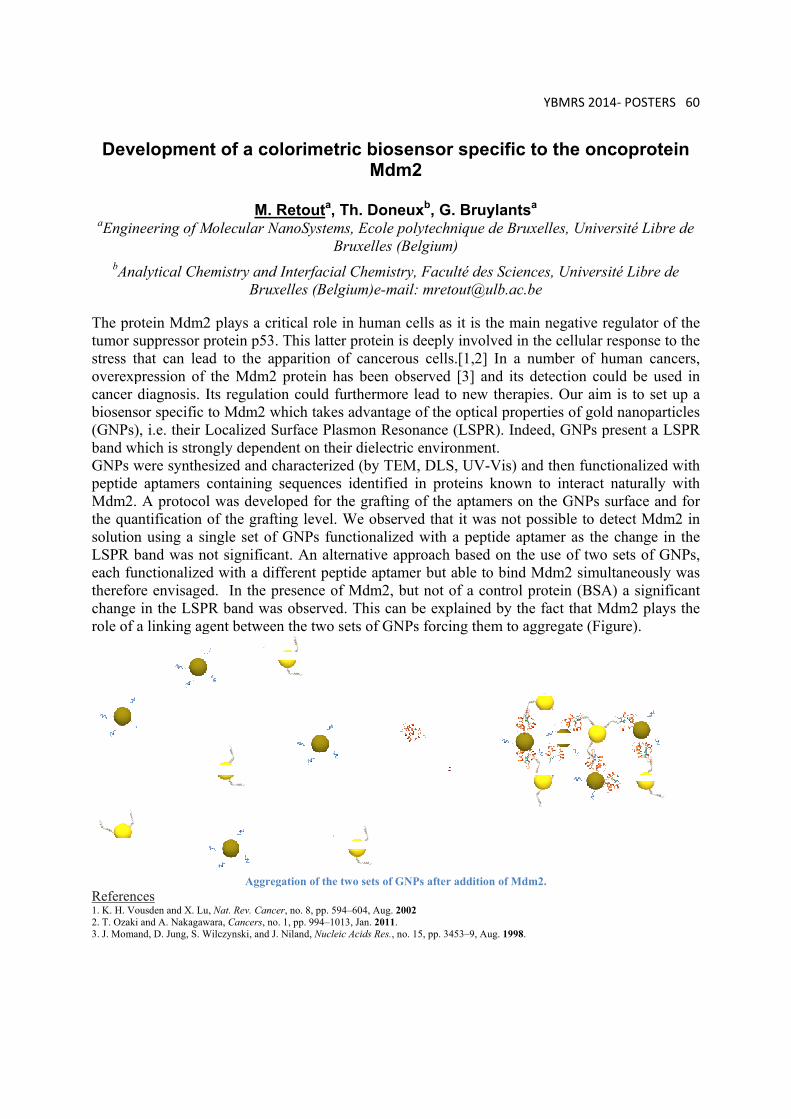
Deve
aEngin
bAna
The protumor sustress thoverexpcancer dbiosenso(GNPs)band whGNPs wpeptide Mdm2. the quansolutionLSPR beach funthereforchange role of a
Referenc1. K. H. Vo2. T. Ozaki 3. J. Moman
elopmen
neering of M
alytical Che
otein Mdm2uppressor phat can leapression of diagnosis. Ior specific t, i.e. their Lhich is stronwere synthe
aptamers cA protocol
ntification on using a siband was nonctionalizedre envisagedin the LSPR
a linking ag
ces ousden and X. Lu
and A. Nakagawnd, D. Jung, S. W
t of a co
M. RMolecular N
emistry and Brux
2 plays a criprotein p53.ad to the ap
the Mdm2 Its regulatioto Mdm2 wLocalized Sngly dependsized and ccontaining l was develof the graftngle set of ot significand with a difd. In the pR band was
gent between
Aggreg
, Nat. Rev. Cancewara, Cancers, noWilczynski, and J.
olorimetr
Retouta, TNanoSystems
BrInterfacial
xelles (Belgi
itical role in This latter
pparition ofprotein ha
on could furwhich takes Surface Plasdent on theirharacterizedsequences oped for th
ting level. Wf GNPs funcnt. An alterfferent peptpresence of s observed.n the two se
gation of the tw
er, no. 8, pp. 594–. 1, pp. 994–1013 Niland, Nucleic
ic biosenMdm2
Th. Doneuxs, Ecole polyruxelles (BeChemistry,
ium)e-mail:
n human ceprotein is d
f cancerouss been obserthermore ladvantage o
smon Resonr dielectric d (by TEMidentified i
he grafting oWe observectionalized rnative apprtide aptamerMdm2, butThis can b
ets of GNPs
wo sets of GNP
–604, Aug. 20023, Jan. 2011. Acids Res., no. 1
nsor spe2
xb, G. Bruylytechnique elgium) Faculté de: mretout@u
ells as it is tdeeply invols cells.[1,2]erved [3] alead to newof the opticnance (LSPRenvironmen, DLS, UV-in proteins of the aptamd that it wawith a peproach basedr but able tot not of a cobe explaineds forcing the
Ps after additio
5, pp. 3453–9, A
YB
ecific to t
ylantsa de Bruxelle
es Sciences, ulb.ac.be
the main nelved in the c In a numb
and its detecw therapies.
al propertieR). Indeed, nt. -Vis) and thknown to
mers on theas not possitide aptame
d on the useo bind Mdmontrol proted by the facem to aggre
on of Mdm2.
Aug. 1998.
BMRS 2014‐ P
the onco
es, Universi
Université
egative regucellular respber of humction couldOur aim is
es of gold naGNPs pres
hen functioninteract nat
e GNPs surfible to detecer as the che of two setm2 simultanein (BSA) act that Mdmegate (Figur
POSTERS 60
oprotein
ité Libre de
Libre de
ulator of theponse to the
man cancersd be used ins to set up aanoparticlessent a LSPR
nalized withturally withface and forct Mdm2 in
hange in thets of GNPsneously wasa significan
m2 plays thee).
0
e e s, n a s
R
h h r n e s, s
nt e
YBMRS 2014‐ POSTERS 61
Using solid-state NMR to characterise pharmaceutical polymorphs
A. S. Tatton1, R. Wechselberger2, H. Nova De Armas2 and C. Damblon1
1 Department of Chemistry, University of Liege 2 Janssen Pharmaceuticals, Beerse
Polymorphism is most commonly described as the possibility for the existence of different crystal structures for the same chemical structure.1 Understanding polymorphism is of particular interest in the pharmaceutical industry, as different polymorphs may have significantly different physical properties, such as changes in solubility or bio-availability. Therefore correct identification of all possible forms is an important step for both patient safety and patent concerns. Herein various solid-state NMR experiments are presented to identify the differences between two pharmaceutical polymorphs, in particular probing intermolecular interactions, such as aromatic π- π effects and hydrogen bonding. The proton chemical shift is well known to be sensitive to changes in intermolecular configurations that govern self-assembly in the solid state. However, the resolution of a typical proton spectrum in the solid state obtained at a moderate Magic-Angle Spinning (MAS) frequency is typically poor, due to the presence of strong, non-commuting homonuclear dipolar couplings. Herein, fast MAS (νr > 30 kHz) or homonuclear decoupling schemes are applied during 2-D correlation experiments to improve resolution and therefore identify differences in intermolecular interactions that govern polymorphism. Clear differences in the aromatic proton chemical shifts were noted between forms I and II via various 1D and 2D experiments, indicating differences in the π-π stacking between the two forms, whereas no significant differences in intermolecular hydrogen bonding interactions were noted.
1. Harris, R. K., Analyst 2006, 131, 351-373.

YBMRS 2014‐LIST OF PARTICIPANTS 62
LIST OF PARTICIPANTS
Last Name First name Affiliation E‐Mail ADRIAENSENS Peter UHasselt [email protected]
ALDIB Iyas ULB [email protected]
ALVAREZ Nadia UAntwerp [email protected]
APRILE Carmela UNamur [email protected]
BRUNETTI Emilio ULB [email protected]
BRUYLANTS Gilles ULB [email protected]
BUYST Dieter UGent [email protected]
CAO PHAM Thanh Trang UCL [email protected]
CAPETTE Loïc UMons [email protected]
CEULEMANS Matthias KU Leuven [email protected]
CHAIN Fernando UGent [email protected]
CHAPON David Solvay [email protected]
CIRCELLI Géraldine UMons [email protected]
COURTIN Tim UGent [email protected]
CUYPERS Bert UAntwerp [email protected]
DAMBLON Christian ULg [email protected]
DE RACHE Aurore Université de Bordeaux
a.de‐[email protected]‐bordeaux.fr
DE ROO Jonathan UGent [email protected]
DE TULLIO Pascal ULg [email protected]
DE VLEESCHOUWER
Matthias UGent [email protected]
DELANGRE Sébastien UMONS [email protected]
DENTAMARO Mario UMONS [email protected]
DESMET Céline UCL [email protected]
DOYEN Matthieu ULB [email protected]
FEHER Krisztina UGent [email protected]
FUSARO Luca UNamur [email protected]
GALLEZ Bernard UCL [email protected]

YBMRS 2014‐LIST OF PARTICIPANTS 63
GARCIA RIBEIRO Rita KU Leuven [email protected]
GEUDENS Niels UGent [email protected]
GILLET Sébastien ULg ‐ GxABT‐ CBI [email protected]
GOOVAERTS Etienne UAntwerp [email protected]
GOSSUIN Yves UMONS [email protected]
GUGGISBERG Mathias Buchem BV [email protected]
HANNECART Adeline UMONS [email protected]
HARRIS Michael KU Leuven [email protected]
HENOUMONT Céline UMONS [email protected]
HENRARD Daniel UMONS [email protected]
HERTSENS Bob Jeol [email protected]
HIMMELREICH Uwe KU Leuven [email protected]
JACQUET Nicolas ULg GxABT [email protected]
JEHASSE Kevin ULg [email protected]
JORDAN Benedicte UCL [email protected]
KAY Jennifer ULg [email protected]
KEYMEULEN Flore ULB [email protected]
KRETSCHMER Axel Dow Corning [email protected]
KUSAKOVSKIJ Jevgenij UGent [email protected]
LAURENT Sophie UMONS [email protected]
LAVENDOMME Roy ULB [email protected]
LE PAIGE Ulric ULg [email protected]
LOUIS Evelyne UHasselt [email protected]
LUCHINAT Claudio University of Florence
LUHMER Michel ULB [email protected]
MARCELIS Lionel ULB [email protected]
MARCHAND Valérie UCL [email protected]
MICHEZ Roman ULB [email protected]
MONTANTE Sylvie UMONS [email protected]
MOREAU Alain EURISO‐TOP [email protected]
MULLER Robert UMONS [email protected]

YBMRS 2014‐LIST OF PARTICIPANTS 64
NEVEU Marie‐Aline UCL marie‐[email protected]
NEVJESTIC Irena UGent [email protected]
NICOLAIJ Klaas TU Eindhoven [email protected]
PENDERS Marc Bruker Belgium [email protected]
REEKMANS Gunter UHasselt [email protected]
REMAUD Gérald Université de Nantes gerald.remaud@univ‐nantes.fr
RETOUT Maurice ULB [email protected]
SAUWEN Nicolas KU Leuven [email protected]
SINGAM Shashi Kanth Reddy
UAntwerp [email protected]
SINNAEVE Davy UGent [email protected]
STANICKI Dimitri UMONS [email protected]
TASSI Marco UHasselt [email protected]
TATTON Andrew ULg [email protected]
VAN DOORSLAER Sabine UAntwerp [email protected]
VAN KERSAVOND Tim UGent [email protected]
VAN LOKEREN Luk VUB [email protected]
VANDER ELST Luce UMONS [email protected]
VUONG Quoc Lam UMONS [email protected]
WAUTERS Céline ULg [email protected]
WEERASEKERA Akila KU Leuven [email protected]
WILLEM Rudolph VUB [email protected]