Embed Size (px)
Citation preview
A Novel Isoform of Sucrose Synthase Is Targeted to theCell Wall during Secondary Cell Wall Synthesis inCotton Fiber[C][W][OA]
Elizabeth Brill, Michel van Thournout, Rosemary G. White, Danny Llewellyn, Peter M. Campbell,Steven Engelen, Yong-Ling Ruan, Tony Arioli, and Robert T. Furbank*
Commonwealth Scientific and Industrial Research Organization Plant Industry, Canberra, Australian CapitalTerritory 2601, Australia (E.B., R.G.W., D.L., Y.-L.R., R.T.F.); Bayer BioScience, 9052 Ghent, Belgium (M.v.T.,S.E.); Commonwealth Scientific and Industrial Research Organization Ecosystem Sciences, Canberra,Australian Capital Territory 2601, Australia (P.M.C.); School of Environmental and Life Sciences, Universityof Newcastle, New South Wales 2308, Australia (Y.-L.R.); and Bayer CropScience, Lubbock, Texas 79423 (T.A.)
Sucrose (Suc) synthase (Sus) is the major enzyme of Suc breakdown for cellulose biosynthesis in cotton (Gossypium hirsutum)fiber, an important source of fiber for the textile industry. This study examines the tissue-specific expression, relativeabundance, and temporal expression of various Sus transcripts and proteins present in cotton. A novel isoform of Sus (SusC) isidentified that is expressed at high levels during secondary cell wall synthesis in fiber and is present in the cell wall fraction.The phylogenetic relationships of the deduced amino acid sequences indicate two ancestral groups of Sus proteins predatingthe divergence of monocots and dicots and that SusC sequences form a distinct branch in the phylogeny within the dicot-specific clade. The subcellular location of the Sus isoforms is determined, and it is proposed that cell wall-localized SusC mayprovide UDP-glucose for cellulose and callose synthesis from extracellular sugars.
In most higher plants, Suc is the major translocatedsugar, requiring cleavage to its component hexosesbefore further biochemical conversion can occur. Thiscleavage can occur via two enzymes: invertase or Sucsynthase (Sus; for review, see Winter and Huber, 2000;Koch, 2004). Sus is often regarded as the more energyconservative of the two enzyme reactions, as it pro-duces Fru and UDP-Glc, the latter being used as aphosphorylated sugar in biosynthetic processes. Sus isbelieved to play a major role in cellulose biosynthesis,where UDP-Glc produced by a membrane-associatedform of Sus is thought to be used directly as a substratefor the cellulose synthase complex (Amor et al., 1995).This so-called “PM” (for plasma membrane) associa-tion of Sus has been studied in a number of species(Winter and Huber, 2000; Haigler et al., 2001; Kominaet al., 2002). Conclusive evidence for substantial par-titioning of cellular Sus to the plasma membrane isdifficult to find in the literature for any species; how-
ever, recent evidence that Sus is an integral componentof the cellulose synthase rosette in bean (Phaseolusvulgaris) hypocotyls has been published (Fujii et al.,2010). It has been shown that suppression of Susexpression in transgenic cotton (Gossypium hirsutum)ovules decreased fiber elongation (Ruan et al., 2003)and affected cellulose deposition (Ruan, 2007), rein-forcing the importance of this enzyme in plant organswhere cellulose is synthesized at high rates. Sus hasalso been implicated as an important gene in wood for-mation,with expression levels correlatingwith second-ary thickening of xylem and wood strength (Hauchand Magel, 1998; Salnikov et al., 2001; Andersson-Gunneras et al., 2006; Nilsson et al., 2010). Sus has beenshown to be important in the pathway of starchbiosynthesis in legumes such as Vicia faba (Heimet al., 1993) and canola (Brassica napus; King et al.,1997) embryos, in the cellularization of cotton seedendosperm (Ruan et al., 2008), and in starch andcellulose synthesis in maize (Zea mays; for review, seeKoch, 2004). In Arabidopsis (Arabidopsis thaliana), sixisoforms of Sus are encoded in the genome, andextensive studies have been made of the tissue-specificexpression patterns of all six isoforms (Baud et al.,2004; Bieniawska et al., 2007). However, despite re-verse genetic and biochemical evidence for crucialroles of Sus in the crop species mentioned above, ithas recently been reported that Arabidopsis Sus geneknockout lines have little or no obvious phenotypes(Bieniawska et al., 2007). This includes analysis of aknockout in the isoform involved in Suc cleavage in
* Corresponding author; e-mail [email protected] author responsible for distribution of materials integral to the
findings presented in this article in accordance with the policydescribed in the Instructions for Authors (www.plantphysiol.org) is:Robert T. Furbank ([email protected]).
[C] Some figures in this article are displayed in color online but inblack and white in the print edition.
[W] The online version of this article contains Web-only data.[OA] Open Access articles can be viewed online without a sub-
scription.www.plantphysiol.org/cgi/doi/10.1104/pp.111.178574
40 Plant Physiology�, September 2011, Vol. 157, pp. 40–54, www.plantphysiol.org � 2011 American Society of Plant Biologists. All Rights Reserved. www.plantphysiol.orgon September 8, 2018 - Published by Downloaded from
Copyright © 2011 American Society of Plant Biologists. All rights reserved.

the embryo and the isoforms present in stems and root,major sites of secondary cell wall synthesis in thisspecies (Bieniawska et al., 2007). It appears that inac-tivation of multiple isoforms of Sus and, in particular,invertase is necessary to impair Suc breakdown in thismodel species and that invertase may work togetherwith UDPG pyrophosphorylase to supply carbon skel-etons to cellulose synthase (Barratt et al., 2009). Intobacco (Nicotiana tabacum), overexpression of bothSus and invertase had little impact on cellulose content(Coleman et al., 2006), while in the woody speciesPopulus alba, overexpression of Sus produced a smallbut reproducible increase in cellulose content and hadan impact on wood properties (Coleman et al., 2009).Thus, despite decades of research, the roles and im-portance of Sus in plant metabolism, and in particularin cellulose biosynthesis, still remain contentious.In cotton, a major source of fiber for the textile
industry, cellulose synthesis is of particular interest.Cotton fiber is composed of single cells initiated fromthe surface of the ovule epidermis around the time offlowering. Over the next 20 d, these cells elongate upto approximately 3 cm before laying down massiveamounts of secondary cell wall (Basra and Malik,1984), and they contain up to 98% cellulose whendesiccated. The flux of Glc moieties into cellulose atthis secondary cell wall synthesis stage is extremelyhigh and comparable in magnitude only to the processof secondary wall formation in xylem tracheids ofwoody species. To date, most research at the biochem-ical and molecular levels in cotton has focused on asingle isoform of Sus expressed at high levels in fiberand seed (Ruan et al., 1997). This Sus has been claimedto represent the predominant isoform in cotton fiber,and the transcript corresponding to this Sus is highlyexpressed in microarray (Arpat et al., 2004) and sub-tractive hybridization experiments on cotton fiber(Haigler et al., 2005). A major focus for research onSus has been the study of the membrane associationof this protein and its proposed role in channelingUDP-Glc to the cellulose synthase complex. The hy-pothesis proposed by Amor et al. (1995) and subse-quently explored in many other laboratories (forreview, see Winter and Huber, 2000; Haigler et al.,2001; Koch, 2004) has been that a single, highlyexpressed Sus isoform can exist either in a free cyto-solic state or as a membrane-bound form and that thisequilibrium can be regulated by protein phosphory-lation and/or other posttranslational processes. Theexact mechanism responsible for this membrane asso-ciation and the biochemical control of the proportion ofmembrane-bound versus soluble protein has thus farremained elusive.In this study, we examine the abundance and tem-
poral expression of various Sus transcripts presentduring fiber development and identify a novel isoformthat is expressed at high levels during secondary cellwall synthesis. The gene sequences and tissue-specificexpression patterns of this Sus isoform and other genesin the cotton Sus family are described, along with
subcellular location, predicted protein structure, andproposed role of this novel Sus isoform.
RESULTS
Identification of the Sus Gene Family in Cotton
It has previously been suggested that a single iso-form of Sus is expressed during both cotton fiberelongation and secondary cell wall synthesis, and it iswidely believed that posttranslational modification ofSus is important in the secondary cell wall synthesisstage to direct UDP-Glc to the cellulose synthasecomplex (Amor et al., 1995; for review, see Winterand Huber, 2000). However, very little work has beendone on elucidation of the Sus genes expressed overfiber development. We examined this issue by creatinga small library of Sus partial cDNA sequences derivedby reverse transcription (RT)-PCR of fiber RNA ex-tracted from fibers undergoing rapid elongation (ap-proximately 10 d after flowering [DAF]) and in thesecondary cell wall synthesis stages (approximately 20DAF). Primers were designed to hybridize to a regionof the Sus sequence highly conserved across all higherplants (see “Materials and Methods”). Full-lengthcDNAs of representatives of the classes of sequenceswere then obtained by 5# RACE. The sequences ofpartial cDNAs obtained from the fiber RNA pools fellinto four classes, termed SusA, SusB, SusC, and SusD.Full-length deduced amino acid sequences of these areshown in Figure 1. Deduced amino acid sequences forSusB and -D were 93% and 95.5% similar to SusA,respectively. Sequence A is identical to the sequencereported in the literature by Amor et al. (1995) andRuan et al. (1997) and is the isoform commonly stud-ied in the literature (GenBank accession no. U73588).As cotton is tetraploid, it is possible that SusA, -B, and-D are homeologous, but this cannot be definitivelydetermined from these data. In contrast, the deducedamino acid sequence of SusCwas only 76% identical tothat of the SusA protein, with regions of markedsequence divergence at both the N and C termini.
To examine the likely genomic origins and related-ness of the four classes of Sus genes, genomic clones ofeach class were isolated and the gene structures werecompared. Figure 2 shows the intron-exon distributionof the Sus genes isolated from cotton. All four classesof Sus have complex gene structures, 10 exons in thecase of SusC, 12 in SusA and -D, and 13 in the case ofSusB. This is broadly similar to the gene structure ofthe Arabidopsis Sus genes, the only other dicot Susgene family sequenced, which ranges from 12 to 15exons (Baud et al., 2004).
The phylogenetic relationship between the deducedamino acid sequences of the SusA, -B, -C, and -Dproteins from cotton, SusC proteins from a range ofdiploid and tetraploid Gossypium species, and Sus se-quences from other dicot and monocot plant specieswith fully sequenced genomes are shown in Figure 3
SusC in Cotton
Plant Physiol. Vol. 157, 2011 41 www.plantphysiol.orgon September 8, 2018 - Published by Downloaded from
Copyright © 2011 American Society of Plant Biologists. All rights reserved.

(the alignments are provided as Supplemental Fig.S1). There are clearly two ancestral groups of Susproteins that predate the divergence of monocots anddicots but also more specialized forms of Sus that arespecific to either dicots or monocots. SusC sequencesform a distinct branch in the phylogeny within thedicot-specific clade and have evolved away fromthe other members of the cotton Sus gene family.The major regions of divergence are in the N andC termini of the SusC protein rather than in regionsresponsible for enzymatic activity. The SusA, -D,and -B isoforms are most closely related to eachother than to SusC. It is notable that there seems tobe no direct homolog of SusC in the Arabidopsisor Populus genomes. Within the Gossypium species,SusC appears to be quite ancestral, with distinctclades in the A and D genome diploids (Fig. 3B). Inthe AD tetraploids, the A genome form of SusC ap-pears to have been lost at some time after polyploid-ization, except for Gossypium mustelinum, which hasretained both homeologues. This was confirmed bySouthern blotting using SusC-specific probes (datanot shown).
Expression of Sus Genes in Cotton Fiber andOther Tissues
The expression levels of the Sus isoforms in variousorgans of cotton plants and across fiber developmentwere examined using semiquantitative PCR. Gene-specific primers for SusA, SusB, SusC, and SusDtranscripts were used (Fig. 4, A–D), along with ubiq-uitin as a normalization control (Fig. 4E; see “Materialsand Methods”). All four transcripts were detected athigh levels in stem and fiber RNA pools (Fig. 4).Transcripts for SusA, -B, and -D were also detected atlow levels in leaf and at higher levels in petal samples.During fiber development, transcripts encoding SusA,-B, and -D were detected at all six stages of fiberdevelopment, with transcript levels generally fallingfrom 8 to 21 DAF (Fig. 4). In contrast, SusC transcriptabundance increased with developmental age, declin-ing slightly at 21 DAF. Public G. hirsutum EST collec-tions were also searched using the unique peptidesfrom the N and C termini of the SusC protein se-quence; no SusC ESTs were found in libraries isolatedfrom the fiber initiation, elongation, and primary cell
Figure 1. Deduced amino acid sequence comparison of cotton SusA, -B, -D, and -C isoforms (generated using GeneticsComputer Group Wisconsin Package Pileup software). Note the sequence divergence at the N and C termini of SusC comparedwith the A,B,D sequences. The line below the SusC sequence indicates the oligopeptide synthesized to raise antiserum specific tothe SusC isoform.
Brill et al.
42 Plant Physiol. Vol. 157, 2011 www.plantphysiol.orgon September 8, 2018 - Published by Downloaded from
Copyright © 2011 American Society of Plant Biologists. All rights reserved.

wall biosynthesis stages of development (0–15 DAF).In contrast, in EST libraries constructed from fibersat 20 to 40 DAF, 30 SusC EST sequences were identi-fied using the C-terminal region as a query sequenceand 35 using the N-terminal region as a query. Con-sistent with the phylogenetic analysis of the SusCamino acid sequence, no ESTs were identified fromgenera other thanGossypium using the N- or C-terminalregion of the SusC protein as query sequence (data notshown).
Immunological Detection and Subcellular Targeting ofSus Isoforms
As discussed above, it has previously been proposedthat during secondary cell wall synthesis, Sus binds tothe plasma membrane, channeling UDPG directly tothe cellulose synthase complex (for review, see Huberand Winter, 2000; Haigler et al., 2001). Sus has alsobeen proposed to associate with the actin cytoskeleton(Winter et al., 1998), mitochondria (Subbaiah et al.,2006), and the tonoplast (Etxeberria and Gonzalez,2003). It has been proposed that a single Sus isoform canbe targeted to these diverse subcellular compartments,although no overarching mechanism for controllingthis targeting and association has been resolved. Pro-
tein phosphorylation has been implicated in partition-ing the protein between the plasma membrane and thecytosol and between the cytosol and the cytoskeleton(for review, see Winter and Huber, 2000), and regionsof both the C and N terminus and the presence of apleckstrin-like domain have been implicated in mem-brane binding in maize Sus (Hardin et al., 2006). Thedata above show that Sus gene family members havedistinctly different expression profiles during fiberdevelopment and that a single protein does not dom-inate. This presents the possibility that the individualisoforms may play distinct roles in fiber metabolism,possibly in separate subcellular compartments. Toexplore this possibility, antibodies were raised againstoligopeptides specific to the SusC protein to produceantiserum that would not cross-react with the A/D orB isoform. Separation of the other protein isoformsimmunologically was not possible due to their highsequence similarity. To test the specificity of the SusCantibody, western blotting was carried out using pro-tein prepared from fiber samples in the elongationphase (8–13 DAF) and the secondary cell wall stage (20DAF; Fig. 5). Western blots probed with an antiserumraised against purified rice Sus protein, which detectsmultiple Sus isoforms (Fallahi et al., 2008), detecteda single band at 92 kD in fiber sampled at 8 to 13 DAF
Figure 2. Intron/exon distributions inthe genomic sequences of the four Susisoforms present in the cotton genome(A) compared with the gene structuresof the six isoforms present in the Arab-idopsis genome (B; Baud et al., 2004).
SusC in Cotton
Plant Physiol. Vol. 157, 2011 43 www.plantphysiol.orgon September 8, 2018 - Published by Downloaded from
Copyright © 2011 American Society of Plant Biologists. All rights reserved.

(Fig. 5A). In 20-DAF fiber, two bands were detectedat approximately 90 and 92 kD (Fig. 5A). When probedwith the specific SusC antibody, a single band at 90 kDwas detected in 20-DAF fiber, corresponding to thelower molecular mass band detected by the rice Susantiserum, which is consistent with the predictedmolecular mass of the SusC protein being smallerthan the A, B, and D isoforms (Fig. 1). No band was
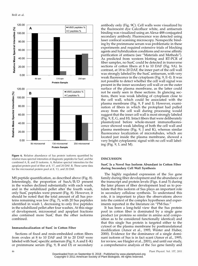
detected with specific SusC antiserum in fiber at theelongation stage at 8 DAF (Fig. 5B). To confirm theidentity of the proteins recognized by these antibodies,a Coomassie blue-stained gel slice corresponding tothese two protein bands was excised from duplicateSDS-PAGE gels and subjected to tryptic digestionand subsequent liquid chromatography-tandem massspectrometry (LC-MS/MS). Amixture of peptides fromthe A, B, and D isoforms was detected in both the 8-to 13-DAF and 20-DAF fiber samples, while the spe-cific lower molecular mass protein recognized by theSusC antiserum in the 20-DAF fiber sample also con-tained peptides unique to the SusC isoforms (data notshown). This LC-MS/MS result is also confirmed inthe apoplast and microsomal protein fractions from8-DAF and 20- to 25-DAF fiber, showing no SusC pep-tides in 8-DAF fiber samples and numerous uniqueSusC peptides in 20-DAF fiber (Fig. 6; SupplementalTable S1).
The SusC-specific antiserum was used to investigatethe subcellular location of SusC both through westernblotting and LC-MS/MS of tryptic peptide from pro-teins prepared from fiber-cell fractions (Figs. 6–8) orby immunohistochemistry (Fig. 9). Cotton fibers har-vested at 8 to 13 DAF (elongation phase) and 17 to 25DAF (secondary cell wall stage) were frozen in liquidnitrogen, ground to a fine powder, and then extractedin buffer (see “Materials and Methods”) intended toextract cytosolic proteins plus proteins loosely boundto cell wall or membrane components. This extractwas subjected to centrifugation to remove particulatematerial, mostly cell wall debris (“cell wall fraction”).The supernatant fraction from this first centrifugationalso contained suspended membrane componentsthat were removed by ultracentrifugation. The super-natant from this ultracentrifugation was termed “sol-uble fraction” and the pellet was termed “microsomalfraction,” the latter being enriched in plasma mem-brane and other membranous cell fractions (Carlsonand Chourey, 1996). The microsomal fraction was alsofurther purified by density gradient ultracentrifuga-tion to enrich the proportion of plasma membranes(“PM fraction”; Carlson and Chourey, 1996). The cellwall pellet was either further extracted or solubilizedfor SDS-PAGE, western blotting, and subsequent LC-MS/MS. The supernatant, microsomal, and PM frac-tions were subjected to SDS-PAGE and subsequentLC-MS/MS. Consistent with previous experimentsfrom total soluble fiber fractions (Fig. 5), SusC proteinwas only detected in fiber microsomal extracts fromapproximately 13 to 17 DAF onward, increasing in thesecondary cell wall synthesis stage (20–25 DAF) bothin western blotting experiments (data not shown) andLC-MS/MS (Fig. 6).
Over the whole experiment and considering all theobserved peptides identified to Sus isoforms A, B, C,and D, 19 distinct peptides were unique to SusC,whereas the remaining peptides could have beenderived from SusA, -B, or -D or subsets of that group(Supplemental Table S1; Supplemental Fig. S3). Only
Figure 3. Phylogenetic tree of Sus protein sequences generated usingMEGA 4.0 and the neighbor-joining method (see “Materials andMethods”). A, Unrooted tree of 28 plant Sus proteins from plantswith sequenced genomes: the dicots Arabidopsis (AtSUS1 to -6),Populus trichocarpa (POPTRDRAFT), and Ricinus communis (RCOM_xxxxxxx), the monocotsOryza sativa japonica group (Osxxxxxxxx) andSorghum bicolor (SORBIDRAFTxxxxxxxx), together with the four cot-ton Sus proteins reported in this paper, and with the moss (Physcomi-trella patens subspecies patens) Sus protein (Pp-EDQ65205.1) as anoutgroup. The cotton Sus proteins are surrounded by dotted lines, andthe SusC protein is shaded in gray (alignment files are provided asSupplemental Fig. S1). B, Phylogenetic tree of the SusC proteins fromselected diploid and tetraploid Gossypium species (alignment filesshown in Supplemental Fig. S2). The G. kirkii SusC sequence was usedas the outgroup. Tetraploid cotton species names are indicated byasterisks. The numbers on the interior branches refer to the bootstrapvalues for 1,000 replications. Bootstrap values less than 50% are notgiven. The scale at the bottom is in units of amino acid substitutionsper site.
Brill et al.
44 Plant Physiol. Vol. 157, 2011 www.plantphysiol.orgon September 8, 2018 - Published by Downloaded from
Copyright © 2011 American Society of Plant Biologists. All rights reserved.

one peptide was observed that could have been de-rived from SusC and also could have been derivedfrom SusB and -D. For an estimate of the relativeabundance of Sus isoforms in each sample, we com-pared the total mass spectrum intensities of peptidesunique to SusC with the total intensities for peptidesindicating SusA, -B, or -D expressed as a proportion ofthe total spectral intensity for all peptides indicatingSus isoforms. For the purposes of this estimation, theone peptide observed that could have been derived bySusB, -C, or -D was excluded. The high similarity ofthe SusA, -B, and -D amino acid sequences meant thatthese proteins could not be estimated individually.This approach does not allow absolute amounts of the
proteins to be estimated without the use of standards(Perkins et al., 2005), but it was sufficient for ourpurposes here to indicate where and when SusA/B/Ddominated relative to SusC and vice versa.
Relative abundance of the Sus isoforms in cellularfractions at 25 DAF was estimated by quantification ofthe spectral intensities of unique, diagnostic peptidesfor SusC found in each sample, relative to those ofisoforms A/B/D and normalized to the total spectralintensity of all Sus peptides in that sample (Fig. 6).Because of the high similarity of the SusA, -B, and -Damino acid sequences, these proteins could not beindividually quantified using this method. SusA,B,Dand SusC were all present in the soluble samples andthe microsomal fractions at similar abundance in 25-DAF fiber (Fig. 6); however, SusC was barely detect-able in the PM fraction (data not shown).
Two approaches were used to examine the presenceof Sus isoforms in the cell wall fraction. First, ovuleswere excised from cotton fruits with attached fibersintact, washed briefly in cold isotonic buffer, and thenproteins that were loosely bound to the cell walls wereextracted by gentle washing in isotonic buffer contain-ing a variety of protease inhibitors and stabilizingagents. Proteins recovered by concentrating this washwere subjected to SDS-PAGE, the region of the gelcontaining Sus proteins was excised, and the trypticpeptides were identified byMS/MS.While SusA/B/Dpeptides were present and dominant in the earlystages of fiber development, SusC diagnostic peptidesonly began to appear in the Sus tryptic peptide profileby 17 DAF and dominated the profile from 20 DAFonward in the cell wall fraction (Fig. 6B). This wasconfirmed by western blotting of duplicate gels withthe SusC-specific antibody (data not shown).
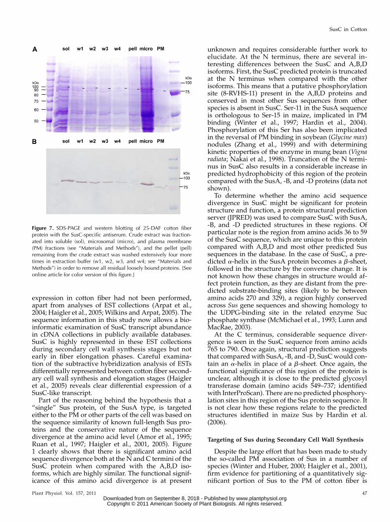
The second approach to extracting cell wall-associ-ated proteins involved sequential washing of the “cellwall pellet” fraction from 25-DAF fibers followingcentrifugation and subjecting these washes to SDS-PAGE (Fig. 7A), western blotting (Fig. 7B), and MS/
Figure 4. Semiquantitative PCR of Sus RNA prepared from differentplant organs and over fiber development: young fully expanded leaf (L),petal (P), and stem (S) and fiber RNA from bolls at 8, 10, 13, 17, and21 DAF. Primers specific to SusA, -B, -C, and -D were used (A–D,respectively) and to a member of the ubiquitin family as a quantitativecontrol (E). F compares relative expression levels of Sus isoforms shownin A to E by normalizing the fluorescence intensity of each PCR producton the gel to the respective Ubiquitin control and presenting this as apercentage of the maximum expression level for the Sus isoform in thattissue sample and developmental stage.
Figure 5. Western analysis of cotton fiber proteins extracted from bollsat 8 and 13 DAF (elongation stage) and 20 DAF (secondary cell wallstage). A, Protein from these three stages probed with the nonselectiveantiserum, with the Sus protein bands indicated by arrows. Note theappearance of labeling of a lower molecular mass band at 20 DAFshown by MS/MS to be SusC. B, Western blot of cotton fiber proteins at8 and 20 DAF probed with the SusC-specific antiserum, detecting asingle protein band shown by MS/MS to be SusC.
SusC in Cotton
Plant Physiol. Vol. 157, 2011 45 www.plantphysiol.orgon September 8, 2018 - Published by Downloaded from
Copyright © 2011 American Society of Plant Biologists. All rights reserved.
MS peptide quantification, as described above (Fig. 8).Interestingly, the proportion of SusA/B/D presentin the washes declined substantially with each wash,and in the solubilized pellet after the fourth wash,only SusC peptides were present (Fig. 8). However, itshould be noted that the total amount of all Sus pro-teins remaining was low (Fig. 7), with 20 Sus peptidesidentified in wash 1, decreasing to only five peptidesin the solubilized pellet after four washes. At this stageof development, microsomal and apoplast fractionsalso contained more SusC than the other isoformscombined.
Immunolocalization of SusC in Cotton Fiber
Sections of fixed and resin-embedded cotton fibersfrom ovules at 8 to 10 DAF and 18 to 20 DAF werelabeledwith SusC-specific antiserum (Fig. 9, A and E–K)or preimmune serum (Fig. 9, B and D) or secondary
antibody only (Fig. 9C). Cell walls were visualized bythe fluorescent dye Calcofluor white, and antiserumbinding was visualized using an Alexa-488-conjugatedsecondary antibody. Fluorescence was detected usinglaser confocal scanning microscopy. Nonspecific bind-ing by the preimmune serumwas problematic in theseexperiments and required extensive trials of blockingagents and hybridization conditions and reverse affinitypurification of antisera (see “Materials and Methods”).As predicted from western blotting and RT-PCR offiber samples, no SusC could be detected in transversesections of cotton fibers at 8 to 10 DAF (Fig. 9A). Incontrast, at 18 to 20 DAF, the inner part of the cell wallwas strongly labeled by the SusC antiserum, with veryweak fluorescence in the cytoplasm (Fig. 9, E–I). It wasnot possible to detect whether the cell wall signal waspresent in the inner secondary cell wall or on the outersurface of the plasma membrane, as the latter couldnot be easily seen in these sections. In glancing sec-tions, there was weak labeling of cytoplasm close tothe cell wall, which could be associated with theplasma membrane (Fig. 9, F and I). However, exami-nation of fibers in which the protoplast had pulledaway from the cell wall during processing wouldsuggest that the inner cell wall is most strongly labeled(Fig. 9, E, G, and H). Intact fibers that were deliberatelyplasmolyzed before whole-mount immunofluores-cence showed weak labeling of both the cell wall andplasma membrane (Fig. 9, J and K), whereas similarfluorescence localization of microtubules, which arelocated just inside the plasma membrane, showed avery bright cytoplasmic signal with no cell wall label-ing (Fig. 9, L and M).
DISCUSSION
SusC Is a Novel Sus Isoform Abundant in Cotton Fiberduring Secondary Cell Wall Synthesis
The highly regulated expression of the Sus genefamily during fiber development and the abundance atthe transcript and protein levels (Figs. 4 and 5) duringthe later phases of fiber development lead us to pos-tulate that this isoform of Sus plays an important rolein secondary cellulose synthesis. To understand thisrole, it is important to place the observations aboveinto the context of the complex hypotheses and exper-iments reported in the literature on “PM-Sus.”
It has been a long-held view that the Sus proteinpool in cotton fiber is dominated by a single geneproduct (or proteins so similar in amino acid compo-sition as to be considered functionally identical) andthat this single Sus protein is targeted either to thecytosol or the plasma membrane by posttranslationalmodification (Amor et al., 1995; Winter and Huber,2000). Evidence for the dominance of a single domi-nant isoform of Sus is in fact scant (Amor et al., 1995;for review, see Haigler et al., 2001), and until our study,a comprehensive analysis of the Sus gene family and
Figure 6. Relative abundance of Sus protein isoforms quantified byrelative mass spectral intensities of diagnostic peptides for SusC and thecombined A, B, and D isoforms. A, Relative spectral intensities for theapoplast protein pool of fiber at 8, 17, and 20 DAF. B, These same datafor the microsomal protein pool at 8, 13, and 20 DAF.
Brill et al.
46 Plant Physiol. Vol. 157, 2011 www.plantphysiol.orgon September 8, 2018 - Published by Downloaded from
Copyright © 2011 American Society of Plant Biologists. All rights reserved.
expression in cotton fiber had not been performed,apart from analyses of EST collections (Arpat et al.,2004; Haigler et al., 2005; Wilkins and Arpat, 2005). Thesequence information in this study now allows a bio-informatic examination of SusC transcript abundancein cDNA collections in publicly available databases.SusC is highly represented in these EST collectionsduring secondary cell wall synthesis stages but notearly in fiber elongation phases. Careful examina-tion of the subtractive hybridization analysis of ESTsdifferentially represented between cotton fiber second-ary cell wall synthesis and elongation stages (Haigleret al., 2005) reveals clear differential expression of aSusC-like transcript.Part of the reasoning behind the hypothesis that a
“single” Sus protein, of the SusA type, is targetedeither to the PM or other parts of the cell was based onthe sequence similarity of known full-length Sus pro-teins and the conservative nature of the sequencedivergence at the amino acid level (Amor et al., 1995;Ruan et al., 1997; Haigler et al., 2001, 2005). Figure1 clearly shows that there is significant amino acidsequence divergence both at the N and C termini of theSusC protein when compared with the A,B,D iso-forms, which are highly similar. The functional signif-icance of this amino acid divergence is at present
unknown and requires considerable further work toelucidate. At the N terminus, there are several in-teresting differences between the SusC and A,B,Disoforms. First, the SusC predicted protein is truncatedat the N terminus when compared with the otherisoforms. This means that a putative phosphorylationsite (8-RVHS-11) present in the A,B,D proteins andconserved in most other Sus sequences from otherspecies is absent in SusC. Ser-11 in the SusA sequenceis orthologous to Ser-15 in maize, implicated in PMbinding (Winter et al., 1997; Hardin et al., 2004).Phosphorylation of this Ser has also been implicatedin the reversal of PM binding in soybean (Glycine max)nodules (Zhang et al., 1999) and with determiningkinetic properties of the enzyme in mung bean (Vignaradiata; Nakai et al., 1998). Truncation of the N termi-nus in SusC also results in a considerable increase inpredicted hydrophobicity of this region of the proteincompared with the SusA, -B, and -D proteins (data notshown).
To determine whether the amino acid sequencedivergence in SusC might be significant for proteinstructure and function, a protein structural predictionserver (JPRED) was used to compare SusC with SusA,-B, and -D predicted structures in these regions. Ofparticular note is the region from amino acids 36 to 59of the SusC sequence, which are unique to this proteincompared with A,B,D and most other predicted Sussequences in the database. In the case of SusC, a pre-dicted a-helix in the SusA protein becomes a b-sheet,followed in the structure by the converse change. It isnot known how these changes in structure would af-fect protein function, as they are distant from the pre-dicted substrate-binding sites (likely to be betweenamino acids 270 and 329), a region highly conservedacross Sus gene sequences and showing homology tothe UDPG-binding site in the related enzyme Sucphosphate synthase (McMichael et al., 1993; Lunn andMacRae, 2003).
At the C terminus, considerable sequence diver-gence is seen in the SusC sequence from amino acids765 to 790. Once again, structural prediction suggeststhat comparedwith SusA, -B, and -D, SusCwould con-tain an a-helix in place of a b-sheet. Once again, thefunctional significance of this region of the protein isunclear, although it is close to the predicted glycosyltransferase domain (amino acids 549–737; identifiedwith InterProScan). There are no predicted phosphory-lation sites in this region of the Sus protein sequence. Itis not clear how these regions relate to the predictedstructures identified in maize Sus by Hardin et al.(2006).
Targeting of Sus during Secondary Cell Wall Synthesis
Despite the large effort that has been made to studythe so-called PM association of Sus in a number ofspecies (Winter and Huber, 2000; Haigler et al., 2001),firm evidence for partitioning of a quantitatively sig-nificant portion of Sus to the PM of cotton fiber is
Figure 7. SDS-PAGE and western blotting of 25-DAF cotton fiberprotein with the SusC-specific antiserum. Crude extract was fraction-ated into soluble (sol), microsomal (micro), and plasma membrane(PM) fractions (see “Materials and Methods”), and the pellet (pell)remaining from the crude extract was washed extensively four moretimes in extraction buffer (w1, w2, w3, and w4; see “Materials andMethods”) in order to remove all residual loosely bound proteins. [Seeonline article for color version of this figure.]
SusC in Cotton
Plant Physiol. Vol. 157, 2011 47 www.plantphysiol.orgon September 8, 2018 - Published by Downloaded from
Copyright © 2011 American Society of Plant Biologists. All rights reserved.

difficult to find in the literature. The standard tech-nique of extraction of a “pellet” fraction with EGTAfollowing centrifugation varies in the efficacy of ex-traction of Sus in the literature, even from the samegroups working in cotton (Amor et al., 1995; Haigleret al., 2001, Komina et al., 2002). There is little conclu-sive evidence of “docking” of Sus with the cellulosesynthase rosette on the plasma membrane using eitherthe standard techniques of immunoprecipitation ortechniques such as fluorescence resonance energytransfer (Dixit et al., 2006); however, recently, evidencehas been published that Sus is an integral componentof the cellulose synthase rosette using detergent-soluble granular particles from the plasma membraneof bean epicotyls (Fujii et al., 2010). Localization ofa significant proportion of SusC to the cell wall, sug-gested from both the protein mass spectrometry ofsubcellular fractions and the immunolocalization ex-periments shown heremay shed light on some of theseearlier observations. It is not clear why EGTA shouldspecifically release Sus from the plasma membrane.It has been suggested that the interaction with themembrane may involve calcium, although evidencefor this is circumstantial and the membrane binding ofSus varies in reversibility markedly between species(Winter and Huber 2000; Komina et al., 2002). It is wellknown that EGTA loosens the interactions betweenpectins and other cell wall components and as such is astandard reagent in buffers used to elute proteins fromcell walls (Zhao et al., 2008, and refs. therein). Clearly,from Figure 5, a simple buffer excluding EGTA elutedSusA and -C from the cell wall fraction of cotton fiber,but it is difficult to elute all the SusC protein in the cellwall pellets, suggesting that this isoform is moretightly bound to the cell wall fraction. Little or noSusA, -B, or -D is present in successive washes of cellwall material, suggesting that these proteins are likelyto be either loosely bound or even cytosolic isoforms
binding nonspecifically to this fraction (Figs. 7 and 8).It would be difficult to design definitive experimentsto determine which of these options is correct. How-ever, irrespective of this uncertainty, the presence ofsignificant amounts of Sus in the cell wall fractionwould certainly call into question the standardmethod of estimating PM-Sus levels by comparingsoluble Sus levels with EGTA washes of the pelletfraction of crude cotton fiber extracts, or other tissuesfor that matter.
The immunolocalization of SusC using the isozyme-specific antiserum (Fig. 9) also indicated that a largeproportion of SusC is present in the cell wall of fiber at18 to 20 DAF. This is entirely consistent with the cellwall pellet washing experiments described above. Theother isoforms of Sus, in particular SusA, were alsoshown to be present in this compartment at all stagesof development using a nondiscriminatory anitiserum(data not shown). Localization of Sus to the fiber cellwall of cultured ovules has been reported severaltimes (Amor et al., 1995; Haigler et al., 2001; Salnikovet al., 2003); however, the significance of this observa-tion has not been emphasized, seemingly because theobservations were interpreted to be artifacts of fixationwith the cell wall label, indicating plasma membrane-bound Sus (Amor et al., 1995; Haigler et al., 2001).Recent work on immunolocalization of Sus in pollentubes has also suggested that Sus is present in cellwalls (Persia et al., 2008). This evidence, however, isnot compelling, based on the presence of the protein inthe postnuclear supernatant fraction of cell extractsand antibody labeling of detergent-treated pollen tubes,in which the site of labeling could be in the cell wall,plasmalemma, or cytosol. (Persia et al., 2008). In cottonfiber, immunolocalization experiments at later stagesof fiber development reported here and in the litera-ture (Salnikov et al., 2003) are much clearer, consistentlyshowing almost no Sus labeling in the cytoplasm,
Figure 8. MS/MS of Sus proteins quantified byrelative mass spectral intensity of SusC and SusA,-B, and -D diagnostic peptides (as in Fig 6) butderived from the 25-DAF fiber protein samples(described in Fig 7). Note the persistence of SusCpeptides in the protein pool in later washes andthe dominance of this isoform in the final exten-sively washed and solubilized pellet.
Brill et al.
48 Plant Physiol. Vol. 157, 2011 www.plantphysiol.orgon September 8, 2018 - Published by Downloaded from
Copyright © 2011 American Society of Plant Biologists. All rights reserved.

despite reasonable preservation of this compartment. Inour experiments and in those of Salnikov et al. (2003),greater than 90% of Sus labeling is in the “exoplasmiczone” (i.e. cell wall apoplast). Quantification of thislabeling at the plasma membrane surface (internally orexternally) is difficult; however, both the fluorescentimmunolocalization carried out here and the previousimmunogold work (Salnikov et al., 2003; Ruan, 2007)show that a large percentage of Sus is external to theplasma membrane “zone” as defined by Salnikov et al.(2003).There is convincing evidence that “particulate” or
microsomal Sus is also strongly associated with micro-tubules and F-actin filaments (for review, seeWinter andHuber, 2000). The region of SusC corresponding to theputative actin-binding site identified in other species(Winter and Huber 2000) has the following consensus
amino acid sequence: [*,E,D]-D-[V,A]-[A,G,S,T]-X-E-[L,V,I]-[T,S,A,M]-[K,R,G,M,L]-E-[*,L,M]-[Q,N], whereboldface amino acids are polar-hydrophylic and italicamino acids are nonpolar-hydrophobic at neutral pH.The sequence motif corresponding to this F-actin-binding domain in SusC is 383-KDVAAEITKEFQ-394. The amino acids highlighted in boldface areunique to SusC. Of interest is that 383-K (Lys) has abasic side chain while E (Glu) and D (Asp) have acidicside chains. The unique K substitution in the SusCsequence may indicate that this protein may no longerbind to the F-actin filaments. This is also compoundedby the presence of F (Phe) in this motif, also unique toSusC, containing a side chain more voluminous thanL and M, which are present in SusA, -B, and -D andtheir counterparts in other species, potentially leadingto added instability in any actin-SusC interaction.
Figure 9. Confocal laser scanning micrographs of cotton fiber cross sections (A–I) or whole fibers (J–M) labeled with SusCaffinity-purified antibodies (A and E–K) or preimmune serum (B and D) and then Alexa-488 secondary antibody, secondaryantibody only (C), or monoclonal anti-a-tubulin followed by Alexa-488 secondary antibody (L and M). A and B, Sections of10-DAF fibers showing nonspecific autofluorescence (green, red; arrowheads) and weak cellulose fluorescence (blue) afterincubation in SusC antibody (A) or preimmune serum (B). C to I, Sections of 18-DAF fibers showing background autofluorescence(mostly red; arrowheads in C, D, and G) and strong cellulose fluorescence (blue). Very little background label was seen insections treated with secondary antibody only (C) or preimmune serum and then secondary antibody (D). SusC (punctate greenfluorescence) was detected primarily on the inner surface of the cell wall (E–I; arrows) and was occasionally detected in thecytoplasm, where the plasma membrane and cytoplasm have retracted from the cell wall (F, G, and I; double arrowheads). Aglancing section (F and I) shows diffuse autofluorescence in the cytoplasm (F; arrowhead) as well as SusC label. H and I, SusCfluorescence (green) from E and F, respectively, overlaid on bright-field images. J, Intact 18-DAF fibers treatedwith 0.3 Mmannitolbefore SusC labeling (green) show fluorescence associated with contracted cytoplasm (arrowhead) and with the cell walls(arrows). K, Overlay of SusC label in J with cellulose fluorescence (blue) showing colocalization with the cell wall (arrows). L,Microtubules (green) in 18-DAF fibers treated with 0.6 M mannitol are contracted away from the cell walls (arrowhead), with nodetectable wall-associated background fluorescence (arrow). M, Overlay of microtubule label in L with cellulose fluorescence(blue) showing no colocalization with the cell wall (arrow). Bars = 10 mm (A–I) or 20 mm (in J for J–M).
SusC in Cotton
Plant Physiol. Vol. 157, 2011 49 www.plantphysiol.orgon September 8, 2018 - Published by Downloaded from
Copyright © 2011 American Society of Plant Biologists. All rights reserved.

The Roles of SusA and -C in Cotton Fiber Cell
Wall Synthesis
Conclusive elucidation of the role of SusC in fibercellulose biosynthesis awaits the results of isoform-specific gene suppression and overexpression experi-ments currently underway for cotton; however, thedata presented here may support some informedspeculation. Previous cotton Sus gene suppressionexperiments in our laboratory, using a construct tar-geted to SusA, showed a clear disruption of fiber andseed development, producing shorter fibers (Ruanet al., 2003) containing less cellulose (Ruan, 2007).Due to the severity of phenotypic effects, it wasdifficult to isolate the impact of low SusA on second-ary cell wall synthesis from pleiotropic developmentaleffects on fiber growth and primary wall synthesis,and it is also unknown to what degree this geneconstruct cross-silenced SusC gene expression. Thetranscriptional analysis of Sus expression during fiberdevelopment shown here and the analysis of SusA/B/D and SusC abundance in cDNA libraries fromcotton fiber suggest that the SusA/B/D isoforms areexpressed highly during fiber elongation, falling offduring secondary cell wall synthesis (Fig. 4). In con-trast, we have shown that SusC is absent at both thetranscript and protein levels in early fiber develop-ment but highly expressed in later fiber development(Figs. 4, 5, and 9). This high-level expression of SusCspecifically during secondary cell wall synthesis incotton fibers, its targeted expression in other tissuesthat undergo secondary cell wall synthesis (such asstem; Fig. 4), and its distant phylogenetic relationshipto Sus in most herbaceous species (Fig. 3) providestrong circumstantial evidence for its specific involve-ment in secondary cell wall cellulose synthesis.
If we accept that the data shown here cast doubt onthe precise location of SusC (and SusA) but accept thatparticulate or “P-Sus” is important for secondary cellwall synthesis, we can calculate how much P-Susmight be required in cotton fiber to support flux.Haigler et al. (2005) calculated that 80% of sugar fluxto cotton fibers is used for cellulose synthesis duringthe peak secondary wall synthesis stage. Their modelpredicts that greater than 80% of Sus should be“membrane associated.” Extraction of cell wall/mem-brane fractions isolated with EGTA washing from avariety of species commonly yields figures of only 10%to 50% of total Sus protein associated with the partic-ulate fraction, with microsomal and more purifiedpreparations yielding estimates at the lower end of thisrange (Amor et al., 1995; Winter et al., 1997; Haigleret al., 2001, and refs. therein). This disparity betweenstandard biochemical methods for determining Sussubcellular targeting and cell biological techniquesmay be explained by the data shown here. It is appar-ent from Figure 9 that the major Sus isoforms presentin cotton fiber bind differentially to cell walls. Thesoluble fraction, described by Amor et al. (1995) andsubsequent authors, may in fact be contaminated with
a large proportion of loosely bound SusA and SusC. Asignificant proportion of SusC will also be retained inthe pellet fraction and discarded when microsomalfractions are prepared. Thus, membrane-bound Sus,on which a great deal of research has been conducted,may be a very minor proportion of particulate Sus, themajority of which is bound to varying degrees withinthe cell wall apoplast. The role of SusC would beclearer if this isoform alone were targeted to the cellwall. This is not the case, however, and SusA/B/D arealso found in this fraction to varying degrees, shownin both destructive and in situ (data not shown)immunological approaches and by diagnostic proteinmass spectrometry carried out here.
The implications of a large pool of cell wall-associ-ated, extracellular Sus are manifold, regardless of theidentity of the Sus isoforms involved. An active apo-plastic Sus could function in much the same way ascell wall-associated invertase (for review, see Koch,2004); however, it would require apoplastic UDP as asubstrate in addition to Suc. The sugar composition ofthe apoplastic fluid of cotton locules 18 to 20 wasdetermined here using a protocol developed for leaves(Nadwodnik and Lohaus, 2008; data not shown) andHPLC-pulsed-amperometric detection (Ruuska et al.,2006). While UDP could not be measured in thissystem, Suc, Glc, and Fru were abundant (data notshown). High sugar levels are not unusual in theapoplast of other tissues, particularly leaves (Canny,1995), and observations of high extracellular Suc levelsin leaf apoplast gave rise to the theory of apoplasticloading of leaf phloem (for review, see Giaquinta, 1983;Van Bel, 1993; Turgeon and Wolf, 2009). In cotton fiber,the detailed postphloem pathway of sugar import intothe fiber over development has only recently beenelucidated (Ruan et al., 2000, 2001). Suc is proposed tomove from the phloem through the outer seed coatsymplastically to the fiber foot, where it either entersthe fiber cell through plasmodesmata or via activetransport, depending on developmental stage (Ruanet al., 2001). The origin of sugars in the fiber apoplast,contiguous with the boll cavity, however, is less cer-tain. When cotton bolls are dissected for fiber removal,liquid is obviously present in this space, decliningwith fiber maturity (data not shown). Further work isrequired to determine if apoplastic Suc can be used bya cell wall-localized Sus and whether this is a majoralternative pathway for the generation of UDP-Glc forcellulose synthesis.
An alternative role for Sus in the apoplasm couldalso be the synthesis of callose, and this has been sug-gested for the Sus5 and Sus6 isoforms in Arabidopsis(Barratt et al., 2009). Callose synthesis begins near theonset of secondary wall deposition in cotton, and theamount of callose remains high throughout second-ary wall deposition (Maltby et al., 1979; Waterkeyn,1981). Experiments with developmentally similar Gos-sypium arboreum showed that callose synthesis per-sists throughout secondary wall deposition in cottonfiber (Pillonel et al., 1980). However, callose flux is
Brill et al.
50 Plant Physiol. Vol. 157, 2011 www.plantphysiol.orgon September 8, 2018 - Published by Downloaded from
Copyright © 2011 American Society of Plant Biologists. All rights reserved.

comparatively low compared with cellulose, and thereported timing of the onset of synthesis does notcoincide with the period when SusC dominates thefiber Sus protein profile (Figs. 5–9).What triggers cell wall targeting of Sus remains
unresolved. One possibility is that heterotetramericSusA-SusC protein is required for secretion to the cellwall or for the activity of Sus in the apoplasm. Sus isknown to form heterotetramers in maize (Duncanet al., 2006), and the unique nature of the N and Ctermini of the SusC protein may provide targetinginformation, although no secretion signals or obvioustargeting domains are present. Differential targeting ofthe heterotetramer would explain observations in theliterature that particulate Sus is only amajor isoform oftotal fiber Sus during secondary wall synthesis, andthis hypothesis would not require developmentallycontrolled phosphorylation. While we observed someSusA, -B, and -D in the apoplasm at all developmentalstages, the MS measurements made here only allowrelative, not absolute, comparisons to be made. Re-gardless of mechanism, the strict developmental reg-ulation of SusC expression would suggest a pivotalrole at the secondary cell wall synthesis stage.
CONCLUSION
The role of Sus in secondary cell wall cellulose syn-thesis has been a complex area of research, and mech-anisms controlling the partitioning of Sus betweensoluble and particulate or membrane-associated Sushave remained elusive. Despite assertions that one Susfamily, more than 93% identical in amino acid se-quence between its members, dominates the Sus iso-forms found in developing cotton fiber (Haigler et al.,2001), we have isolated a novel Sus from cotton fiber(termed SusC) that is present at high levels in the fibercell wall apoplast during secondary cell wall synthe-sis. While this isoform dominates the Sus protein poolduring this stage, it is not exclusive in its partitioningbetween cellular compartments. The precise role of thisSus in cell wall biosynthesis remains to be elucidated.
MATERIALS AND METHODS
Plant Material
Cotton (Gossypium hirsutum ‘Coker 315’) seeds were grown under naturally
lit greenhouse conditions with partial temperature control (25�C–30�C during
the day and 18�C–22�C during the night). About 100 g per pot of controlled-
release fertilizer (Osmocote; Scotts) was applied once every 14 d. The plants
were watered once per day. Standard pest- and disease-control practices were
used. Cotton boll age was determined by tagging the flowering truss when the
flower was fully opened.
RNA and Protein Extraction
Cotton tissues were harvested from glasshouse-grown plants, snap frozen
in liquid nitrogen, and stored at 280�C until needed. The cotton fibers were
separated from seed under liquid nitrogen using fine-grinding techniques and
forceps. The remaining fiber tissue was then ground to a fine powder under
liquid nitrogen with a mortar and pestle.
Protein was extracted by adding frozen ground powder to 4 volumes of
extraction buffer (25 mM HEPES-KOH, pH 7.3, 0.1% polyethylene glycol-6000,
250 mM Suc, 10 mM leupeptin, 100 mM phenylmethylsulfonyl fluoride, and
1 mM dithiothreitol) and grinding for a further 1 min. This extract was then
centrifuged for 15 min at 4,000 rpm at 4�C. The supernatant was filtered
through one layer of Miracloth (Calbiochem), and the resultant filtrate is the
crude protein extract. The remaining pellet was subsequently washed four
times with 2 volumes of extraction buffer to remove any remaining soluble
proteins, with centrifugation for 15 min at 4,000 rpm at 4�C after each wash.
After four washes, the pellet was further solubilized using solubilization
buffer containing 0.1% Triton X-100, 5 mM EDTA, 1 mM phenylmethylsulfonyl
fluoride, 25 mMHEPES, pH 7.5, 1 M NaCl, and 1mM dithiothreitol. Microsomal
vesicles and plasma membrane were extracted from the prepared crude
extract by published methods (Carlson and Chourey, 1996) using a Beckman
TL-100 tabletop ultracentrifuge and a TLS-55 swing-out rotor at 55,000 rpm
(201,078 relative centrifugal force average) for 2 h. Apoplastic protein was
isolated by placing freshly harvested, intact cotton locules into a tube con-
taining 4 volumes of extraction buffer. This was gently shaken on a platform
rocker (Seoulin Mylab shaker SLS4) set at 20 rpm at 4�C overnight. The locules
and buffer were filtered through one layer of Miracloth, and the resultant
supernatant was the putative apoplastic fraction. Contamination of the
apoplastic fraction with cytoplasmic components was assessed in two ways.
First, Glc-6-P dehydrogenase activity was assayed in the apoplastic washes
and in the intact fiber; apoplast activities were less than 1% of whole fiber
values. Second, fibers were labeled with the membrane-permeable dye
6-carboxyfluorescein diacetate (Ruan et al., 2003), subjected to the apoplastic
wash, and then the appearance of dye in the wash was monitored by fluores-
cence microscopy over time. This dye was cleaved intracellularly to become
fluorescent andmembrane impermeable. Dye passing out of fibers into thewash
medium can only do so if fiber plasma membranes are compromised. Fluores-
cence due to dye leakage was undetectable in the apoplastic wash.
RNAwas extracted by previously published methods (Wu et al., 2002) and
treated with DNaseI (Qiagen) to remove any remaining contaminating ge-
nomic DNA.
cDNA Cloning and Sequence Analysis
Sus amino acid and deduced amino acid sequences from soybean (Glycine
max; accession no. AF030231), mung bean (Vigna radiata; D10266), bean
(Phaseolus vulgaris; AF315375), cotton (U73588), satsuma mandarin (Citrus
unshiu; AB022092), Arabidopsis (Arabidopsis thaliana; NM_122090), potato
(Solanum tuberosum; M18745), and maize (Zea mays; X02382, X02400, L22296,
and AY124703) were aligned using the Pileup program from Genetics Com-
puter Group Wisconsin Package to find any possible regions of good amino
acid homology between all the sequences. An area of homology (FDPKFNI)
was identified that corresponded to amino acid 516 in the cotton sequence.
This region was then compared at the nucleotide level across all the
sequences, and good homology was also found at this level. Two oligonucle-
otides were designed based on this homology: primer A (5#-TTTGATCC-
CAAATTCAACAT-3#) and primer B (5#-TTTGATCCTAAATTCAACAT-3#).These oligonucleotides were used in combination with an oligo(dT)30 primer
that included an EcoRI restriction site (underlined; 5#-CGGAATTCT30N-3#) toisolate partial Sus cDNA sequences.
Five micrograms of total RNA from 10-DAF fiber and 20-DAF fiber was
reverse transcribed to make first-strand cDNA using ThermoScript reverse
transcriptase (Invitrogen) and 50 mmol of oligo(dT)30 primer and incubating at
50�C for 1 h. Aliquots of cDNA, equivalent to 0.1 mg of total RNA, were PCR
amplified with 20 pmol of either primer A or primer B in combination with
the oligo(dT)30 primer using HotStarTaq DNA polymerase (Qiagen). Low-
stringency PCR amplification was performed with a Corbett FTS 4000 thermal
sequencer using the following program: 95�C for 15 min (one cycle); 94�C for
45 s, 48�C for 45 s, and 72�C for 1.5 min (30 cycles); and then 72�C for 10 min
and 25�C for 1 min (one cycle). The products obtained from these reactions
were purified using the Wizard PCR Preps DNA Purification System
(Promega). The products were subcloned into pGEMT-Easy (Promega) and
transformed into Escherichia coli TOP10F# competent cells (Invitrogen) fol-
lowing the manufacturer’s protocols and sequenced using Big Dye Terminator
sequencing (Applied Biosystems).
Three novel Sus cDNA sequences (Sus B, C, and D) were identified, and
specific primers complementary to these sequences were designed in order to
isolate the full-length cDNA clones. The primers used were as follows: SusB,
5#-GGAAATCACAATCTTTTGTTGGAATCCAGG-3#; SusC, 5#-GCAATCA-
ATGGGACCAAACCCAGAGTTC-3#; SusD, 5#-CAGATGTTGAAACAATG-
CCCAAAACATGAAC-3#. Full-length clones of SusB, -C, and -D were
isolated from 20-DAF total RNA by 5#-RACE using the gene-specific primers
SusC in Cotton
Plant Physiol. Vol. 157, 2011 51 www.plantphysiol.orgon September 8, 2018 - Published by Downloaded from
Copyright © 2011 American Society of Plant Biologists. All rights reserved.

and BD SMART RACE technology (BD Biosciences Clontech). The products
were subcloned into pGEMT-Easy (Promega) and transformed into E. coli
TOP10F# competent cells (Invitrogen) following the manufacturer’s protocols
and sequenced using Big Dye Terminator sequencing (ABI). Genomic se-
quences for SusC were amplified from various diploid and tetraploid
Gossypium species, including G. arboreum, G. raimondii, G. herbaceum, G. kirkii,
G. hirsutum (FM966),G. barbadense (Pima S7),G. darwinii,G. tomentosum, andG.
mustelinum, using the primers C-type-5#UTR (5#-CCCTTCTGCCATTTCAG-
GAACC-3#) and C-type-3#UTR (5#-AATGGGACCAAACCCAGAGTTC-3#),and PCR products cloned into the pCR4-TOPO vector (Invitrogen) and four
independent clone inserts were sequenced to make a consensus sequence. In all
the tetraploids except G. mustelinum, which had two different SusC genes (one
more similar to the diploidD genome SusC and the other to theA genome SusC),
only one version (more similar to the D genome form) of SusC was recovered.
A total of 29 putative plant Sus protein sequences from dicots, monocots,
and mosses whose complete genomes have been sequenced (GenBank RefSeq
genomes) were retrieved from the National Center for Biotechnology Infor-
mation and aligned with the cotton SusA, -B, -C, and -D sequences using
ClustalW within the MEGA 4.0 software package (Molecular Evolutionary
Genetics Analysis; Kumar et al., 2001). A phylogenetic tree was drawn with
the same package using the neighbor-joining method with complete deletion;
1,000 replicates were used for bootstrap analysis, and the cutoff value was
50%. Phylogenetic comparisons between the different SusC proteins from
tetraploid and diploid cotton species were treated in the same way.
Semiquantitative RT-PCR
To determine relative transcript levels of each of the Sus genes, specific
primers were designed to amplify fragments specific for each gene. The
sequences of the primer pairs for each Sus gene were as follows: Sus A (62 bp
of 3#-untranslated region [UTR]), 5#-GACAAGATGAAATACAAAGGAGC-3#and 5#-CATTGGGCCGGTTTTTCTTGGAG-3#; Sus B (105 bp of 3#-UTR),
5#-GGCTTTTTCTTGTCCGACCATA-3# and 5#-AAAGGAAGAGGCGGGTTT-
TCC-3#; Sus C (150 bp of 3#-UTR), 5#-CAATGGGACCAAACCCAGAGTTC-3#and 5#-AGCAAAAGGCTGCTTGGAAAC-3#; Sus D (1,030 bp), 5#-TTTGATCC-TAAATTCAACAT-3# and 5#-CAGATGTTGAAACAATGCCCAAAACATG-
AAC-3#.Five micrograms of total RNA from leaf, petal, stem, and fiber from 8, 10,
and 13 DAF (spanning the elongation and primary cell wall synthesis stages)
and from 17 and 21 DAF (representing secondary cell wall synthesis stages)
was reverse transcribed to make first-strand cDNA using 50 mmol of oligo
(dT)30 primer and ThermoScript reverse transcriptase (Invitrogen) and incu-
bating at 50�C for 1 h. Aliquots of cDNA, equivalent to 0.1 mg of total RNA,
were PCR amplified using 20 pmol of both primers from each primer set and
HotStarTaq DNA polymerase (Qiagen). PCR amplification was performed
with a Corbett FTS 4000 thermal sequencer using the following program: 95�Cfor 15 min (one cycle); 94�C for 30 s, 48�C for 30 s, and 72�C for 1.5 min (30
cycles); and then 72�C for 10 min and 25�C for 1 min (one cycle). Normalization
of all the templates was done using primers for a cotton ubiquitin gene
(CK738219), producing a 200-bp fragment: 5#-CAAGACAAGGAAGGCATCC-
CAC-3# and 5#-TCGGAACTCTCCACCTCCAAAG-3#. Conditions for normal-
izing PCRwere the same as for the Sus genes, but using only 10 pmol of primers,
an annealing temperature of 65�C, a 1-min extension time, and only 20 cycles.
Protein Separation and Identification by
Mass Spectrometry
One-dimensional SDS-PAGE of standards and samples (as above) was
performed on precast gradient gels (Invitrogen: MultiMark Multi-Colored
Standard [LC5725], NuPAGE 4–12% Bis-Tris Gel, 1-mm3 10 well [NP0321Box],
and NuPAGEMOPS running buffer [NP0001]) according to the manufacturer’s
instructions for about 50 min at 200 V; then they were transferred to nitro-
cellulose membranes (Amersham Biosciences) by western blotting. The mem-
braneswere probedwith either a nonspecific antibody raised against a rice SUS2
peptide (provided by T. Hirose, National Agricultural Research Center, Japan),
used at a dilution of 1:10,000 in Tris-buffered saline buffer, or an antibody
raised against oligopeptides specific to the SusC protein, used at a dilution of
1:5,000 in Tris-buffered saline buffer.
Sus protein bands, identified by western blotting of duplicate gels, were
excised from Coomassie blue-stained gels. Proteins were digested in-gel with
trypsin, and the resultant peptides were analyzed using an Agilent 1100
capillary liquid chromatography system and an Agilent XCT ion-trap mass
spectrometer as described by Campbell et al. (2008).
Mass spectral data sets were used to search sequence databases using
Agilent’s Spectrum Mill software (Revision A.03.02.060). False-positive iden-
tifications were avoided by using the software’s stringent “autovalidation”
default settings. This includes a requirement for the peptide matches to be
considerably better than the best match against the reversed database and
various weightings favoring more probable ionization and fragmentation
patterns (“protonmobility scoring”). The data were first used to search a small
database of possible contaminants such as keratins and trypsin. Autovalida-
tion with this data set removed from consideration spectra of contaminant
peptides that might otherwise have produced low-quality matches to cotton
sequences. The unmatched data were then used to search an in-house
database of cotton cDNA sequences combined with public domain sequences
from cotton and its near relatives, again with a round of autovalidation. Sus
isoforms were identified with autovalidation by at least two distinct peptides
in all samples reported here. Only tryptic peptides were considered, allowing
for the possibility of one missed cleavage and oxidized Met.
Immunolocalization
For immunolocalization of Sus in resin sections, cotton ovules were
dissected into a drop of the fixative, comprising 2% (v/v) paraformaldehyde
and 0.1% (v/v) glutaraldehyde in 25 mM phosphate buffer, pH 7.2, and fixed
for 2 h at room temperature. After washing in buffer, the ovules were
dehydrated in an ethanol series and embedded in LR White resin (medium
grade; Alltech). Semithin sections were incubated with Sus antibody or
preimmune serum in phosphate-buffered saline (PBS) that had been reverse
affinity purified by filtering through cotton wool, which removes background
labeling caused by serum components binding to cellulose. After incubation
in Sus antibody for 2 h, sections were rinsed in PBS and incubated in Alexa-
488-tagged anti-mouse antibody (Invitrogen). Cell walls were highlighted by
staining in 0.05% Calcofluor white for 2min prior to imaging on a SP2 confocal
laser scanning microscope (Leica Microsystems)
For analysis of plasmolyzed fibers, whole locules, containing several
developing seeds, were dissected from 10-DAF or 18- to 20-DAF cotton bolls
and processed as described by Preuss et al. (2003). Briefly, the locules were
fixed in 4% formaldehyde and 0.1% glutaraldehyde in 50 mM PIPES, 1 mm
MgSO4, and 5 mm EGTA, pH 6.9, containing 0.1% Tween 20 and 0.3 M man-
nitol for 1 h at room temperature. Some replicates contained 0.6 M mannitol to
induce plasmolysis. After three washes in appropriate buffer, individual
ovules were carefully dissected into individual wells of a multiwell plate for
further processing. Ovules were then incubated in 2% cellulase (Onozuka)
R-10 (Yakult Pharmaceutical) and 0.1% macerozyme R-10 (Yakult Pharma-
ceutical) in buffer for 20 to 30 min for 10-DAF fibers and for 45 to 60 min for 18-
to 20-DAF fibers. After washing in buffer, tissue was incubated for 1 h in 1%
Triton X-100 in buffer at room temperature. After three more washes in buffer,
the tissues were plunged into methanol at220C for 10 min. After rehydration
in PBS, ovules were incubated in Sus antibody diluted 1:50 in PBS for 2 h,
rinsed in PBS, incubated in Alexa-488 anti-mouse secondary antibody for 2 h,
rinsed in PBS, and then immediately imaged on Leica SP2 confocal laser
scanning microscope.
Sequence data from this article can be found in the GenBank/EMBL data
libraries under accession numbers JN248431 to JN248440 and JN376125 to
JN376127.
Supplemental Data
The following materials are available in the online version of this article.
Supplemental Figure S1. Sequence alignments of Sus protein sequences
used to generate Figure 3A.
Supplemental Figure S2. Sequence alignments of cotton Sus protein
sequences used to generate Figure 3B.
Supplemental Figure S3. Sequence alignments of SusA, -B, -C, and -D
proteins showing sequence coverage of diagnostic peptides used for
mass spectrometry.
Supplemental Table S1. All known Sus peptides identified in individual
experiments identifying which isoform(s) they are present in; spectral
intensities shown were used to calculate the total spectral intensity for
SusC versus SusA, -B, and -D in each experiment for Figures 6 and 8.
Brill et al.
52 Plant Physiol. Vol. 157, 2011 www.plantphysiol.orgon September 8, 2018 - Published by Downloaded from
Copyright © 2011 American Society of Plant Biologists. All rights reserved.

ACKNOWLEDGMENTS
We thank Colin Jenkins (CSIRO Plant Industry, Australia) for assistance
with sugar analysis and Mark Talbot (CSIRO Plant Industry, Australia) for
microscopy work. We thank Dr. Tatsuro Hirose (Department of Rice Research,
National Agricultural Research Center, Japan) for providing the anti-rice SUS2
antibody.
Received May 4, 2011; accepted July 11, 2011; published July 14, 2011.
LITERATURE CITED
Amor Y, Haigler CH, Johnson S, Wainscott M, Delmer DP (1995) A
membrane-associated form of sucrose synthase and its potential role in
synthesis of cellulose and callose in plants. Proc Natl Acad Sci USA 92:
9353–9357
Andersson-Gunneras S, Mellerowicz EJ, Love J, Segerman B, Ohmiya Y,
Coutinho PM, Nilsson P, Henrissat B, Moritz T, Sundberg B (2006)
Biosynthesis of cellulose-enriched tension wood in Populus: global
analysis of transcripts and metabolites identifies biochemical and
developmental regulators in secondary wall biosynthesis. Plant J 45:
144–165
Arpat AB, Waugh M, Sullivan JP, Gonzales M, Frisch D, Main D, Wood T,
Leslie A, Wing RA, Wilkins TA (2004) Functional genomics of cell
elongation in developing cotton fibers. Plant Mol Biol 54: 911–929
Barratt DH, Derbyshire P, Findlay K, Pike M, Wellner N, Lunn J, Feil R,
Simpson C, Maule AJ, Smith AM (2009) Normal growth of Arabidopsis
requires cytosolic invertase but not sucrose synthase. Proc Natl Acad Sci
USA 106: 13124–13129
Basra AS, Malik CP (1984) Development of the cotton fiber. Int Rev Cytol
89: 65–113
Baud S, Vaultier MN, Rochat C (2004) Structure and expression profile
of the sucrose synthase multigene family in Arabidopsis. J Exp Bot 55:
397–409
Bieniawska Z, Paul Barratt DH, Garlick AP, Thole V, Kruger NJ, Martin
C, Zrenner R, Smith AM (2007) Analysis of the sucrose synthase gene
family in Arabidopsis. Plant J 49: 810–828
Campbell PM, Cao AT, Hines ER, East PD, Gordon KHJ (2008) Proteomic
analysis of the peritrophic matrix from the gut of the caterpillar,
Helicoverpa armigera. Insect Biochem Mol Biol 38: 950–958
Canny MJ (1995) Apoplastic water and solute movement: new rules for an
old space. Annu Rev Plant Physiol Plant Mol Biol 46: 215–236
Carlson SJ, Chourey PS (1996) Evidence for plasma membrane-associated
forms of sucrose synthase in maize. Mol Gen Genet 252: 303–310
Coleman HD, Ellis DD, Gilbert M, Mansfield SD (2006) Up-regulation of
sucrose synthase and UDP-glucose pyrophosphorylase impacts plant
growth and metabolism. Plant Biotechnol J 4: 87–101
Coleman HD, Yan J, Mansfield SD (2009) Sucrose synthase affects carbon
partitioning to increase cellulose production and altered cell wall
ultrastructure. Proc Natl Acad Sci USA 106: 13118–13123
Dixit R, Cyr R, Gilroy S (2006) Using intrinsically fluorescent proteins for
plant cell imaging. Plant J 45: 599–615
Duncan KA, Hardin SC, Huber SC (2006) The three maize sucrose
synthase isoforms differ in distribution, localization, and phosphory-
lation. Plant Cell Physiol 47: 959–971
Etxeberria E, Gonzalez P (2003) Evidence for a tonoplast-associated form
of sucrose synthase and its potential involvement in sucrose mobiliza-
tion from the vacuole. J Exp Bot 54: 1407–1414
Fallahi H, Scofield GN, Badger MR, Chow WS, Furbank RT, Ruan Y-L
(2008) Localization of sucrose synthase in developing seed and siliques
of Arabidopsis thaliana reveals diverse roles for SUS during development.
J Exp Bot 59: 3283–3295
Fujii S, Hayashi T, Mizuno K (2010) Sucrose synthase is an integral
component of the cellulose synthesis machinery. Plant Cell Physiol 51:
294–301
Giaquinta RT (1983) Phloem loading of sucrose. Annu Rev Plant Physiol
34: 347–387
Haigler CH, Ivanova-Datcheva M, Hogan PS, Salnikov VV, Hwang S,
Martin K, Delmer DP (2001) Carbon partitioning to cellulose synthesis.
Plant Mol Biol 47: 29–51
Haigler CH, Zhang D, Wilkerson CG (2005) Biotechnological improve-
ment of cotton fibre maturity. Physiol Plant 124: 285–294
Hardin SC, Duncan KA, Huber SC (2006) Determination of structural
requirements and probable regulatory effectors for membrane associa-
tion of maize sucrose synthase1. Plant Physiol 141: 1106–1119
Hardin SC, Winter H, Huber SC (2004) Phosphorylation of the amino
terminus of maize sucrose synthase in relation to membrane association
and enzyme activity. Plant Physiol 134: 1427–1438
Hauch S, Magel E (1998) Extractable activities and protein content of
sucrose-phosphate synthase, sucrose synthase and neutral invertase in
trunk tissues of Robinia pseudoacacia L. are related to cambial wood
production and heartwood formation. Planta 207: 266–274
Heim U, Weber H, Baumlein H, Wobus U (1993) A sucrose-synthase gene
of Vicia faba L.: expression pattern in developing seeds in relation to
starch synthesis and metabolic regulation. Planta 191: 394–401
King SP, Lunn JE, Furbank RT (1997) Carbohydrate content and enzyme
metabolism in developing canola (Brassica napus L.) siliques. Plant
Physiol 114: 153–160
Koch KE (2004) Sucrose metabolism: regulatory mechanisms and pivotal
roles in sugar sensing and plant development. Curr Opin Plant Biol 7:
235–246
Komina O, Zhou Y, Sarath G, Chollet R (2002) In vivo and in vitro
phosphorylation of membrane and soluble forms of soybean nodule
sucrose synthase. Plant Physiol 129: 1664–1673
Kumar S, Tamura K, Jakobsen IB, Nei M (2001) MEGA2: molecular
evolutionary genetics analysis software. Bioinformatics 17: 1244–1245
Lunn JE, MacRae E (2003) New complexities in the synthesis of sucrose.
Curr Opin Plant Biol 6: 208–214
Maltby D, Carpita NC, Montezinos D, Kulow C, Delmer DP (1979) b-1,3-
Glucan in developing cotton fibers: structure, localization and relationship of
synthesis to that of secondary wall cellulose. Plant Physiol 63: 1158–1164
McMichael RW Jr, Klein RR, Salvucci ME, Huber SC (1993) Identification
of the major regulatory phosphorylation site in sucrose-phosphate
synthase. Arch Biochem Biophys 307: 248–252
Nadwodnik J, Lohaus G (2008) Subcellular concentrations of sugar alco-
hols and sugars in relation to phloem translocation in Plantago major,
Plantago maritima, Prunus persica, and Apium graveolens. Planta 227:
1079–1089
Nakai T, Konishi T, Zhang XQ, Chollet R, Tonouchi N, Tsuchida T,
Yoshinaga F, Mori H, Sakai F, Hayashi T (1998) An increase in apparent
affinity for sucrose of mung bean sucrose synthase is caused by in vitro
phosphorylation or directed mutagenesis of Ser-11. Plant Cell Physiol
39: 1337–1341
Nilsson R, Bernfur K, Gustavsson N, Bygdell J, Wingsle G, Larsson C
(2010) Proteomics of plasma membranes from poplar trees reveals tissue
distribution of transporters, receptors, and proteins in cell wall forma-
tion. Mol Cell Proteomics 9: 368–387
Perkins PD, Miller CA, Kuhlmann FE (2005) Comparison of different ap-
proaches for the label-free relative quantitation of proteins. Application note,
Agilent Technologies. www.agilent.com/chem (August 3, 2011)
Persia D, Cai G, Del Casino C, Faleri C, Willemse MT, Cresti M (2008)
Sucrose synthase is associated with the cell wall of tobacco pollen tubes.
Plant Physiol 147: 1603–1618
Pillonel C, Buchala AJ, Meier H (1980) Glucan synthesis by intact cotton
fibres fed with different precursors at the stages of primary and
secondary cell wall formation. Planta 149: 306–312
Preuss ML, Delmer DP, Liu B (2003) The cotton kinesin-like calmodulin-
binding protein associates with cortical microtubules in cotton fibers.
Plant Physiol 132: 154–160
Ruan Y-L (2007) Rapid cell expansion and cellulose synthesis regulated by
plasmodesmata and sugar: insights from the single-celled cotton fibre.
Funct Plant Biol 34: 1–10
Ruan Y-L, Chourey PS, Delmer DP, Perez-Grau L (1997) The differential
expression of sucrose synthase in relation to diverse patterns of carbon
partitioning in developing cotton seed. Plant Physiol 115: 375–385
Ruan Y-L, Llewellyn DJ, Furbank RT (2000) Pathway and control of
sucrose import into initiating cotton fibre cells. Aust J Plant Physiol 27:
795–800
Ruan Y-L, Llewellyn DJ, Furbank RT (2001) The control of single-celled
cotton fiber elongation by developmentally reversible gating of plas-
modesmata and coordinated expression of sucrose and K+ transporters
and expansin. Plant Cell 13: 47–60
Ruan Y-L, Llewellyn DJ, Furbank RT (2003) Suppression of sucrose
synthase gene expression represses cotton fiber cell initiation, elonga-
tion, and seed development. Plant Cell 15: 952–964
SusC in Cotton
Plant Physiol. Vol. 157, 2011 53 www.plantphysiol.orgon September 8, 2018 - Published by Downloaded from
Copyright © 2011 American Society of Plant Biologists. All rights reserved.

Ruan Y-L, Llewellyn DJ, Liu Q, Xu X-M, Wu L-M, Wang L, Furbank RT
(2008) Expression of sucrose synthase in developing endosperm is essential
for early seed development in cotton. Funct Plant Biol 35: 382–393
Ruuska SA, Rebetzke GJ, van Herwaarden AF, Richards RA, Fettell NA,
Tabe L, Jenkins CLD (2006) Genotypic variation in water-soluble
carbohydrate accumulation in wheat. Funct Plant Biol 33: 799–809
Salnikov VV, Grimson MJ, Delmer DP, Haigler CH (2001) Sucrose
synthase localizes to cellulose synthesis sites in tracheary elements.
Phytochemistry 57: 823–833
Salnikov VV, Grimson MJ, Seagull RW, Haigler CH (2003) Localization of
sucrose synthase and callose in freeze-substituted secondary-wall-stage
cotton fibers. Protoplasma 221: 175–184
Subbaiah CC, Palaiappan A, Duncan DM, Rhoads DM, Huber SC, Sachs
MM (2006) Mitochondrial localization and putative signaling function
of sucrose synthase in maize. J Biol Chem 281: 15625–15635
Turgeon R, Wolf S (2009) Phloem transport: cellular pathways and molec-
ular trafficking. Annu Rev Plant Biol 60: 207–221
Van Bel AJE (1993) Strategies of phloem loading. Annu Rev Plant Physiol
Plant Mol Biol 44: 253–281
Waterkeyn L (1981) Cytochemical localisation and function of the 3-linked
glucan callose in the developing cotton fibre cell wall. Protoplasma 106:
49–67
Wilkins TA, Arpat AB (2005) The cotton fiber transcriptome. Physiol Plant
124: 295–300
Winter H, Huber S (2000) Regulation of sucrose metabolism in higher
plants: localisation and regulation of activity of key enzymes. Crit Rev
Plant Sci 19: 31–67
Wu Y, Llewellyn DJ, Dennis ES (2002) A quick and easy method for
isolating good quality RNA from cotton (Gossypium hirsutum L.) tissues.
Plant Mol Biol Rep 20: 213–218
Zhang XQ, Lund AA, Sarath G, Cerny RL, Roberts DM, Chollet R (1999)
Soybean nodule sucrose synthase (nodulin-100): further analysis of its
phosphorylation using recombinant and authentic root-nodule en-
zymes. Arch Biochem Biophys 371: 70–82
Zhao Q, Yuan S, Wang X, Zhang Y, Zhu H, Lu C (2008) Restoration of
mature etiolated cucumber hypocotyl cell wall susceptibility to expan-
sin by pretreatment with fungal pectinases and EGTA in vitro. Plant
Physiol 147: 1874–1885
Brill et al.
54 Plant Physiol. Vol. 157, 2011 www.plantphysiol.orgon September 8, 2018 - Published by Downloaded from
Copyright © 2011 American Society of Plant Biologists. All rights reserved.