Embed Size (px)
Citation preview
Progestin Levels in Corpora Ovarian Venous and Jugular of the Pregnant Bovine ~
Lutea and Progesterone in Vein Blood Plasma
R. E. ERB,: V. L. ESTERGREEN, JR., 3 W. R. GOMES, : ' ' E. D. PLOTKA, 2'~ and O. L. FROST 6
Department of Animal Sciences, Purdue University, Lafayette, Indiana, and the Departments of Animal Sciences and Veterinary Clinical Medicine
and Surgery, Washington State University, PulJman
Abstract
Progesterone and 20B-hydroxy-A'-preg- nene-3-one (20fl-ol) were assayed in ]25 corpora lutes (CL), 78 ovarian venous blood plasma, and 61 jugular venous blood plasma samples from cows 11 to 284 days af ter estrus, breeding, or pregnancy. The objective was to evaluate luteal function during pregnancy. Weight of the CL (P < 0.005), progesterone content (P < 0.025), 2019-o1 content (P < 0.01), and concentra- tion (P < 0.05) increased significantly as age of the animal increased, but progester- one concentration did not. Also, CL from Guernsey cows were heavier than CL from Jersey cows (P < 0.01) on an age-adjusted basis. Other breed differences were non- significant.
Progesterone concentration in ovarian vein plasma declined during pregnancy (r = --0.38; P < 0.0]), while jugu]ar plasma levels increased (r = 0.28; P 0.05). Weight and progesterone content and concentration of the CL did not change significantly (P < 0.25), but content and concentration of 20fl-ol were high at 52 days, then declined and increased to a sec- ond peak at 204 days and again declined at 232 and 252 days (P < 0.005).
Weight of the CL was not highly corre- lated with any of the hormone measure- ments. The part ial com'elations (age o£
Received for publication July 7, 1967. ~Journa] paper no. 3117, Purdue University
Agricultural Experiment Station, 47907, Projects 1306 (Indiana) and 1585 (Washington). A con- tribution from Western Regional Research, Proj- ect W-49.
2 Department of Animal Sciences, Purdue UnL versity.
3Department of Animal Sciences, Washington State University.
4 Present address: Department o£ Dairy Science, Ohio State University, Columbus.
Present address: Department of Biochemistry, University of Georgia, Athens.
~Department of Veterinary Clinical Medicine and Surgery, Washington State University.
animal held constant) between content and concentration of progesterone, 20fl-ol, and total progestin in the CL were 0.87, 0.94, and 0.87 (P < 0.01), respectively. Though CL progesterone and 20fl-ol were signifi- cantly correlated (P < 0.01), the relation- ship was considered too low to use one hor- mone to predict quantities of the other. Ovarian venous plasma progesterone con- centration was correlated with progesterone (r = 0.25; P < 0.05) and progestin (r = 0.23; P ( 0 . 0 5 ) content of the CL. No other correlations involving blood plasma progesterone, ovarian or jugular~ were sig- nificant. Though the CL remains active throughout pregnancy, weight and levels of progesterone of the CL do not reflect the decline in ovarian venous plasma proges- terone, the changes in CL 20fl-ol or the in- creasing Ievels of progesterone in jugular plasma.
Average --+ standard errors for the study were: CL--weight 6.5 ± 0.15 g; proges- terone content 199 --+ 9.9 ~g and concentra- tion 31.1 ----- 1.4 gg /g ; 20fl-ol content 41.0
3.0 ~g and concentration 6.2 --+ 0.44/zg/ g; and ovarian and jugular venous plasma concentrations 2.9 --+ 0.21 /~g/ml and 30.3 ----- 1.7 ng/ml, respectively. The average age of the animals was 54 ± 2.2 mouths.
As recently reviewed, it is clear that the bovine corpus luteum (CL) contains relatively large quantities of progesterone and measurable amounts of 20fl-hydroxy-A'-pregnene-3-one (20fl-ol) throughout pregnancy (12). More- over, the bovine requires a functional corpus luteum for maintenance of pregnancy for 180 to 200 days (10, 40) and throughout pregnancy for normal length of gestation, normal delivery of the calf, and expulsion of the fetal mem- branes (10).
Progestin (progesterone and 20fl-ol) levels have been reported for bovine CL (2, 8, 17-19, 25, 36, 43-46) and peripheral blood plasma (2, 14, 24, 32, 33) during pregnancy, but data on
401

402 ERB, E S T E R G R E E N JR., GOMES, PLOTKA, AND FROST
ovarian venous blood levels during pregnancy are available for only six cows (14).
The CL of pregnancy is capable of synthesiz- h~g (31) and releasing (14) progesterone, and is required for normal parturi t ion (10), but the relationships between level of progesterone in the CL, secretion of the hormone into the blood, and level in the general circulation are poorly defined for the duration of pregnancy. In con- trast, it is known that progesterone levels in jugular and ovarian venous plasma and in CL each reflect the stage of the normal estrous cycle in the nonpregnant cow (13, 29), sow (23), and ewe (38).
The present study was undertaken to assess luteal function during pregnancy by measuring progestin in CL and progesterone in ovarian venous and jugular venous blood plasma.
Experimental Procedure Animals. This study included 39 Guernsey,
59 Holstein, and 27 Jersey cows and heifers from the University herds of Idaho, Purdue, and Washington State. The animals. ]5 to 116 months of age and weighing 284 to 817 kg, were sampled 11 to 284 days after estrus. All were pregnant except for 20 animals, a group which included twelve unconfirmed pregnancies 14 to 18 days after breeding and eight sampled 11 to 15 days after estrus. Twenty-five CL of preg- nancy were collected and analyzed prior to 1961. Beginning in 1961 and continuing for four years, 101 animals were unilaterally or bilater- ally ovariectomized after breeding. The term reproductive cycle will be used subsequently when collectively referring to these data. Ob- servations relative to maintenance of pregnancy subsequent to ovariectomy have been reported (9, 10, 17).
Sample collection. During early phases of the work more than one liter of blood was collected from the jugular vein pr ior to prepar ing ani- mals for laparotomy. Later this quantity was reduced to 250 ml. Following laparotomy, ovarian venous blood was collected by section- ing one of the ovarian veins above the anasta- mosis with uterine vessels and within 2 cm from the ovary and allowing the blood to flow into a 50-ml sterile beaker containing 1 ml of anti- coagulant solution (potassium oxalate or heparin) . The ar tery was located by manual palpation and care taken to prevent sectioning it during blood collection. All blood was chilled in ice water, then centrifuged to remove the plasma and frozen at --10 C until assayed. Since mul- tiple veins drain the bovine ovary, no attempt was nmde to measure flow rate. The ovary was surgically removed after collecting ovarian ve-
J. DAIRY SCIENCE ~'OL. 51. NO. 3
nous blood. The C1 was enucleated from the ovary and both were frozen and stored at --10 C until assayed.
Chemical methods. Progesterone and 20B-ol (collectively called progestin, hereafter)were spectrophotometrically assayed in CL at Wash- ington State University, using the procedures outlined by Stormshak et al. (37). Progesterone was nmasured in ovarian venous plasma either spectrophotometrically as described by Gomes et al. (13) or by the double-isotope derivative (DID) reaction of Woolever and Goldfein (42) as previously described (15, 29). Jugular plasma was assayed by the DID reaction.
Standard curves and progesterone levels in assayed samples were routinely calculated using the For t ran program developed by Plotka et al. (30). All blood assays were done at Purdue University. The frozen samples were shipped between laboratories by air express and kept frozen in transit with an adequate supply of solid C02.
Collection of ovarian venous blood was not always successful and some samples were lost due to contamination during assay. Init ial at- tempts to use the ultraviolet absorption method for assay of jugular plasma were unsatisfactory because of" lack of sensitivity. All of the sam- ple was extracted in these attempts so that nothing remained for reassay after changing to the DID reaction.
Assays were completed on 76 samples of ovarian venous plasma from ovaries containing a CL. Progesterone was determined in 60 sam- ples of jugular plasma, and 49 of these were from cows with assays of progesterone in ovar- ian venous plasma.
Details concerning the sensitivity, precision, and specificity of the assay methods have been reported (13, 15, 29, 42), as have procedures used for the qualitative identification of the hormones (13, 15, 29).
Data analysis. Statistical techniques included step-wise regression and analysis of variance (1, 28). Pr ior to final analysis, a study was made to evaluate the relative importance of measuring progestin in ovarisn tissue other than CL tissue and progesterone in ovarian venous plasma from the ovary not containing a CL. First , all combinations of available data were mechanically plotted using a correlation with transgeneration For t ran program to detect im- portant relationships and note obvious depar- tures from linearity, i f any (1). Effects of variables involving stage of the reproductive period, breed, age, and weight of the animal on CL weight, progesterone concentration in ovar- ian and jugular venous plasma were measured,

P R O G E S T E R O N E D U R I N G P R E G N A N C Y 403
using a build-up step-wise regression computer program in Fortran (1). From these data, Erb et al. (6) reported that the part-whole correla- tion between progestin content of the CL and progcstin in the ovary, including the CL, was 0.99. Similarly, progestiu concentration in the CL was also highly significantly correlated with progestin content of the ovary (r ---- 0.86). Moreover, the ovary minus the CL contained only 6% of the progestin and the opposite ovary (without a CL) contained less than 3% of the total pr0gestins in both ovaries (6). Progesterone concentrations in ovarian venous plasnm from each ovary were not significantly correlated nor were any significant correlations found by comparing progestin content of both ovaries with concentrations of progesterone in ovarian and jugular venous plasma (6). I t was concluded that progestin in the CL adequately reflects the content in both ovaries (6). There- fore, the results presented in this paper exclude further consideration of ovary tissue other than CL tissue.
Results and Discussion
Age, liveweight, and breed. Age of the ani- mal was significantly related to increased pro- gesterone (P < 0.025) and progestin (P < 0.005) content and 20fi-ol content (P < 0.01) and concentration (P < 0.05) in the CL and weight of the CL (P < 0.005) (Table 1). In addition, holding age of animal constant, the CL from Guernseys averaged 0.7 g heavier than Jersey CL (P < 0.01) and Jersey CL aver- aged 0.3 g lighter than Holstein CL (P < 0.25). No other effects due to age or breed of animal were observed, nor were any of the linear par- tial regressions for live weight and days of the reproductive cycle, independent of age and
breed, significant (P < 0.25) for the measure- ments shown in Table 1.
Org(l**ization of quantitative data. A major objective of this research project was to relate luteal function to pregnancy maintenance (6, 9, 10, 17, 36). Therefore, as results accumulated on pregnancy maintenance additional cows were selected to test more thoroughly those periods of gestation where changes appeared to occur. Methodology improved during the time-period of this project to an extent that observations on changes in ovarian and jugular venous plasma and ul~ine were included during the later phases, The data have been grouped into ten periods relative to the reproductive cycles, starting with Day 11 post-estrus and continuing throughout gestation. The ranges and average days of the time-periods and the number of animals in each are shown in Table 2. The changes in hormone levels are summarized in Fig. 1 and 2, using averages of individual animal values adjusted for the significant differences due to age (Table 1) and, additionally, breed in the case of CL weight.
Progesterone in ovarian venous plasma (n ~-- 78). A comparison of the results obtained by the two methods of assay of ovarian venous plasma showed no difference due to method either between over-all averages (2.5 and 2.6 /~g/ml for the ultraviolet and DID methods, re- spectively), averages within stages of preg- nancy or means of assays of the same sample by both methods. Therefore, differences in method were not considered for purposes of final summary.
Progesterone levels in blood plasma draining the ovary are shown in Figure 1. Concentration declined from the 14-day level of 4.5 +--- 0.77
TABLE 1 Average (5), linear regression coefficients (b) to adjust for significant differences (P (0 .05) due to
age of the animal and their standard errors
Unadjusted Age regression*
Measurement No. of cows 5 SE b SE
Corpus luteum Progesterone (t~g) 125 199.3 9.93 1.02 ~ Progesterone (rig~g) 125 31.1 1.39 NS 20f~-ol (t~g) 125 41.0 3.02 0.36 b 20fl-ol (rig~g) 125 6.2 0.44 0.04 b Progestin (/tg) 125 240.2 11.38 1.35 Progestin (rig~g) 125 37.2 1.52 NS Weight (g) 125 6.5 0.15 0.02 °
Blood plasma Ovarian vein (tLg/ml) 76 2.9 0.21 NS Jugular vein (ng/ml) 60 30.3 1.69 NS
0.39
0.12 0.017 0.441
0.006 e
* Average age was 54 ± 2.2 months; NS z nonsignificant. Partial regression with days in reproductive cycle held constant.
e g/month; also significant differences were observed between breeds--see text. J. DAIRY SCIF~NCE VOL. 51, NO. 3

4 0 4 ERB, E S T E R G R E E N JR., GOMES, PLOTKA, AND FROST
T A B L E 2 Number of cows and d a y s i n the reproduetive cycle f o r t h e p e r i o d s s h o w n l n Fig. l a~ad 2
Corpus luteum ~ Ovarian vein J u g u l a r
Per iod Period Period
Mean Range Cows Mean Range Cows Mean Range Cows
- - ( d a y s ) . - - (no.) - - ( d a y s ) - - (no.) - - ( d a y s ) ~ (no.) 11 11 4 I b 0-28 19 14 13-15 14 ' i i 13 :~ ~ 14 ~ 14 8 14 23 16-34 15 22 16-30 8 21 15-30 6 52 37-81 18 52 37-73 7 51 39-75 6
105 95-112 13 106 95-112 12 103 95-112 6 124 115-144 12 123 115-140 9 123 115-140 9 173 165-185 11 172 165-181 8 172 165-181 9 204 199-212 10 204 199-212 8 204 199-212 10 232 226-238 13 232 226-237 7 232 226-237 11 258 247-284 15 262 25~284 8 264 251-284 4
" All corpus ]uteum measurements. b Post-estrus (Plotka and Erb, 29).
511 6O
45
30
IS
r ,3ooi 2 4 0
180 o
120
60 ' 00t; li 240 r I II 180
Ij 120
Ii 6o ',
60 (ESTRUS)
ill . . . . . . . . BL OD LA M . . . . . . . . OVARIAN VEIN - - - - JUGULAR VEIN
~4o b
l ~ b '] . . . . . . . ~ , o , , i , ,
CONTENT - - CORPUS LUTEUM - - CONCENTRATION
E i n "lh :~ I PROGESTRONE PROGESTRONE ~ 50
L i i i J i L L I I
PROGESTJN PROGESTIN
i 120
30
20
'°~i I ISO 2 4 0 0 60 120 180
J
24O
DAYS OF REPRODUCTIVE CYCLE
FIG. 1. Changes in progesterone, concentration in blood plasma f rom the ovarian and jugu la r veins and progesterone, 20fl-ol, and progest in in the CL during pregnancy. ~ Adjus ted values were used (Table 1) and other details are shown in Table. 2. The slng]e lines extending into and above each h is togram represent the mean ± one s tandard error, b Plotka et al. (29).
J. I)AII%¥ SCIENCE ~¢~OL. 51, NO. 3
PROGESTERONE DURING PREGNANCY 4 0 5
3600
m 3000
24OC
180C
120C
E 0 60¢
I I J [ I I I
URINARY ESTROGEN EXCRETION RATE ~ /
4° I 32
24 1. i
Z5
4.~
3.C
I,~
i 30
(ESTRUS
2 0 B - eL
1
CORPUS LUTEUM WEIGHT
60 120 150 180 210
DAYS OF REPRODUCTIVE CYCLE
, , i J
2 4 0 270
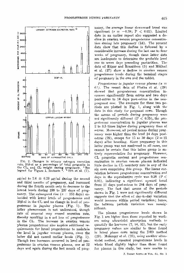
FIG. 2. Changes in urinary estrogen excretion rate, 20fl-ol as a percentage of total progestin in the CL, and CL weight during pregnancy. ~ See legend for Figure 1, footnote % b Erb et al. (7).
/~g/ml to 2.6 4- 0.29 /~g/ml during the second and third months of pregnancy, and increased during the fourth month only to decrease to the lowest levels during 199 to 237 days of preg- nancy. The subsequent rise (5 = 258 days) co- incided with lower levels of progesterone and 20fl-ol in the CL and no change in level of pro- gesterone in jugular plasma (Fig. 1). The latter phenomenon is not understood, though rate of removal may exceed secretion rate, thereby resulting in a net loss of progesterone in the CL. The increase in ovarian venous plasma progesterone also implies additional re- quirements for luteal progesterone to maintain the level in jugular venous plasma, since the latter did not exceed earlier levels (Fig. 1). Though two increases occurred in level of pro- gesterone in ovarian venous plasma, one at 52 days and again during the last month o£ preg-
nancy, the average linear downward trend was significant (r = --0.38; P < 0.01). Limited data in an earlier report also suggested a de- cline in ovarian venous progesterone eoncentra- tion during late pregnancy (14). The present data show that this decline is followed by a considerable increase during the last one to four weeks of pregnancy, though these latter data are inadequate to determine the probable level one to seven days preceding parturition. The data of Edgar and Ronaldson (5) and Mikhail et al. (27) show a decline in ovarian venous progesterone levels during the terminal stages of pregnancy in the ewe and the rabbit.
Progesterone in jugular venous plasma (n = 61). The recent data of Plotka et al. (29) showed that progesterone concentration in- creases significantly from estrus and two days post-estrus to 14 days post-estrus in the non- pregnant cow. The averages for these two pe- riods are plotted in Fig. 1, along with the data in this study for pregnant cows. Though the means of periods during pregnancy were not significantly different (P < 0.25), the pro- gesterone concentration in jugular plasma was 2 to 3.5 times higher during pregnancy than at estrus. Moreover, all period means during preg- nancy were higher than the level 14 days post- estrus (29), except for 15 to 30 days (~ = 21 days) after breeding. Since pregnancy in this latter group was not confirmed in all cases, one cannot be certain that this latter group is en- tirely representative for pregnancy. However, CL progestin content and progesterone con- centration in ovarian venous plasma indicated little decline in CL secretory rate in any of the six cows comprising this group. The linear cor- relation between progesterone concentration and days in the reproductive cycle was 0.28 (P < 0.05), indicating a significant upward trend from 11 days post-estrus to 284 days of preg- nancy. The fact that means of the periods shown in Fig. 1 were not significantly different suggests that the effect of day-to-day variation would increase within period variation; hence, the between periods variation was nonsig- nificant.
The plasma progesterone levels shown in Fig. 1 are higher than those reported by work- ers using' ultraviolet absorption methods to quantify the hormone (2, 14, 32), but the earty pregnancy values are similar to those found in luteal phase cows using the DID method (29). Melampy et al. (25), using another ultra- violet method, reported progesterone levels in whole blood slightly higher than those found for plasnm in this study, but were unable to
J . DAIlgY S C I E N C E VOIJ. 51 , NO. 3

406 ERB, E S T E R G R E E N JR., GOMES, PLOTKA, AND FROST
repeat their findings using a conventional ultra- violet method (2).
The differences found in progesterone con- centration by using different quantitative pro- cedures are difficult to understand; both the ultraviolet and DID methods appear to be adequately specific and accurate, since both give similar results when progesterone is more concentrated, as is the case for ovarian venous plasma. However, many of the earlier studies did not utilize individual sample recovery es- timates (2, 24, 32) and thus reported levels uncorrected for method losses. Furthermore, the ultraviolet method requires the extraction of large amounts of plasma (500 ml or more) compared to the DID method (10 to 20 ml), necessitating the removal of additional mate- rials which interfere with ultraviolet absorption analysis. Comparison of plasnm progesterone levels found in women by workers using various methods (16, 21, 34, 41) suggest that ultra- violet methods may give slightly lower values at very low concentrations of the hormone, but not at higher levels.
The results presented here also differ from those presented earlier, in that no decline in jugular plasma progesterone levels was found during the last month of pregnancy (Fig. 1). Short (32, 33) reported declining levels of the hormone in peripheral plasma during the last month before parturit ion. Bowerman and Melampy (2) showed an apparent decline in progesterone after 185 days of pregnancy, but data were too few to confirm this. Progesterone concentration has been reported to increase duing human pregnancy (16, 21, 33, 34).
Progestins in the CL (n = 125). The average concentration of progesterone in the CL was fair ly constant af ter the first month of preg- nancy, though both concentration and content increased to 210 days of pregnancy and then declined slightly (Fig. 1). The present data are similar to those summarized by Gomes and Erb (12) and to the data of Labhsetwar (19) and Kristoffersen (18), if the lat ter are cor- rected for procedural losses. However, they are slightly higher than the late pregnancy values reported by Kristoffersen (18) and consider- ably higher than those reported by Bowerman and Melampy (2) and Melampy et al. (25).
Though content and concentration of 20fl-ol in the CL changed significantly (P < 0.005) during the reproductive cycle, combined pro- gesterone and 20fl-ol (progestin) did not differ significantly (P < 0.25) (Fig. 1).
The content and concentration of 20fl-ol in- creased steadily during the first three months
J. DAIRY SCIENCE ~rOL. 51, NO. 3
of pregnancy and then declined and remained low for the next three months. The amounts were increased from 199 to 238 days of preg- nancy, but were low during the last month of pregnancy (Fig. 1). The means for the pe- riods shown in Fig. 1 were highly significantly different (P < 0.005). The amount of 20fi-ol in the CL averaged 21% of the CL progestin and varied from 8.5% at 11 and 14 days to 28% at 52 days (P < 0.005) (Fig. 2). Though several workers have measured 20fl-ol in the bovine CL (12), its importance in, or relation- ship to, reproductive processes is not under- stood. Levels of 20fl-ol are generally low in CL of the nonpregnant bovine during the growth phase and increase in the regressing CL (8, 12, 13, 22, 35, 36, 43). Progesterone may be converted to 20fi-ol by in vitro incubation with bovine blood (3) or by incubation of CL slices (39). Levels of 20fi-ol generally have been higher when CL were removed af ter slaughter (8, 35, 36) than when removed surgically (22, Fig. 1). Therefore, it seems possible that 20fl- ol nmy increase when synthetic activity and secretion rate of progesterone by the CL de- clines, late in the estrous cycle. These early changes in level and proport ion of 20fl-ol to CL progestin content have been histologically associated with decreasing percentages of Types I and I I lutein cells and increasing percentages of Type I I I lutein cells as defined by Foley and Greenstein (11) from 14 to 42 days of preg- nancy (11, 20, 44).
Zimbehnan et al. (44) have reported that Types I and I I lutein cells were more common at 14 days in the nonpregnant than in the pregnant animal and that Type I I I cells in- creased between 14 and 18 days of pregnancy, with further increases by 22 to 23 days and 28 days. No change was observed when 28 days was compared to 42 days of pregnancy. Zim- belman et al. (44) also stated that proportions of functional lutein cells were lower at 56 days of pregnancy, as compared to proportions re- ported by Loy ct al. (20) for 14 days after estrus in nonpregnant animals. This aspect deserves nmre careful study, since measurement of 20fi-ol in the CL would be a relatively easy procedure for assessing luteal secretory activ- ity, providing that 20fl-ol levels could be proven an adequate indicator. I f the concept that 20fl- ol content reflects CL activity is tenable, the changing concentrations of progesterone in ovarian venous plasnm also reflects CL secre- tion rate, because these concentrations increase when content and concentration of 20fl-ol decreases (Fig. 1).

P R O G E S T E R O N E D U R I N G P R E G N A N C Y 407
Weight of the CL. Although average CL weight varied somewhat during the reproduc- tive cycle, the standard error for each period overlaps the means of each of the other periods (Fig. 2). From this and other work (12, 36, 46), it is doubtful that the differences observed in CL weight during pregnancy represent more than random error due to differences among cows and age and breed of cow, at least, through the first 8 to 8.5 months of pregnancy.
Correlations. Erb eta] . (6) have shown that progestin content of the CL adequately reflects the progestin content of both ovaries, since the latter tissue contains a relatively small per- centage of the total progestin. The partial linear correlations comparing relationships be- tween CL weight and progestin levels, and ovarian and jugular venous plasma progesterone concentration are shown in Table 3.
Weight of the CL was not highly correlated with any of the hormone measurements shown in Table 3, though the relationships with con- tent of progesterone, 20fi-o], and progestin in the CL were significant (P < 0.01). Because CL weight was not highly correlated with hor- mone content, the partial correlations between content and concentration were high, being 0.87, 0.94, and 0.87, respectively, for proges- terone, 20fl-ol, and progestln in the CL (P < 0.01). Because of the predominance of pro-
gesterone in the CL, the part-whole partial correlations between its content and concentra- tion and the respective progestin measurements were 0.97 and 0.96 (P < 0.01).
From these results, it is attractive to consider measuring only CL progesterone when assess- ing luteal function. Though CL 20fl-ol is sig- nificantly correlated with CL weight (P < 0.01) and CL progesterone content (P < 0.01) and concentration (P < 0.05), the relationships are too low to satisfactorily predict 20fl-ol status in the individual CL from measurement of progesterone.
Progesterone concentration in ovarian but not jugular venous plasma was significantly correlated (P < 0.05) with CL progesterone and progestin content. No other comparisons involving the two sources of plasma proges- terone were significant (Table 3), including those using total ovary progesterone and pro- gestin content (6). The correlation between progesterone concentration in ovarian and jugu- lar venous plasma was negative (--0.16) but not significant. This relationship was expected, since progesterone concentration in jugular plasma increased slightly during pregnancy and ovarian venous plasma decreased (P < 0.01).
The degree of association between ovarian venous plasma progesterone and CL proges-
TABLE 3 Partial linear correlations between progestln measurements and weight of the corpus luteum and pro-
gesterone concentration in ovarian vein and jugular plasma
Dependent variable
Corpus ]uteum Ovaxian Independent vein
variable (1) (2) (3) (4) (5) (6) (7) (8)
(Partial linear correlation) a Corpus luteum b
(1) Weight (g) (2) Progesterone (~g) 0.28 e (3) Progesterone (~g/g) --0.16 0.87 e (4) 20fl-ol (~g) 0.31 ~ 0.32 ~ 0.17 (5) 20fl-ol (~g/g) 0.08 0.26" 0.22 f 0.94 ~ (6) Progestin (~g) 0.33 e 0.97 ~ 0.82 ~ 0.55 e 0.48 ~ (7) Progestin (~g/g) --0.12 0.85 e 0.96 ~ 0.42 e 0.48 e 0.87 ~ . .....
Ovarian vein ¢ (8) Progesterone
(~g/ml) 0.18 0.25 ~ 0.15 0.14 0.07 0.23 ~ 0.14 ...... Jugular vein d
(9) Progesterone (rig~rot) 0.09 0.11 0.05 0.11 0.10 0.12 0.07 --.167
*Breed and age held constant for corpus luteum (CL) weight; age held constant for CL proges- terone, 20fl-ol and progestin content and concentration; days of reproductive cycle held constant for ovarian and jugular vein progesterone.
b'¢'dN-~ 125, 76 and 60 pairs, respectively. e., Significant at the 1 and 5% level Of probability, respectively.
N : 49 pairs.
J. DAIRY SCIENCE VOL. 51, NO. 3

408 ERB, ESTERGREEN JR., GOMES, PLOTKA, AND FROST
terone during pregnancy is considerably less than during the estrous cycle of nonpregnant cows (13). In the latter study ovarian venous plasma progesterone was more highly correlated with the CL progcstin concentration and con- tent, being 0.55 and 0.44 (P < 0.01), respec- tively. Moreover, morphology (4, 20) weight, and progestin content of the CL (12) and progesterone in ovarian venous plasma (13, 15) and jugular plasma (12, 13, 15, 29) reflect the stages of the estrous cycle and presumably CL secretion rate.
General disc.ussion. It seems clear that the CL in the bovine is functional throughout preg- nancy (Fig. 1). Though most cows maintain pregnancy following ovariectomy after 200 to 237 days of pregnancy, gestations are generally short and the placenta is retained (10). Since these two abnornmlities can be avoided by ad- ministration of progesterone until 274 days of pregnancy (17), extra-ovarian sources of pro- gesterone during late pregnancy appear inade- quate in the ovarieetomized cow. The nature of of these sources has not been determined, but the adrenal and placenta have been implicated (12). Extra-ovarian sources of progesterone may be considerable after 200 days of preg- nancy, since the concentration of progesterone in jugular venous plasma remained high after the third month, even though the concentration in ovarian venous plasma was surprisingly low from 199 to 237 days (Fig. 1). Increased se- cretory activity by the CL during the last month of pregnancy was indicated by a greater con- ccntration of progesterone in ovarian venous plasma and considerably less 20B-ol in the CL. On the other hand, weight and levels of pro- gesterone in the CL declined after 200 days, though the period means for these measure- ments were not significantly (P < 0.25) differ- ent during the reproductive cycle (Fig. I and 2).
The most dramatic change in female sex ste- roids near parturition is the rapid increase in rate of urinary excretion of estrogens (7, 26, Fig. 2). Though it is generally believed that progesterone secretion by the CL declines dur- ing late pregnancy (2, 25, 33) in preparation for parturition, the increasing concentrations of progesterone in ovarian venous plasma and a continued high level of progesterone in jugu- lar venous plasma (Fig. 1) suggest that this may not or need not be an important change. Admittedly, the present data are inadequate to evaluate progesterone changes during the last few days preceding parturition, tIowever, urinary estrogen excretion doubles during the last month (7, Fig. 2) and shows a further
increase of 29% during the eight-hour period centered on parturition (26). Under these con- ditions estrogens could dominate progestins and function in the mechanism of parturition, even for cows with increasing luteal function during late gestation. The variability between cows relative to the indicators of female sex steroid activity during pregnancy is one of the most striking features of these and other data accumulated in our laboratories (7, 8, 12, 26, 36). Under these conditions it may be an- ticipated that further research may reveal a rather wide range of progestin and estrogen levels within which pregnancy and parturition can occur normally.
Acknowledg me.ts The authors gratefully acknowledge the as-
sistance of R. B. Harrington, Department of Ani- mal Sciences, Purdue University, for design of the IBM 7094 phases of the statistical analyses, and J. F. Bullard, School of Veterinary Medicine, Purdue University, for performing ovariectomy on 11 of the pregnant animals used in the study, and to R. D. Randel for assistance with data tabulation.
References (1) Biomedical Computer Programs. 1964. W . J .
Dixon, ed. Health Sciences Computing Facility. Dept. Preventative Medicine and Public Itealth, School of Medicine, Univ. of California, Los Angeles.
(2) Bowerman, A. M., and Melampy, R. M. 1962. Progesterone and Ad-Pregnene-20fl-ol - 3-one in Bovine Reproductive Organs and Body Fluids. Proe. Soe. Exptl. Biol. Med., 109 : 45.
(3) Coyle, M. G., and Romanoff, E. B. 1964. Transformation of 4-Cl~-Progesterone by Bo- vine Blood. (Abstract.) Federation Proc., 23 : 462.
(4) Cupps, 1 ). T., Laben, R. C., a~d Mead, S. W. 1959. Histology of the Pituitary, Adrenal, and Reproductive Organs in Normal Cattle and Cattle with Lowered Reproductive Ef- ficiency. Hilgardia, 29: 383.
(5) Edgar, D. G., and Ronaldson, J. W. 1958. Blood Levels of Progesterone in the Ewe. J. Endocrlnol., 16: 378.
(6) Erb, R. E., Estergreen, V. L., Jr., Gomes, W. R., Flotka, E. D., aa~d Frost, O. L. 1968. Progestin Content of Ovaries and the Effect on Assessment of Luteal Activ- ity in the Bovine. J. Dairy Sci., 51:411.
(7) Erb, R. E., Randel, R. D., Mellin, T. N., and Estergreen, V. L., Jr. 1968. Urinary Estrogen Excretion Rates During Preg- nancy in the Bovine. J. Dairy Sci., 51: 416.
(8) Erb, R. E., and Stormshak, F. 1961. Fro- gestins in Corpora Lutea, Ovaries and
J. DAIRY SCIElZCE VOL. 51, NO. 3

P R O G E S T E R O N E D U R I N G P R E G N A N C Y 409
Adrenals After Estrus and Breeding of Normal and Abnormal Cows. J. Dairy Sei., 44: 888.
(9) Estergreen, V. L., Jr . 1966. Effect of 20fl- Hydroxy--~4-pregnene-3-one on Maintenance of Pregnancy in Ovariectomized Cows. (Abstract .) J. Dairy Sci., 49: 732.
(10) Estergreen, V. L., Jr., Frost, O. L., Gomes, W. R., Erb, R. E., and Bullaxd, J. F. 1967. The Effect of Ovariectomy on Preg- nancy Maintenance and Par tur i t ion in Dairy Cows. J. Dairy Sci., 50:1293.
(11) Foley, R. C., and Greenstein, J. S. 1958. Cytological Changes in the Bovine Corpus Luteum During Early Pregnancy. Proc. I I I r d Symposium Reproduction and Infer- tility, pp. 89-96. P. X. Gassner, ed. Per- gamon Press, London.
(12) Comes, W. R., and Erb, R. E. 1965. Pro- gesterone in Bovine Reproduction: A Re- view. J. Dairy Sei., 48: 314.
(13) Domes, W. R., Estergreen, V. L., Jr., Frost, O. L., and Erb, R. E. 1963. Progestin Levels in Jugular and Ovaa-ian Venous Blood, Corpora Lutea, and Ovaries of the Nonpregnant Bovine. J. Dairy Sei., 46: 553.
(14) Comes, W. R., Frost, O. L., and Estergreen, V. L., Jr . 1962. Progestins in Ovarian and Peripheral Blood of Cows During Late Pregnancy. (Abstract . ) J . Dairy Sol., 45: 670.
(15) Comes, W R., Hersehler, R. C., and Erb, R. E. 1965. Progesterone Levels in Ovarian Venous Effluent of the Nonpregnant Sow. J. Anlm. Sci., 24: 722.
(16) Grieg, M., Coyle, M. G., Cooper, W., and Walker, J . 1962. Plasma Progestero~m in the Mother and Foetus in the Second Hal f of Human Pregnancy. J. Obstet. Gynaecol. Brit. Cmwlth., 69: 772.
(17) Johnson, K. R., and Erb, R. E. 1962. Main- tenance of Pregnancy in Ovariectomized Cattle with Progest in Compounds and Their Effect on Progestin Levels in the Corpus Luteum. J. Dairy Sei., 45: 633.
(18) Kristoffersen, J. 1960. Gestogens in Corpus Luteum of Cattle. Acta Endocrinol., 33 : 417.
(19) Labhsetwar, A. P., Collins, W. E., Tyler, W. J., and Casida, L. E. 1964. Some Pitui- tary-OvariaJ~ l~elationships in the Peri- par tur ient Cow. J. t~eprod. Fertil. , 8:85.
(20) Loy, R. G., Zimbclman, R. G., and Casida, L. E. 1960 Effects of Injected Ovarian Hormones on the Corpus Luteum of the Estrual Cycle in Cattle. J. Anita. SCl., 19: 175.
(21) Lurie, A. O., Vi]lee, C. A., and Reid, D. E. 1966. Progesterone in the Blood. A Qugnti- tative Method Employing Gas Liquid Chro- matography. J. Clin. Endocrinol. Metab., 26:742.
(22) Mares, S. E., Zimbelman, R. G., and Caslda, L. E. 1962. Variations in Progesterone Con-
tent of the Corpus Luteum of the Estrual Cycle. J. Anita. Sci., 21: 266.
(23) Masuda, H., Anderson, L. L., Henricks, D. M., and Melampy, R. M. 1967. Pro- gesterone in Ovarian Venous Plasma and Corpora Lntea of the Pig. Endocrinology, 80 : 240.
(24) McCracken, J. A. 1964. Progesterone in the Body F a t of the Dairy Cow. J. En- docrinol., 28: 339.
(25) Melampy, R. M., Hearn, W. R., and Rakes, J. M. 1959. Progesterone Content of Bo- vine Reproductive Organs and Blood Dur- ing Fregnaucy. J. Anim. Sci., 18:307.
(26) Mcllin, T. N., Erb, R. E., and Estergreen, V. L., Jr . 1966. Urinary Excretion of Estrogen by the Bovine Before and After Parturi t ion. J. Anim. Sei., 25: 955.
(27) Mikhail, J., NoaH, M. W., and Allen, W. M. 1961. Progesterone Levels in the Rabbi t Ovarian Vein Blood Throughout Preg- nancy. Endocrinology, 69: 504.
(28) Ostle, B. 1963. Statistics in Research. 2ud ed. Tile Iowa. State University Press, Ames.
(29) P]otka, E. D., Erb, R. E., Callahan, C. J., and Comes, W. R. 1967. Levels of Pro- gesterone in Peripheral Blood Plasma Dur- ing the Estrous Cycle of the Bovine. J . Dairy Sci., 50: 1158.
(30) Plotka, E. D., Stant, E. G., Jr.. Waltz, F. A., Garwood, V. A., and Erb, R. E. 1966. A Computer Program for Double Ra.dionu- elide Assay Data. Int . J. Appl. Radiat ion and Isotopes, 17: 637.
(31) Savard, K., and Telegdy, G. 1965. Steroid Formation in the Bovine Corpus Luteum. Steroids, Suppl., 2: 205.
(32) Short, R. V. 1958. Progesterone in Blood. IL Progesterone in the Peripheral Blood of Pregnant Cows. J. Endocrino]., 16: 426.
(33) Short, R. V. 1960. Blood Progesterone Levels in Relation to Parturi t ion. d. Reprod. Fertil. , 1: 61.
(34) Short, R. V., and Levitt , L 1962. The F]uorometric Determination of Progesterone in Human Plasma During Pregnancy and the Menstrual Cycle. J. Endocrfiml., 25: 239.
(35) Staples, R. E., and Hansel, W. 1961. Luteal Funct ion and Embryo Survival in the Bo- vine. J. Dairy Sci., 44:2040.
(36) Stormshak, F., and Erb, R. E. 1961. Pro- gestins in Bovine Corpora Lutea, Ovaries, and Adrenals During Pregnancy. J. Dairy Sci., 44: 310.
(37) Stormshsk, F., Hunt, M. L., and Erb, R. E. 1961. Qua~xtit~tive Methods for Deter- mining Progesterone in Corpora Lutea, Ovaries, and Adrenals of the Cow and Sow Using Progesterone-4-C *~ as a Measure of Recovery of Extracted Hormone. J. Dairy Sei., 44: 300.
J, DAII~¥ SCIENCE VOL. 51, NO. 3
410 ERB, ESTERGREEN JR.. GOMES, PLOTKA, AND FROST
(38) Stormshak, F., Inskcep, E. I(., Lynn, J. E., Pope, A. L., and Casida, L. E. 1963. Pro- gesterone Levels in Corpora Lutea and Ovarian Effluent Blood of the Ewe. J . Anita. Sci., 22: 1021.
(39) Sweat, M. L., Berliner, D. L., Bryson, M. J., Nabors, C., Jr., Haskell, J., and Holmstrom, E G. 1960. The Synthesis and Metabolism of Progesterone in the Human and Ovine Ovary. Biochim. et Biophys. Acta, 40: 289.
(40) Tanabe, T. Y. 1966. Essentiality of the Corpus Luteum for Maintenance of Preg- nancy in Dairy Cows. (Abstract.) J. Dairy Sci., 49: 731.
(41) Woolever, C. A. 1963. Daily Plasma Pro- gesterone Levels During the Menstrual Cycle. Am. J. Obstet. Gynecol., 81: 981.
(42) Woolever, C. A., and Goldfein, A. 1963. A Double-Isotope Derivative Method for Plus-
ma Progesterone Assay. Int. J. Appl. Ra- diation and Isotopes, 14: 163.
(43) Zimbelman, R. G., Loy, R. G., and Casida, L. E. 1959. Biochemical Studies of the Bovine Corpus Luteum of Early Pregnancy. J. Anim. Sei., 18: 1551.
(44) Zimbelman, R. G., Loy, R. G., and Casida., L. E. 1961. Variation in Some Biochemical and Histological Characteristics of Bovine Corpora Lutea During Early Pregnancy. J. Anita. Sci., 20: 99.
(45) Zimbehnan, R. G., Loy, R. G., and Casida, L. E. 1961. Effect of Exogenous Proges- terone on the Bovine Corpus Luteum of Early Pregnancy. J. Anim. Sci., 20: 106.
(46) Zimbelman, R. G., McShan, W. I~., Tyler, W. J., and Casida, L. E. 1961. Effect of a Pituitary Extract on the Bovine Corpus Luteum of Late Pregnancy. J. Anita. Sci., 20 : 246.
J. DAIRY SOIENCE VOL. 51, NO. 3