Embed Size (px)
Citation preview
— 1 —
EUROIMMUNM e d i z i n i s c h eL a b o r d i a g n o s t i k aA G
Product Cataloguediagnostics for the determination of Autoantibodies,
for Infectious Serology and Allergology
Indirect Immunofl uorescence — ELISA — RIA — WesternblotEUROASSAY — EUROLINE — EUROPLUS — EUROArray
2011
EU
RO
IMM
UN
P
rod
uct
Cat
alo
gu
e �0
11

— � —
EUROIMMUNM e d i z i n i s c h eL a b o r d i a g n o s t i k aA G
Table of Contents
EUROIMMUN – Company Profile ...............................................................................................................................................3
Techniques for the Serological Investigation of Antibodies .....................................................................................................5 Indirect Immunofluorescence: An Easy and Modern Method .......................................................................................................................... 6 BIOCHIP Mosaics™ .............................................................................................................................................................................................. 9 The Indirect Immunofluorescence Test, Performed Using the TITERPLANE™ Technique ........................................................................... 10 Recommended Serum Dilutions for Indirect Immunofluorescence ............................................................................................................... 1� Diagnostically Relevant Systemic Autoantibodies ........................................................................................................................................... 16 Organ-/Tissue-Specific Autoantibodies ............................................................................................................................................................. 17 Antibodies for Infectious Serology .................................................................................................................................................................... 18 Antibodies for Allergology ................................................................................................................................................................................. 19 EUROIMMUN Microplate ELISA ........................................................................................................................................................................ 18 ELISA Automation using the EUROIMMUN Analyzer I ................................................................................................................................... �� Incubating the Microplate ELISA ....................................................................................................................................................................... �3 EUROASSAY: Line Blots in TITERPLANE™Technique Format ....................................................................................................................... �4 Incubating the EUROASSAY (TITERPLANE™Technique) ................................................................................................................................ �5 The EUROLINE: A New Technique for Extensive Antibody Profiles .............................................................................................................. �6 EUROLINE Automation Using EUROBlotMaster and EUROLineScan ............................................................................................................ �7 Westernblots/EUROLINE-WB: Reliable Differentiation of Antibodies Present ............................................................................................... �8 Incubating the EUROLINE/Westernblot/EUROLINE-WB ................................................................................................................................... �9 EUROIMMUN Radioimmunoassays (RIA/IRMA) .............................................................................................................................................. 30
EUROIMMUN Products for the Determination of Autoantibodies ...........................................................................................31 Autoantibodies against Cell Nuclei (ANA) ........................................................................................................................................................ 3� Autoantibodies against Double-Stranded DNA (dsDNA) ................................................................................................................................ 35 Autoantibodies against CCP and Sa .................................................................................................................................................................. 36 Autoantibodies against Mitochondria (AMA) ................................................................................................................................................... 37 Autoantibodies against Liver Antigens ............................................................................................................................................................. 38 Autoantibodies against Thyroid Gland Antigens / Antigen Detections .......................................................................................................... 40 Autoantikörper against Antigens of the Skin ................................................................................................................................................... 41 Autoantibodies against Neuronal Antigens ...................................................................................................................................................... 4� Autoantibodies against Islet Cell Antigens ....................................................................................................................................................... 44 Autoantibodies against Parietal Cells (PCA) ..................................................................................................................................................... 45 Autoantibodies against Granulocyte Cytoplasm (cANCA/pANCA) ................................................................................................................. 46 Antibodies against Endomysium and Gliadin .................................................................................................................................................. 48
EUROIMMUN Products for Infectious Serology ......................................................................................................................49 Antibodies against Borrelia ................................................................................................................................................................................ 50 Antibodies against Epstein-Barr Virus (EBV) .................................................................................................................................................... 5� Antibodies against Helicobacter Pylori ............................................................................................................................................................. 54 Antibodies against Herpes Simplex Virus (HSV) ............................................................................................................................................. 55 Antibodies against Chlamydia ........................................................................................................................................................................... 56 Antibodies against Emerging Viruses ............................................................................................................................................................... 57 BIOCHIP Mosaics™ for Infectious Serology ..................................................................................................................................................... 58 Additional Reagents for the Determination of Acute Infections ..................................................................................................................... 60 EUROIMMUN Products for Allergology ...................................................................................................................................61
Order Information and Product Data .......................................................................................................................................64 Fluorescence-Labelled Antibodies: Fluorescein (FITC) for EUROIMMUN IIFT ............................................................................................... 65 Controls for EUROIMMUN IIFT: Organ-Specific Autoantibodies .................................................................................................................... 66 Controls for EUROIMMUN IIFT: Systemic Autoantibodies ............................................................................................................................. 68 Controls for EUROIMMUN IIFT: Infectious Serology ....................................................................................................................................... 70 Controls for EUROIMMUN IIFT: Determination of Further Antibodies .......................................................................................................... 75 Controls for EUROIMMUN Westernblots/EUROLINE-WB................................................................................................................................ 76 EUROASSAY for the Determination of Autoantibodies (Test Systems) ........................................................................................................ 78 EUROASSAY for Allergology (Test Systems) ................................................................................................................................................... 79 EUROLINE for the Determination of Autoantibodies (Test Systems) ............................................................................................................. 80 EUROLINE for Infectious Serology (Test Systems) .......................................................................................................................................... 81 EUROLINE for Allergology (Test Systems) ....................................................................................................................................................... 81 Westernblot/EUROLINE-WB for the Determination of Autoantibodies (Test Systems) ................................................................................ 83 Westernblot/EUROLINE-WB for Infectious Serology (Test Systems) ............................................................................................................. 84 Microplate ELISA for the Determination of Autoantibodies (Test Systems) ................................................................................................. 86 Latex Agglutination tests for the Determination of Autoantibodies (Test Systems) .................................................................................... 89 EUROArray for Molecular Genetic Determinations (Test Systems) ............................................................................................................... 89 Radioimmunoassay (RIA) for the Determination of Autoantibodies / Autoantigens / Hormone Determination (Test Systems) .............. 89 Microplate ELISA for Infectious Serology (Test Systems) .............................................................................................................................. 91 Microplate ELISA for the Determination of Antibodies against Other Antigens (Test Systems) ................................................................. 96 Microplate ELISA for the Determination of Hormones and Proteins (Test Systems) ................................................................................... 96 Allercoat™ 6 System .......................................................................................................................................................................................... 97 Diagnostics for Indirect Immunofluorescence: Organ-Specific Autoantibodies ..........................................................................................117 Diagnostics for Indirect Immunofluorescence: Systemic Autoantibodies ...................................................................................................1�7 Diagnostics for Indirect Immunofluorescence: Infectious Serology .............................................................................................................135 Diagnostics for Indirect Immunofluorescence: Other Antigens ....................................................................................................................149 Further Reagents for EUROIMMUN IIFT ..........................................................................................................................................................149 Other Items for EUROIMMUN IIFT ...................................................................................................................................................................150 General Delivery Conditions .............................................................................................................................................................................151
Index ......................................................................................................................................................................................152

— 3 —
EUROIMMUNM e d i z i n i s c h eL a b o r d i a g n o s t i k aA G
EUROIMMUN Ag – COMPANY PROfILE
EUROIMMUN was founded in September 1987 and today has its headquarters in Luebeck, Germany. Branches are situated in groß groenau near Luebeck (Schleswig-Holstein), in dassow (Mecklenburg-Western Pomerania), Rennersdorf (Upper Lusatia, Saxony), and in Pegnitz (Upper Franconia, Bavaria). Further EUROIMMUN subsidiaries can be found in Canada (Mississauga), China (Beijing, Hangzhou), great britain (Pontypool in Wales), Italy (Padua), Lebanon (Bei rut), Poland (Wroc law), Switzerland (Lucerne), Singapore, South Africa (Cape town), Turkey (Istanbul) and the USA (New Jersey). At present EUROIMMUN has 775 employees in Germany, 990 worldwide. The company is ISO-certified (EN ISO 9001:�008, EN ISO 13485:�003/CMDCAS).
EUROIMMUN produces reagents for medical laboratory diagnostics. In the foreground are test systems for the determination of various antibodies in patient serum in the diagnosis of autoimmune diseases, infectious diseases and allergies.
The test methods employed are predominantly indirect immunofluorescence, microplate ELISA, various blot techniques (Westernblot, EUROASSAY, EUROLINE, EUROLINEWb) and all molecular biology techniques. The company is based on worldwide-patented state-of-the-art production methods and microanalysis techniques and is one of the world’s leading manu facturers of medical laboratory diagnostics.
The bIOChIPs are one of EUROIMMUN’s many inventions: paper-thin sheets of glass are coated with cells or tissue sections and then cut automatically into millimetre-sized fragments which are subsequently glued onto slides using a fully automated device. This bIOChIP technology allows extreme miniaturization and standardization of immun bio chemical analyses. With bIOChIP Mosaics made from 30 or more different organ sections, cell substrates or defined antigens (EUROPLUS) only mini mum incubation efforts are necessary to obtain a detailed antibody profile.
In EUROIMMUN enzyme immunoassays (ELISA) defined antigens, purified using state-of-the-art bio-technological processes, are employed as the antigen substrate. Some of these antigens are synthesized in the company’s molecular biology laboratories. EUROIMMUN ELISA are characterized by their excellent stability, simple handling and short incubation times, and they are ideal for automated use. All reagents are delivered ready-to-use and are exchangeable between different lots. EUROIMMUN offers the largest and most differentiated arsenal of enzyme immunoassays worldwide for the diagnosis of autoimmune and infectious diseases.
Company headquarters Luebeck Company branch dassow
Company branch Pegnitz Company branch Rennersdorf
Company site groß groenau
EUROIMMUN worldwide

— 4 —
EUROIMMUNM e d i z i n i s c h eL a b o r d i a g n o s t i k aA G
The innovative procedures EUROASSAY and EUROLINE developed by EUROIMMUN follow the same test principle as the ELISA methods, with the use of the BIOCHIP technology. At EUROIMMUN purified antigens are printed in parallel lines at defined positions on membrane strips. Following incubation, users can evaluate results visually without additional equipment. These reagents allow in particular the differentiation of antibodies that are not clearly defined microscopically by indirect immunofluorescence. EUROASSAY and EUROLINE are also employed in laboratories where no sophisticated laboratory instruments are available.
EUROIMMUN produces an extensive range of Westernblot strip test systems and corresponding reagents for confirmation of positive fluorescence and ELISA results as well as clarification of difficult-to-interpret results in autoimmune diagnostics, infectious serology and allergology. Refined electrophoretical processes have been developed to allow the precise separation of diagnostically relevant proteins from one another. Lot-specific evaluation templates are produced for the evaluation of band patterns. The program ”EUROLineScan“ enables fully automated evaluation of membrane-based test systems and simplifies the archiving of results with large sample series.
One of the company’s main strengths is its technical expertise. This encompasses not just the manufacture and sale of medical laboratory diagnostics, but also the diagnostic application of the products in a reference laboratory which provides highly differential diagnostics. This reference laboratory has set standards in Germany, and worldwide is unequalled in the whole field of autoimmune diagnostics. The diagnostic spectrum of the laboratory also covers the areas of infectious serology and serological allergy diagnostics. The reference laboratory receives hundreds of serum samples daily from all over Germany as well as from many other countries. It helps EUROIMMUN customers to secure their results: a large proportion of serum samples sent to EUROIMMUN for evaluation are analysed free of charge in order to maintain high standards in the laboratories of EUROIMMUN customers. Customers can obtain further technical information from experienced scientists in the company, with whom they can also discuss serological problem cases. The “Institute for Quality Assurance”, an institution newly founded by EUROIMMUN, organises unbiased quality assessments and provides advice in the area of quality management. Moreover EUROIMMUN has established the “Institute for experimental Immunology”, which is engaged in basic research.
In October �010, the EUROIMMUN workforce included 150 university and college graduates, among these biologists, biochemists, chemists, engineers and medical doctors (48 of them holding a doctor’s degree). Medical technicians are particularly strongly represented with 103 people, corresponding to EUROIMMUN’s activities, as well as biology/chemistry laboratory technicians (81). At present the company is training 55 young people as biology laboratory assistants, industrial clerks, IT specialists, electronic system technicians, electronic technicians for devices and systems, industrial mechanics, lathe operators and cooks as well as business information technology specialists and business economists (dual system). At EUROIMMUN great value is placed on advising customers and prospective customers in a factual, technical and commercially restrained manner and fully supporting them in the use of our diagnostically demanding products. EUROIMMUN products are backed by an energetic and competent sales force, qualified information material, didactic test instructions and scientifically based, but nevertheless understandable advertisements in technical journals. Advertising material is produced in-house using the latest desktop publishing methods, right up until the fully digitalised ready-for-exposure documents. The most important publications and posters are translated into many languages. EUROIMMUN has set up an informative homepage on the internet (www.euroimmun.com) which is visited extensively internationally.
Over 3,000 laboratories worldwide use EUROIMMUN diagnostics. 400 of these are in Germany. The company’s development is shaped by continuous growth. Although the diagnostic market in Germany stagnated and has become particularly strongly competitive, EUROIMMUN has been able to continue its strong expansion. The company is achieving an ever increasing independence from the German market, since more and more products are sold abroad. With their quality and standardization, EUROIMMUN products are capturing the leading position in the world.
— 5 —
EUROIMMUNM e d i z i n i s c h eL a b o r d i a g n o s t i k aA G
Tec
hn
iqu
es f
or
the
Ser
olo
gic
alIn
vest
igat
ion
of
An
tib
od
ies
TEChNIqUES fOR ThE SEROLOgICAL INvESTIgATION Of ANTIbOdIES

— 6 —
FITC
FITC
EUROIMMUNM e d i z i n i s c h eL a b o r d i a g n o s t i k aA G
Tech
niq
ues fo
r the S
erolo
gical
Investig
ation
of A
ntib
od
ies
cell with antigen
specific human antibody
FITC-labelled anti-human antibody
Principle of the Test• For the determination of autoantibodies or antibodies against infectious agents,
cells, tissue sections or purified, biochemically characterized substances are used as antigen substrates.
• If the sample is positive, specific antibodies in the diluted serum sample attach to the antigens coupled to a solid phase.
• In a second step, the attached antibodies are stained with fluorescein-labelled anti-human antibodies and visualized with the fluorescence microscope.
• Positive samples can be titrated in steps. The most suitable titration interval is provided by the dilution factor 3.16� (square root of 10). In this way, every second step represents in its denominator an integral power of 10 (1 : 10, 1 : 3�, 1 : 100, 1 : 3�0, 1 : 1000, 1 : 3�00, 1 : 10000 etc.).
Indirect Immunofluorescence: A Standardized Technique for the Determination of Autoantibodies and Antibodies against Infectious Agents• High specificity: positive and negative samples produce a large difference in
signal strength. Each bound antibody shows a typical fluorescence pattern depending on the location of the individual antigens.
• The entire antigen spectrum of the original subtrate is available, thus allowing the detection of a large number of antibodies and achieving a higher detection rate.
• Immunofluorescence enables simultaneous detection of antibodies against several biochemically different antigens on one single biological substrate.
• The indirect immunofluorescence test is the analytical method of choice when it would be too difficult or too complicated to prepare the test antigens indi-vidually for enzyme immunoassays.
Pattern homog. Anti-dsDNA? Anti-Histones?
EUROIMMUN’s Innovations for the Standardization and Modern-ization of Indirect Immunofluorescence • Activation technique: physically or chemically activated cover glasses are coated
with cultured cells or tissue sections. Frozen tissue sections are fixed to the glass surface by covalent bonding, increasing adhesion more than 100 times and thus preventing the substrates from being detached.
• BIOCHIP Technology: cover glasses coated with biological substrates are cut into millimetre-sized fragments (BIOCHIPs) on a machine. This makes it possible to obtain ten or more first-class preparations of homogeneous quality per tissue section, in the case of cultured cell substrates even several thousands.
• BIOCHIP Mosaics™: using several BIOCHIPs coated with different substrates side by side on one and the same reaction field, antibodies against various organs or infectious agents can be investigated simultaneously. Detailed anti-body profiles can thus be established with comparatively little effort, allowing the reciprocal determination of the results on different substrates.
• TITERPLANE™ Technique: samples or reagents are applied to the reaction fields of a reagent tray. The BIOCHIP Slides are then placed into the recesses of the reagent tray, where all BIOCHIPs come into contact with the fluids, and the individual reactions commence simultaneously. As the fluids are confined in a closed space, there is no need for the use of a conventional „humidity chamber“.
Pattern fine-granular. Anti-SS-A? Anti-SS-B?
Pattern nucleolar. Anti-PM-Scl?
Pattern cytoplasmic. AMA M�?
Differentiation of antibodies using HEp-� cells.
Indirect Immunofluorescence: An Easy and Modern Method
tissue sections
antigen dots
culture cells
transf. cells

— 7 —
EUROIMMUNM e d i z i n i s c h eL a b o r d i a g n o s t i k aA G
Tec
hn
iqu
es f
or
the
Ser
olo
gic
alIn
vest
igat
ion
of
An
tib
od
ies
Indirect Immunofluorescence: An Easy and Modern Method
Chemically Activated Cover Glasses for Histochemistry• For diagnostics of organ-specific or tissue-specific autoantibodies frozen tissue
sections of various organs are used. However, formerly, the morphology of tissues suffered during incubation in aqueous medium, tissue parts occasionally became detached from slides, and the interpretation of results was difficult.
• Using the activation technique for the first time in histology, we have applied solid phase techniques. Firstly, the surface of cover glasses is coated with spontaneously reactive aldehyde groups. In a second step, the tissue sections are applied to the chemically activated cover glasses (Stöcker, W: European Patent No. 0 117 262; U.S. Patent No. 4,647,543). Free amino groups of the tissue sections, especially of the hydroxylysine contained in the collagen, bind to the carrier material by covalent bonding.
• This results in an increased adhesion of frozen tissue sections more than a hundredfold and prevents them from being detached during incubation. Furthermore, in some cases the activation technique results in a significantly better conservation of tissue structures, especially in organs which previously exhibited a generally low level of adhesion. Therefore, the tests can be evaluated with considerably greater confidence.
Fixation of frozen tissue sections to glass surfaces by covalent bonding.
Determination of Low-Avidity Antibodies• An alternative principle for the serological diagnosis of fresh infections has been
established by investigating the antibody avidity.
• The first reaction of the immune system following an infection is the formation of low-avidity antibodies. As the infection proceeds, increasingly antigen-adapted IgG is formed, and avidity grows. As long as high-avidity IgG is not yet detected in the serum, it can be assumed that the infection is still in an early stage.
• To identify low-avidity antibodies in a patient’s serum, two immunofluorescence tests are performed in parallel: one test is carried out in the conventional way, the other one includes urea treatment between incubations with patient’s serum and peroxidase-labelled anti-human IgG, resulting in the detachment of low-avidity antibodies from the antigens.
• Low-avidity antibodies are present if the fluorescence intensity is significantly reduced (two intensity levels ore more) by urea treatment.
• The following test kits for avidity determination are available: Toxoplasma gondii, Rubella virus, West-Nile virus, CMV, EBV-EA, EBV-CA.
high-avide Ab against EBV-CA
low-avide Ab against EBV-CA
without urea with urea
Si
(Ch2)3
Nh
(Ch2)2
N
(Ch2)3
hC
hC
N
frozen tissue section
O
OSi
(Ch2)3
Nh
(Ch2)2
N
(Ch2)3
hC
hC O
Nh2
frozen tissue section
O
OSi
(Ch2)3
Nh
(Ch2)2
Nh2
(Ch2)3
hC O
hC O
glutardialdehyde
O
O
Si
(Ch2)3
Nh
(Ch2)2
Nh2
OCh3
OCh3
h3CO
glass
Aminoethylaminoproyl
trimethoxysilane
Oh
h2O 3 Ch3Oh h2O
EUROPLUS™ System: Combination of conventional immuno-fluorescence substrates and monospecific tests• In EUROPLUS™ immunofluorescence tests antibody detection is performed using
both tissue sections/cell substrates and monospecifically reacting antigens.
• Antibodies detected in IFT screening tests can therefore be differentiated or confirmed with one and the same reaction field. In some cases, the antigens help to extend the antigen spectrum, offering a wider range for screening.
• BIOCHIPs coated with purified or recombinant antigens are used as monospecific substrates.
• In case of a positive result the antigens fluoresce green in defined areas under the microscope.
• In some EUROPLUS™ test systems several different antigens are coated on one BIOCHIP in separate antigen rows. In this manner, several monospecific analyses can be performed using a single BIOCHIP.
• Available EUROPLUS™ substrates: MPO, PR3, gliadin GAF-3X, OspC, VlsE, Plasmodium falciparum und vivax, gp1�5, p19.
EUROPLUS™: BIOCHIP combination of tissue sections / cell substrates (left) and purified antigens (right).
— 8 —
EUROIMMUNM e d i z i n i s c h eL a b o r d i a g n o s t i k aA G
Tech
niq
ues fo
r the S
erolo
gical
Investig
ation
of A
ntib
od
ies
Indirect Immunofluorescence: An Easy and Modern Method
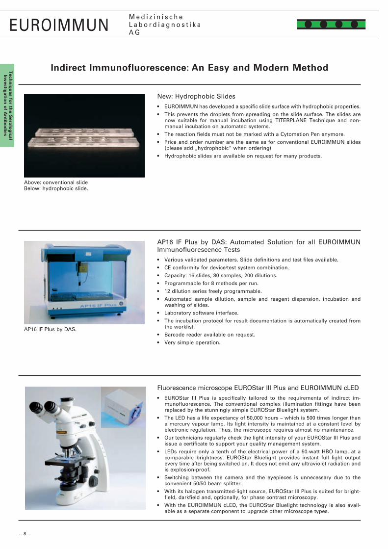
New: Hydrophobic Slides• EUROIMMUN has developed a specific slide surface with hydrophobic properties.
• This prevents the droplets from spreading on the slide surface. The slides are now suitable for manual incubation using TITERPLANE Technique and non-manual incubation on automated systems.
• The reaction fields must not be marked with a Cytomation Pen anymore.
• Price and order number are the same as for conventional EUROIMMUN slides (please add „hydrophobic“ when ordering)
• Hydrophobic slides are available on request for many products.
AP16 IF Plus by DAS: Automated Solution for all EUROIMMUN Immunofluorescence Tests• Various validated parameters. Slide definitions and test files available.
• CE conformity for device/test system combination.
• Capacity: 16 slides, 80 samples, �00 dilutions.
• Programmable for 8 methods per run.
• 1� dilution series freely programmable.
• Automated sample dilution, sample and reagent dispension, incubation and washing of slides.
• Laboratory software interface.
• The incubation protocol for result documentation is automatically created from the worklist.
• Barcode reader available on request.
• Very simple operation.
Fluorescence microscope EUROStar III Plus and EUROIMMUN cLED• EUROStar III Plus is specifically tailored to the requirements of indirect im-
munofluorescence. The conventional complex illumination fittings have been replaced by the stunningly simple EUROStar Bluelight system.
• The LED has a life expectancy of 50,000 hours – which is 500 times longer than a mercury vapour lamp. Its light intensity is maintained at a constant level by electronic regulation. Thus, the microscope requires almost no maintenance.
• Our technicians regularly check the light intensity of your EUROStar III Plus and issue a certificate to support your quality management system.
• LEDs require only a tenth of the electrical power of a 50-watt HBO lamp, at a comparable brightness. EUROStar Bluelight provides instant full light output every time after being switched on. It does not emit any ultraviolet radiation and is explosion-proof.
• Switching between the camera and the eyepieces is unnecessary due to the convenient 50/50 beam splitter.
• With its halogen transmitted-light source, EUROStar III Plus is suited for bright-field, darkfield and, optionally, for phase contrast microscopy.
• With the EUROIMMUN cLED, the EUROStar Bluelight technology is also avail-able as a separate component to upgrade other microscope types.
Above: conventional slideBelow: hydrophobic slide.
AP16 IF Plus by DAS.

— 9 —
EUROIMMUNM e d i z i n i s c h eL a b o r d i a g n o s t i k aA G
Tec
hn
iqu
es f
or
the
Ser
olo
gic
alIn
vest
igat
ion
of
An
tib
od
ies
bIOChIP Mosaics™
Slides for indirect immunofluorescence can be produced with single substrates or bIOChIP Mosaics™ from up to 45 different substrates according to your individual requirements: thyroid gland, parathyroid gland, pancreas, adrenal gland, ovary, placenta*, testis, spermatozoa, pituitary gland, hypothalamus*, hippocampus, cerebrum, cerebellum, brain stem, pons*, lobus temporalis, substantia nigra*, peripheral nerve, spinal cord, optic nerve (AQP-4, NMO IgG) eye, granulocytes (fixed with EOH, HCHO or MOH), DNA-bound lactoferrin, lymphocytes, monocytes*, thrombocytes, kidney (primate, rat, mouse), lung*, liver (primate, rat, mouse), mouth mucosa, stomach (corpus, antrum), jejunum, colon, intestinal goblet cells, umbilical cord, mamma, lacrimal gland, parotid gland, prostate, vesicula seminalis, skeletal muscle, F-actin (VSM47), heart muscle, thymus, lipocytes, cartilage*, epidermis, oesophagus (primate, rat), tongue, lip, melanocytes, HEp-� cells, HEp-�0-10 cells, HUVEC, Crithidia luciliae sensitive etc. Adenovirus, Afipia felis*, Bartonella henselae, B. quintana, Bordetella parapertussis, B. pertussis, Borrelia afzelii, B. burgdorferi sensu stricto (strains CH, USA), B. garinii, Campylobacter coli*, C. jejuni, Candida albicans, C. glabrata*, C. krusei*, C. parapsilosis*, C. tropicalis*, Chikungunya virus, Chlamydia pneumoniae, C. trachomatis, C. psittaci, CMV, Coxsackievirus (A7, A9, A16, A�4, B1 to B6), Dengue virus type 1 to 4, EBV-CA, EBNA, EBV-EA, Echinococcus granulosus, ECHO virus, Hantavirus, Haemophilus influenzae*, Helicobacter pylori, HHV-6, HSV-1, HSV-�, Influenza virus A (strains H3N�, H1N1, H5N1), Influenza virus B, Japanese encephalitis virus, Klebsiella pneumoniae*, Legionella bozemanii*, L. dumoffii*, L. gormanii*, L. jordanis*, L. longbeachae, L. micdadei*, L. pneumophila (serotypes 1 to 14), Leishmania donovani, Listeria monocytogenes (1/�a and 4b)*, measles virus, mumps virus, Mycoplasma hominis, M. pneumoniae, Parainfluenza virus type 1 to 4, Rift valley fever virus*, RSV, rubella virus, Saccharomyces cerevisiae, SARS-CoV, Sindbis virus, TBE virus, TO.R.C.H. profile, Toxoplasma gondii, Treponema pallidum, T. phagedaenis, Ureaplasma urealyticum, VZV, West Nile virus, Yellow fever virus, Yersinia enterocolitica (O:3, O:4, O:6 and O:9)*. EUROPLUS: HEp-�/liver + RNP/Sm, Sm, SS-A, SS-B, Scl-70, rib. P-proteins, Jo-1; granulocytes + MPO, PR3; primate stomach (parietal cells) + intrinsic factor; primate liver (endomysium) + gliadin (GAF-3X); rat kidney + AMA M�; thyroid gland + thyroglobulin; renal glomeruli and tubules + GBM + PLA�R; BP180 (NC16A-4X); Borrelia burgdorferi and afzelii + OspC and VlsE; EBV-CA + gp1�5 + p19; Plasmodium falciparum (HRP-�, MSP-�); P. vivax (MSP, CSP). Transfected cells: rPAg 1 + � (pancreas antigen 1 + �), AQP-4, glutamate receptor (type NMDA, AMPA), Caspr�, Lgi1, AMPA, GABABR, PLA�R, desmoglein 1 + 3, BP�30 gc, Crimean Congo fever virus (GPC, N). * Currently not available in the European Union.

— 10 —
EUROIMMUNM e d i z i n i s c h eL a b o r d i a g n o s t i k aA G
Tech
niq
ues fo
r the S
erolo
gical
Investig
ation
of A
ntib
od
ies
(Reaction fields 5 x 5 mm)
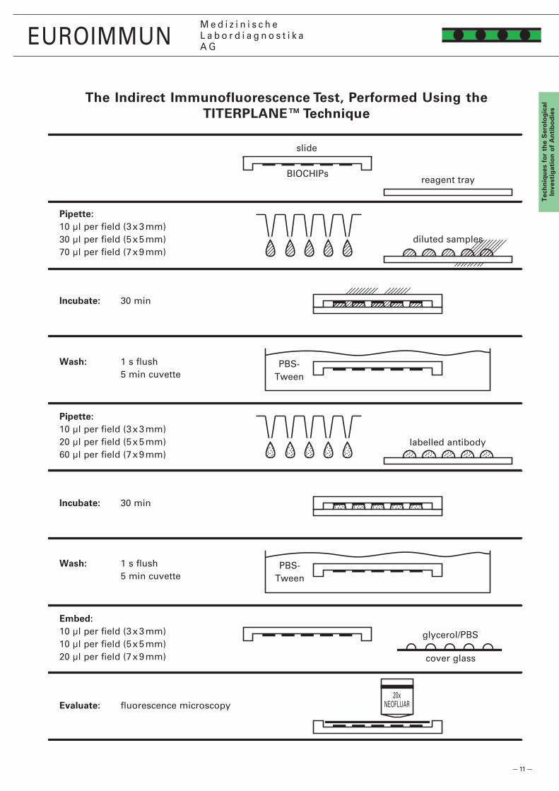
The TITERPLANE™ Technique was developed by EUROIMMUN in order to standardize immunological analyses: Samples or labelled antibodies are applied to the reaction fields of a reagent tray. The bIOChIP Slides are then placed into the recesses of the reagent tray, where all BIOCHIPs of the slide come into contact with the fluids, and the individual reactions commence simultaneously. Position and height of the droplets are exactly defined by the geometry of the system. As the fluids are confined in a closed space, there is no need for the use of a conventional ”humidity chamber”. It is possible to incubate any number of samples next to each other and simultaneously under identical conditions.
Prepare: Check the reagent tray: Are the reaction fields hydrophiIic and the surrounding coating hydrophobic? If not, rub with a wet paper towel, using normal household detergent or Extran MA 01 (Merck) if necessary, and rinse thoroughly with water. For occasional disinfection, immerse for 1 h in 3% Sekusept Extra (Henkel) in water. Open the individual packets containing the BIOCHIP Slides only after they have reached room temperature. Do not touch the BIOCHIPs. Mark BIOCHIP Slides as required with a felt pen.
dilute: Dilute serum samples according to the user’s test protocol. Include positive and negative controls with every test procedure. Mix control sera before use.
Pipette: Apply 30 µl of diluted serum to each reaction field of the reagent tray, avoiding air bubbles. Transfer all samples to be tested before starting the incubation (up to �00 droplets). Use a polystyrene pipetting template.
Incubate: Start reactions by fitting the BIOCHIP Slides into the corresponding recesses of the reagent tray. Ensure that each sample makes contact with its BIOCHIP and that the individual samples do not come into contact with each other. Incubate for 30 min at room temperature.
Wash: Rinse the BIOCHIP Slides with a flush of PBS-Tween using a beaker, and immerse them immediately afterwards in a cuvette containing PBS-Tween for at least 5 min.
Pipette: Apply �0 µl of fluorescein-labelled anti–human immunoglobulin (conjugate) onto each reaction field of a clean reagent tray. Add all drops (reagent for a maximum of 50 slides) before continuing incubation. Use a stepper pipette. The labelled anti-human serum should be mixed with a pipette before use. To save time, conjugate can be pipetted onto separate reagent trays during incubation with the diluted serum.
Incubate: Remove one BIOCHIP Slide from the PBS-Tween and within five seconds blot only the back and the long edges with a paper towel and immediately put the BIOCHIP Slide into the recesses of the reagent tray. Do not dry the areas between the reaction fields. Check for correct contact between the BIOCHIPs and liquids. Then continue with the next BIOCHIP Slide. From now on, protect the slides from direct sunlight. Incubate for 30 min at room temperature.
Wash: Rinse the BIOCHIP Slides with a flush of PBS-Tween using a beaker and put them in a cuvette containing PBS-Tween for at least 5 min. 10 drops of Evans Blue (150 µl) for each 150 ml phosphate buffer can be added for counterstaining.
Embed: Place glycerol/PBS onto a cover glass – drops of 10 µl per reaction field. Use a polystyrene embedding template. Remove one BIOCHIP Slide from the PBS-Tween and dry the back and all four edges as well as the surface around, but not between, the reaction fields with a paper towel. Put the BIOCHIP Slide, with the BIOCHIPs facing downwards, onto the prepared cover glass. Check immediately that the cover glass is properly fitted into the recesses of the slide. Correct the position if necessary. Now proceed in the same way with the next BIOCHIP Slide.
Evaluate: Read the fluorescence under the microscope.
The Indirect Immunofluorescence Test, Performed Using the TITERPLANE™ Technique
— 11 —
�0xNEOFLUAR
EUROIMMUNM e d i z i n i s c h eL a b o r d i a g n o s t i k aA G
Tec
hn
iqu
es f
or
the
Ser
olo
gic
alIn
vest
igat
ion
of
An
tib
od
ies
The Indirect Immunofluorescence Test, Performed Using the TITERPLANE™ Technique
Pipette:10 µl per field (3 x 3 mm)30 µl per field (5 x 5 mm)70 µl per field (7 x 9 mm)
Pipette:10 µl per field (3 x 3 mm)�0 µl per field (5 x 5 mm)60 µl per field (7 x 9 mm)
Embed:10 µl per field (3 x 3 mm)10 µl per field (5 x 5 mm)�0 µl per field (7 x 9 mm)
Incubate: 30 min
Incubate: 30 min
Evaluate: fluorescence microscopy
Wash: 1 s flush 5 min cuvette
Wash: 1 s flush 5 min cuvette
slide
reagent trayBIOCHIPs
diluted samples
PBS-Tween
labelled antibody
PBS-Tween
glycerol/PBS
cover glass

— 1� —
EUROIMMUNM e d i z i n i s c h eL a b o r d i a g n o s t i k aA G
Tech
niq
ues fo
r the S
erolo
gical
Investig
ation
of A
ntib
od
ies
Recommended Serum dilutions for Indirect Immunofluorescence– Autoimmunity –
Antibodies against Substrate IgA Igg IgM IgAgM
*) In addition to the preferential analysis or as a plausibility check.**) Hepatitis Mosaic: liver, monkey / heart, monkey / HEp-� cells / liver, rat / kidney, rat / stomach, rat.
acetylcholine receptor *skeletal muscle, monkey / heart, monkey 100actin Hepatitis Mosaic**, VSM47 10 10 100ADH-producing cells nucl. supraopticus & paraventricularis, monkey 10 10adrenal cortex adrenal gland, monkey 10alveolar basement membrane lung, monkey / kidney, monkey 10
aquaporin-4 Neurology Mosaic 10asialoglycoprotein receptors *Hepatitis Mosaic** 100basic myelin protein (BMP) cerebellum, monkey / nerves, monkey / intestinal tissue, fetal monkey 10 + 100bile canaliculi Hepatitis Mosaic** 100 100bile duct epithelium Hepatitis Mosaic** 10 + 100
BP180 Dermatology Mosaic (EUROPLUS) 10BP�30 Dermatology Mosaic (transfected cells) 10brain: grey matter cerebellum, monkey / intestinal tissue, fetal monkey 10 + 100 10brain: white matter cerebellum, monkey / intestinal tissue, fetal monkey 10 + 100 10cANCA granulocytes (EOH), human / liver, monkey 10 10 + 100 1
cartilage trachea, fetal monkey 10 10cell nuclei (ANA) HEp-� cells / liver, monkey 100 + 1000 + 10000 100CENP-F *HEp-� cells / liver, monkey 100 + 1000 + 10000 100centromere HEp-� cells / liver, monkey 100 + 1000 + 10000 100cerebrum gyrus precentralis, monkey 10 + 100 10
chondroitin sulfate trachea / cartilage, monkey 10 10 10 collagen type VII oesophagus, monkey / transfected cells 10 10collagenous connective tissue pancreas, monkey 10 10colon (epithelial cells) intestinal tissue, fetal monkey 10 10 cornea eye, monkey 10 10
CUZD1 (rPAg1) CIBD Mosaic 10 10cyclin I (PCNA) HEp-� cells / liver, monkey 100 + 1000 + 10000 100cyclin II (mitosin) HEp-� cells / liver, monkey 100 + 1000 + 10000 100cytoskeleton HEp-� cells / liver, monkey 100 100 + 1000 + 10000 100desmoglein 1+3 Dermatology Mosaic (transfected cells) 10
desmosomes oesophagus, monkey or tongue, monkey 10 dsDNA *HEp-� cells / liver, monkey 100 + 1000 + 10000 100dsDNA Crithidia luciliae 10 10 dsDNS-bound laktoferrin CIBD Mosaic 10elastin stomach, rat / kidney, rat 10
endocardium endocardium, monkey / intestinal tissue, fetal monkey 10endomysium liver, monkey 10 10 endoplasmatic reticulum HEp-� cells / liver, monkey 100 + 1000 + 10000 100endothelial cells lung, monkey 10 10endplates *skeletal muscle, monkey / heart, monkey 100
enterocytes intestinal tissue, fetal monkey 10 10 eosinophilic granulocytes granulocytes (EOH) / liver, monkey 10 10 + 100 1epidermal basement membrane oesophagus, monkey or tongue, monkey 10 10eye muscle eye, monkey 10fibrillarin (U3-nRNP) HEp-� cells / liver, monkey 100 + 1000 + 10000 100
filaggrin oesophagus, rat 10 10GABA-receptor B1 transfected cells 10ganglion cells ganglion stellatum, monkey / intestinal tissue, fetal monkey 10 10gastric mucosa stomach, monkey 10gastrin-producing cells stomach (antrum), monkey / stomach (corpus), monkey 10 10
glandula suprarenalis adrenal gland, monkey 10gliadin *intestinal tissue, fetal monkey / gliadin dots 10 10 glomerular basement membrane (GBM) kidney, monkey 10 10glutamate receptor type AMPA transfected cells 10glutamate receptor type NMDA transfected cells 10

— 13 —
EUROIMMUNM e d i z i n i s c h eL a b o r d i a g n o s t i k aA G
Tec
hn
iqu
es f
or
the
Ser
olo
gic
alIn
vest
igat
ion
of
An
tib
od
ies
glutamic acid decarboxylase (GAD) cerebellum, monkey / pancreas, monkey 10 + 100 10goblet cells goblet cells (culture) 10 10 Golgi apparatus HEp-� cells / liver, monkey 100 + 1000 + 10000 100GP� (rPAg�) CIBD Mosaic 10 10hair follicle epidermis, monkey 10 10
heart muscle skeletal muscle, monkey / heart, monkey 100 100heart valves mitral valve, monkey 10 10histones *HEp-� cells / liver, monkey 100 + 1000 + 10000 100Hu cerebellum, monkey / nerves, monkey / intestinal tissue, fetal monkey 10 + 100 10 + 100hypothalamus nucl. supraopticus & paraventricularis, monkey 10 10
inner ear inner ear, rat or guinea pig 10 10intercalated discs heart, monkey 100 100intestinal epithelial tissue intestinal tissue, fetal monkey 10 10 intrinsic factor *stomach, monkey / intrinsic factor 10 10 Jo-1 *HEp-� cells / liver, monkey 100 + 1000 + 10000 100
keratin oesophagus, monkey or tongue, monkey 10 10keratin, RA-associated (filaggrin) oesophagus, rat 10 10Ku *HEp-� cells / liver, monkey 100 + 1000 + 10000 100labyrinth inner ear, rat or guinea pig 10 10lacrimal gland (excretory ducts and acini) lacrimal gland, monkey 10 10
laminin epidermis, monkey / lung, monkey / kidney, monkey 10 10lamins HEp-� cells / liver, monkey 100 + 1000 + 10000 100lipocytes fat tissue, monkey 10 10liver membrane (LMA) Hepatitis Mosaic** 100liver-kidney microsomes (LKM) Hepatitis Mosaic** 100
liver-pancreas antigen (LP) Hepatitis Mosaic** / pancreas, monkey 100liver-specific protein (LSP) Hepatitis Mosaic** 100lymphocytes Lymphocytes 10 + 100 10 + 100lysosomes HEp-� cells / liver, monkey 100 + 1000 + 10000 100M� *Hepatitis Mosaic** 100 + 1000 + 10000 100
M3 *Hepatitis Mosaic** 100 + 1000 + 10000 100M4 *Hepatitis Mosaic** 100 + 1000 + 10000 100M5 *Hepatitis Mosaic** 100 + 1000 + 10000 100M6 *Hepatitis Mosaic** 100 + 1000 + 10000 100M7 *Hepatitis Mosaic** 100 + 1000 + 10000 100
M8 *Hepatitis Mosaic** 100 + 1000 + 10000 100M9 *Hepatitis Mosaic** 100 + 1000 + 10000 100medullated nerves cerebellum, monkey / nerves, monkey 10 + 100melanocytes retina, monkey 10 10Mi-1 HEp-� cells / liver, monkey 100 + 1000 + 10000 100
Mi-� HEp-� cells / liver, monkey 100 + 1000 + 10000 100mitochondria (AMA) kidney, rat / stomach, rat / M� dots / HEp-� cells 100 + 1000 + 10000 100mouth mucosa mouth mucosa 10 10 10 myelin cerebellum, monkey / nerves, monkey 10 + 100myelin-associated glycoprotein (MAG) cerebellum, monkey / nerves, monkey 10 + 100
myeloperoxidase (MPO) granulocytes (EOH), human / liver, monkey 10 10 + 100 1myocardium skeletal muscle, monkey / heart, monkey 100 100myolemma skeletal muscle, monkey / heart, monkey 10 + 100 10 + 100myosin skeletal muscle, monkey / heart, monkey 100 100native collagen pancreas, monkey 10
nerves cerebellum, monkey / nerves, monkey / intestinal tissue, fetal monkey 10 + 100 10 + 100neuroendothelium cerebellum, monkey / nerves, monkey / intestinal tissue, fetal monkey 10 + 100 10 + 100neurofilaments cerebellum, monkey / nerves, monkey / intestinal tissue, fetal monkey 10 + 100 10 + 100NOR HEp-� cells / liver, monkey 100 + 1000 + 10000 100nRNP HEp-� cells / liver, monkey 100 + 1000 + 10000 100
Recommended Serum dilutions for Indirect Immunofluorescence– Autoimmunity –
Antibodies against Substrate IgA Igg IgM IgAgM
*) In addition to the preferential analysis or as a plausibility check.**) Hepatitis Mosaic: liver, monkey / heart, monkey / HEp-� cells / liver, rat / kidney, rat / stomach, rat.

— 14 —
EUROIMMUNM e d i z i n i s c h eL a b o r d i a g n o s t i k aA G
Tech
niq
ues fo
r the S
erolo
gical
Investig
ation
of A
ntib
od
ies
ovary ovary, monkey 10 10pANCA granulocytes (EOH), human / liver, monkey 10 10 + 100 1pancreas acini (Crohn’s disease autoantigen) pancreas, monkey 10 10 pancreas islets pancreas, monkey (1st step: 18 hours) 10 10pancreas, excretory duct epithelium pancreas, monkey 10 10 10
parathyroid gland parathyroid gland, monkey 10 10parietal cells stomach (corpus), monkey 10 10 10 + 100parotid gland parotid gland, monkey 10 10peripheral nerves cerebellum, monkey / nerves, monkey / intestinal tissue, fetal monkey 10 + 100 10 + 100pituitary gland, anterior lobe pituitary gland, monkey 10 10
placenta placenta, human 10PM-1 HEp-� cells / liver, monkey 100 + 1000 + 10000 100proinsulin *pancreas, monkey 10 10prostate prostate, monkey 10proteinase 3 (PR3) granulocytes (EOH), human / liver, monkey 10 10 + 100 1
Purkinje cell cytoplasm (Yo) cerebellum, monkey 10 + 100 10 + 100RANA Raji cells / HEp-� cells 10 10reticulin intestinal tissue, fetal monkey 10 10 10 reticulocytes bone marrow, monkey 10 10retina retina, monkey 10 10
Ri cerebellum, monkey / nerves, monkey / intestinal tissue, fetal monkey 10 + 100 10 + 100ribosomal P-proteins HEp-� cells / liver, monkey 100 + 1000 + 10000 100ribosomes HEp-� cells / liver, monkey 100 + 1000 + 10000 100RNA polymerase HEp-� cells / liver, monkey 100 + 1000 + 10000 100RNA HEp-� cells / liver, monkey 100 + 1000 + 10000 100
salivary glands (acini and excretory ducts) parotid gland, monkey 10 10sarcolemma skeletal muscle, monkey / heart, monkey 10 + 100 10 + 100Scl-70 HEp-� cells / liver, monkey 100 + 1000 + 10000 100signal recognition particle (SRP) HEp-� cells / liver, monkey 100 + 1000 + 10000 100skeletal muscle skeletal muscle, monkey / heart, monkey 100 100
Sm HEp-� cells / liver, monkey 100 + 1000 + 10000 100smooth muscles (ASMA) stomach, rat / kidney, rat 100 100spermatozoa spermatozoa smear, human 10 10 10 spindle fibers HEp-� cells / liver, monkey 100 + 1000 + 10000 100spleen spleen, monkey 10 + 100
SS-A (Ro) *HEp-� cells / liver, monkey 100 + 1000 + 10000 100SS-B (La) *HEp-� cells / liver, monkey 100 + 1000 + 10000 100ssDNA *HEp-� cells / liver, monkey 100 + 1000 + 10000 100striated muscles skeletal muscle, monkey / heart, monkey 100 100substantia nigra substantia nigra, monkey 10 10 + 100
synovialis joint cartilage, monkey 10 10testis testis, monkey 10 10thrombocytes (bound antibodies) thrombocyte smear – – – thrombocytes (free antibodies) thrombocytes, human 10 10 10 thymus thymus, monkey 10 10
thyroglobulin thyroid gland, monkey or struma, human/TG 10 + 100 10thyroid colloid type II thyroid gland, monkey or struma, human 10 10thyroid microsomes thyroid gland, monkey or struma, human 10 + 100 10trachea trachea, monkey 10 10tubular basement membrane kidney, monkey 10 10
VGKC (CASPR� + Lgi1) transfected cells 10U1-nRNP *HEp-� cells / liver, monkey 100 + 1000 + 10000 100vasopressin-producing cells nucl. supraopticus & paraventricularis, monkey 10 10vestibular organ inner ear, rat or guinea pig 10 10vimentin *HEp-� cells / liver, monkey 100 + 1000 + 10000 100
”xANCA” granulocytes (EOH, acetone, HCHO) / liver, monkey 10 10 + 100 1
Recommended Serum dilutions for Indirect Immunofluorescence– Autoimmunity –
Antibodies against Substrate IgA Igg IgM IgAgM
*) In addition to the preferential analysis or as a plausibility check.
— 15 —
EUROIMMUNM e d i z i n i s c h eL a b o r d i a g n o s t i k aA G
Tec
hn
iqu
es f
or
the
Ser
olo
gic
alIn
vest
igat
ion
of
An
tib
od
ies
Antibodies against IgA Igg IgM
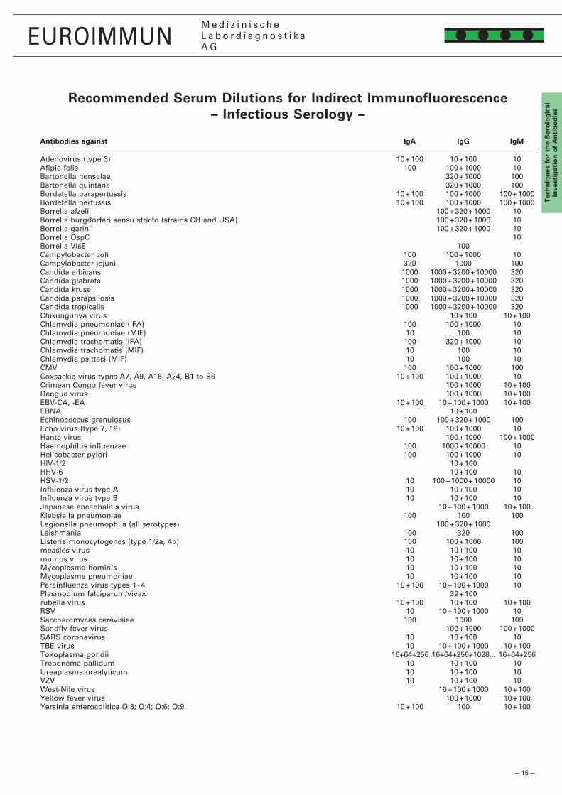
Recommended Serum dilutions for Indirect Immunofluorescence– Infectious Serology –
Adenovirus (type 3) 10 + 100 10 + 100 10Afipia felis 100 100 + 1000 10Bartonella henselae 3�0 + 1000 100Bartonella quintana 3�0 + 1000 100Bordetella parapertussis 10 + 100 100 + 1000 100 + 1000Bordetella pertussis 10 + 100 100 + 1000 100 + 1000Borrelia afzelii 100 + 3�0 + 1000 10Borrelia burgdorferi sensu stricto (strains CH and USA) 100 + 3�0 + 1000 10Borrelia garinii 100 + 3�0 + 1000 10Borrelia OspC 10Borrelia VlsE 100 Campylobacter coli 100 100 + 1000 10Campylobacter jejuni 3�0 1000 100Candida albicans 1000 1000 + 3�00 + 10000 3�0Candida glabrata 1000 1000 + 3�00 + 10000 3�0Candida krusei 1000 1000 + 3�00 + 10000 3�0Candida parapsilosis 1000 1000 + 3�00 + 10000 3�0Candida tropicalis 1000 1000 + 3�00 + 10000 3�0 Chikungunya virus 10 + 100 10 + 100Chlamydia pneumoniae (IFA) 100 100 + 1000 10Chlamydia pneumoniae (MIF) 10 100 10Chlamydia trachomatis (IFA) 100 3�0 + 1000 10Chlamydia trachomatis (MIF) 10 100 10Chlamydia psittaci (MIF) 10 100 10CMV 100 100 + 1000 100Coxsackie virus types A7, A9, A16, A�4, B1 to B6 10 + 100 100 + 1000 10Crimean Congo fever virus 100 + 1000 10 + 100Dengue virus 100 + 1000 10 + 100EBV-CA, -EA 10 + 100 10 + 100 + 1000 10 + 100EBNA 10 + 100Echinococcus granulosus 100 100 + 3�0 + 1000 100Echo virus (type 7, 19) 10 + 100 100 + 1000 10Hanta virus 100 + 1000 100 + 1000Haemophilus influenzae 100 1000 + 10000 10Helicobacter pylori 100 100 + 1000 10HIV-1/� 10 + 100 HHV-6 10 + 100 10HSV-1/� 10 100 + 1000 + 10000 10Influenza virus type A 10 10 + 100 10Influenza virus type B 10 10 + 100 10Japanese encephalitis virus 10 + 100 + 1000 10 + 100Klebsiella pneumoniae 100 100 100Legionella pneumophila (all serotypes) 100 + 3�0 + 1000 Leishmania 100 3�0 100Listeria monocytogenes (type 1/�a, 4b) 100 100 + 1000 100measles virus 10 10 + 100 10mumps virus 10 10 + 100 10Mycoplasma hominis 10 10 + 100 10Mycoplasma pneumoniae 10 10 + 100 10Parainfluenza virus types 1 - 4 10 + 100 10 + 100 + 1000 10Plasmodium falciparum/vivax 3� + 100 rubella virus 10 + 100 10 + 100 10 + 100RSV 10 10 + 100 + 1000 10Saccharomyces cerevisiae 100 1000 100Sandfly fever virus 100 + 1000 100 + 1000SARS coronavirus 10 10 + 100 10TBE virus 10 10 + 100 + 1000 10 + 100Toxoplasma gondii 16+64+�56 16+64+�56+10�8... 16+64+�56Treponema pallidum 10 10 + 100 10Ureaplasma urealyticum 10 10 + 100 10VZV 10 10 + 100 10West-Nile virus 10 + 100 + 1000 10 + 100Yellow fever virus 100 + 1000 10 + 100Yersinia enterocolitica O:3; O:4; O:6; O:9 10 + 100 100 10 + 100

— 16 —
EUROIMMUNM e d i z i n i s c h eL a b o r d i a g n o s t i k aA G
Tech
niq
ues fo
r the S
erolo
gical
Investig
ation
of A
ntib
od
ies
diagnostically Relevant Autoantibodies
Dr. Winfried StöckerClinical Pathologist
Clinical Immunology LaboratorySeekamp 31
D-23560 Lübeck (Germany) Telephone +49 451 58 55 986
Fax 58 55 134
Systemic Autoantibodies against
Grey boxes: standard analysis *) ANCA diagnostics in acute cases within one hour, at any hour **) Use special procedure for taking sample(s) ***) Send frozen sample(s)
Ig- AGM A G M SYST. LUPUS ERYTHEMATOSUS (SLE)151 ANA (cell nuclei) IF global testing1574 nucleosomes SLE specific1571 dsDNA ELISA SLE specific1572 dsDNA-NcX ELISA SLE specific1572 dsDNA IFT (C. luciliae) SLE specific1572 dsDNA RIA SLE specific159 ENA PoolPlus ELISA1591 U1-nRNP (70K, A, C)1593 Sm SLE specific1595 SS-A (Ro) 60 kDa: native159s Ro-52: recombinant1597 SS-B (La)1640 ribosomal P proteins SLE specific1605 Ku1601 cyclin I (PCNA)156 histones (global testing)1576 ssDNA (single-stranded DNA)121 pANCA* (granulocytes) vasculitis
CIRC. IMMUNE COMPLEXES1818 C1q ELISA
Ig- AGM A G M BASIS SPECTRUM151 ANA (cell nuclei) IF global testing152 ANA profile differentiation1572 dsDNA-NcX ELISA1572 dsDNA IFT SLE specific1590 ENA ProfilePlus ELISA 1 nRNP/Sm, Sm, SS-A, SS-B, Scl-70, Jo-11590 ENA ProfilePlus ELISA 2 rib. P proteins, RNP/Sm, Sm, SS-A, SS-B, Scl-70, Jo-1, CENP B1590 SLE Profile ELISA (dsDNA, histones, rib. P prot., nRNP/Sm, Sm, SS-A, SS-B, Scl-70)162 AMA (mitochondria)171 ASMA (smooth muscle)120 cANCA* (granulocytes) Wegener’s dis.121 pANCA* (granulocytes) vasculitis1030 autoantibody profile 30 IF substrates
Ig- AGM A G M POLYMYOSITIS, DERMATOMYOSITIS151 ANA (cell nuclei) IF global testing1530 Myositis Profile 3 EUROLINE (Mi-2, Ku, PM-Scl100, PM-Scl75, SRP, Jo-1, PL-7, PL-12, OJ, EJ, Ro-52)1661 Jo-11662 PL-71663 PL-121664 OJ1665 EJ1584 PM-Scl75, PM-Scl1001616 SRP (signal recognition particle)159s Ro-52: recombinant1605 Ku1607 Mi-21635 serotonin ab1636 PMR (polymyalgia rheumatica factor)
Ig- AGM A G M OTHER AUTOANTIBODIES1612 centrioles1613 MSA-1 (NuMa, spindle fibres)1614 MSA-2 (midbody)1615 MSA-3 (chromosome-ass. antigen)1617 centromer F protein (CENP-F)1641 ribosomes1642 Golgi apparatus1643 lysosomes165 cytoskeleton1651 actin1652 vimentin1653 cytokeratin1654 tropomyosin1655 vinculin1656 desmin1659 laminin (basal membranes)1947 collagen type VII1950 elastin196 vessel endothelium
Ig- AGM A G M SYSTEMIC SCLEROSIS (DIFFUSE + LIMITIERTE FORM)151 ANA (cell nuclei) IF global testing1532 Systemic Sclerosis Profile EUROLINE (Scl-70, CENP A, CENP B, RP11, RP155, Fibrillarin, NOR90, Th/To, PM-Scl100, PM-Scl75, Ku, PDGFR, Ro-52)1599 Scl-70 (DNA topoisomerase I)1584 PM-Scl75 (75 kDa)1584 PM-Scl100 (100 kDa)1611 centromeres1611 centromere A and B protein (rec.)1582 U3-nRNP (fibrillarin)1583 RNA polymerase I, II, III1585 7-2-RNP (Th/To)1586 4-6-S-RNA1587 NOR (nucleolus organizer region)1605 Ku
Ig- AGM A G M (RHEUMATOID) ARTHRITIS1505 CCP (cyclic citrullinated peptides)151a Sa1814 RF (class. rheumatoid factor)1508 filaggrin (RA keratin)1219 GS ANA (granulocyte specific ANA)151 ANA (cell nuclei) IF global testing1604 RANA (rheum. arthritis nuclear antigen)121 pANCA* (granuloc.) RF-ass. vasculitis148 cartilaginous subst. polychondritis1947 collagen type VII
Ig- AGM A G M THERAPY CONTROL1821 interferon alpha***1822 interferon beta1824 erythropoetin1572 dsDNA RIA1818 CIC-C1q ELISA
Ig- AGM A G M ANA DIAGNOSTICS, WESTERNBLOT1520 PM-Scl, CENP A/B, Ku 86 and 72 kDa, M2 74 kDa, RNP 70 kDa, RNP A/C, Sm B/B’/D, SS-A 60 and 52 kDa, Ro-52, SS-B 52, 47, 44 and 43 kDa, ribosomal P proteins P0/P1/P2, Scl-70, Jo-1)
FURTHER RHEUMATOID RELEVANT Ig- AGM A G M ANALYSES2011 anti-streptolysin2012 anti-streptokinase2013 anti-streptodornase2014 anti-DPNase (anti-NADase)2031 anti-staphylolysin2034 anti-hyaluronidase213 Borrelia burgdorferi2171 Yersinia enterocolitica O:32191 Chlamydia trachomatis
IMMUNOGLOBULINS: Ig- AGM A G M ANTI-1811 human IgA1813 human IgE1814 human IgG1815 human IgM
Ig- AGM A G M SJÖGREN’S SYNDROME151 ANA (cell nuclei) IF global testing1595 SS-A (Ro) 60 kDa: native159s Ro-52: recombinant1597 SS-B (La)
Ig- AGM A G M ANTI-PHOSPHOLIPID SYNDROME (APS)1621 cardiolipin1632 ß-2-glycoprotein 11631 lupus anticoagulant (plasma)**162a phosphatidylserine
Ig- AGM A G M SHARP’S SYNDROME MCTD1591 U1-nRNP (70K, A, C)151 ANA (cell nuclei) IF global testing
Comments (diagnosis, presumptive diagnosis, medication, major results, etc.):
Date of sample: Type of sample:
Serum .................................................................
Sample number:
Bill
ing
det
ails
Medical insurance
Doctor/hospital
Patient Name and address:
..................................................................................................................................................................................................
..................................................................................................................................................................................................
Pat
ien
t
Surname: First name: Date of birth: Sex: ......................................................................................................................................................................................................................................................... Address:
................................................................................................................................................................................................................................................................................................................
Doctor’s stamp/signature
Order Form for Autoimmune Diagnostic Tests
Female Male
For results
E-mail:
Fax:

— 17 —
EUROIMMUNM e d i z i n i s c h eL a b o r d i a g n o s t i k aA G
Tec
hn
iqu
es f
or
the
Ser
olo
gic
alIn
vest
igat
ion
of
An
tib
od
ies
diagnostically Relevant Autoantibodies
Dr. Winfried StöckerClinical Pathologist
Clinical Immunology LaboratorySeekamp 31
D-23560 Lübeck (Germany) Telephone +49 451 58 55 986
Fax 58 55 134
Grey: standard *) ANCA diagn. in acute cases within 1 h **) Special procedure for taking sample ***) CV2 partial protein, which only contains the N-terminally localised epitopes of the antigenSA_0000_I_UK_A06, 10/2010
Organ-/Tissue-Specifi c Autoimmunity: Autoantibodies againstIg- AGM A G M AUTOANTIBODY PROFILE1030 30 IF substrates (BIOCHIPs)
Ig- AGM A G M THYROID GLAND1015 TRAb (TSH receptors)1012 TPO ab (thyroidea peroxidase)1013 TAb (thyroglobulin)1014 colloid antigen II ab1011 MAb (microsomes)1016 T3 ab1017 T4 ab
Ig- AGM A G M DIABETES MELLITUS1021 ICA (islet cell antibodies)1022 GAD (glutamic acid decarboxylase)1023 IA-2 (tyrosine phosphatase)1024 insulin ab human1025 insulin receptor1026 glucagon-producing cells1027 zink transporter 8147 lipocytes
Ig- AGM A G M (POLY-)ENDOCRINOPATHY1051 adrenal cortex Addison’s dis.1053 21-hydroxylase Addison’s dis.1061 ovary: theca cells1062 ovary: corpus luteum1081 testis: Leydig cells105 steroid hormon-producing cells104 parathyroid gland1021 ICA (islet cell antibodies)1012 TPO ab (thyroidea peroxidase)1361 H+/K+-ATPase ab ELISA1361 PCA (parietal cells)1091 pituitary gland: anterior lobe1092 pituitary gland: posterior lobe1011 MAb (thyroid microsomes)1052 adrenal medulla107 placenta110 VPZ (vasopr.-prod. cells) D. insipudus
Ig- AGM A G M INFERTILITY1621 cardiolipin1060 ovary: theca c., c. luteum, z. pellucida1081 testis: Leydig cells1086 spermatozoa1091 pituitary gland: anterior lobe107 placenta1401 prostate
Ig- AGM A G M NERVOUS SYSTEM111 Neuronal Ab IFT global testing
Paraneoplastic neurol. syndromes1111 Neuronal Antigens Profile 2 EUROLINE amphiphysin, CV2.1***, PNMA2 (Ma-2), Ri, Yo, Hu1112 Tr (Purkinje cell cytoplasm)1113 Yo (Purkinje cell cytoplasm; PCA-1)1114 PCA-2 (Purkinje cell cytoplasm)1115 Ri (neurone nuclei; ANNA-2)1116 Hu (neurone nuclei; ANNA-1)112d NMDA receptors112k AMPA receptors (GluR1, GluR2)112l GABAB receptors1117 Ma1/Ma2 (neurone nuclei; Ta)1119 CV2 (CRMP-5)1022 GAD stiff-person syndr.112e amphiphysin stiff-person syndr.112a AGNA (anti-glia nuclear antigen; SOX-1)112b ANNA-31439 potassium channels (VGKC)1439 LGI11439 CASPR2
further parameters1154 aquaporin-4 neuromyelitis optica1157 glycine receptors1156 MOG (myelin-oligodendroc. glykoprot.)1121 myelin1122 MBP (myelin-basic protein)1123 MAG (myelin-assoc. glycoprotein)1124 myelin of peripheral nerves1126 neuroendothelium1127 neurofilaments1128 GFAP (glial fibrillary acidic protein)1129 non-medullated nerves112f astrocytes112g basal ganglia112h ganglion stellatum112i plexus myentericus1130 ganglioside profile GM1, GM2, GM3, GD1a, GD1b, GT1b, GQ1b113 single ganglioside analysis: GM1 GM2 GM3 GD1a GD1b GT1b GQ1b151 ANA (cell nuclei) IF global testing213 Borrelia burgd.: serum CSF
Ig- AGM A G M ANCA-ASSOC. VASCULITIDES (WEGENER’S DIS., MICR. ARTERITIS, CHURG-STRAUSS SYNDROME)*120 cANCA IFT* granuloc. Wegener’s dis.1201 PR3 (proteinase 3)1202 BPI (CAP 57)121 pANCA IFT* granulocytes vasculitis1211 MPO (myeloperoxidase)1212 elastase1213 cathepsin G1215 lactoferrin120 ANCA Profile ELISA PR3, MPO, elastase, cath. G, BPI, lactoferrin151 ANA (cell nuclei) IF global testing195 elastin196 vessel endothelium
Ig- AGM A G M EYE1178 recoverin1177 tunica choroidea chron. chorioretinitis1171 cornea1172 retina1173 lens oculi1174 corpus ciliare1175 eye muscles1176 retro bulbar connective tissue120 cANCA* (granulocytes) Wegener’s dis.151 ANA (cell nuclei) IF global testing
Ig- AGM A G M IMMUNOHAEMATOLOGY124 erythrocytes (global testing)1209 granulocyte membrane1221 lymphocytes1231 thrombocytes: indirect test (free ab)1232 thrombocytes: direct test (bound ab)**1361 H+/K+-ATPase ab ELISA1361 PCA (parietal cells)
Ig- AGM A G M SKELETAL MUSCLE, THYMUS1435 acetylcholine receptors M. gravis1434 MuSK M. gravis1437 calcium channels (VGCC) LEMS1439 potassium ch. (VGKC) neuromyotonia1439 CASPR2 neuromyotonia144 thymus M. gravis, thymoma1431 titin M. gravis143 skeletal muscle M. gravis1432 sarcolemma1436 myosin
Ig- AGM A G M LIPODYSTROPHY147 lipocytes
Ig- AGM A G M EPIDERMIS1501 desmosomes pemphigus1495 desmoglein 1 pemphigus1496 desmoglein 3 pemphigus1491 envoplakin paraneopl. pemphigus1502 epiderm. basal membr. pemphigoid1502 BP180 bullous pemphigoid1502 BP230 bullous pemphigoid135 oral mucosa Behçet’s/ Crohn’s dis.1503 basal membrane (urinary bladder)1509 epidermal keratin191 endomysium GSE, Duhring’s dis.3011 gliadin GSE, Duhring’s dis.1502 herpes gestationis factor1504 melanocytes150h hair follicle1947 collagen type VII NC1
Ig- AGM A G M LIVER, BILIARY DUCTS130 Liver Ab IFT global testing, 6 BIOCHIPs130 Autoimmune Liver Dis. Ab Profile EUROLINE AMA-M2, 3E (BPO), Sp100, PML, gp210, LC-1, LKM-1, SLA/LP, Ro-52
Autoimmune hepatitis (AIH)1302 SLA/LP (soluble liver antigen)1651 F-actin151 ANA (cell nuclei) IF global testing1307 LC-1 (liver cytosol)132 LKM (liver kidney microsomes)1321 LKM-1 ELISA1322 LKM-21323 LKM-31303 ASGPR (asialoglycoprotein receptors)171 ASMA (smooth muscles)1301 LSP (liver-specific protein)1304 LMA (liver cell membrane)
Primary biliary cirrhosis (PBC)162 AMA (mitochondria)1622 AMA-M2 (PDH + BPO)1624 AMA-M4 (sulfitoxidase)1629 AMA-M9 (glycogen phosphorylase)1603 Sp100, PML (nuclear dots)1608 gp210 (nuclear membrane, lamin)
Primary-sclerosing cholangitis (PSC)121 pANCA (granulocytes)
further antibodies1305 bile ducts1306 bile canaliculi1609 coilin; P80 (few nuclear dots)
Ig- AGM A G M STOMACH, INTESTINE1361 PCA (parietal cells) atroph. gastritis 1361 H+/K+-ATPase ab atroph. gastritis1362 intrinsic factor ab vit. B12 deficiency1366 gastrin (G) cells1391 pancreas acinus cells Crohn’s dis.1391 CUZD1 Crohn’s dis.1392 GP2 Crohn’s dis.2841 Saccharomyces cerev. Crohn’s dis.1392 pankreas secretion Crohn’s dis.135 mouth mucosa Behçet’s/ Crohn’s dis.1381 intestinal goblet cells ulc. colitis121 pANCA (granulocytes) ulc. colitis1382 enterocytes Crohn’s dis. , ulc. colitis191 endomysium GSE, Duhring’s dis.191 transglutaminase GSE, Duhring’s dis.3011 deamidated gliadin (Z-AGFA) GSE192 reticulin GSE, Duhring’s dis.
EXOCRINE GLANDS, PANCREATITIS,Ig- AGM A G M SJÖGREN’S SYNDROME139 exocrine pancreas 1391 pancreas acini1393 pancreas excretory ducts142 salivary glands (parotid gland)1421 parotid gland acini1423 parotid gland excretory ducts141 lacrimal gland
Sjögren’s syndrome151 ANA (cell nuclei) IF global testing1595 SS-A (Ro)1597 SS-B (La)1576 ssDNA (single-stranded DNA)
further antibodies1401 prostate1406 mamma
Ig- AGM A G M ANTIBODIES AGAINST ANIMAL IgG3811 HAMA (human anti-mouse IgG)
Ig- AGM A G M KIDNEY, LUNG120 cANCA IFT* granuloc. Wegener’s dis.121 pANCA IFT* granulocytes vasculitis125 kidney IF global testing1251 GBM ELISA glomerular basal membrane1254 PLA2R idiop. membr. nephropathy151 ANA (cell nuclei) IF global testing1572 dsDNA IFT1252 TBM (tubular basal membrane)1271 lung alveolar basal membrane
Ig- AGM A G M HEART1627 AMA-M7 (myocard-specific)146 heart muscle1462 heart: intercalated disk1463 heart: myolemma
— 18 —
EUROIMMUNM e d i z i n i s c h eL a b o r d i a g n o s t i k aA G
Tech
niq
ues fo
r the S
erolo
gical
Investig
ation
of A
ntib
od
ies
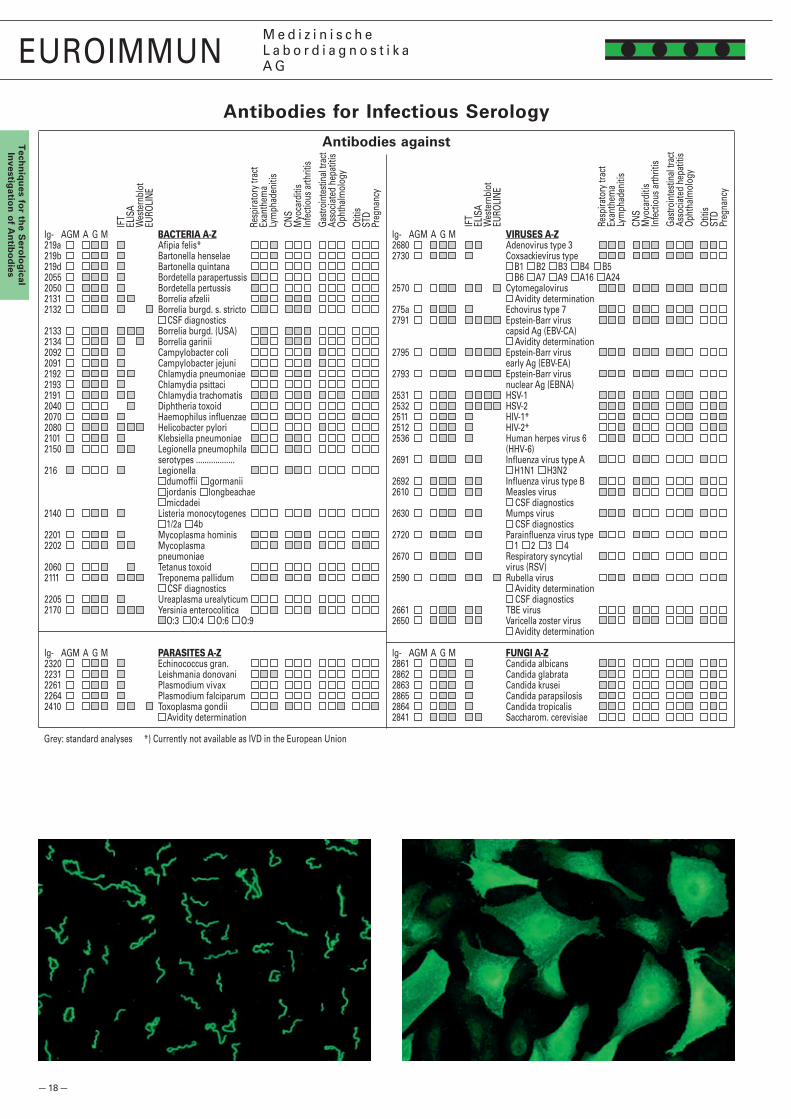
Antibodies for Infectious Serology
Dr. Winfried StöckerClinical Pathologist
Clinical Immunology LaboratorySeekamp 31
D-23560 Lübeck (Germany) Telephone +49 451 58 55 986
Fax 58 55 134
Grey: standard analyses *) Currently not available as IVD in the European UnionSI_0000_I_UK_A05, 08/2009
Comments (diagnosis, presumptive diagnosis, medication, major results, etc.):
Date of sample: Type of sample:
Serum .................................................................
Sample number:
Bill
ing
det
ails
Medical insurance
Doctor/hospital
Patient Name and address:
..................................................................................................................................................................................................
..................................................................................................................................................................................................
Pat
ien
t
Surname: First name: Date of birth: Sex: ......................................................................................................................................................................................................................................................... Address:
................................................................................................................................................................................................................................................................................................................
Doctor’s stamp/signature
Order Form for Infectious Serology Tests
Female Male
Antibodies against
Ig- AGM A G M BACTERIA A-Z219a Afi pia felis* 219b Bartonella henselae 219d Bartonella quintana 2055 Bordetella parapertussis 2050 Bordetella pertussis 2131 Borrelia afzelii 2132 Borrelia burgd. s. stricto CSF diagnostics2133 Borrelia burgd. (USA) 2134 Borrelia garinii 2092 Campylobacter coli 2091 Campylobacter jejuni 2192 Chlamydia pneumoniae 2193 Chlamydia psittaci 2191 Chlamydia trachomatis 2040 Diphtheria toxoid 2070 Haemophilus infl uenzae 2080 Helicobacter pylori 2101 Klebsiella pneumoniae 2150 Legionella pneumophila serotypes ..................216 Legionella dumoffi i gormanii jordanis longbeachae micdadei2140 Listeria monocytogenes 1/2a 4b2201 Mycoplasma hominis 2202 Mycoplasma pneumoniae2060 Tetanus toxoid 2111 Treponema pallidum CSF diagnostics2205 Ureaplasma urealyticum 2170 Yersinia enterocolitica O:3 O:4 O:6 O:9
Ig- AGM A G M PARASITES A-Z2320 Echinococcus gran. 2231 Leishmania donovani 2261 Plasmodium vivax 2264 Plasmodium falciparum 2410 Toxoplasma gondii Avidity determination
Ig- AGM A G M VIRUSES A-Z2680 Adenovirus type 3 2730 Coxsackievirus type B1 B2 B3 B4 B5 B6 A7 A9 A16 A24 2570 Cytomegalovirus Avidity determination275a Echovirus type 7 2791 Epstein-Barr virus capsid Ag (EBV-CA) Avidity determination2795 Epstein-Barr virus early Ag (EBV-EA)2793 Epstein-Barr virus nuclear Ag (EBNA)2531 HSV-1 2532 HSV-2 2511 HIV-1* 2512 HIV-2* 2536 Human herpes virus 6 (HHV-6) 2691 Infl uenza virus type A H1N1 H3N22692 Infl uenza virus type B 2610 Measles virus CSF diagnostics2630 Mumps virus CSF diagnostics2720 Parainfl uenza virus type 1 2 3 42670 Respiratory syncytial virus (RSV)2590 Rubella virus Avidity determination CSF diagnostics2661 TBE virus 2650 Varicella zoster virus Avidity determination
Ig- AGM A G M FUNGI A-Z2861 Candida albicans 2862 Candida glabrata 2863 Candida krusei 2865 Candida parapsilosis 2864 Candida tropicalis 2841 Saccharom. cerevisiae
IFT
ELIS
AW
este
rnbl
otEU
ROLI
NE
Resp
irato
ry tr
act
Exan
them
aLy
mph
aden
itis
CNS
Myo
card
itis
Infe
ctio
us a
rthrit
isGa
stro
inte
stin
al tr
act
Asso
ciate
d he
patit
isOp
htha
lmol
ogy
Otiti
sST
DPr
egna
ncy
IFT
ELIS
AW
este
rnbl
otEU
ROLI
NE
Resp
irato
ry tr
act
Exan
them
aLy
mph
aden
itis
CNS
Myo
card
itis
Infe
ctio
us a
rthrit
isGa
stro
inte
stin
al tr
act
Asso
ciate
d he
patit
isOp
htha
lmol
ogy
Otiti
sST
DPr
egna
ncy

— 19 —
EUROIMMUNM e d i z i n i s c h eL a b o r d i a g n o s t i k aA G
Tec
hn
iqu
es f
or
the
Ser
olo
gic
alIn
vest
igat
ion
of
An
tib
od
ies
Antibodies for Allergology
Allercoat™ 6 System: Antibodies of class IgE against 600 different allergens of the areas animal allergens, environmental allergens, food, grasses, herbal and flower pollen, house dust, insects, mites, moulds, parasites, pharmaceutical drugs, trees.
Diagnosis:
Date of sample: Type of sample:
Serum .................................................................
Sample number:
bill
ing
det
ails
Medical insurance
Doctor/hospital
PatientName and address:
...................................................................................................................................................................................................
...................................................................................................................................................................................................
Pat
ien
t
Surname: First name: Date of birth:
...................................................................................................................................................................................................Address:
...................................................................................................................................................................................................
Doctor’s stamp/signature
Order form for Allergology
Allergen Profiles: Antibodies of class IgE against
Dr. Winfried StöckerClinical Pathologist
Clinical Immunology LaboratorySeekamp 31
D-23560 Lübeck (Germany) Telephone +49 451 58 55 986
Fax 58 55 134
GLOBAL TEST
3840 Determination of total IgE (ELISA)
INHALATION
3110 Allergy Profile Inhalation(g1, g3, g6, g1�, t�, t3, t4, t7, w1, w6, w9,d1, d�, e1, e�, e3, m1, m�, m3, m6, CCD)
3110 Allergy Profile Inhalation 2(g6, g1�, t�, t3, t4, w6, w9, d1, d�, e1, e�, e3, e6,e8�, e84, es4, m1, m�, m3, m6, CCD)
3111 Allergy Profile Pediatric Inhalation(g6, g1�, t�, t3, t4, w6, w8, w9, d1, d�, e1,e�, e3, e6, e8�, e84, m1, m�, m3, m6, CCD)
311� Allergy Profile Mediterranean Inhalation(g�, g6, t3, t4, t9, t11, t�3, t�10, w1, w6, w9, w19,d1, d�, d70, e1, e�, e3, m�, m6, CCD)
3113 Allergy Profile Inhalation "South East Asia"(ts19, t104, t19, t��3, gs1, ds1, i6, u134, e1,e�, es17�, e6, e71, e8�, e84, ms1, ms4, m5,m1�, m45, CCD)
3116 Allergy Profile Inhalation "China"(gs�3, ts�1, t3, t8, t11, t1�, t14, t70, ws18, w1, w6,w9, es1, d1, d�, i6, ms5, m1, u73, u80, CCD)
3117 Allergy Profile Inhalation "Middle East"(g1, g6,g1�, t�, t3, t7, t9, w1, w6, w8, d1, d�,i6, e1, e84, m1, m�, m3, m5, m6, CCD)
3118 Allergy Profile Inhalation "Gulf"(g6, g1�, t�, t3, t7, t9, w1, w6, d1, d�, i6, e1,e�, e3, e17, m1, m�, m3, m5, m6, CCD)
3119 All. Profile Inhalation “Mix-Screen Turkey 1”(ts�3, ts�4, ts�5, gs1�, gs15, gs�1, ws17, ws18,ws19, ws�0, ms11, ms1�, CCD)
3119 Allergy Profile Inhalation “Turkey 1”(gs1�, gs15, gs�1, g1�, ts�3, ts�4, t9, t70, ws18,ws19, ws�0, d1, d�, i6, es�, es17�, e1, e�, e3, e4,e80, e81, e84, ms11, ms1�, m1, m�, m3, m6, CCD)
3119 Allergy Profile Inhalation “Turkey 2”(m1, m�, m3, m6, ds1, i6, e1, e�, e3, e4,e81, e84, CCD)
31�0 Allergy Profile Inhalation "India"(g6, g1�, g�0, t18, w4, w�7, w�9, ds1, d�, i6, e1, e�,e11, e85, m3, m37, u81, u1�6, u1�9, u140, CCD)
31�1 Allergy Profile Inhalation "Screen France"(hs1�, es1, gs4, ts4, ws�, ms1, CCD)
FOOD
3410 Allergy Profile Food(f1, f75, f�, f45, f4, f5, f9, f13, f14,f17, f�0, f49, f84, f�37, f�5, f31,f35, f85, f3, f�3, CCD)
3410 Allergy Profile Food 2(f1, f75, f�, f78, f4, f5, f14, f10, f13,f17, f�0, f49, f84, f95, f�5, f31, f35,f85, f3, f�3, CCD)
3411 Allergy Profile Food "South East Asia 1"(f1, f75, f�, f4, f9, f10, f14, f13, f17,f63, f64, f83, fs10, fs14, f�3, f�4,f80, f179, f105, f336, CCD)
3411 Allergy Profile Food "South East Asia 2"(f1, f75, f�, f4, f9, f10, f14, f13, f17, f63,f340, f83, fs10, fs14, f�3, f�4, f80, f179,f105, f336, CCD)
3414 Allergy Profile Food "China"(f1, f�, f4, f7, f�7, f88, fs35, f13, f14,fs40, f�5, f�9�, fs4�, f�3, f�34, f3,f41, f56, fs41, fs77, CCD)
3415 Allergy Profile Food "Middle East"(f1, f75, f�, f78, e�04, f4, f14, f45, f13,f17, f�0, f33, f49, f9�, f�5, f31, f85,f48, f88, f89, CCD)
3416 Allergy Profile Food "Gulf"(f1, f75, f�, f105, f4, f14, f45, fs36, f13,f�9, f33, f44, f93, f�5, f31, f48, f83,f88, f3, f�3, CCD)
34�0 Allergy Profile Food “Mix Screen Turkey 1”(f�45, fs8, fs50, fs38, fs37, fs46, fs47, fs48,fs49, fs45, fs10, fs16, CCD)
34�0 Allergy Profile Food “Turkey 1”(f1, f75, f�, f169, f78, f4, f79, f9, f14, f10, f13, f17,f144, u87, f���, f73, f33, f44, f49, f9�, f84, f146,f3�8, f�5, f31, f35, f48, f95, f97, f1��, f13�, fs14,fs10, fs43, f83, CCD)
34�0 Allergy Profile Food “Turkey 2”(f3, f�1, f�06, f437, f438, f436, f71, f177, f3�4,f�7, f83, f88, CCD)
34�0 Allergy Profile Food “Turkey 3”(f1, fs51, f14, fx�0, f13, fs35, f49, f44, f�5, f31,fs10, fs16, CCD)
34�1 Allergy Profile Food "India"(f�, f75, f168, f4, f9, f14, f13, f36, f49, f50,f35, f38, f48, f�44, f83, f89, f74, f�40,f�3, f�4, CCD)
ATOPY, POLLEN-ASSOCIATEDFOOD ALLERGIES
3710 Allergy Profile Atopy "Top Screen"(rs1, rs�, fx5, fs5�, CCD)
3710 Allergy Profile Atopy(g6, g1�, t3, w6, d1, e1, e�, e3, m�, m6, f1, f�,f3, f4, f9, f14, f17, f31, f35, f49, CCD)
3710 Allergy Profile Atopy 3(g6, t3, t4, w6, d1, d�, e1, e�, e3, m�, m3, f1,f75, f�, f3, f4, f13, f14, f31, f49, CCD)
3711 Allergy Profile Pollen-FoodCross Reactions(g6, t3, w6, f4, f5, f13, f17, f�0, f48, f89,f�71, f�75, f44, f49, f348, f�37, f3�8, f31,f35, f85, CCD)
371� Allergy Profile Pediatrics(gx, t3, w6, d1, e1, e�, e3, m�, m6, f1, f75, f3, f�,f76, f77, f78, e�04, f4, f9, f14, f13, f17, f31, f35,f49, CCD)
3713 Allergy Profile Atopy "China"(ts�0, w1, w6, ds1, h1, e1, e�, i6, ms1, u80, f1, f�,f13, f14, f�7, f88, fs33, fs34, f�3, f�34, CCD)
3713 Allergy Profile Atopy "China 3"(ts��, w6, us1, ds1, es1, h1, ms5, f�45,fs9, fs43, fs56, fs34, fs44, CCD)
3719 Allergy Profile Atopy “Mix Screen Turkey 1”(hs1�, ms1, gs�, ws�1, ts�6, ts�5, fx5, fx�0,es5, es6, es17�, es�, CCD)
3719 Allergy Profile Atopy “Turkey 1”(d1, m�, es5, g6, t3, w6, f1, f�, f13,f14, f4, fs16, CCD)
INSECT VENOMS
37�0 Allergy Profile Insect Venoms(i1, i3, CCD)
EUROASSAY ALLERGY PROFILES
3710 Allergy Profile Inhalation(g1, g3, g6, g1�, t�, t3, t4, t7, w1, w8, w6, w9, d1, d�,es4, e�, e3, e6, e1, e8�, e84, m1, m�, m3, m6, CCD)
3410 Allergy Profile Food(f1, f75, f�, f78, f45, f4, f5, f9, f14, f10, f13, f17, f�0,f84, f95, f�37, f�5, f31, f35, f85, f3, f�3, f49, CCD)
371� Allergy Profile Pediatrics/Atopy(g6, g1�, t3, w6, d1, d�, e1, e�, e3, m�, m3, m6,f1, f75, f3, f�, f76, f77, f78, e�04, f4, f9, f14, f13,f17, f31, f35, f49, CCD)
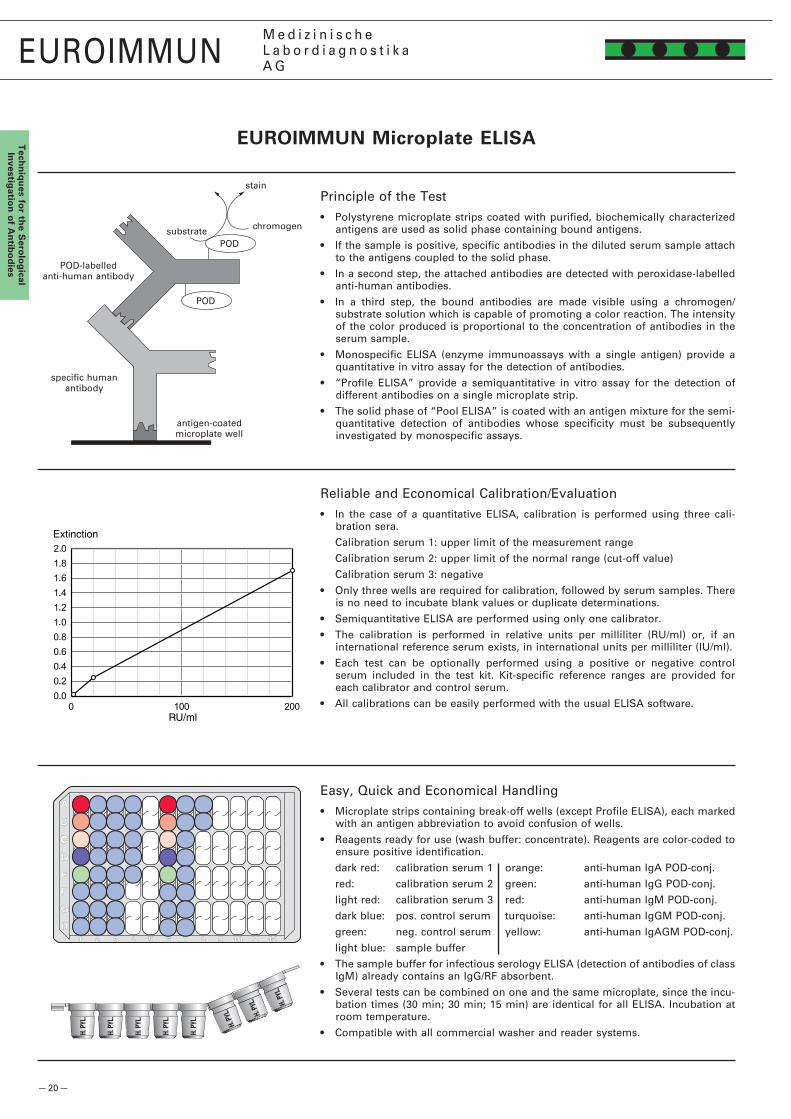
— �0 —
AA
B
C
D
E
F
G
H1 2 3 4 5 6 7 8 9 10 11 12
h. P
YL.
h. P
YL.
h. P
YL.
h. P
YL.
h. P
YL.
h. P
YL.
h. P
YL.
h. P
YL.
EUROIMMUNM e d i z i n i s c h eL a b o r d i a g n o s t i k aA G
Tech
niq
ues fo
r the S
erolo
gical
Investig
ation
of A
ntib
od
ies
EUROIMMUN Microplate ELISA
Principle of the Test• Polystyrene microplate strips coated with purified, biochemically characterized
antigens are used as solid phase containing bound antigens.
• If the sample is positive, specific antibodies in the diluted serum sample attach to the antigens coupled to the solid phase.
• In a second step, the attached antibodies are detected with peroxidase-labelled anti-human antibodies.
• In a third step, the bound antibodies are made visible using a chromogen/substrate solution which is capable of promoting a color reaction. The intensity of the color produced is proportional to the concentration of antibodies in the serum sample.
• Monospecific ELISA (enzyme immunoassays with a single antigen) provide a quantitative in vitro assay for the detection of antibodies.
• “Profile ELISA” provide a semiquantitative in vitro assay for the detection of different antibodies on a single microplate strip.
• The solid phase of “Pool ELISA” is coated with an antigen mixture for the semi-quantitative detection of antibodies whose specificity must be subsequently investigated by monospecific assays.
Reliable and Economical Calibration/Evaluation• In the case of a quantitative ELISA, calibration is performed using three cali-
bration sera.
Calibration serum 1: upper limit of the measurement range
Calibration serum �: upper limit of the normal range (cut-off value)
Calibration serum 3: negative
• Only three wells are required for calibration, followed by serum samples. There is no need to incubate blank values or duplicate determinations.
• Semiquantitative ELISA are performed using only one calibrator.
• The calibration is performed in relative units per milliliter (RU/ml) or, if an international reference serum exists, in international units per milliliter (IU/ml).
• Each test can be optionally performed using a positive or negative control serum included in the test kit. Kit-specific reference ranges are provided for each calibrator and control serum.
• All calibrations can be easily performed with the usual ELISA software.
Easy, Quick and Economical Handling• Microplate strips containing break-off wells (except Profile ELISA), each marked
with an antigen abbreviation to avoid confusion of wells.
• Reagents ready for use (wash buffer: concentrate). Reagents are color-coded to ensure positive identification.
dark red: calibration serum 1 orange: anti-human IgA POD-conj.
red: calibration serum � green: anti-human IgG POD-conj.
light red: calibration serum 3 red: anti-human IgM POD-conj.
dark blue: pos. control serum turquoise: anti-human IgGM POD-conj.
green: neg. control serum yellow: anti-human IgAGM POD-conj.
light blue: sample buffer
• The sample buffer for infectious serology ELISA (detection of antibodies of class IgM) already contains an IgG/RF absorbent.
• Several tests can be combined on one and the same microplate, since the incu-bation times (30 min; 30 min; 15 min) are identical for all ELISA. Incubation at room temperature.
• Compatible with all commercial washer and reader systems.
antigen-coated microplate well
specific human antibody
POD-labelled anti-human antibody
PODsubstrate
stain
chromogen
POD

— �1 —
0
�
4
6
8
10
0 �5 50 75 100
EUROIMMUNM e d i z i n i s c h eL a b o r d i a g n o s t i k aA G
Tec
hn
iqu
es f
or
the
Ser
olo
gic
alIn
vest
igat
ion
of
An
tib
od
ies
Determination of Low-Avidity Antibodies• An alternative principle for the serological diagnosis of fresh infections has been
established by investigating the antibody avidity.
• The first reaction of the immune system following an infection is the formation of low-avidity antibodies. As the infection proceeds, increasingly antigen-adapted IgG is formed, and avidity grows. As long as high-avidity IgG is not yet detected in the serum, it can be assumed that the infection is still in an early stage.
• To identify low-avidity antibodies in a patient’s serum, two microplate ELISA are performed in parallel: one test is carried out in the conventional way, the other one includes urea treatment between incubations with patient’s serum and peroxidase-labelled anti-human IgG, resulting in the detachment of low-avidity antibodies from the antigens.
• Low-avidity antibodies are present if the ELISA extinction is significantly reduced by urea treatment. For an objective interpretation, the relative avidity index (RAI) can be calculated out of the measured values with and without urea incubation.
• Serum dilution 1 : 100, conjugate class anti-human IgG, POD-labelled.
• 3-point calibration, quantitative (IgG).
• The following test kits for avidity determination are available: Toxoplasma gondii, CMV, rubella virus, VZV, West-Nile virus, EBV-CA.
Avidity of antibodies against EBV-CA (IgG)
Relative Avidity Index (RAI) in %
Nu
mb
er o
f P
atie
nts
RAI =Ewith urea
Ewithout urea
Primary Infection
Previous Infection
EUROIMMUN Microplate ELISA
Antibody Determination in CSF• Indication: local infections of the brain.
• CSF dilution 1 : �, serum dilution 1 : 404. Conjugate classes anti-human IgG or IgM, POD-labelled.
• Easy to conduct: ready-for-use reagents.
• 4-point calibration, quantitative. Identical incubation conditions and times (room temperature; 60 min / 60 min / 15 min): all EUROIMMUN ELISA for CSF dia-gnostics can be combined without difficulty on one and the same microplate.
• The antibody concentration in the patient’s serum is determined in parallel to the antibody concentration in CSF on one and the same microplate. The CSF/serum quotient CSQpath.-spec. is calculated from both measured values.
• An intrathecal synthesis of specific antibodies is present if the CSF/serum quotient of the specific antibodies CSQpath.-spec. is significantly higher than the CSF/serum quotient of the whole IgG (CSQtotal) or if necessary the CSQlim.. The relation of both values indicates the relative CSF/serum quotient CSQrel. (synonym: antibody specificity index, ASI).
• Interpretation of results (according to the recommendations of Prof. Reiber):
CSQrel. < 1,3: standard range
CSQrel. 1,3 – 1,5: borderline range
CSQrel. > 1,5: Indication of pathogen-specific antibody production in the CNS
• For the automatic calculation of the CSQrel. EUROIMMUN provides a specific Excel table free of charge.
• Highest sensitivity, specificity and reproducibility. Antibody concentrations in the serum and CSF are determined within the linear range of the test.
• The following test kits for CSF diagnostics are available: Borrelia burgdorferi, Toxoplasma gondii, HSV-1, HSV-�, HSV-1/� Pool, CMV, rubella virus, measles virus, mumps virus, VZV, TBE, EBV-CA.
• All test systems for CSF diagnostics can also be used only for serology.
• Perfectly adapted for the automated incubation in incubation devices.
CFS/serum quotient diagram according to Reiber and Lange (1991)
ELISA incubation scheme
Table-based evaluation of the CSQrel.
tota
l
CSQalb.
CS
Q
Reference range for normal values, intact blood-CSF barrier function
Blood-CSF barrier dysfunction, no Ig production in CNS
Blood-CSF barrier dysfunction, additional Ig production in CNS
Clear Ig production in CNS, no disturbance in blood-CSFbarrier function
Error in blood extraction or analysis
1 2 3 4 5 6
A CA C31:�
b CB S31:404
C CC C41:�
d CD S41:404
E C11:�
C51:�
f S11:404
S51:404
g C�1:�
C61:�
h S�1:404
S61:404
CA-DCSF-Calibrators C CSF S serum

— �� —
ap
proved
approved
EUROIMMUNM e d i z i n i s c h eL a b o r d i a g n o s t i k aA G
Tech
niq
ues fo
r the S
erolo
gical
Investig
ation
of A
ntib
od
ies
ELISA Automation Using the EUROIMMUN Analyzers
EUROIMMUN Analyzer I and EUROIMMUN Analyzer I-�P: Broad Parameter Spectrum, Proven Reliability, Variable Throughput• One for all: fully automated processing of all EUROIMMUN ELISA – autoimmune
diagnostics, infectious serology and allergology – with only one system
• Flexibility for your benefit: open system with more than 800 validated EUROIMMUN parameters for serum, plasma and cerebrospinal fluid, over 50 or 30 parameters in parallel.
• Convenient, simple and reliable operation: e.g. by scanning QC certificates using a �D-hand barcode scanner – ready-for-use reagents and preprogrammed assay protocols enable you to start immediately.
• Capacity and throughput: quick loading and efficient time management allow processing of up to 70 or 50 tests per hour – up to 7 or 3 plates, 180 or 144 samples at the start of a run.
• Security for patients: validated test systems and the comprehensive safety kit provide the basis for reliable diagnostics.
• Reliability and service: instruments and reagents from one manufacturer, quick and targeted support from our personnel for a smooth workflow in your laboratory.
Modular System: A Highly Sophisticated Solution• High convenience, fast and reliable loading through barcode identification of
samples and reagents: automatic scanning when racks are inserted, reading of lot-specific QC data via �D hand barcode scanner.
• Dilution area: �88 or 19� dilution positions (Deepwell, � ml).
• Liquid level detection (capacitive), multi-shot (dispensing) mode, automatic tip detection, clot detection.
• Pipetting possible during plate transport due to separation of transport and pipetting unit.
• 4 or � incubators with heating and shaking function, 4 or 3 incubators at room temperature.
• Standard Windows software: familiar user interface, all relevant statistical functions provided, available in different languages.
• Efficient use and convenient handling of tips and dilution plates through memory function.
A Convincing and Reliable Package: EUROIMMUN Analyzer, EUROIMMUN ELISA, EUROIMMUN Service• Comprehensive test system validation for the EUROIMMUN Analyzer: all
parameters validated in accordance with the 98/79/EC directive and based on EN ISO 13485:�003/CMDCAS.
• All ELISA test systems are manufactured according to the European Quality Standards (IVD).
• National and international conformity (standardisation): CE, IVD, FDA and CMDCAS.
• Programming and set-up of automated system, including introduction to the system with customer training, performed by qualified personnel.
• Reliable and fast delivery of consumables and reagents.
• Connection to in-house computer system via communication protocol ASTM.
• Maintenance contract with EUROIMMUN on request.
*) soon available for the EUROIMMUN Analyzer I-�P

— �3 —
EUROIMMUNM e d i z i n i s c h eL a b o r d i a g n o s t i k aA G
Tec
hn
iqu
es f
or
the
Ser
olo
gic
alIn
vest
igat
ion
of
An
tib
od
ies
Incubate: 30 min
Wash: 300 µl wash buffer per well
microplate wells coated with antigens
diluted samples
enzymeconjugate
chromogen/ substrate solution
Pipette: 100 µl per well
Pipette: 100 µl per well
Incubate: 30 min
Evaluate: photometric measurement at a wavelength of 450 nm
Wash: 300 µl wash buffer per well
Pipette: 100 µl per well
Incubate: 15 min
Pipette: 100 µl per well
stop solution
Incubating the Microplate ELISA

— �4 —
nRNP/Sm
Sm
SS-A
SS-B
Scl-70
Jo-1
EUROIMMUNM e d i z i n i s c h eL a b o r d i a g n o s t i k aA G
Tech
niq
ues fo
r the S
erolo
gical
Investig
ation
of A
ntib
od
ies
stain
APsubstratechromogen
AP
membrane coated with antigen
specific humanantibody
AP-labelled anti-human
antibody
EUROASSAY: Line blot in TITERPLANE™ Technique format
Principle of the Test• Membrane strips coated with thin parallel lines of several purified, biochemically
characterized antigens are used as solid phase. The membrane strips are fixed as BIOCHIPs in the fields of microscope slides.
• If the sample is positive, specific antibodies in the diluted serum sample attach to the antigens coupled to the solid phase.
• In a second incubation step, the attached antibodies react with AP-labelled anti-human antibodies.
• In a third step, the bound antibodies are stained with a chromogen/substrate solution which is capable of promoting a color reaction. An intense dark band at the line of the corresponding antigen appears if the serum sample contains specific antibodies.
• The microscope slides are incubated using the TITERPLANE™ Technique: samples and reagents are applied to the reaction areas of a reagent tray. The slides are then placed into the recesses of the reagent tray, where all test strips of one slide come into contact with the liquids, and the individual reactions begin simultaneously.
• Depending on the spectrum of antigens used, it is possible to analyze several antibodies next to each other and simultaneously under identical conditions.
Easy Handling• Several serum samples can be analyzed simultaneously on one and the same
slide.
• Total time for performing the EUROASSAY test is about 100 minutes. During the washing procedure, reagents for the next incubation step can be applied to reagent trays.
• All incubation steps proceed at room temperature. Shaking the slides together with the reagent tray on a circulatory shaker ensures the best possible sensitivity.
• Low reagent consumption. Only 50 µl each of diluted serum and reagent are needed per test field.
• Reagents ready for use (wash buffer: concentrate).
Reliable and Simple Evaluation• Since results are evaluated visually, there is no investment required for
photometers, etc.
• The antigen bands are located at exactly defined positions, which means that evaluation of the test is much simpler than for Westernblots.
• Correct completion of the individual incubation steps in each test field is indicated by staining of the control band.
• Positive and negative results can be easily and reliably differentiated from each other. The intensity of the antigen bands correlates with the antibody titer.
• The antigens used are highly pure, mostly isolated by affinity chromatography. The membrane strips do not contain any superfluous proteins which might cause unspecific positive results.
• The incubated microscope slides can be stored for long periods. Results can be easily documented.
EUROASSAY Anti-ENA ProfilePlus: detection of antibodies against SS-A and SS-B in a case of Sjögren’s syndrome.
Control band

— �5 —
EUROIMMUNM e d i z i n i s c h eL a b o r d i a g n o s t i k aA G
Tec
hn
iqu
es f
or
the
Ser
olo
gic
alIn
vest
igat
ion
of
An
tib
od
ies
Incubate: 30 min shaking on a circulatory shaker (300 rpm)
Wash: 5 s flush 15 min cuvette
slide
reagent traymembrane strips
diluted samples
washbuffer
enzyme conjugate
chromogen/substrate solution
washbuffer
Pipette: 50 µl per field
Pipette: 50 µl per field
Incubate: 30 min shaking on a circulatory shaker (300 rpm)
Wash: 5 s flush 15 min cuvette
Pipette: 50 µl per field
Incubate: 10 min shaking on a circulatory shaker (300 rpm)
Wash: flush with deionized or distilled water, air dry
Evaluate: visual assessment of color intensity
Incubating the EUROASSAY (TITERPLANE™ Technique)

— �6 —
PM-Scl100
Mi-�
Ku
Ro-5�
PL-7
Jo-1
PL-1�
nRNP/Sm
SS-ARo-5�
SS-B
Sm
Scl-70
PM-Scl
Jo-1
CENP B
PCNA
AMA-M�
Sp100
M�-3E (BPO)
PML
gp�10
LKM-1
LC-1
SLA/LP
Ro-5�
PM-Scl75
SRP
EJ
OJ
EUROIMMUNM e d i z i n i s c h eL a b o r d i a g n o s t i k aA G
Tech
niq
ues fo
r the S
erolo
gical
Investig
ation
of A
ntib
od
ies
Principle of the Test• Membrane strips coated with thin parallel lines of several purified, biochemically
characterized antigens are used as solid phase. The membranes are fixed as BIOCHIPs onto synthetic foil.
• If the sample is positive, specific antibodies in the diluted serum sample attach to the antigens coupled to the solid phase.
• In a second incubation step, the attached antibodies react with alkaline-phosphatase-labelled anti-human antibodies.
• In a third step, the bound antibodies are stained with a chromogen/substrate solution which is capable of promoting a color reaction. An intense dark band at the line of the corresponding antigen appears if the serum sample contains specific antibodies.
• Depending on the spectrum of antigens used, it is possible to analyze several antibodies next to each other and simultaneously under identical conditions.
Easy Handling, Reliable and Simple Evaluation• A separate membrane strip is incubated for each serum sample.
• Total time for performing the analysis is about 105 minutes.
• The incubation can be automated using the EUROBlotMaster.
• All incubation steps proceed at room temperature.
• The antigen bands are located at exactly defined positions, which means that the evaluation of the test is much simpler than for Westernblots.
• Correct completion of the individual incubation steps is indicated by staining of the control band on each EUROLINE test strip.
• Positive and negative results can be easily and reliable differentiated from each other. The intensity of the antigen bands correlates with the antibody titer.
• The antigens used are highly pure, mostly isolated by affinity chomatography. The membrane strips do not contain any superfluous proteins which might cause unspecific positive results.
• The incubated EUROLINE test strips can be stored for long periods. Results can be easily documented.
• The program EUROLineScan from EUROIMMUN has been developed to enable quantitative evaluation of EUROLINE test strips, to facilitate management of data, and to provide detailed documentation of results. First, the incubated EUROLINE test strips are scanned using a flatbed scanner. EUROLineScan recognizes the position of the strips, even if they have been placed inexactly, identifies the bands, and measures their intensity. The results are then saved together with the image data. A separate results sheet can be produced for each patient.
stain
APsubstrate chromogen
AP
membrane coatedwith antigen
specific human antibody
AP-labelled anti-human
antibody
The EUROLINE: A New Technique for Extensive Antibody Profiles
Incubated EUROLINE test strips (Autoimmune Liver Diseases Profile, ANA Profile 3, Myositis Profile 3.
ControlControlControl
Rib. P-prot.
Histones
dsDNSNucleos.
AMA M�

— �7 —
EUROBlotMaster
EUROBlotCamera EUROBlotScanner
EUROIMMUNM e d i z i n i s c h eL a b o r d i a g n o s t i k aA G
Tec
hn
iqu
es f
or
the
Ser
olo
gic
alIn
vest
igat
ion
of
An
tib
od
ies
EUROLINE Automation Using EUROblotMaster and EUROLineScan
EUROBlotMaster• Standardised incubation of immunoblot strips – higher precision and repro-
ducibility.
• Automatisation of all EUROIMMUN immunoblot strips (EUROLINE, EUROLINE-WB, Westernblot)
• Over 65 validated parameters available (16 autoantibody, �8 infectious and �1 allergy parameters)
• Up to 30 strips per test run
• Easy operation
• Combination of different conjugates/tests in one run
• Walk-away function – fully automated from the start to the end of processing following loading
• Compatible with modern evaluation systems such as EUROBlotCamera and EUROLineScan
Automatic Evaluation of the Results Using EUROLineScan• For all EUROIMMUN blot systems: EUROLINE, EUROLINE-WB, Westernblot.
• For all areas: autoimmune diagnostics, infectious serology and allergology.
• EUROBlotCamera: digitalisation of strips while in the incubation tray.
• EUROBlotScanner: digitalisation of strips using flatbed scanner.
• Fully automated identification, quantitation and assignment of bands.
• Option to modify results (changes are automatically documented).
• Complete results obtained just a few minutes after finishing the incubation.
• Fully automated administration and documentation of extensive individual data.
• Electronic archiving of all images and data (avoids the need to store potentially infectious blot strips).
• Online connection to laboratory software.
• Network compatible.
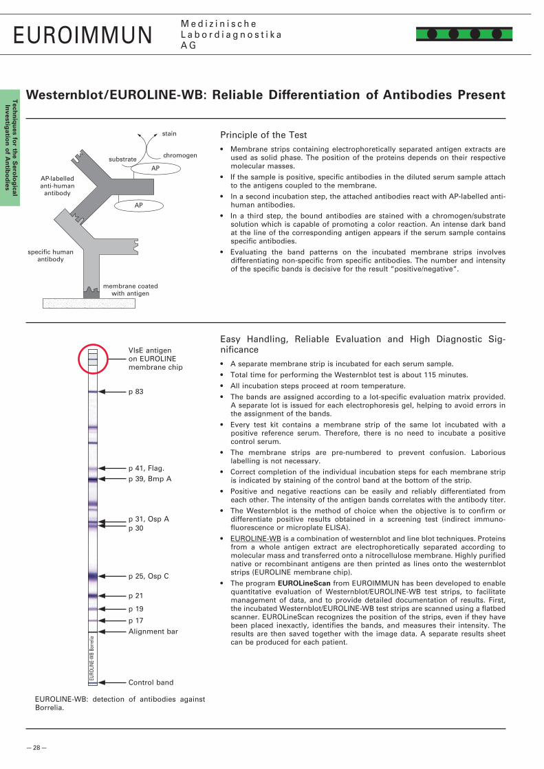
— �8 —
p 83
p 39, Bmp Ap 41, Flag.
p �5, Osp C
p �1
p 30p 31, Osp A
p 19
p 17
EURO
LINE
-WB
Borre
lia
EUROIMMUNM e d i z i n i s c h eL a b o r d i a g n o s t i k aA G
Tech
niq
ues fo
r the S
erolo
gical
Investig
ation
of A
ntib
od
ies
Westernblot/EUROLINEWb: Reliable differentiation of Antibodies Present
Principle of the Test• Membrane strips containing electrophoretically separated antigen extracts are
used as solid phase. The position of the proteins depends on their respective molecular masses.
• If the sample is positive, specific antibodies in the diluted serum sample attach to the antigens coupled to the membrane.
• In a second incubation step, the attached antibodies react with AP-labelled anti-human antibodies.
• In a third step, the bound antibodies are stained with a chromogen/substrate solution which is capable of promoting a color reaction. An intense dark band at the line of the corresponding antigen appears if the serum sample contains specific antibodies.
• Evaluating the band patterns on the incubated membrane strips involves differentiating non-specific from specific antibodies. The number and intensity of the specific bands is decisive for the result “positive/negative“.
stain
AP
substratechromogen
AP
membrane coated with antigen
specific humanantibody
AP-labelled anti-human
antibody
EUROLINE-WB: detection of antibodies against Borrelia.
Control band
Alignment bar
Easy Handling, Reliable Evaluation and High Diagnostic Sig-nificance• A separate membrane strip is incubated for each serum sample.
• Total time for performing the Westernblot test is about 115 minutes.
• All incubation steps proceed at room temperature.
• The bands are assigned according to a lot-specific evaluation matrix provided. A separate lot is issued for each electrophoresis gel, helping to avoid errors in the assignment of the bands.
• Every test kit contains a membrane strip of the same lot incubated with a positive reference serum. Therefore, there is no need to incubate a positive control serum.
• The membrane strips are pre-numbered to prevent confusion. Laborious labelling is not necessary.
• Correct completion of the individual incubation steps for each membrane strip is indicated by staining of the control band at the bottom of the strip.
• Positive and negative reactions can be easily and reliably differentiated from each other. The intensity of the antigen bands correlates with the antibody titer.
• The Westernblot is the method of choice when the objective is to confirm or differentiate positive results obtained in a screening test (indirect immuno-fluorescence or microplate ELISA).
• EUROLINE-WB is a combination of westernblot and line blot techniques. Proteins from a whole antigen extract are electrophoretically separated according to molecular mass and transferred onto a nitrocellulose membrane. Highly purified native or recombinant antigens are then printed as lines onto the westernblot strips (EUROLINE membrane chip).
• The program EUROLineScan from EUROIMMUN has been developed to enable quantitative evaluation of Westernblot/EUROLINE-WB test strips, to facilitate management of data, and to provide detailed documentation of results. First, the incubated Westernblot/EUROLINE-WB test strips are scanned using a flatbed scanner. EUROLineScan recognizes the position of the strips, even if they have been placed inexactly, identifies the bands, and measures their intensity. The results are then saved together with the image data. A separate results sheet can be produced for each patient.
VlsE antigen on EUROLINE membrane chip
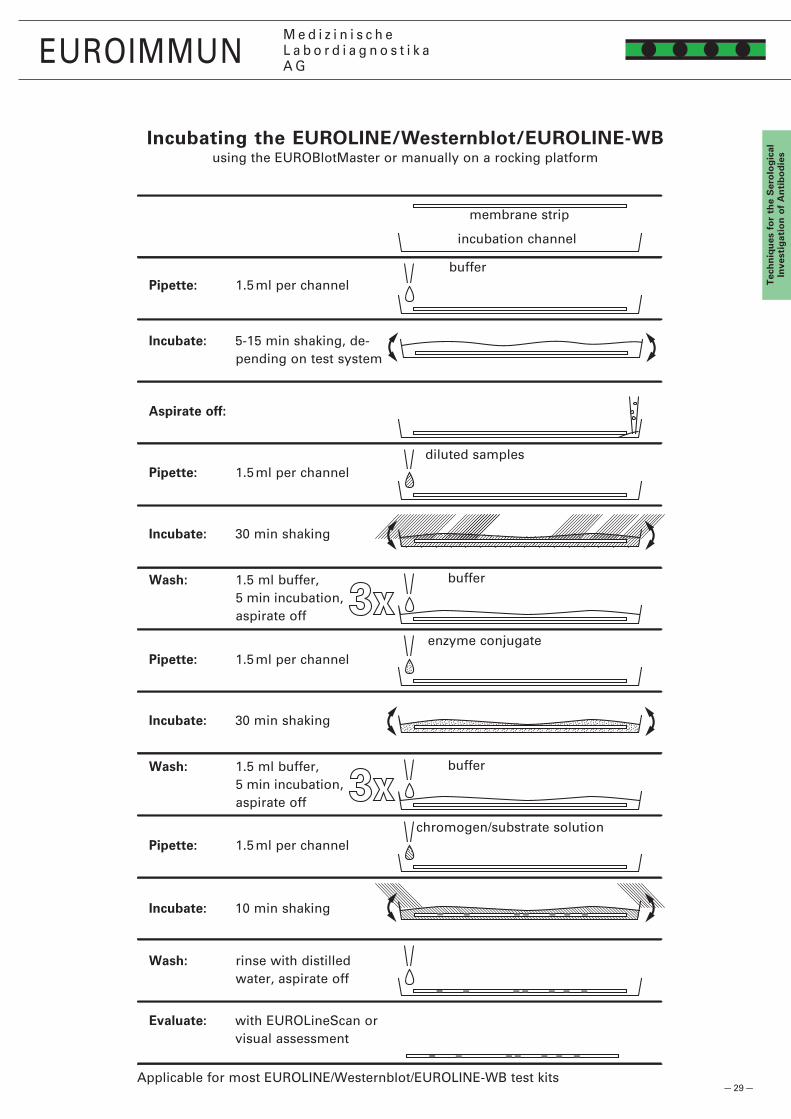
— �9 —
EUROIMMUNM e d i z i n i s c h eL a b o r d i a g n o s t i k aA G
Tec
hn
iqu
es f
or
the
Ser
olo
gic
alIn
vest
igat
ion
of
An
tib
od
ies
Incubate: 5-15 min shaking, de-pending on test system
Aspirate off:
membrane strip
diluted samples
buffer
enzyme conjugate
chromogen/substrate solution
Pipette: 1.5 ml per channel
Pipette: 1.5 ml per channel
Wash: 1.5 ml buffer, 5 min incubation, aspirate off
incubation channel
buffer
Incubate: 30 min shaking
Pipette: 1.5 ml per channel
Incubate: 30 min shaking
Incubate: 10 min shaking
Wash: 1.5 ml buffer, 5 min incubation, aspirate off
Pipette: 1.5 ml per channel
Wash: rinse with distilled water, aspirate off
Evaluate: with EUROLineScan or visual assessment
buffer
Incubating the EUROLINE/Westernblot/EUROLINEWbusing the EUROBlotMaster or manually on a rocking platform
Applicable for most EUROLINE/Westernblot/EUROLINE-WB test kits

— 30 —
1�5I1�5I
1�5I
EUROIMMUNM e d i z i n i s c h eL a b o r d i a g n o s t i k aA G
Tech
niq
ues fo
r the S
erolo
gical
Investig
ation
of A
ntib
od
ies
EUROIMMUN Radioimmunoassays (RIA/IRMA)
Test principle RIA (precipitation techniques)• In the first incubation step patient sera are incubated with 1�5I-labelled antigen in
polystyrene tubes. Any specific antibodies in the sera bind to the antigen.
• In the second incubation step the antigen-antibody complexes are precipitated using a precipitation agent.
• The precipitate is washed with buffer. After centrifugation and decanting of the supernatant, the radioactivity in the precipitate is counted using a gamma counter. The intensity of the radioactivity is proportional to the concentration of specific antibody in the patient serum.
• The antibody concentration is evaluated quantitatively using a calibration curve.
Test principle RIA (coated tubes)• RIA tests (coated tubes) are competitive ligand assays for antibody and antigen
determinations.
• The intensity of radioactive radiation is inversely proportional to the concentration of specific antibodies or antigens in the sample.
• The quantitative evaluation of the antigen/antibody concentration is carried out using a calibration curve.
Test principle IRMA (antigen determination)• With this test principle, monoclonal antibodies which are bound directly or
indirectly to the inner wall of polystyrene tubes are used.
• During the first incubation step, the patient sera to be investigated are incubated with the monoclonal 1�5I-labelled antibodies in the coated tubes.
• The antigen in the sample is bound by the immobilised antibodies and by the 1�5I-labelled antibodies. This results in a solid-phase bound sandwich complex.
• The unbound 1�5I-labelled antibodies are removed by washing and subsequently decanting. The intensity of radioactive radiation is proportional to the concen-tration of antigens in the patient serum.
• The quantitative evaluation of the antigen concentration is carried out using a calibration curve.
Simple and economical handling, reliable analysis• Simple test procedure.
• Synchronous processing of samples, including different tests in parallel.
• Ready-to-use reagents (possible exceptions: tracer, precipitation reagents).
• Different test formats for small and large sample series.
• Because of the large measurement range of EUROIMMUN RIA, further measurements with other sample dilutions are generally not necessary.
• Simple evaluation of test results.
• Each test can be optionally evaluated with controls which are supplied in the test kit. Test kit-specific reference ranges are given for all controls.
• EUROIMMUN radioimmunoassays show the following analytical characteristics:
– High analytical specificity.
– High detection sensitivity.
– High clinical sensitivity and specificity.
– Good reproducibility.
precipitation agent
specific human antibodyradioactively
labelled antigen
(“tracer”)
radioactively labelled antibody
analyte
antibody
solid phase(tube surface)
— 31 —
EUROIMMUNM e d i z i n i s c h eL a b o r d i a g n o s t i k aA G
EU
RO
IMM
UN
Pro
du
cts
for
the
det
erm
inat
ion
of
Au
toan
tib
od
ies
EUROIMMUN PROdUCTS fOR ThE dETERMINATION Of AUTOANTIbOdIES

— 3� —
EUROIMMUNM e d i z i n i s c h eL a b o r d i a g n o s t i k aA G
EU
RO
IMM
UN
Pro
du
cts for th
e d
etermin
ation
of A
uto
antib
od
ies
Autoantibodies against Cell Nuclei (ANA)
Indirect Immunofluorescence Test: EUROPLUS™ ANA Mosaic �0• Screening test for the detection of antibodies against cell nuclei (ANA).
• Indications: rheumatic diseases.
• Initial dilution 1 : 100; conjugate class anti-human IgG, FITC-labelled.
• Using HEp-� cells many antibodies against cell nuclei can be analyzed, e.g. antibodies against DNA, histones, RNA, nRNP, Sm, SS-A, SS-B, nuclear dots, centromeres, nuclear membrane, nucleoli (PM-Scl, fibrillarin, RNA polymerase I, NOR), Scl-70, cyclin I and II, spindle fibers, midbody, centrioles.
• In addition, cytoplasmic autoantibodies are identified with HEp-� cells: antibodies against ribosomes, Jo-1, mitochondria, Golgi apparatus, actin etc.
• The primate liver permits the verification of results between both substrates, makes the pre-differentiation of a large number of ANA possible, and helps to establish titer levels. Moreover, the primate liver often contains additional antigens, allowing the identification of further autoantibodies: antibodies against LMA, LSP, endomysium, bile ducts and endothelium cells, as well as cANCA und pANCA.
• The EUROPLUS™ system is a monospecific test that can be used to confirm the presence of autoantibodies against cell nuclei and cytoplasm with one and the same test kit. The following antigens are currently available as EUROPLUS™ Order No. Formats
FA 1510-####-�0 G page 1�7
Order No. FormatsFA 15��-#### G page 1�8
Order No. FormatsDA 1590-####-1 G page 78
EUROASSAY: Anti-ENA ProfilePlus• Differentiation of anti-nuclear antibodies (ANA).
• Indications: rheumatic diseases.
• Serum dilution 1 : 100; conjugate class anti-human IgG, AP-labelled.
• 6 relevant anti-nuclear antibodies against “extractable nuclear antigens“ (ENA) can be detected simultaneously and monospecifically: antibodies against nRNP/Sm, Sm, SS-A, SS-B, Scl-70, Jo-1.
• Native antigens, purified by affinity chromatography.
• On request EUROASSAY can be produced with special antigen combinations, for example, with dsDNA, histones, nucleosomes, PM-Scl, nRNP/Sm, Sm, SS-A, Ro-5�, SS-B, Scl-70, Jo-1, ribosomal P proteins, centromere protein B, M�, M4, M9, SLA/LP, LC-1, LKM-1.
HEp-�010: antibodies against spindle fibers.
Incubated EUROASSAY Anti-ENA ProfilePlus.
Indirect Immunfluorescence Test: Innovative Cell Line from EURO-IMMUN, HEp-�0-10• Screening test for detection of antibodies against cell nuclei.
• Indications: rheumatic diseases.
• Initial dilution 1 : 100; conjugate class anti-human IgG, FITC-labelled.
• Compared to standard HEp-� cells, HEp-�0-10 cells demonstrate 10-fold more mitotic cells. Antibodies against mitotic-specific structures (centromeres, spindle fibers, midbody, centrioles, NOR) can be more easily identified than with con-ventional preparations.
• At the same time, the cell line HEp-�0-10 offers the full antigen spectrum for cell nuclei antibody diagnostics.
• The BIOCHIP with HEp-�0-10 can be supplemented with additional substrates, for example, primate liver (ANA; Order No. FA 151�-####-1 G) as well as rat kidney and rat stomach (Order No. FA 180�-####-3 G).
EUROPLUS™ ANA Mosaic �0 (HEp-� cells, pri-mate liver, SS-A+SS-B BIOCHIPs, rib. P-prot.+Jo-1 BIOCHIPs: antibodies against SS-A/SS-B.
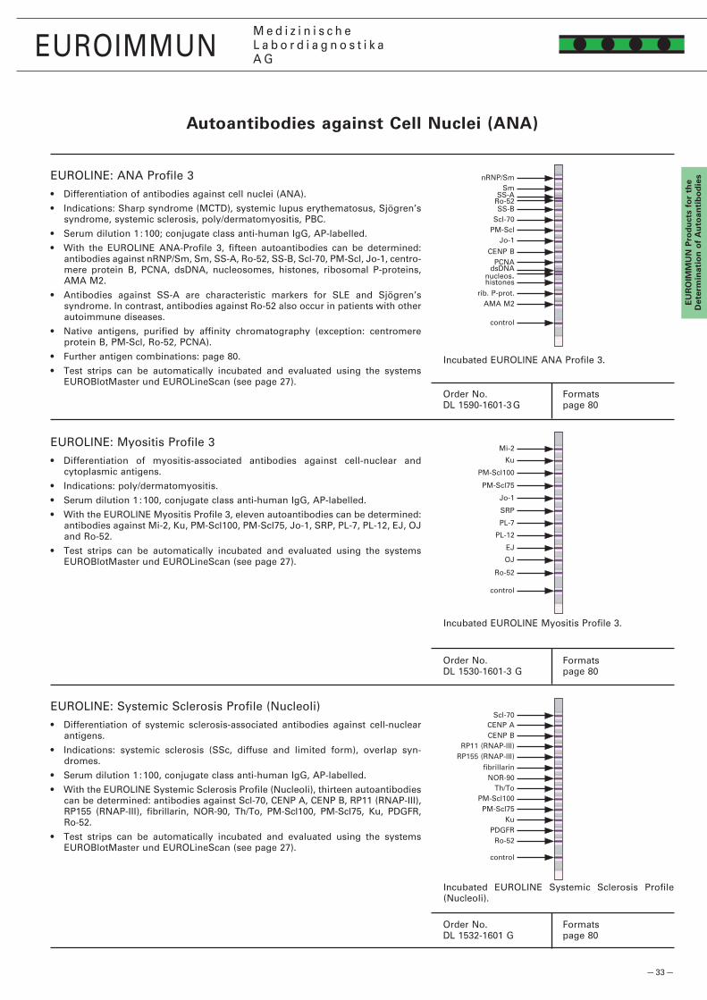
— 33 —
Sm
Jo-1
Scl-70
nRNP/Sm
SS-B
PM-Scl
AMA M�
CENP B
SS-ARo-5�
PCNA
EJ
Ro-5�
OJ
PL-1�
PM-Scl75
PM-Scl100
SRP
PL-7
Jo-1
Ku
Mi-�
PM-Scl100
KuPM-Scl75
Th/To
RP11 (RNAP-III)CENP B
NOR-90
RP155 (RNAP-III)
CENP AScl-70
PDGFRRo-5�
EUROIMMUNM e d i z i n i s c h eL a b o r d i a g n o s t i k aA G
EU
RO
IMM
UN
Pro
du
cts
for
the
det
erm
inat
ion
of
Au
toan
tib
od
ies
Incubated EUROLINE ANA Profile 3.
EUROLINE: ANA Profile 3• Differentiation of antibodies against cell nuclei (ANA).
• Indications: Sharp syndrome (MCTD), systemic lupus erythematosus, Sjögren’s syndrome, systemic sclerosis, poly/dermatomyositis, PBC.
• Serum dilution 1 : 100; conjugate class anti-human IgG, AP-labelled.
• With the EUROLINE ANA-Profile 3, fifteen autoantibodies can be determined: antibodies against nRNP/Sm, Sm, SS-A, Ro-5�, SS-B, Scl-70, PM-Scl, Jo-1, centro-mere protein B, PCNA, dsDNA, nucleosomes, histones, ribosomal P-proteins, AMA M�.
• Antibodies against SS-A are characteristic markers for SLE and Sjögren’s syndrome. In contrast, antibodies against Ro-5� also occur in patients with other autoimmune diseases.
• Native antigens, purified by affinity chromatography (exception: centromere protein B, PM-Scl, Ro-5�, PCNA).
• Further antigen combinations: page 80.
• Test strips can be automatically incubated and evaluated using the systems EUROBlotMaster und EUROLineScan (see page �7).
EUROLINE: Systemic Sclerosis Profile (Nucleoli)• Differentiation of systemic sclerosis-associated antibodies against cell-nuclear
antigens.
• Indications: systemic sclerosis (SSc, diffuse and limited form), overlap syn-dromes.
• Serum dilution 1 : 100, conjugate class anti-human IgG, AP-labelled.
• With the EUROLINE Systemic Sclerosis Profile (Nucleoli), thirteen autoantibodies can be determined: antibodies against Scl-70, CENP A, CENP B, RP11 (RNAP-III), RP155 (RNAP-III), fibrillarin, NOR-90, Th/To, PM-Scl100, PM-Scl75, Ku, PDGFR, Ro-5�.
• Test strips can be automatically incubated and evaluated using the systems EUROBlotMaster und EUROLineScan (see page �7).
Order No. FormatsDL 1590-1601-3 G page 80
Order No. FormatsDL 153�-1601 G page 80
Incubated EUROLINE Systemic Sclerosis Profile (Nucleoli).
Autoantibodies against Cell Nuclei (ANA)
histones
dsDNAnucleos.
rib. P-prot.
control
EUROLINE: Myositis Profile 3• Differentiation of myositis-associated antibodies against cell-nuclear and
cytoplasmic antigens.
• Indications: poly/dermatomyositis.
• Serum dilution 1 : 100, conjugate class anti-human IgG, AP-labelled.
• With the EUROLINE Myositis Profile 3, eleven autoantibodies can be determined: antibodies against Mi-�, Ku, PM-Scl100, PM-Scl75, Jo-1, SRP, PL-7, PL-1�, EJ, OJ and Ro-5�.
• Test strips can be automatically incubated and evaluated using the systems EUROBlotMaster und EUROLineScan (see page �7).
Order No. FormatsDL 1530-1601-3 G page 80
Incubated EUROLINE Myositis Profile 3.
control
fibrillarin
control
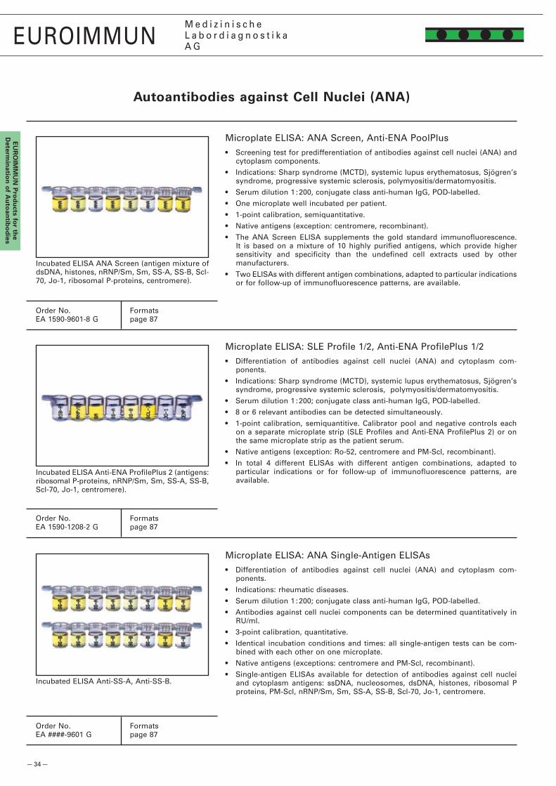
— 34 —
EUROIMMUNM e d i z i n i s c h eL a b o r d i a g n o s t i k aA G
EU
RO
IMM
UN
Pro
du
cts for th
e d
etermin
ation
of A
uto
antib
od
ies
Autoantibodies against Cell Nuclei (ANA)
Microplate ELISA: ANA Screen, Anti-ENA PoolPlus• Screening test for predifferentiation of antibodies against cell nuclei (ANA) and
cytoplasm components.
• Indications: Sharp syndrome (MCTD), systemic lupus erythematosus, Sjögren’s syndrome, progressive systemic sclerosis, polymyositis/dermatomyositis.
• Serum dilution 1 : �00, conjugate class anti-human IgG, POD-labelled.
• One microplate well incubated per patient.
• 1-point calibration, semiquantitative.
• Native antigens (exception: centromere, recombinant).
• The ANA Screen ELISA supplements the gold standard immunofluorescence. It is based on a mixture of 10 highly purified antigens, which provide higher sensitivity and specificity than the undefined cell extracts used by other manufacturers.
• Two ELISAs with different antigen combinations, adapted to particular indications or for follow-up of immunofluorescence patterns, are available.
Order No. FormatsEA 1590-9601-8 G page 87
Order No. FormatsEA 1590-1�08-� G page 87
Order No. FormatsEA ####-9601 G page 87
Microplate ELISA: SLE Profile 1/�, Anti-ENA ProfilePlus 1/�• Differentiation of antibodies against cell nuclei (ANA) and cytoplasm com-
ponents.
• Indications: Sharp syndrome (MCTD), systemic lupus erythematosus, Sjögren’s syndrome, progressive systemic sclerosis, polymyositis/dermatomyositis.
• Serum dilution 1 : �00; conjugate class anti-human IgG, POD-labelled.
• 8 or 6 relevant antibodies can be detected simultaneously.
• 1-point calibration, semiquantitive. Calibrator pool and negative controls each on a separate microplate strip (SLE Profiles and Anti-ENA ProfilePlus �) or on the same microplate strip as the patient serum.
• Native antigens (exception: Ro-5�, centromere and PM-Scl, recombinant).
• In total 4 different ELISAs with different antigen combinations, adapted to particular indications or for follow-up of immunofluorescence patterns, are available.
Microplate ELISA: ANA Single-Antigen ELISAs• Differentiation of antibodies against cell nuclei (ANA) and cytoplasm com-
ponents.
• Indications: rheumatic diseases.
• Serum dilution 1 : �00; conjugate class anti-human IgG, POD-labelled.
• Antibodies against cell nuclei components can be determined quantitatively in RU/ml.
• 3-point calibration, quantitative.
• Identical incubation conditions and times: all single-antigen tests can be com-bined with each other on one microplate.
• Native antigens (exceptions: centromere and PM-Scl, recombinant).
• Single-antigen ELISAs available for detection of antibodies against cell nuclei and cytoplasm antigens: ssDNA, nucleosomes, dsDNA, histones, ribosomal P proteins, PM-Scl, nRNP/Sm, Sm, SS-A, SS-B, Scl-70, Jo-1, centromere.
Incubated ELISA ANA Screen (antigen mixture of dsDNA, histones, nRNP/Sm, Sm, SS-A, SS-B, Scl-70, Jo-1, ribosomal P-proteins, centromere).
Incubated ELISA Anti-ENA ProfilePlus � (antigens: ribosomal P-proteins, nRNP/Sm, Sm, SS-A, SS-B, Scl-70, Jo-1, centromere).
Incubated ELISA Anti-SS-A, Anti-SS-B.
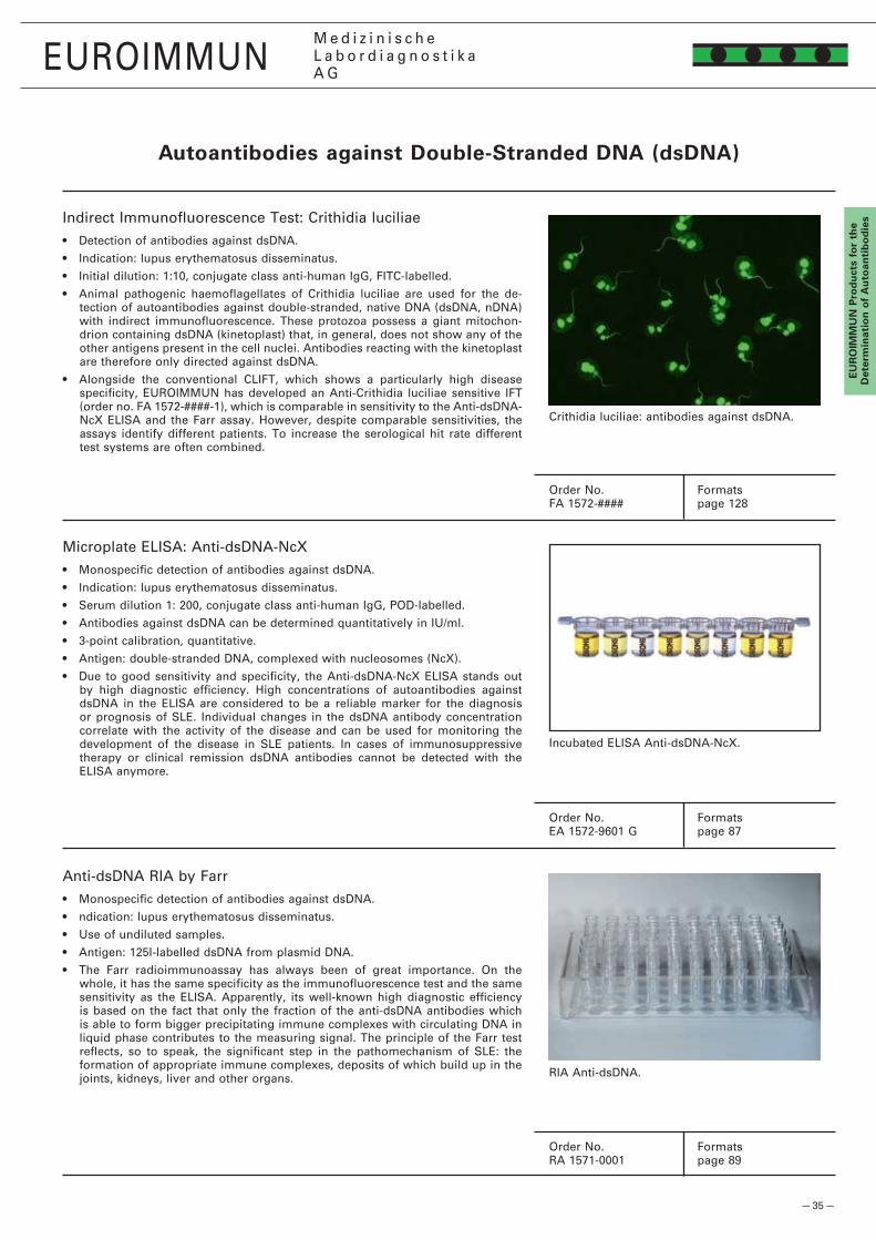
— 35 —
EUROIMMUNM e d i z i n i s c h eL a b o r d i a g n o s t i k aA G
EU
RO
IMM
UN
Pro
du
cts
for
the
det
erm
inat
ion
of
Au
toan
tib
od
ies
Crithidia luciliae: antibodies against dsDNA.
Incubated ELISA Anti-dsDNA-NcX.
RIA Anti-dsDNA.
Order No. FormatsFA 157�-#### page 1�8
Order No. FormatsEA 157�-9601 G page 87
Order No. FormatsRA 1571-0001 page 89
Indirect Immunofluorescence Test: Crithidia luciliae• Detection of antibodies against dsDNA.
• Indication: lupus erythematosus disseminatus.
• Initial dilution: 1:10, conjugate class anti-human IgG, FITC-labelled.
• Animal pathogenic haemoflagellates of Crithidia luciliae are used for the de-tection of autoantibodies against double-stranded, native DNA (dsDNA, nDNA) with indirect immunofluorescence. These protozoa possess a giant mitochon-drion containing dsDNA (kinetoplast) that, in general, does not show any of the other antigens present in the cell nuclei. Antibodies reacting with the kinetoplast are therefore only directed against dsDNA.
• Alongside the conventional CLIFT, which shows a particularly high disease specificity, EUROIMMUN has developed an Anti-Crithidia luciliae sensitive IFT (order no. FA 157�-####-1), which is comparable in sensitivity to the Anti-dsDNA-NcX ELISA and the Farr assay. However, despite comparable sensitivities, the assays identify different patients. To increase the serological hit rate different test systems are often combined.
Microplate ELISA: Anti-dsDNA-NcX• Monospecific detection of antibodies against dsDNA.
• Indication: lupus erythematosus disseminatus.
• Serum dilution 1: �00, conjugate class anti-human IgG, POD-labelled.
• Antibodies against dsDNA can be determined quantitatively in IU/ml.
• 3-point calibration, quantitative.
• Antigen: double-stranded DNA, complexed with nucleosomes (NcX).
• Due to good sensitivity and specificity, the Anti-dsDNA-NcX ELISA stands out by high diagnostic efficiency. High concentrations of autoantibodies against dsDNA in the ELISA are considered to be a reliable marker for the diagnosis or prognosis of SLE. Individual changes in the dsDNA antibody concentration correlate with the activity of the disease and can be used for monitoring the development of the disease in SLE patients. In cases of immunosuppressive therapy or clinical remission dsDNA antibodies cannot be detected with the ELISA anymore.
Anti-dsDNA RIA by Farr• Monospecific detection of antibodies against dsDNA.
• ndication: lupus erythematosus disseminatus.
• Use of undiluted samples.
• Antigen: 1�5I-labelled dsDNA from plasmid DNA.
• The Farr radioimmunoassay has always been of great importance. On the whole, it has the same specificity as the immunofluorescence test and the same sensitivity as the ELISA. Apparently, its well-known high diagnostic efficiency is based on the fact that only the fraction of the anti-dsDNA antibodies which is able to form bigger precipitating immune complexes with circulating DNA in liquid phase contributes to the measuring signal. The principle of the Farr test reflects, so to speak, the significant step in the pathomechanism of SLE: the formation of appropriate immune complexes, deposits of which build up in the joints, kidneys, liver and other organs.
Autoantibodies against doubleStranded dNA (dsdNA)

— 36 —
EUROIMMUNM e d i z i n i s c h eL a b o r d i a g n o s t i k aA G
EU
RO
IMM
UN
Pro
du
cts for th
e d
etermin
ation
of A
uto
antib
od
ies
Order No. (Anti-CCP) FormatsEA 1505-9601 G page 87
EUROIMMUNM e d i z i n i s c h eL a b o r d i a g n o s t i k aA G
EUROIMMUN AG · 23560 Luebeck (Germany) · Seekamp 31 · Telephone +49 451 58550 · Fax 5855591 · E-mail [email protected] · www.euroimmun.com
Antibodies against cyclic citrullinated peptides (CCP): An ELISA for specifi c diagnosis of rheumatoid arthritis
The amino acid citrulline Principle of the anti-CCP ELISA
colourlesschromogen
POD
stain
POD
human antibodyagainst CCP
peroxidase-labelled anti-human antibody
synthetic CCP
Rheumatoid arthritis (RA) is one of the most common autoimmune diseases, affect-ing around 1% of the world population. It is characterised by infl ammation of the syno-vial membrane, which spreads symmetrical-ly from the small to the large joints. Initial symptoms include painful swelling of fi nger joints with morning stiffness in the joints. Early diag nosis and immediate commence-ment of suitable therapy is necessary to keep the disease under control.
The most commonly performed serological test in suspected RA cases was until now the determination of rheumatoid factors (RF). These are antibodies (predominantly of class IgM) which react with gamma globulins and occur in 60-80% of RA patients. RF are a sen-sitive but not very specifi c marker for RA, since they also occur in healthy individuals and in patients with various infections or other autoimmune diseases (systemic lupus erythematosus, Sjögren’s syn drome, sclero-derma and others).
40-60% of RA patients also exhibit auto-anti bodies against epidermal fi l ag grin1 (RA keratin, anti-pe ri nu clear fac tor) in their se-rum. Fil ag grin is a protein of the epi dermis, which links keratin fi laments to one another. Autoanti bodies against fi laggrin are detected by indirect immuno fl uor escence: the antigen substrate rat oesophagus shows staining of the stratum corneum (RA keratin) on the luminal side; anti-perinuclear factors (APF) are apparent in the cyto plasmic inclusion bodies of human epithelial cells of the oral mucosa.
In recent years it has been shown that the rare amino acid citrulline, which is present in fi laggrin, is a substantial component of the antigenic epitope. Enzyme immunoassays which use synthetic citrullinated peptide as the target antigen offer a useful alter-native to indirect immunofl uorescence2. A direct comparison study demonstrated that the sensitivity can be increased from 49% to 68% by using cyclic citrullinated peptide instead of linear citrullinated peptide as an ELISA substrate3. Antibodies against cyc li c citrulli nated pep tides (CCP) are a new and highly specifi c marker for RA.
An ti bodies against CCP are predominantly of class IgG and have a specifi city of 98% for RA. They are observed very early in the disease course and have a high predictive value: patients with anti-CCP antibodies develop signifi cantly more radiologically detectable joint damage than anti-CCP-negative patients4. Antibodies against CCP possess a much higher specifi city than RF (anti-CCP: 97%, RF: 62%) with the same sen-sitivity (anti-CCP: 79%, RF: 78%)5. They can be detected in early stages of the disease in 79% of patients.
EU ROIMMUN offers an innovative micro-plate ELISA for quantitative deter min ation of autoantibodies against CCP. Diluted patient sera are incubated in wells coated with syn-thetic cyclic citrullinated peptides (second gener ation). Specifi c antibodies in the serum bind to the immobi lised antigen and cause a photo metric colour reaction by means of an enzyme-coupled secondary antibody. Five
Anti-CCP ELISA
NH
O NH2
O
N
H
NH
H2N+ NH2
O
N
H
Peptidylarginine-deiminase (PAD)
Ca2+
L-arginine L-citrulline
EA _1505_I_UK_A04, 07/06
calibration sera ensure reliable measure-ment of antibody concentrations. The EURO-IMMUN Anti-CCP ELISA is a highly specifi c and sensitive sero logical test system for the diagnosis of RA.
1) Nogueira et al., Ann. Rheum. Dis. 60: 882 (2001)2) Schellekens et al., J. Clin. Invest. 101: 273-281 (1998)3) Schellekens et al., Arthritis Rheum. 43: 155-163 (2000)4) Kroot et al., Arthritis Rheum. 43: 1831-1835 (2000) 5) Vasishta, Am. Clin. Lab. 21: 34-36 (2002)
Panel nAnti-CCPpositive
Sensitivity RA 419 329 (78.5%)
Asymptomatic blood donors
400 2 (0.5%)
Psoriatic arthritis 28 0
Other arthritides 35 3 (8.6%)
Systemic lupus erythematosus
108 3 (2.8%)
Sjögren‘s syndrome 106 2 (1.9%)
Scleroderma 98 3 (3.1%)
Autoimmune thyroiditis
159 4 (2.5%)
Wegener‘ granulomatosis
25 1 (4.0%)
Anti-parvovirus B19 positive
126 3 (2.4%)
Viral hepatides 54 0
Anti-HIV positive 5 0
Tuberculosis 10 0
Specifi city RA 1154 21 (98.2%)
Autoantibodies against CCP and Sa
Microplate ELISA: Anti-CCP, Anti-Sa
• Screening test for the specific determination of autoantibodies against cyclic citrullinated peptides (CCP) and citrullinated Sa.
• Indication: rheumatoid arthritis (RA).
• Serum dilution 1 : 100; conjugate class anti-human IgG, POD-labelled.
• Antibodies against CCP and Sa can be determined quantitatively in RU/ml. Optional reference control for the determination of ratio values.
• Antigen: synthetic cyclic citrullinated peptides (CCP, second generation), citrullinated Sa.
Antigen Order No.
CCP EA 1505-9601 G
Sa EA 151a-480� GIncubated ELISA Anti-CCP, Anti-Sa.
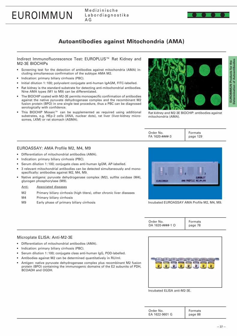
— 37 —
EUROIMMUNM e d i z i n i s c h eL a b o r d i a g n o s t i k aA G
EU
RO
IMM
UN
Pro
du
cts
for
the
det
erm
inat
ion
of
Au
toan
tib
od
ies
Autoantibodies against Mitochondria (AMA)
Indirect Immunofluorescence Test: EUROPLUS™ Rat Kidney and M�-3E BIOCHIPs• Screening test for the detection of antibodies against mitochondria (AMA) in-
cluding simultaneous confirmation of the subtype AMA M�.
• Indication: primary biliary cirrhosis (PBC).
• Initial dilution 1 : 100; polyvalent conjugate anti-human IgAGM, FITC-labelled.
• Rat kidney is the standard substrate for detecting anti-mitochondrial antibodies. Nine AMA types (M1 to M9) can be differentiated.
• The BIOCHIP coated with M�-3E permits monospecific confirmation of antibodies against the native pyruvate dehydrogenase complex and the recombinant M� fusion protein (BPO) in one single test procedure, thus a PBC can be diagnosed serologically with confidence.
• This BIOCHIP Mosaic™ can be supplemented as required using additional substrates, e.g. HEp-� cells (ANA, nuclear dots), rat liver (liver-kidney micro-somes, LKM) or rat stomach (ASMA).
Rat kidney and M�-3E BIOCHIP: antibodies against mitochondria (AMA).
EUROASSAY: AMA Profile M�, M4, M9• Differentiation of mitochondrial antibodies (AMA).
• Indication: primary biliary cirrhosis (PBC).
• Serum dilution 1 : 100; conjugate class anti-human IgGM, AP-labelled.
• 3 relevant mitochondrial antibodies can be detected simultaneously and mono-specifically: antibodies against M�, M4, M9.
• Native antigens: pyruvate dehydrogenase complex (M�), sulfite oxidase (M4), glycogen phosphorylase (M9).
Anti- Associated diseases
M� Primary biliary cirrhosis (high titers), other chronic liver diseases
M4 Primary biliary cirrhosis
M9 Early phase of primary biliary cirrhosis Incubated EUROASSAY AMA Profile M�, M4, M9.
Microplate ELISA: Anti-M�-3E• Differentiation of mitochondrial antibodies (AMA).
• Indication: primary biliary cirrhosis (PBC).
• Serum dilution 1 : 100; conjugate class anti-human IgG, POD-labelled.
• Antibodies against M� can be determined quantitatively in RU/ml.
• Antigen: native pyruvate dehydrogenase complex plus recombinant M� fusion protein (BPO) containing the immunogenic domains of the E� subunits of PDH, BCOADH and OGDH.
Incubated ELISA anti-M�-3E.
Order No. FormatsFA 16�0-####-3 page 1�9
Order No. FormatsDA 16�0-####-1 O page 78
Order No. FormatsEA 16��-9601 G page 88

— 38 —
EUROIMMUNM e d i z i n i s c h eL a b o r d i a g n o s t i k aA G
EU
RO
IMM
UN
Pro
du
cts for th
e d
etermin
ation
of A
uto
antib
od
ies
Indirect Immunofluorescence Test: Liver Mosaic 8• Screening and differentiation test for the detection of liver-specific antibodies,
antibodies against mitochondria (AMA), antibodies against cell nuclei (ANA), antibodies against smooth muscles (ASMA), F-actin and other autoantibodies.
• Indications: autoimmune hepatitis, primary biliary cirrhosis, rheumatic diseases.
• Initial dilution 1 : 100; polyvalent conjugate anti-human IgAGM, FITC-labelled.
• The BIOCHIP Mosaic™ consists of 6 substrates: human epithelial cells (HEp-�), primate liver, rat kidney, rat liver, rat stomach, VSM47. Thus, a broad spectrum of antigens is present, allowing not only targeted serological diagnoses, but also frequently yielding additional results with clinical relevance.
• Antibodies against cell nuclei (ANA) can be particularly well demonstrated using HEp-� cells and primate liver, and are identified according to their fluorescence patterns. However, they also stain the cell nuclei of the other tissues more or less intensely. Clinical significance: rheumatic diseases, primary biliary cirrhosis (antibodies against nuclear dots).
• With primate liver, several liver-specific autoantibodies can be investigated e.g. antibodies against liver cell membrane (anti-LMA) and liver-specific protein (anti-LSP). Clinical significance: autoimmune hepatitis.
• Antibodies against mitochondria (AMA) show a granular cytoplasmic fluo-rescence on all 6 substrates. With the standard substrate rat kidney, the proximal and distal tubule cells fluoresce equally. Clinical significance: primary biliary cirrhosis.
• Autoantibodies against liver-kidney microsomes (anti-LKM) react with rat liver and rat kidney (see below). The other substrates are essentially negative.
• In the case of autoantibodies against smooth muscles (ASMA), the tunica muscularis, the lamina muscularis mucosa as well as the interglandular con-tractile fibrils fluoresce on the rat stomach. ASMA directed against the target antigen F-actin furthermore react with the cytoskeleton of HEp-� cells and the bile canaliculi of primate liver. The substrate VSM47 reacts very specifically, showing a filamentous, needle-like fluorescence. Clinical significance: autoimmune (lupoid) chronic active hepatitis.
• The BIOCHIP Mosaic™ can be supplemented as required with additional sub-strates, e.g. Crithidia luciliae (antibodies against dsDNA), musculus iliopsoas (antibodies against skeletal muscles), heart (antibodies against striated muscles, antibodies against intercalated disks, AMA M7), different EUROPLUS™ sub-strates (AMA-M�-3E, Sp100, gp�10, PML, SLA/LP, LC-1, LKM).
HEp-� cells: anti-nuclear dots. Primate liver: anti-LSP. Rat kidney: AMA. Rat liver: ANA. Rat stomach: ASMA. VSM47: Anti-actin.
Indirect Immunofluorescence Test: BIOCHIP Mosaic™ Rat Liver/ Rat Kidney (Liver Mosaic 1)• Specific detection of antibodies against liver-kidney microsomes (anti-LKM).
• Indications: autoimmune hepatitis, often associated with extrahepatic syndromes such as arthralgias, glomerulonephritis, vitiligo and chronic inflammatory bowel diseases.
• Initial dilution 1 : 100; polyvalent conjugate anti-human IgAGM, FITC-labelled.
• Autoantibodies against liver-kidney microsomes react with rat liver and generate a smooth staining in the cytoplasm of the hepatocytes.
• In rat kidney, particularly in the cortex area, a fine granular fluorescence of the proximal tubules – recognizable by the luminal brush border – is visible. The distal tubules are negative. The fluorescence intensity of the liver cells is normally stronger than that of the proximal renal tubules.
• The differentiation between autoimmune hepatitis and virus-induced hepatitis can additionally be accomplished by investigating the appropriate viral parameters.
Rat liver and rat kidney: antibodies against liver-kidney microsomes (anti-LKM).
Order No. FormatsFA 1300-####-1 page 1�3
Order No. FormatsFA 1300-####-8 page 1�3
Autoantibodies against Liver Antigens

— 39 —
3E (BPO)
gp�10
LKM-1
PML
Sp100
LC-1
SLA/LP
Ro-5�
EUROIMMUNM e d i z i n i s c h eL a b o r d i a g n o s t i k aA G
EU
RO
IMM
UN
Pro
du
cts
for
the
det
erm
inat
ion
of
Au
toan
tib
od
iesEUROLINE: Profile Autoimmune Liver Diseases
• Differentiation of antibodies in autoimmune liver diseases.
• Indications: autoimmune hepatitis, primary biliary cirrhosis, overlap syndromes.
• Serum dilution 1 : 100, conjugate class anti-human IgG, AP-labelled.
• With the EUROLINE Profile Autoimmune Liver Diseases, nine autoantibodies can be determined: antibodies against AMA M�, M�-3E (BPO), Sp100, PML, gp�10, LKM-1, LC-1, SLA/LP and Ro-5�.
• Test strips can be automatically incubated and evaluated using the systems EUROBlotMaster und EUROLineScan (see page �7).
• Further antigen combinations on page 80.
Anti- Associated Diseases
M�, Sp100, PML, gp�10 Primary biliary cirrhosis
LKM-1, SLA/LP, LC-1 Autoimmune hepatitis
Ro-5� Autoimmune hepatitis, rheumatic diseases
Incubated EUROLINE Profile Autoimmune Liver Diseases.
EUROASSAY: Liver Profile (Anti-M�, -LKM-1, LC-1, -SLA/LP)• Determination of mitochondrial antibodies AMA M�, antibodies against liver-
kidney microsomes type 1 (LKM-1), antibodies against liver cytosolic antigen type 1 (LC-1), as well as of antibodies against soluble liver antigen/liver-pancreas antigen (SLA/LP).
• Indication: autoimmune liver diseases.
• Serum dilution 1 : 100, conjugate class anti-human IgG, AP-labelled.
• Antibodies against M�, LKM-1, LC-1 and SLA/LP can be detected simultaneously and monospecifically.
• Antigens: pyruvate dehydrogenase complex (M�, native), cytochrome P450 IID6 (LKM-1, recombinant), formiminotransferase-cyclodeaminase (LC-1, recombi-nant) and soluble liver antigen/liver-pancreas antigen (SLA/LP, recombinant).
Incubated EUROASSAY M�, LKM-1, LC-1, SLA/
Microplate ELISA: Anti-SLA/LP, Anti-LC-1, Anti-LKM-1• Monospecific determination of antibodies against soluble liver antigen/liver-
pancreas antigen (SLA/LP), cytosolic liver antigen type 1 (LC-1) and liver-kidney microsomes type 1 (LKM-1).
• Indication: autoimmune hepatitis.
• Serum dilution 1 : 100, conjugate class anti-human IgG, POD-labelled.
• 3-point calibration, quantitative (exception: Anti-LC-1, semi-quantitative).
• Identical incubation conditions and times: all tests can be combined without difficulty on one and the same microplate.
• Recombinant antigens: soluble liver antigen/liver-pancreas antigen (SLA/LP), formiminotransferase-cyclodeaminase (LC-1) and cytochrome P450 IID6 (LKM-1). The corresponding human cDNA was expressed in E. coli (SLA/LP) or insect cells (LC-1, LKM-1). Incubated ELISA Anti-SLA/LP, Anti-LC-1, Anti-LKM-1.
Order No. FormatsDL 1300-1601-4 G page 80
Order No. FormatsDA 1300-1003-3 G page 78
Order No. (Anti-SLA/LP) FormatsEA 130�-9601 G page 86
Autoantibodies against Liver Antigens
AMA M�
control
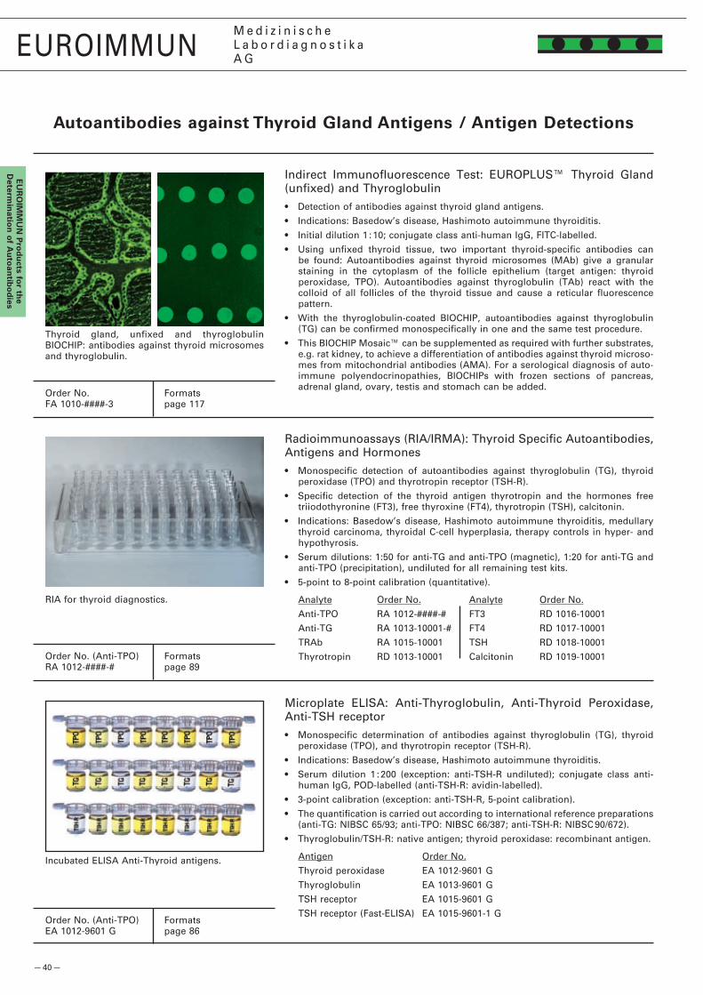
— 40 —
EUROIMMUNM e d i z i n i s c h eL a b o r d i a g n o s t i k aA G
EU
RO
IMM
UN
Pro
du
cts for th
e d
etermin
ation
of A
uto
antib
od
ies
Autoantibodies against Thyroid gland Antigens / Antigen detections
Indirect Immunofluorescence Test: EUROPLUS™ Thyroid Gland (unfixed) and Thyroglobulin• Detection of antibodies against thyroid gland antigens.
• Indications: Basedow’s disease, Hashimoto autoimmune thyroiditis.
• Initial dilution 1 : 10; conjugate class anti-human IgG, FITC-labelled.
• Using unfixed thyroid tissue, two important thyroid-specific antibodies can be found: Autoantibodies against thyroid microsomes (MAb) give a granular staining in the cytoplasm of the follicle epithelium (target antigen: thyroid peroxidase, TPO). Autoantibodies against thyroglobulin (TAb) react with the colloid of all follicles of the thyroid tissue and cause a reticular fluorescence pattern.
• With the thyroglobulin-coated BIOCHIP, autoantibodies against thyroglobulin (TG) can be confirmed monospecifically in one and the same test procedure.
• This BIOCHIP Mosaic™ can be supplemented as required with further substrates, e.g. rat kidney, to achieve a differentiation of antibodies against thyroid microso-mes from mitochondrial antibodies (AMA). For a serological diagnosis of auto-immune polyendocrinopathies, BIOCHIPs with frozen sections of pancreas, adrenal gland, ovary, testis and stomach can be added.
Thyroid gland, unfixed and thyroglobulin BIOCHIP: antibodies against thyroid microsomes and thyroglobulin.
Radioimmunoassays (RIA/IRMA): Thyroid Specific Autoantibodies, Antigens and Hormones• Monospecific detection of autoantibodies against thyroglobulin (TG), thyroid
peroxidase (TPO) and thyrotropin receptor (TSH-R).
• Specific detection of the thyroid antigen thyrotropin and the hormones free triiodothyronine (FT3), free thyroxine (FT4), thyrotropin (TSH), calcitonin.
• Indications: Basedow’s disease, Hashimoto autoimmune thyroiditis, medullary thyroid carcinoma, thyroidal C-cell hyperplasia, therapy controls in hyper- and hypothyrosis.
• Serum dilutions: 1:50 for anti-TG and anti-TPO (magnetic), 1:�0 for anti-TG and anti-TPO (precipitation), undiluted for all remaining test kits.
• 5-point to 8-point calibration (quantitative).
Analyte Order No. Analyte Order No.
Anti-TPO RA 101�-####-# FT3 RD 1016-10001
Anti-TG RA 1013-10001-# FT4 RD 1017-10001
TRAb RA 1015-10001 TSH RD 1018-10001
Thyrotropin RD 1013-10001 Calcitonin RD 1019-10001
RIA for thyroid diagnostics.
Microplate ELISA: Anti-Thyroglobulin, Anti-Thyroid Peroxidase, Anti-TSH receptor• Monospecific determination of antibodies against thyroglobulin (TG), thyroid
peroxidase (TPO), and thyrotropin receptor (TSH-R).
• Indications: Basedow’s disease, Hashimoto autoimmune thyroiditis.
• Serum dilution 1 : �00 (exception: anti-TSH-R undiluted); conjugate class anti-human IgG, POD-labelled (anti-TSH-R: avidin-labelled).
• 3-point calibration (exception: anti-TSH-R, 5-point calibration).
• The quantification is carried out according to international reference preparations (anti-TG: NIBSC 65/93; anti-TPO: NIBSC 66/387; anti-TSH-R: NIBSC 90/67�).
• Thyroglobulin/TSH-R: native antigen; thyroid peroxidase: recombinant antigen.
Antigen Order No.
Thyroid peroxidase EA 101�-9601 G
Thyroglobulin EA 1013-9601 G
TSH receptor EA 1015-9601 G
TSH receptor (Fast-ELISA) EA 1015-9601-1 G
Incubated ELISA Anti-Thyroid antigens.
Order No. FormatsFA 1010-####-3 page 117
Order No. (Anti-TPO) FormatsRA 101�-####-# page 89
Order No. (Anti-TPO) FormatsEA 101�-9601 G page 86

— 41 —
EUROIMMUNM e d i z i n i s c h eL a b o r d i a g n o s t i k aA G
EU
RO
IMM
UN
Pro
du
cts
for
the
det
erm
inat
ion
of
Au
toan
tib
od
iesIndirect Immunofluorescence test: Dermatology Mosaic 7
• Screening and differentiation test for detection of skin-specific antibodies.
• Indication: autoimmune bullous dermatoses.
• Initial dilution 1 : 10; conjugate class anti-human IgG, FITC-labelled.
• The BIOCHIP Mosaic consists of 6 substrates: primate oesophagus, salt-split skin, desmoglein-1-expressing cells, desmoglein-3-expressing cells, BP�30-expressing cells (gc) and BP180 (EUROPLUS). Thus a comprehensive antigen spectrum is available in a single analysis, allowing targeted serological diagnosis.
• Antibodies against prickle cell desmosomes react with surface antigens of keratinocytes. Tissue sections of oesophagus and tongue show a granular fluorescence of the intercellular matter in the whole stratum spinosum, but differentiation between desmoglein 1 and desmoglein 3 is difficult. When specific transfected cells are employed in addition, a targeted diagnosis in a single test run is possible.
• Antibodies against basal membrane structures react with salt-split skin. Anti-BP180, anti-BP�30 and anti–LAD97 cause staining of the epidermal side, and antibodies against laminin 5, collagen VII and other antigens staining of the dermal side of salt-split skin.
• When autoantibodies against BP180 or BP�30 are present, the epidermal basal membrane in the oesophagus or tongue is visible as a fine linear colouring between the stratum basale and the connective tissue. These antibodies can be differentiated by means of BP180-NC16A-4X coated BIOCHIPs and cells specifically transfected with BP�30 (globular C-terminal domain (gC)).
• This BIOCHIP Mosaic can be customised with further substrates if required, e.g. tongue (antibodies against prickle cell desmosomes, epidermal basal membrane), bladder (antibodies against plakins).
Substrate Order No.
Desmoglein 1 (transfected / non-transfected cells) FA 1495-####-50
Desmoglein 3 (transfected / non-transfected cells) FA 1496-####-50
Oesophagus FA 1501-####
Oesophagus / Tongue FA 1501-####-1
Tongue FA 150�-####
Bladder mucosa FA 1507-####
Salt split skin FA 150b-####
BP180-NC16A-4X / BP�30gC FA 150�-####-1
Envoplakin FA 1491-####-50
Collagen type VII NC1 FA 1947-####-50
Microplate ELISA: Anti-Desmoglein 1, Anti-Desmoglein 3, Anti-Envoplakin, Anti-BP180-NC16A-4X, Anti-BP�30-CF• Monospecific detection of antibodies against desmoglein 1, desmoglein 3, envo-
plakin, BP180 und BP�30.
• Indication: autoimmune bullous dermatoses.
• Serum dilution 1 : 100; conjugate class anti-human IgG, POD-labelled.
• 3-point calibration, quantitative (exception: anti-envoplakin, semiquantitative).
• Identical incubation conditions and times: all tests can be combined on one microplate.
• Recombinant antigens: extracellular domain of desmoglein 1 or 3, envoplakin, tetramer of NC16A domain of BP180 protein, C-terminal segment of BP�30 protein. The corresponding human cDNA is produced in E. coli (BP180-NC16A-4X, BP�30-CF, envoplakin) or in mammalian cells (desmoglein 1, desmoglein 3).
Oesophagus and salt-split skin (top left, middle left): ab against epid. basal membrane. BP�30 gc (top right). BP180 (NC16A-4X) (middle right). Desmoglein 1 + 3 (bottom left and right).
Order No. FormatsFA 1501-####-7 page 1�5
Order No. (Anti-Dsg-1) FormatsEA 1495-4801 G page 87
Incubated ELISA Anti-Desmoglein 1 and 3, Anti-BP180-NC16A-4X, Anti-BP�30-CF.
Autoantibodies against Antigens of the Skin
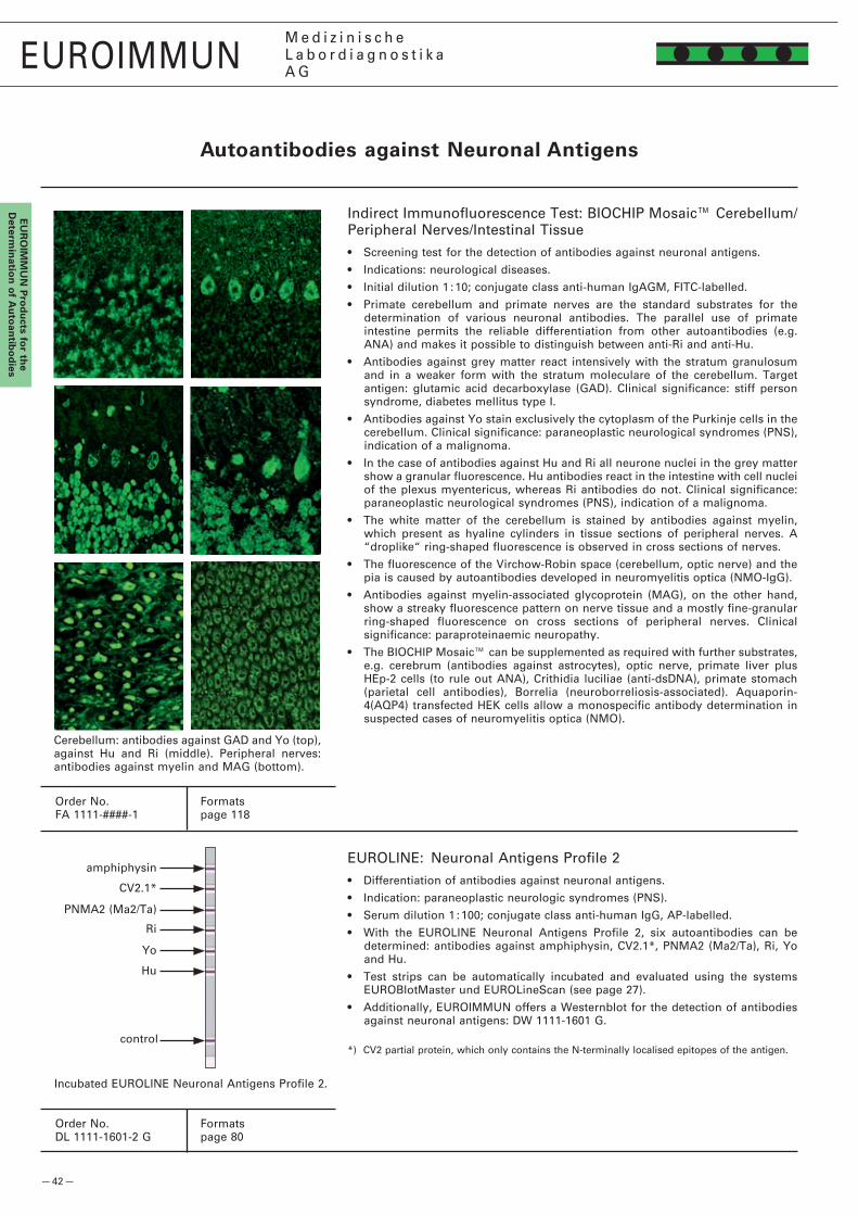
— 4� —
CV�.1*
Ri
Yo
Hu
PNMA� (Ma�/Ta)
EUROIMMUNM e d i z i n i s c h eL a b o r d i a g n o s t i k aA G
EU
RO
IMM
UN
Pro
du
cts for th
e d
etermin
ation
of A
uto
antib
od
ies
Autoantibodies against Neuronal Antigens
Indirect Immunofluorescence Test: BIOCHIP Mosaic™ Cerebellum/Peripheral Nerves/Intestinal Tissue• Screening test for the detection of antibodies against neuronal antigens.
• Indications: neurological diseases.
• Initial dilution 1 : 10; conjugate class anti-human IgAGM, FITC-labelled.
• Primate cerebellum and primate nerves are the standard substrates for the determination of various neuronal antibodies. The parallel use of primate intestine permits the reliable differentiation from other autoantibodies (e.g. ANA) and makes it possible to distinguish between anti-Ri and anti-Hu.
• Antibodies against grey matter react intensively with the stratum granulosum and in a weaker form with the stratum moleculare of the cerebellum. Target antigen: glutamic acid decarboxylase (GAD). Clinical significance: stiff person syndrome, diabetes mellitus type I.
• Antibodies against Yo stain exclusively the cytoplasm of the Purkinje cells in the cerebellum. Clinical significance: paraneoplastic neurological syndromes (PNS), indication of a malignoma.
• In the case of antibodies against Hu and Ri all neurone nuclei in the grey matter show a granular fluorescence. Hu antibodies react in the intestine with cell nuclei of the plexus myentericus, whereas Ri antibodies do not. Clinical significance: paraneoplastic neurological syndromes (PNS), indication of a malignoma.
• The white matter of the cerebellum is stained by antibodies against myelin, which present as hyaline cylinders in tissue sections of peripheral nerves. A “droplike“ ring-shaped fluorescence is observed in cross sections of nerves.
• The fluorescence of the Virchow-Robin space (cerebellum, optic nerve) and the pia is caused by autoantibodies developed in neuromyelitis optica (NMO-IgG).
• Antibodies against myelin-associated glycoprotein (MAG), on the other hand, show a streaky fluorescence pattern on nerve tissue and a mostly fine-granular ring-shaped fluorescence on cross sections of peripheral nerves. Clinical significance: paraproteinaemic neuropathy.
• The BIOCHIP Mosaic™ can be supplemented as required with further substrates, e.g. cerebrum (antibodies against astrocytes), optic nerve, primate liver plus HEp-� cells (to rule out ANA), Crithidia luciliae (anti-dsDNA), primate stomach (parietal cell antibodies), Borrelia (neuroborreliosis-associated). Aquaporin-4(AQP4) transfected HEK cells allow a monospecific antibody determination in suspected cases of neuromyelitis optica (NMO).
Cerebellum: antibodies against GAD and Yo (top), against Hu and Ri (middle). Peripheral nerves: antibodies against myelin and MAG (bottom).
EUROLINE: Neuronal Antigens Profile �• Differentiation of antibodies against neuronal antigens.
• Indication: paraneoplastic neurologic syndromes (PNS).
• Serum dilution 1 : 100; conjugate class anti-human IgG, AP-labelled.
• With the EUROLINE Neuronal Antigens Profile �, six autoantibodies can be determined: antibodies against amphiphysin, CV�.1*, PNMA� (Ma�/Ta), Ri, Yo and Hu.
• Test strips can be automatically incubated and evaluated using the systems EUROBlotMaster und EUROLineScan (see page �7).
• Additionally, EUROIMMUN offers a Westernblot for the detection of antibodies against neuronal antigens: DW 1111-1601 G.
*) CV� partial protein, which only contains the N-terminally localised epitopes of the antigen.
Incubated EUROLINE Neuronal Antigens Profile �.
Order No. FormatsFA 1111-####-1 page 118
Order No. FormatsDL 1111-1601-� G page 80
amphiphysin
control
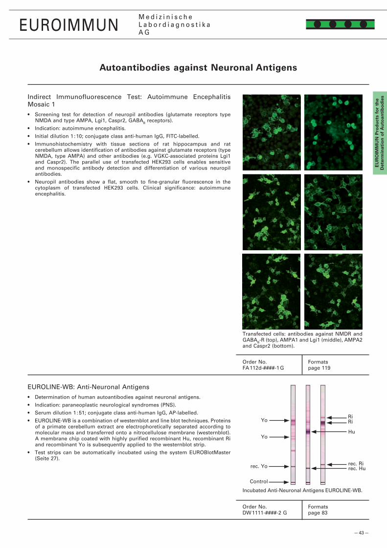
— 43 —
Yo
Yo RiRi
Hu
EUROIMMUNM e d i z i n i s c h eL a b o r d i a g n o s t i k aA G
EU
RO
IMM
UN
Pro
du
cts
for
the
det
erm
inat
ion
of
Au
toan
tib
od
ies
EUROLINE-WB: Anti-Neuronal Antigens• Determination of human autoantibodies against neuronal antigens.
• Indication: paraneoplastic neurological syndromes (PNS).
• Serum dilution 1 : 51; conjugate class anti-human IgG, AP-labelled.
• EUROLINE-WB is a combination of westernblot and line blot techniques. Proteins of a primate cerebellum extract are electrophoretically separated according to molecular mass and transferred onto a nitrocellulose membrane (westernblot). A membrane chip coated with highly purified recombinant Hu, recombinant Ri and recombinant Yo is subsequently applied to the westernblot strip.
• Test strips can be automatically incubated using the system EUROBlotMaster (Seite �7).
Order No. FormatsDW 1111-####-� G page 83
rec. Yo
Control
rec. Rirec. Hu
Incubated Anti-Neuronal Antigens EUROLINE-WB.
Order No. FormatsFA 11�d-####-1 G page 119
Autoantibodies against Neuronal Antigens
Indirect Immunofluorescence Test: Autoimmune Encephalitis Mosaic 1• Screening test for detection of neuropil antibodies (glutamate receptors type
NMDA and type AMPA, Lgi1, Caspr�, GABAB receptors).
• Indication: autoimmune encephalitis.
• Initial dilution 1 : 10; conjugate class anti-human IgG, FITC-labelled.
• Immunohistochemistry with tissue sections of rat hippocampus and rat cerebellum allows identification of antibodies against glutamate receptors (type NMDA, type AMPA) and other antibodies (e.g. VGKC-associated proteins Lgi1 and Caspr�). The parallel use of transfected HEK�93 cells enables sensitive and monospecific antibody detection and differentiation of various neuropil antibodies.
• Neuropil antibodies show a flat, smooth to fine-granular fluorescence in the cytoplasm of transfected HEK�93 cells. Clinical significance: autoimmune encephalitis.
Transfected cells: antibodies against NMDR and GABAB-R (top), AMPA1 and Lgi1 (middle), AMPA� and Caspr� (bottom).

— 44 —
EUROIMMUNM e d i z i n i s c h eL a b o r d i a g n o s t i k aA G
EU
RO
IMM
UN
Pro
du
cts for th
e d
etermin
ation
of A
uto
antib
od
ies
Autoantibodies against Islet Cell Antigens
Indirect Immunofluorescence Test: Primate Pancreas• Detection of antibodies against islet cells.
• Indications: Differentiation between a late manifestation of diabetes type 1 (latent autoimmune diabetes in adulthood, LADA) and diabetes type �.
• For a reliable determination of antibodies against islet cells an extended incubation time of 18 hours for the patient serum must be observed. The incubation time may be reduced to � hours but this will lead to a decrease in the sensitivity of the antibody detection test.
• Standardised control with JDF units available (order no. CA 10�1-0101-1).
• With indirect immunofluorescence autoantibodies against pancreas islets (ICA) can be detected in 80% of patients with new-onset diabetes type 1. Two target antigens of ICA have been identified so far: the enzymes glutamic acid decarboxylase (GAD) and tyrosine phosphatase (IA�).
• This BIOCHIP may be supplemented with further substrates, e. g. primate cerebellum for the detection of antibodies against GAD.
• The microscopic evaluation can be significantly simplified by using small BIOCHIPs (1 x 1 mm). The BIOCHIPs appear almost completely in the field of view and facilitate finding the islet cells, thus rendering a time-consuming search unnecessary, especially in negative samples.
Primate pancreas: antibodies against islet cells.
Microplate ELISA: Anti-GAD, Anti-IA�, Anti-GAD/IA� Pool• Monospecific detection of antibodies against glutamic acid decarboxylase (GAD),
tyrosine phosphatase (IA�) or bispecific detection of both antibodies in a single reagent well.
• Indications: early diagnosis of diabetes mellitus type 1, risk prediction in first grade relatives, prognosis of the clinical progression of diabetes type 1 for prediction of insulin dependence, differential diagnosis in gestational diabetes, differentiation between a late manifestation of diabetes type 1 (latent autoimmune diabetes in adulthood, LADA) and diabetes type �.
• Use of undiluted samples. Similar incubation conditions and times. Manual or automated test performance.
• Multipoint calibration. The quantitation is based on an international reference preparation (NIBSC 97/550).
• GAD and IA�: human, recombinant antigens.
Antigen Order no.
Glutamic acid decarboxylase (GAD) EA 10��-9601 G
Tyrosine phosphatase (IA�) EA 10�3-9601 G
GAD/IA� Pool EA 10��-9601-1 G
Incubated ELISA anti-GAD.
RIA: Anti-GAD, Anti-IA�, Anti-Insulin• Monospecific detection of antibodies against glutamic acid decarboxylase (GAD),
tyrosine phosphatase (IA�) and insulin.
• Indications: Early diagnosis of diabetes mellitus type 1, risk prediction in first grade relatives, prognosis of the clinical progression of diabetes type 1 for prediction of insulin dependence, differential diagnosis in gestational diabetes, differentiation between a late manifestation of diabetes type 1 (latent autoimmune diabetes in adulthood, LADA) and diabetes type �.
• Use of undiluted samples. Similar incubation conditions and times. Manual or automated test performance.
• Test kit formats for 50 or 100 determinations.
• GAD and IA�: human, recombinant, 1�5I-labelled antigens, insulin: human, synthetic, 1�5I-labelled antigen.
Antigen Order no.
Glutamat-Decarboxylase (GAD) RA 10��-####
Tyrosin-Phosphatase (IA�) RA 10�3-####
Insulin RA 10�4-####
RIA Anti-IA�.
Order No. FormatsFA 10�0-#### page 117
Order No. (Anti-GAD) FormatsEA 10��-9601 G page 86
Order No. (Anti-IA�) FormatsRA 10�3-#### page 89

— 45 —
EUROIMMUNM e d i z i n i s c h eL a b o r d i a g n o s t i k aA G
EU
RO
IMM
UN
Pro
du
cts
for
the
det
erm
inat
ion
of
Au
toan
tib
od
iesIndirect Immunfluorescence Test: Primate Stomach with Urea
Pretreatment• Screening test for detection of antibodies against parietal cells.
• Indications: forms of chronic atrophic gastritis, pernicious anemia, funicular myelosis, various autoimmune endocrinopathies such as Basedow’s and Addison’s diseases.
• Initial dilution 1 : 10; polyvalent conjugate anti-human IgAGM, FITC-labelled.
• Primate stomach is the standard substrate for detection of parietal cell antibodies. For titration, stomach tissue from rat or mouse is sufficient.
• With positive results the parietal cells show a course granular to clumpy fluorescence, and the surrounding areas are usually dark.
• Parietal cell antibodies (PCA) are often mixed up with antibodies against mitochondria (AMA) in microscopic analysis. The latter give an even fine granular fluorescence of the parietal cell cytoplasm, with the surrounding region showing a (weaker) reaction.
• For reliable differentiation of both types of antibody, a 30-minute pretreatment of the tissue sections with urea-glycine buffer (order no. ZF 1140-0101, see page 151) is recommended.
• The cytomplasmic fluorescence of parietal cells resulting from PCA occurs with the same intensity with or without urea pretreatment. The typical pattern of mitochondrial antibodies is almost completely supressed by urea pretreatment, greatly facilitating PCA diagnostics.
• In some AMA-positive samples it is possible to detect PCA that are obscured by the AMA pattern in conventional tissue sections.
• The urea-pretreated tissue shows a significantly darker background, enabling specific fluorescence to be more easily and reliable identified.
• This BIOCHIP can be supplemented with additional substrates, for example, thyroid (thyroid peroxidase, thyroglobulin), pancreas (pancreas islets), adrenal gland (adrenal cortex), ovary (ovary antigens), testis (Leydig cells), and intrinsic factor.
Primate stomach: antibodies against parietal cells. With urea-pretreatment (top) and without urea-pretreatment (bottom).
Microplate ELISA: Anti-Parietal Cells• Monospecific detection of antibodies against parietal cells (PCA).
• Indications: forms of chronic atrophic gastritis, pernicious anemia, funicular myelosis, various autoimmune endocrinopathies such as Basedow’s and Addison’s diseases
• Serum dilution 1 : �00; conjugate class anti-human IgG, POD-labelled.
• 3-point calibration, quantitative.
• Native antigen: H+/K+-ATPase, purified by affinity chromatography.
Incubated ELISA Anti-Parietal Cells.
Order No. FormatsFA 1360-#### G page 1�3
Order No. FormatsEA 1361-9601 G page 86
Primate stomach: antibodies against mitochon-dria. With urea-pretreatment (top) and without urea-pretreatment (bottom).
Autoantibodies against Parietal Cells (PCA)
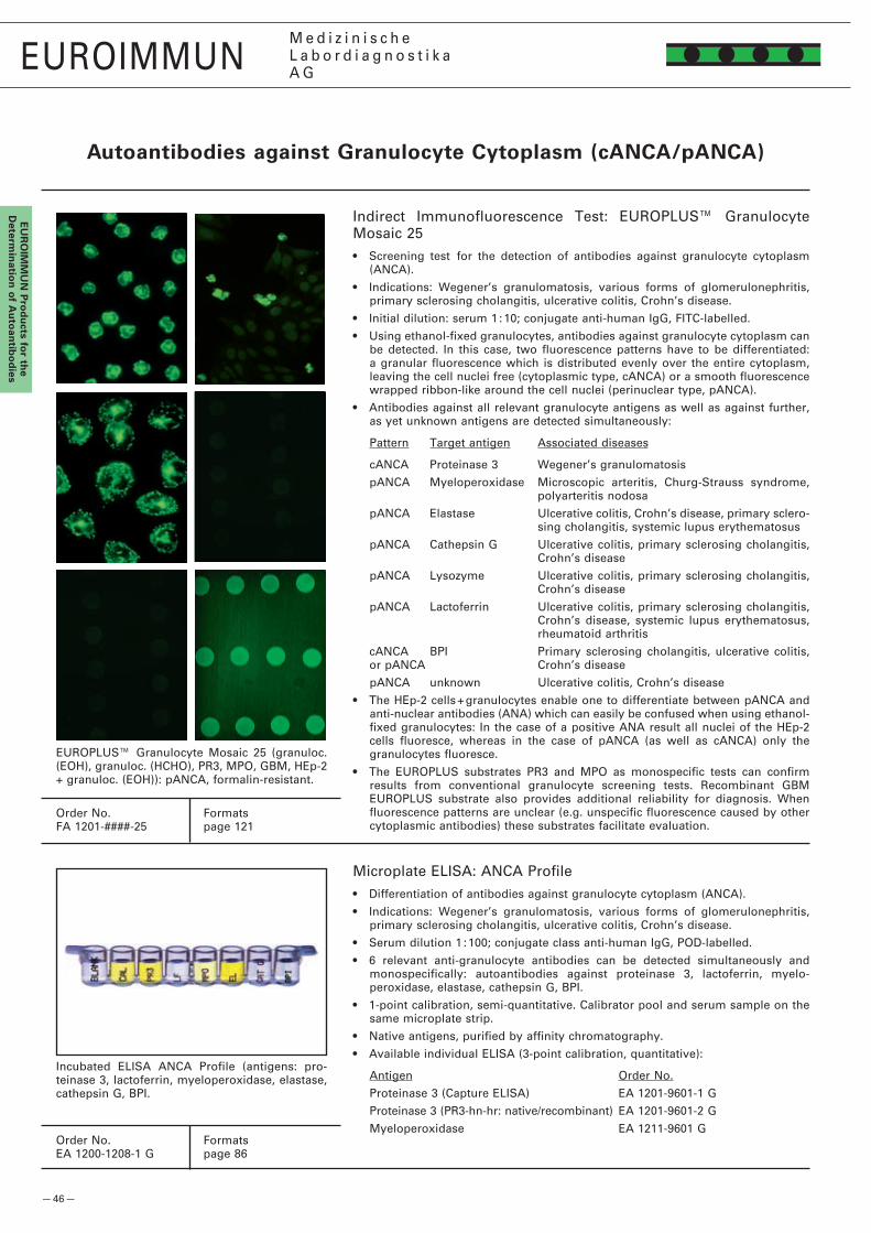
— 46 —
EUROIMMUNM e d i z i n i s c h eL a b o r d i a g n o s t i k aA G
EU
RO
IMM
UN
Pro
du
cts for th
e d
etermin
ation
of A
uto
antib
od
ies
Autoantibodies against granulocyte Cytoplasm (cANCA/pANCA)
Indirect Immunofluorescence Test: EUROPLUS™ Granulocyte Mosaic �5• Screening test for the detection of antibodies against granulocyte cytoplasm
(ANCA).
• Indications: Wegener’s granulomatosis, various forms of glomerulonephritis, primary sclerosing cholangitis, ulcerative colitis, Crohn’s disease.
• Initial dilution: serum 1 : 10; conjugate anti-human IgG, FITC-labelled.
• Using ethanol-fixed granulocytes, antibodies against granulocyte cytoplasm can be detected. In this case, two fluorescence patterns have to be differentiated: a granular fluorescence which is distributed evenly over the entire cytoplasm, leaving the cell nuclei free (cytoplasmic type, cANCA) or a smooth fluorescence wrapped ribbon-like around the cell nuclei (perinuclear type, pANCA).
• Antibodies against all relevant granulocyte antigens as well as against further, as yet unknown antigens are detected simultaneously:
Pattern Target antigen Associated diseases
cANCA Proteinase 3 Wegener’s granulomatosis
pANCA Myeloperoxidase Microscopic arteritis, Churg-Strauss syndrome, polyarteritis nodosa
pANCA Elastase Ulcerative colitis, Crohn’s disease, primary sclero-sing cholangitis, systemic lupus erythematosus
pANCA Cathepsin G Ulcerative colitis, primary sclerosing cholangitis, Crohn’s disease
pANCA Lysozyme Ulcerative colitis, primary sclerosing cholangitis, Crohn’s disease
pANCA Lactoferrin Ulcerative colitis, primary sclerosing cholangitis, Crohn’s disease, systemic lupus erythematosus, rheumatoid arthritis
cANCA BPI Primary sclerosing cholangitis, ulcerative colitis, or pANCA Crohn’s disease
pANCA unknown Ulcerative colitis, Crohn’s disease
• The HEp-� cells + granulocytes enable one to differentiate between pANCA and anti-nuclear antibodies (ANA) which can easily be confused when using ethanol-fixed granulocytes: In the case of a positive ANA result all nuclei of the HEp-� cells fluoresce, whereas in the case of pANCA (as well as cANCA) only the granulocytes fluoresce.
• The EUROPLUS substrates PR3 and MPO as monospecific tests can confirm results from conventional granulocyte screening tests. Recombinant GBM EUROPLUS substrate also provides additional reliability for diagnosis. When fluorescence patterns are unclear (e.g. unspecific fluorescence caused by other cytoplasmic antibodies) these substrates facilitate evaluation.
Microplate ELISA: ANCA Profile• Differentiation of antibodies against granulocyte cytoplasm (ANCA).
• Indications: Wegener’s granulomatosis, various forms of glomerulonephritis, primary sclerosing cholangitis, ulcerative colitis, Crohn’s disease.
• Serum dilution 1 : 100; conjugate class anti-human IgG, POD-labelled.
• 6 relevant anti-granulocyte antibodies can be detected simultaneously and monospecifically: autoantibodies against proteinase 3, lactoferrin, myelo-peroxidase, elastase, cathepsin G, BPI.
• 1-point calibration, semi-quantitative. Calibrator pool and serum sample on the same microplate strip.
• Native antigens, purified by affinity chromatography.
• Available individual ELISA (3-point calibration, quantitative):
Antigen Order No.
Proteinase 3 (Capture ELISA) EA 1�01-9601-1 G
Proteinase 3 (PR3-hn-hr: native/recombinant) EA 1�01-9601-� G
Myeloperoxidase EA 1�11-9601 G
Incubated ELISA ANCA Profile (antigens: pro-teinase 3, lactoferrin, myeloperoxidase, elastase, cathepsin G, BPI.
Order No. FormatsFA 1�01-####-�5 page 1�1
Order No. FormatsEA 1�00-1�08-1 G page 86
EUROPLUS™ Granulocyte Mosaic �5 (granuloc. (EOH), granuloc. (HCHO), PR3, MPO, GBM, HEp-� + granuloc. (EOH)): pANCA, formalin-resistant.

— 47 —
EUROIMMUNM e d i z i n i s c h eL a b o r d i a g n o s t i k aA G
EU
RO
IMM
UN
Pro
du
cts
for
the
det
erm
inat
ion
of
Au
toan
tib
od
ies
EUROIMMUNM e d i z i n i s c h eL a b o r d i a g n o s t i k aA G
Screening test indirect immunofluorescence: BIOCHIP sextet granulocytes (EOH), granulocytes (HCHO), EUROPLUSTM microdots PR3, EUROPLUSTM microdots MPO, human epithelial cells (HEp-2), and primate liver
AAb againstPR3-hn-hr
WG 80-90 %
AAb againstBPI
WG 5 %
ELISA: anti-PR3-hn-hr / ANCA Profile (BPI)
cANCA
Cytoplasmic fluorescenceof granulocytes (A, B, F)
WG 80-90 %MPA 10-15 %CSS 10-20 %PAN < 9 %
pANCA
Perinuclear fluorescence of granulocytes (A, F)
MPA 42-70 %CSS 18-60 %SLE 9-25 %RA 3-25 %
pANCA, formalin-resistant
At B cytoplasmicfluorescence
pANCA, formalin-sensitive
At B no cytoplasmicfluorescence
only ANA
Fluorescence of all cell nuclei(A, E, F)
pANCA, formalin-res. & ANA
Cytoplasmic fluorescenceof granulocytes (B)
pANCA, formalin-sens. & ANA
Nuclei of granuloc. brighter thannuclei of hepatocytes (F), B neg.
pANCA, formalin-sens.? & ANA
ANCA reaction at A,ANA reaction at E & F
Qualified ANA diagnostics: screening test usingHEp-2 cells/primate liver (E, F), differentiationusing ELISA, EUROASSAY, EUROLINE, Westernblot
See EUROIMMUN poster: "Strategy for Determinationof Autoantibodies against Cell Nuclei (ANA)
and Cytoplasm Components"
AAb againstMPO
ELISA: anti-MPO
AAb against PR3, MPO,elastase, cathepsin G,
BPI, lactoferrin
WG 5 % (BPI)MPA 53 % (MPO)MPA 3 % (LF)RA, vasculitides 45 % (LF)SLE 6 % (EL)
ELISA: ANCA Profile
ANA and pANCA
Fluorescence of all cell nuclei(A, E, F), granuloc. accent. (F)
MPA 42-70 %
CU 67 %MC 7 %PSC 87 %
MPA 53 %
F
Primateliver
E
HEp-2cells
B
Granulocytes(HCHO-fixed)
D
EUROPLUSTM
MPOmicrodots
C
EUROPLUSTM
PR3microdots
A
Granulocytes(EOH-fixed)
AAb against dsDNA, ssDNA, nucleosomes, histones, nuclear membrane, nRNP/Sm, Sm, SS-A, Ro-52, SS-B, Ku, cyclin I (PCNA), mitosin (CENP-F, cyclin II), nuclear dots, centromeres (CENP B), spindle fibres, midbody, centrioles, Scl-70, PM-Scl, fibrillarin, RNA polymerase I, NOR, ribosomal P-proteins, Jo-1, PL-7, PL-12, Mi-2, mitochondria (AMA), lysosomes, Golgi apparatus, vimentin, tropomyosin, actin.
Cost-effective Strategy for the Detection ofAutoantibodies against Granulocyte Cytoplasm (ANCA)
HA_1200_I_UK_A03, 10/2008
ANA: anti-nuclear antibodies BPI: bactericidal permeability increasing protein cANCA: anti-neutrophil cytoplasmic antibodies, cytoplasmic type CD: Crohn‘s disease CSS: Churg-Strauss syndrome EL: elasta-se EOH: ethanol HCHO: formalin HEp-2: human epithelial cells IFT: immunofluorescence test LF: lactoferrin MPA: microscopic polyangiitis MPO: myeloperoxidase PAN: polyarteritis nodosa pANCA: anti-neutro-phil cytoplasmic antibodies, perinuclear type PR3: proteinase 3 PSC: primary sclerosing cholangitis RA: rheumatoid arthritis SLE: systemic lupus erythematosus UC: ulcerative colitis WG: Wegener‘s granulomatosis
The highest diagnostic sensitivity in the determination of autoantibodies against neutrophil granulocytes (ANCA) is achieved by using indirect immunofluorescence and assays with defined target antigens (particularly PR3 and MPO) simultaneously at the start. However, under the pressure of cost optimisation, an immunofluo-rescence test may be performed on its own and then followed up by specific ELISA tests only if the result is positive. Ethanol-fixed human granulocytes are the standard substrate for indirect immunofluorescence. With this substrate two relevant fluorescence patterns can be differentiated: the cytoplasmic type (cANCA) associated with Wegener’s granulomatosis and the perinuclear type (pANCA), which indicates a range of various diseases. The differentiation of pANCA from antibodies against cell nuclei (ANA) is often difficult. Therefore, HEp-2 cells (possibly with sedimented granulocytes) or primate liver are used as an additional substrate. If ANA and pANCA occur together, the granulocytes show a much brighter fluorescence than the cell nuclei. Thanks to EUROIMMUN BIOCHIPs it is not necessary to incubate human epithelial cells on a second slide in parallel for the exclusion of cell nucleus antibodies, since all substrates are present in one and the same test field. A fourth BIOCHIP with formalin-fixed human granulocytes detects a large proportion of the diagnostically relevant antibodies against myeloperoxidase, whereas other pANCA (which are particularly important in gastroenterology) and almost all antibodies against cell nuclei (whose differentiation is a separate chapter in autoantibody diagnostics) are generally completely suppressed. The EUROPLUSTM substrates PR3 and MPO help to confirm diagnosis and allow a quick and reliable interpretation of results even in problematic cases.
EUROIMMUN AG · D-23560 Luebeck (Germany) · Seekamp 31 · Phone +49 451 58550 · Fax 5855591 · E-mail [email protected]
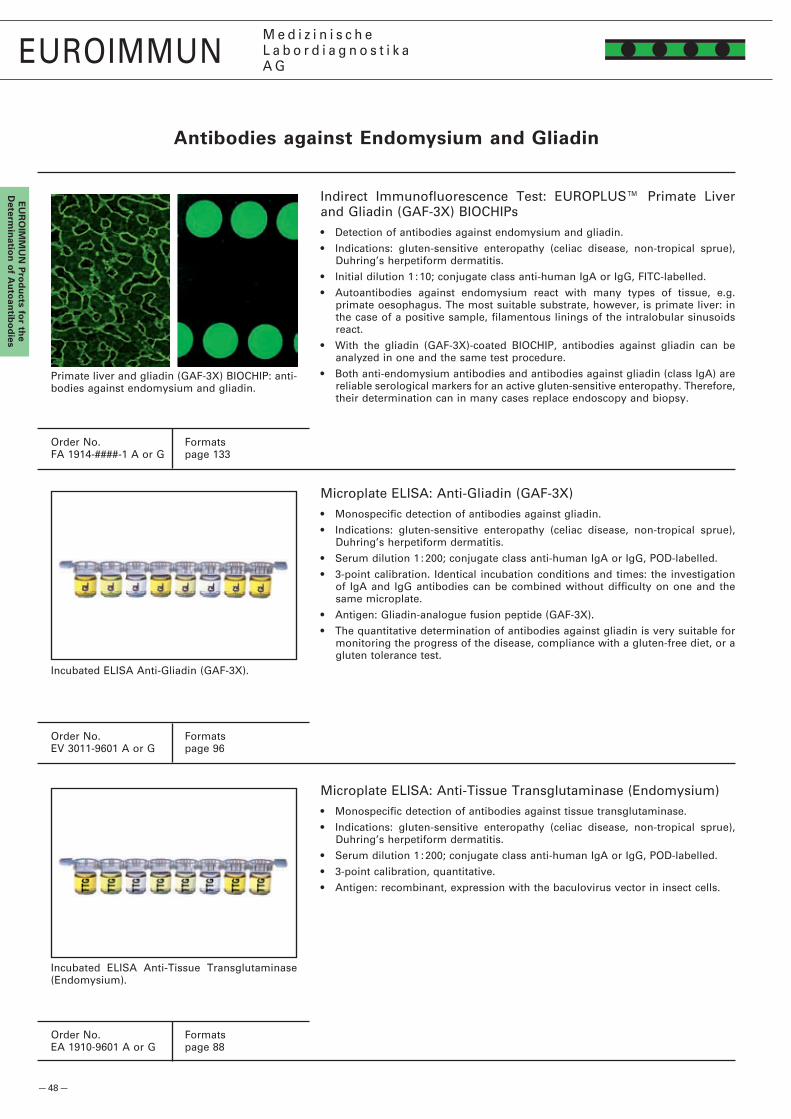
— 48 —
EUROIMMUNM e d i z i n i s c h eL a b o r d i a g n o s t i k aA G
EU
RO
IMM
UN
Pro
du
cts for th
e d
etermin
ation
of A
uto
antib
od
ies
Antibodies against Endomysium and gliadin
Indirect Immunofluorescence Test: EUROPLUS™ Primate Liver and Gliadin (GAF-3X) BIOCHIPs• Detection of antibodies against endomysium and gliadin.
• Indications: gluten-sensitive enteropathy (celiac disease, non-tropical sprue), Duhring’s herpetiform dermatitis.
• Initial dilution 1 : 10; conjugate class anti-human IgA or IgG, FITC-labelled.
• Autoantibodies against endomysium react with many types of tissue, e.g. primate oesophagus. The most suitable substrate, however, is primate liver: in the case of a positive sample, filamentous linings of the intralobular sinusoids react.
• With the gliadin (GAF-3X)-coated BIOCHIP, antibodies against gliadin can be analyzed in one and the same test procedure.
• Both anti-endomysium antibodies and antibodies against gliadin (class IgA) are reliable serological markers for an active gluten-sensitive enteropathy. Therefore, their determination can in many cases replace endoscopy and biopsy.
Primate liver and gliadin (GAF-3X) BIOCHIP: anti-bodies against endomysium and gliadin.
Microplate ELISA: Anti-Gliadin (GAF-3X)• Monospecific detection of antibodies against gliadin.
• Indications: gluten-sensitive enteropathy (celiac disease, non-tropical sprue), Duhring’s herpetiform dermatitis.
• Serum dilution 1 : �00; conjugate class anti-human IgA or IgG, POD-labelled.
• 3-point calibration. Identical incubation conditions and times: the investigation of IgA and IgG antibodies can be combined without difficulty on one and the same microplate.
• Antigen: Gliadin-analogue fusion peptide (GAF-3X).
• The quantitative determination of antibodies against gliadin is very suitable for monitoring the progress of the disease, compliance with a gluten-free diet, or a gluten tolerance test.
Incubated ELISA Anti-Gliadin (GAF-3X).
Incubated ELISA Anti-Tissue Transglutaminase (Endomysium).
Microplate ELISA: Anti-Tissue Transglutaminase (Endomysium) • Monospecific detection of antibodies against tissue transglutaminase.
• Indications: gluten-sensitive enteropathy (celiac disease, non-tropical sprue), Duhring’s herpetiform dermatitis.
• Serum dilution 1 : �00; conjugate class anti-human IgA or IgG, POD-labelled.
• 3-point calibration, quantitative.
• Antigen: recombinant, expression with the baculovirus vector in insect cells.
Order No. FormatsFA 1914-####-1 A or G page 133
Order No. FormatsEV 3011-9601 A or G page 96
Order No. FormatsEA 1910-9601 A or G page 88

— 49 —
EUROIMMUNM e d i z i n i s c h eL a b o r d i a g n o s t i k aA G
EU
RO
IMM
UN
Pro
du
cts
for
Infe
ctio
us
Ser
olo
gy
EUROIMMUN PROdUCTS fOR INfECTIOUS SEROLOgY
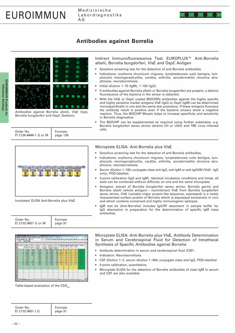
— 50 —
EUROIMMUNM e d i z i n i s c h eL a b o r d i a g n o s t i k aA G
EU
RO
IMM
UN
Pro
du
cts for
Infectio
us S
erolo
gy
Antibodies against borrelia
Indirect Immunofluorescence Test: EUROPLUS™ Anti-Borrelia afzelii, Borrelia burgdorferi, VlsE and OspC Antigen• Sensitive screening test for the detection of anti-Borrelia antibodies.
• Indications: erythema chronicum migrans, lymphadenosis cutis benigna, lym-phocytic meningoradiculitis, carditis, arthritis, acrodermatitis chronica atro-phicans, neuroborreliosis.
• Initial dilution 1 : 10 (IgM), 1 : 100 (IgG).
• If antibodies against Borrelia afzelii or Borrelia burgdorferi are present, a distinct fluorescence of the bacteria in the smear is obtained.
• With the VlsE or OspC coated BIOCHIPs antibodies against the highly specific and highly sensitive marker antigens VlsE (IgG) or OspC (IgM) can be determined monospecifically in one and the same test procedure. If these antigens fluoresce the antibody result is positive even if the bacteria smears show a negative reaction. Thus, the BIOCHIP Mosaic helps to increase specificity and sensitivity in Borrelia diagnostics.
• The BIOCHIP can be supplemented as required using further substrates, e.g. Borrelia burgdorferi sensu stricto (strains CH or USA) and TBE virus infected cells.
Antibodies against Borrelia afzelii, VlsE (top), Borrelia burgdorferi and OspC (bottom).
Incubated ELISA Anti-Borrelia plus VlsE.
Microplate ELISA: Anti-Borrelia plus VlsE• Sensitive screening test for the detection of anti-Borrelia antibodies.
• Indications: erythema chronicum migrans, lymphadenosis cutis benigna, lym-phocytic meningoradiculitis, carditis, arthritis, acrodermatitis chronica atro-phicans, neuroborreliosis.
• Serum dilution 1 : 100; conjugate class anti-IgG, anti-IgM or anti-IgAGM (VlsE: -IgG only), POD-labelled.
• 3-point calibration (IgG and IgM). Identical incubation conditions and times: all tests can be combined without difficulty on one and the same microplate.
• Antigens: extract of Borrelia burgdorferi sensu stricto, Borrelia garinii and Borrelia afzelii (whole antigen) / recombinant VlsE from Borrelia burgdorferi sensu stricto. VlsE (variable major protein-like sequence, expressed) is a newly characterized surface protein of Borrelia which is expressed exclusively in vivo and which contains conserved and highly immunogenic epitopes.
• IgM test kit (Anti-Borrelia) includes IgG/RF absorbent in sample buffer for IgG absorption in preparation for the determination of specific IgM class antibodies.
Microplate ELISA: Anti-Borrelia plus VlsE, Antibody Determination in Serum and Cerebrospinal Fluid for Detection of Intrathecal Synthesis of Specific Antibodies against Borrelia• Antibody determination in serum and cerebrospinal fluid (CSF).
• Indication: Neuroborreliosis.
• CSF dilution 1 : �, serum dilution 1 : 404; conjugate class anti-IgG, POD-labelled.
• 4-point calibration, quantitative.
• Microplate ELISA for the detection of Borrelia antibodies of class IgM in serum and CSF are also available.
Order No. FormatsFI �136-####-1 G or M page 136
Order No. FormatsEI �13�-9601 G or M page 91
Order No. FormatsEI �13�-9601 L G page 91
Table-based evaluation of the CSQrel.
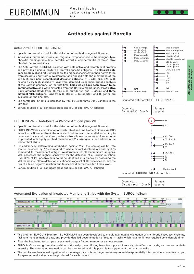
— 51 —
p 83
p 31, Osp A
p 39, Bmp Ap 41, Flag.
p �5, Osp C
p 30
p 19p 17
p �1
VlsE
VlsE B. afzelii
VlsE B. gariniiVlsE B. burgdorferi
Lipid B. afzeliiLipid B. burgdorferip83 B. afzeliip41 B. gariniip39 B. garinii
OspC B. garinii
p58p�1p�0p19p18
IggIgM
OspC B. burgd.OspC B. garinii
OspC B. afzelii
p41 B. afzeliip39 B. afzelii
VlsE B. burgd.
EUROIMMUNM e d i z i n i s c h eL a b o r d i a g n o s t i k aA G
EU
RO
IMM
UN
Pro
du
cts
for
Infe
ctio
us
Ser
olo
gy
Automated Evaluation of Incubated Membrane Strips with the System EUROLineScan
• The program EUROLineScan from EUROIMMUN has been developed to enable quantitative evaluation of membrane based test systems, facilitate management of data, and provide detailed documentation of results — tasks which have until now required considerable time.
• First, the incubated test strips are scanned using a flatbed scanner or camera system.
• EUROLineScan recognizes the position of the strips, even if they have been placed inexactly, identifies the bands, and measures their intensity. The automated evaluation can be monitored, and it is possible to supplement the data manually.
• The results are then saved together with the image data. It is no longer necessary to archive (potentially infectious) incubated test strips. A separate results sheet can be produced for each patient.
Antibodies against borrelia
Incubated Anti-Borrelia EUROLINE-RN-AT .
Order No. FormatsDN �131-3�01 G or M page 81
Control band
Anti-Borrelia EUROLINE-RN-AT• Specific confirmatory test for the detection of antibodies against Borrelia.
• Indications: erythema chronicum migrans, lymphadenosis cutis benigna, lym-phocytic meningoradiculitis, carditis, arthritis, acrodermatitis chronica atro-phicans, neuroborreliosis.
• The Anti-Borrelia EUROLINE is coated with both native and recombinant proteins and provides a unique mixture of Borrelia specific antigens: The classical antigens OspC, p83 and p39, which show the highest specificity in their native form, were accurately cut from a Westernblot and applied onto the membrane of the line blot. five new, recombinant designer antigens (p18, p19, p�0, p�1, p58) having a very high specificity (IgG) were identified using bioinformatic analysis of the Borrelia genome. For the first time, lipids which have been proven to be immunoreactive and were extracted from the Borrelia membranes, three native OspC antigens (IgM) from B. afzelii, B. burgdorferi and B. garinii and three different vlsE antigens (IgG) from B. afzelii, B. burgdorferi and B. garinii are available on the line blot.
• The serological hit rate is increased by 10% by using three OspC variants in the IgM test.
• Serum dilution 1 : 50; conjugate class anti-IgG or anti-IgM, AP-labelled.
Incubated EUROLINE-WB Anti-Borrelia.
Order No. FormatsDY �131-1601-1 G or M page 85
Control band
EUROLINE-WB: Anti-Borrelia (Whole Antigen plus VlsE)• Specific confirmatory test for the detection of antibodies against Borrelia.
• EUROLINE-WB is a combination of westernblot and line blot techniques. An SDS extract of a Borrelia afzelii strain is electrophoretically separated according to molecular mass and transferred onto a nitrocellulose membrane. A membrane chip coated with highly purified recombinant VlsE-Antigen is then added to the westernblot strips.
• By additionally determining antibodies against VlsE the serological hit rate can be increased by �0% compared to whole extract Westernblots and by 30% compared to recombinant antigen Westernblots. Of all recombinant antigens, VlsE possesses the highest sensitivity for the detection of a Borrelia infection. Over 85% of IgG-positive sera could be identified at a glance by assessing the VlsE band. VlsE allows detection of antibodies against all Borrelia species, and the risk of a false negative reaction due to species differences is ten times lower.
• Serum dilution 1 : 50; conjugate class anti-IgG or anti-IgM, AP-labelled.
Control band

— 5� —
EUROIMMUNM e d i z i n i s c h eL a b o r d i a g n o s t i k aA G
EU
RO
IMM
UN
Pro
du
cts for
Infectio
us S
erolo
gy
Antibodies against Epsteinbarr virus (Ebv)
Microplate ELISA: Anti-EBV-CA, Anti-EBNA-1, Anti-EBV-EA• Specific confirmatory test for antibodies against EBV-CA, EBNA-1 or EBV-EA.
• Indications: infectious mononucleosis, Burkitt’s lymphoma, nasopharyngeal carcinoma.
• Serum dilution 1 : 100; conjugate class anti-IgA, -IgG or IgM, POD-labelled.
• 1-point calibration, semi-quantitative (IgA, IgM) or 3-point calibration, quantita-tive (IgG). Identical incubation conditions and times: all tests can be combined without difficulty on one and the same microplate.
• EBV-CA: native antigen, purified by affinity chromatography; EBNA-1 and EBV-EA: recombinant antigen.
• IgM test kit includes IgG/RF absorbent in sample buffer for IgG absorption in preparation for the determination of specific IgM class antibodies.
• Available individual ELISA:
Antigen Order No.
EBV-CA EI �791-9601 A, G or M
EBNA-1 EI �793-9601 G
EBV-EA EI �795-9601 A, G or M
Incubated ELISA Anti-EBNA-1.
Order No. FormatsFI �799-####-�1 X page 147
Order No. (anti-EBNA-1) FormatsEI �793-9601 G page 95
Patient 1: EBV-CA (IgG) EBV-CA (IgG): urea-treated EBV-CA (IgM) EBV-EA (IgG) EBNA (IgG)
Patient �: EBV-CA (IgG) EBV-CA (IgG): urea-treated EBV-CA (IgM) EBV-EA (IgG) EBNA (IgG)
fres
h in
fect
ion
prev
ious
infe
ctio
n
Indirect Immunofluorescence Test: BIOCHIP Sequence EBV• Gold standard for the determination of antibodies against the EBV-CA antigens (Epstein-Barr virus capsid antigen), EBV-EA (Epstein-
Barr early antigen) and EBNA (Epstein-Barr nuclear antigen).
• Indication: infectious mononucleosis, Burkitt‘s lymphoma, nasopharyngeal carcinoma (NPC).
• IgG antibodies against EBV-CA indicate an EBV infection. An at least twofold increase in titer and the absence of antibodies against EBNA at the same time is characteristic for the early phase of the infection. IgM antibodies against EBV-CA and antibodies against EBV-EA can also be found in acute infections, but they do not necessarily always occur. The presence of antibodies against EBNA generally indicates the late phase of an EBNA infection.
• In cases of an acute EBV infection which cannot be reliably discriminated from a relapse or reinfection, the determination of the antibody avidity using a modified immunofluorescence test as an additional parameter is useful. This requires an additional incubation with urea solution (ZF 1130-0801). The determination of low-avidity antibodies against EBV-CA indicates an acute infection.
• For the monospecific confirmation of EBV-CA antibodies in the same test procedure the BIOCHIP containing ECV-CA is supplemented with the antigen substrates gp1�5 antigen (native) and p19 antigen (recombinant) (EUROPLUS FI �791-####-�0 G or M).
• For highly differentiated diagnostics the BIOCHIP Sequence EBV (FI �799-####-1 X) can be supplemented by using the antigens gp1�5 and p19 (EUROPLUS FI �799-####-�1 X).
• Due to the high prevalence of anti-EBV-CA IgA in NPC patients, the parameter is well suited for screening. Confirmation of the result by determination of IgA antibodies against EBV-EA is recommended. Further anti-EBV test kits for indirect immunofluorescence:
Substrate Order No. Substrate Order No.
EBV-CA FI �791-#### A, G or M EBV-EA FI �795-#### A or G
EBNA FI �793-#### G EBV-CA & EBV-EA FI �791-####-� A or G

— 53 —
0
�
4
6
8
10
0 �5 50 75 100
VCA gp1�5VCA p19EBNA-1
p��EA-D
VCA 1�5
VCA 65
VCA 40/41/4�
VCA 33
VCA ��
EA REBNA-1
EA D
EUROIMMUNM e d i z i n i s c h eL a b o r d i a g n o s t i k aA G
EU
RO
IMM
UN
Pro
du
cts
for
Infe
ctio
us
Ser
olo
gy
Antibodies against Epsteinbarr virus (Ebv)
EUROLINE: EBV Profile �• Differentiation of antibodies against Epstein-Barr virus antigens.
• Indications: infectious mononucleosis, Burkitt’s lymphoma, nasopharyngeal carcinoma.
• Serum dilution 1 : 100; conjugate class anti-human IgG or IgM, AP-labelled.
• With the EUROLINE EBV Profile �, five different antibodies can be determined: antibodies against VCA gp1�5, VCA p19, EBNA-1, p��, EA-D.
• Recombinant antigens (exception: VCA gp1�5, native, purified by affinity chro-matography).
• EBNA-1 (IgG) negative und VCA (IgG and IgM) positive: acute (fresh) infection.
• EBNA-1 (IgG) and VCA (IgG) positive and VCA (IgM) negative: late phase of infection.
• EBNA-1 (IgG) negative, but band p�� (IgG) and VCA (IgG) positive: late phase of infection with loss of anti-EBNA-1.
• Test strips can be automatically incubated and evaluated using the systems EUROBlotMaster und EUROLineScan (see page �7).
Westernblot: Anti-EBV• Specific confirmatory test for the detection of antibodies against Epstein-Barr
virus antigens, differentiation of antibodies against Epstein-Barr virus antigens.
• Indications: infectious mononucleosis, Burkitt’s lymphoma, nasopharyngeal carcinoma.
• Serum dilution 1 : 50; conjugate class anti-IgG or anti-IgM, AP-labelled.
• Antigens: whole antigen, SDS extract.
• Bands from all specific antigens are included and clearly separated.
• EBNA-1 (IgG) negative und VCA (IgG and IgM) positive: acute (fresh) infection.
• EBNA-1 (IgG) and VCA (IgG) positive and VCA (IgM) negative: late phase of infection.
• EBNA-1 (IgG) negative, but band p�� (IgG) and VCA (IgG) positive: late phase of infection with loss of anti-EBNA-1.
• Test strips can be automatically incubated and evaluated using the systems EUROBlotMaster und EUROLineScan (see page �7).
Incubated Westernblot Anti-EBV.
Order No. FormatsDN �790-1601-� G or M page 81
Order No. FormatsDY �790-1501 G or M page 85
Control band
Avidity of antibodies against EBV-CA (IgG).
Relative Avidity Index (RAI) in %
Nu
mb
er o
f P
atie
nts
RAI =Ewith urea
Ewithout urea
Primary Infection
Previous Infection
Determination of Low-Avidity Antibodies against EBV-CA • An alternative principle for the serological diagnosis of fresh infections with EBV
has been established by investigating the antibody avidity.
• The first reaction of the immune system following an infection is the formation of low-avidity antibodies. As the infection proceeds, increasingly antigen-adapted IgG is formed, and avidity grows. As long as high-avidity IgG is not yet detected in the serum, it can be assumed that the infection is still in an early stage.
• To identify low-avidity antibodies against EBV-CA in a patient’s serum, two microplate ELISA or immunofluorescence tests are performed in parallel: one test is carried out in the conventional way, the other one includes urea treatment between incubations with patient’s serum and peroxidase-labelled anti-human IgG, resulting in the detachment of low-avidity antibodies from the antigens.
• Low-avidity antibodies against EBV-CA are present if the ELISA extinction is significantly reduced by urea treatment. For an objective interpretation, the relative avidity index (RAI) can be calculated out of the measured values with and without urea incubation.
• With the immunofluorescence test the presence of low-avidity antibodies has been proved if the test using urea treatment gives a far weaker fluorescence than the two-step test.
Control
Incubated EUROLINE EBV Profile �.
Order No. (IIFT) FI �791-#### XOrder No. (ELISA) FormatsEI �791-####-1 G page 95 and 146

— 54 —
p 1�0, CagA
p 33
p 95, VacA
p 30p �9, UreAp �6
p 19, OMPp 17
p 66, UreB
EUROIMMUNM e d i z i n i s c h eL a b o r d i a g n o s t i k aA G
EU
RO
IMM
UN
Pro
du
cts for
Infectio
us S
erolo
gy
Antibodies against helicobacter pylori
Indirect Immunofluorescence Test: Anti-Helicobacter pylori• Sensitive screening test for the detection of antibodies against Helicobacter
pylori.
• Indications: gastritis, ulcus ventriculi et duodeni. Late consequences: MALT lymphomas and adenocarcinomas.
• Initial dilution 1 : 10 (IgM), 1 : 100 (IgG), 1 : 3� (IgA).
• If antibodies against Helicobacter pylori are present, a distinct fluorescence of the bacteria in the smear is obtained.
• A positive IgA result correlates well with the activity of a gastritis. An increased IgG antibody titer is considered to be a marker for chronic infections. A signifi-cant drop in the IgG antibody titer about 6 months after therapy is a sign of success.
• The BIOCHIP can be supplemented as required with further substrates, e.g. other infectious agents or tissue sections of primate stomach.
Antibodies against Helicobacter pylori.
Microplate ELISA: Anti-Helicobacter pylori• Sensitive screening test for the detection of antibodies against Helicobacter
pylori.
• Indications: gastritis, ulcus ventriculi et duodeni. Late consequences: MALT lymphomas and adenocarcinomas.
• Serum dilution 1 : 100; conjugate class anti-IgA or anti-IgG, POD-labelled.
• Antibodies against Helicobacter pylori antigens can be determined quantitatively in RU/ml.
• 1-point calibration, semi-quantitative (IgA) or 3-point calibration, quantitative (IgG). Identical incubation conditions and times: both tests can be combined without difficulty on one and the same microplate.
• Native antigens.Incubated ELISA Anti-Helicobacter pylori.
EUROLINE-WB: Anti-Helicobacter pylori• Specific confirmatory test for the detection of antibodies against Helicobacter
pylori.
• Indications: gastritis, ulcus ventriculi et duodeni. Late consequences: MALT lymphomas and adenocarcinomas.
• Serum dilution 1 : 50; conjugate class anti-IgA or anti-IgG, AP-labelled.
• EUROLINE-WB is a combination of westernblot and line blot techniques. An SDS extract of a Helicobacter pylori strain is electrophoretically separated according to molecular mass and transferred onto a nitrocellulose membrane (westernblot). Two membrane chips coated with highly purified recombinant CagA and VacA are subsequently applied to the westernblot strips.
• Bands from all specific antigens are included and clearly separated.
• Test strips can be automatically incubated and evaluated using the systems EUROBlotMaster und EUROLineScan (see page �7).
Incubated EUROLINE-WB Anti-Helicobacter pylori.
Order No. FormatsFI �080-#### A or G page 134
Order No. FormatsEI �080-9601 A or G page 91
Order No. FormatsDY �080-1601-1 A or G page 84
Control band
Alignment bar

— 55 —
gG 1
gC 1
gG �
EUROIMMUNM e d i z i n i s c h eL a b o r d i a g n o s t i k aA G
EU
RO
IMM
UN
Pro
du
cts
for
Infe
ctio
us
Ser
olo
gy
Antibodies against herpes Simplex virus (hSv)
Indirect Immunofluorescence Test: BIOCHIP Mosaic™ HSV-1/HSV-�• Sensitive screening test for the detection of antibodies against herpes simplex
viruses.
• Indication: herpes simplex.
• Initial dilution 1 : 10 (IgM), 1 : 100 (IgG).
• If antibodies against herpes simplex-� virus are present in the sample, a typical fluorescence of the infected cells is obtained – mainly in the outspread cyto-plasm, less in the cell nuclei.
• As HSV-1 and HSV-� are morphologically and immunologically closely related, cross-reactions can occur. Differentiation may be attempted by testing a serum against both antigen substrates and comparing the titers.
• The BIOCHIP Mosaic™ can be supplemented as required with further substrates, e.g. other infectious agents.
• Anti-HSV individual tests for indirect immunofluorescence:
Substrate Order No.
HSV-1 FI �531-#### G or M
HSV-� FI �53�-#### G or M
HSV-1 and -� FI �531-####-1 G or M
Antibodies against HSV-1 and HSV-�.
Microplate ELISA: Anti-HSV-1, Anti-HSV-�• Specific confirmatory tests for antibodies against HSV-1 or HSV-�.
• Indication: herpes simplex.
• Serum dilution 1 : 100; conjugate class anti-IgG or anti-IgM, POD-labelled.
• 1-point calibration, semiquantitative (IgM) or 3-point calibration, quantitative (IgG). Identical incubation conditions and times: all tests can be combined without difficulty on one and the same microplate.
• Antigens: type-specific glycoproteins C1 or G�, purified by affinity chromato-graphy. Cross-reactions do not occur.
• IgM test kit includes IgG/RF absorbent in sample buffer for IgG absorption in preparation for the determination of specific IgM class antibodies.
• Available individual ELISA:
Antigen Order No.
HSV-1 EI �531-9601-� G or M
HSV-� EI �53�-9601-� G or M
HSV-1/�-Pool EI �531-9601-1 G or M
Incubated ELISA Anti-HSV-1.
EUROLINE-WB: Anti-HSV• Specific confirmatory test for the differentiation of antibodies against HSV-1 and
HSV-�.
• Indication: herpes simplex.
• Serum dilution 1 : 50; conjugate class anti-IgG or anti-IgM, AP-labelled.
• EUROLINE-WB is a combination of westernblot and line blot techniques. Proteins from an SDS extract of HSV-1 are electrophoretically separated according to molecular mass and transferred onto a nitrocellulose membrane. A membrane chip coated with HSV-� type-specific glycoprotein G� (gG �), purified by affinity chromatography, is then added to the westernblot strips.
• Bands from all specific antigens are included and clearly separated.
• The gG � band allows the simple differentiation between HSV-1 and HSV-� infections.
• Test strips can be automatically incubated and evaluated using the systems EUROBlotMaster und EUROLineScan (see page �7).
Incubated EUROLINE-WB Anti-HSV.
Order No. FormatsFI �531-####-1 G or M page 141
Order No. (anti-HSV-1) FormatsEI �531-9601-� G or M page 93
Order No. FormatsDY �531-1601-1 G or M page 85
Alignment bar
Control band

— 56 —
EUROIMMUNM e d i z i n i s c h eL a b o r d i a g n o s t i k aA G
EU
RO
IMM
UN
Pro
du
cts for
Infectio
us S
erolo
gy
Antibodies against Chlamydia
Indirect Immunofluorescence test: Anti-Chlamydia MIF (Micro-Immunofluorescence test)• Serological gold standard for the determination of antibodies against Chlamydia.
• Indication: trachoma, urogenital infections, lymphogranuloma venereum, laryn-gitis, sinusitis, bronchitis, pneumonia, psittacosis.
• Initial dilution 1 : 10 (IgA), 1 : 100 (IgG), 1 : 10 (IgM).
• The micro-immunofluorescence test uses purified elementary bodies of the species C. trachomatis, C. pneumoniae and C. psittaci as the antigen. The mutual lipopolysaccharide (LPS) antigen is inactivated to minimise cross reactivity.
• The evaluation of the MIF could be significantly facilitated compared to con-ventional test systems by using a cell-based substrate.
• The fourth BIOCHIP with non-infected cells allows a reliable differentiation between unspecific and specific fluorescence.Anti-Chlamydia MIF.
Microplate ELISA: Anti-Chlamydia trachomatis• Monospecific detection of antibodies against Chlamydia trachomatis.
• Indication: trachoma, conjunctivitis, urogenital infections, pneumonia in infants, lymphogranuloma venereum.
• Serum dilution 1 : 100, conjugate class anti-human IgA, IgG or IgM, POD-labelled.
• 1-point calibration, semiquantitative (IgA and IgM) or 3-point calibration, quanti-tative (IgG).
• Antigen: native MOMP antigen (major outer membrane protein). MOMP is a transmembrane protein and represents the main part of the outer membrane of the elementary bodies. Protein purification starts with BGM cells infected with Chlamydia trachomatis of the serotype K.
• IgM test kit includes IgG/RF absorbent in sample buffer for IgG absorption in preparation for the determination of specific IgM class antibodies.
Incubated ELISA Anti-Chlamydia trachomatis.
Incubated ELISA Anti-Chlamydia pneumoniae.
Microplate ELISA: Anti-Chlamydia pneumoniae• Monospecific detection of antibodies against Chlamydia pneumoniae.
• Indication: laryngitis, sinusitis, bronchitis, pneumonia.
• Serum dilution 1: 100, conjugate class anti-human IgA, IgG or IgM, POD-labelled.
• 1-point calibration, semiquantitative (IgA and IgM) or 3-point calibration, quanti-tative (IgG).
• Antigen: cell lysate from HL cells, strain CDC/CWL-0�9.
• IgM test kit includes IgG/RF absorbent in sample buffer for IgG absorption in preparation for the determination of specific IgM class antibodies.
Order No. FormatsFI �191-####-3 A, G or M page 138
Order No. FormatsEI �191-9601 A, G or M page 9�
Order No. FormatsEI �19�-9601 A, G or M page 9�

— 57 —
EUROIMMUNM e d i z i n i s c h eL a b o r d i a g n o s t i k aA G
EU
RO
IMM
UN
Pro
du
cts
for
Infe
ctio
us
Ser
olo
gy
Antibodies against Emerging viruses
Indirect Immunofluorescence Test• Over the past years it has been observed that a number of new viruses (
„emerging“
or „reemerging viruses“) and other pathogens has spread worldwide, introducing
since then unknown diseases into previously unaffected regions.
• EUROIMMUN offers a broad spectrum of indirect immunofluorescence tests for the detection of specific antibodies against:
• Many of these substrates are available as single substrate with non-infected cells or as useful combinations (syndrome or geographically orientated) for the investigation of serum samples.
• IgG absorption as preparatory step for the determination of specific antibodies of class IgM: page 60.
• Cross reactions within the virus family, especially with Flaviviruses, should be taken into consideration since they may cause false-positive results. The in-fectious agent can be determined by titration of the sample and comparison of
Antibodies against Plasmodium, Rift valley fever virus, Chikungunya virus.
Microplate ELISA: Anti-TBE Virus, Anti-West Nile Virus, Anti-Dengue Virus• Monospecific determination of antibodies against TBE, West Nile and Dengue
virus.
• Serum dilution 1: 100, conjugate class anti-human IgG or IgM, POD-labelled.
• 1-point calibration, semiquantitative (IgM) or 3-point calibration, quantitative (IgG). Similar incubation conditions and times: All tests can be combined on one and the same microplate.
• IgM test kit with IgG/RF absorbent in the sample buffer for IgG absorption as preparatory step for the determination of specific IgM antibodies.
• Available single ELISA:
Antigen Order No.
TBE EI �661-9601 G or M, avidity, Ab determination in CSF
TBE „Vienna“ EI �661-9601-9 G
WNV EI �66�-9601 G or M, avidity
Dengue EI �66b-9601 G or M
Incubated ELISA Anti-TBE virus, Anti-WNV, Anti-Dengue virus.
Order No. (Anti-TBE) FormatsEI �661-9601 G or M page 94
Antibodies against SARS CoV, TBE virus, West Nile virus.
Antibodies against Japanese encephalitis virus, Yellow fever virus, Dengue virus.
Antibodies against Sandfly fever virus, Hanta-virus, Crimean-Congo fever virus.
Pathogen Disease, syndromes see page
Corona virus SARS corona virus (SARS-CoV) Severe acute respiratory syndrome (SARS) 14�
flaviviruses
TBE virus (TBEV) Tick-borne encephalitis 14�
West Nile virus (WNV) West Nile fever, encephalitis 14�, 143
Japanese encephalitis virus (JEV) Japanese encephalitis 14�, 143, 148
Yellow fever virus (YFV) Yellow fever, hepatitis, haemorrhagic fever, arthritis 14�, 143
Dengue virus (DENV, types 1-4) Dengue fever, haemorrhagic fever 14�, 143, 148
bunyaviruses
Hantavirus (types Hantaan, Puumala, Seoul, Saaremaa, Dobrava, Sin Nombre and Andes)
HFRS (haemorrhagic fever with renal syndrome); HCPS (Hantaviral cardio-pulmonary syndrome)
145
Sandfly fever virus (types Sicilian, Naples, Toscana and Cyprus)
Pappataci fever, meningitis, encephalitis 145
Rift valley fever virus (RVFV) Rift valley fever, haemorrhagic fever, hepatitis 147
Crimean-Congo fever virus (CCHFV-GPC and -N) Crimean-Congo haemorrhagic fever 147
TogavirusesChikungunya virus (CHIKV) Chikungunya fever, arthritis 148, 149
Sindbis virus (SINV) Sindbis fever 148
Kinetoplastida Leishmania donovani Visceral leishmaniasis 140
haemosporinaPlasmodium falciparum Malaria tropica 140, 148
Plasmodium vivax Malaria tertiana 140, 148

— 58 —
EUROIMMUNM e d i z i n i s c h eL a b o r d i a g n o s t i k aA G
EU
RO
IMM
UN
Pro
du
cts for
Infectio
us S
erolo
gy
fI 282110011 g fI 282210011 g RESPIRATORY TRACT PROfILE 1 EXANThEMA PROfILE 1 Igg IgM Igg IgM
field A 1 : 10 1 : 10 field A 1 : 10 1 : 10V Verification BIOCHIP*** V Verification BIOCHIP***1. RSV 1. HHV-6 �. Adenovirus type 3 �. Rubella virus* 3. Influenza virus type A (H1N1) 3. Measles virus 4. Influenza virus type A (H3N�) 4. Mumps virus
field b 1 : 10 1 : 10 field b 1 : 10 1 : 105. Influenza virus type B 5. VZV 6. Parainfluenza virus type 1 6. EBV-CA** 7. Parainfluenza virus type � 7. EBV-EA 8. Parainfluenza virus type 3 8. Treponema pallidum
field C 1 : 10 1 : 10 field C 1 : 100 1 : 109. Parainfluenza virus type 4 9. HSV-1 10. Bordetella pertussis** 10. HSV-� 11. Bordetella parapertussis** 11. Coxsackie virus type B1 1�. Mycoplasma pneumoniae 1�. Coxsackie virus type A9
field d 1 : 100 1 : 10 field d 1 : 100 1 : 1013. Coxsackie virus type B1 13. Echo virus type 7 14. Coxsackie virus type A7 14. Borrelia afzelii 15. Echo virus type 7 15. Borrelia burgdorferi sensu stricto (CH) 16. Chlamydia pneumoniae 16. Borrelia garinii
field E 1 : 100 1 : 100 field E 1 : 100 1 : 10017. Haemophilus influenzae*,** 17. CMV 18. Klebsiella pneumoniae* 18. Candida albicans** 19. Legionella pneumophila serotype 1*,** 19. Candida krusei*,** �0. Legionella pneumophila serotype 1�*,** �0. Candida tropicalis*,**
fI 282310011 g fI 282410011 g LYMPhAdENITIS PROfILE 1 CENTRAL NERvOUS SYSTEM PROfILE 1 Igg IgM Igg IgM
field A 1 : 10 1 : 10 field A 1 : 10 1 : 10V Verification BIOCHIP*** V Verification BIOCHIP***1. HIV-1* 1. Rubella virus* �. HIV-�* �. Measles virus 3. HHV-6 3. Mumps virus 4. Rubella virus* 4. VZV
field b 1 : 10 1 : 10 field b 1 : 10 1 : 105. Measles virus 5. Adenovirus type 3 6. Mumps virus 6. EBV-CA** 7. Adenovirus type 3 7. Treponema pallidum 8. Parainfluenza virus type 1 8. Toxoplasma gondii**
field C 1 : 10 1 : 10 field C 1 : 100 1 : 109. EBV-CA** 9. HSV-1 10. EBV-EA 10. HSV-� 11. Toxoplasma gondii** 11. Coxsackie virus type B1 1�. Treponema pallidum 1�. Coxsackie virus type A7
field d 1 : 100 1 : 10 field d 1 : 100 1 : 1013. HSV-1 13. Echo virus type 7 14. HSV-� 14. Borrelia afzelii 15. CMV** 15. Borrelia burgdorferi sensu stricto (CH) 16. Coxsackie virus type B5 16. Borrelia garinii
field E 1 : 100 1 : 10 field E 1 : 100 1 : 10017. Coxsackie virus type A9 17. CMV 18. Bartonella henselae** 18. Haemophilus influenzae*,** 19. Chlamydia trachomatis** 19. Listeria monocytogenes 1/�a* �0. Chlamydia pneumoniae �0. Listeria monocytogenes 4b*
bIOChIP Mosaics™ for Infectious Serology(For formats see pages 147-148)
* For clinical evaluation the results must be confirmed with a CE approved test.** For practical reasons the recommended incubation differs from the standard incubation for this substrate.*** Field A contains a verification BIOCHIP. The incubation was performed correctly if the dots show a visible colour reaction.BIOCHIP Mosaics™ containing fewer substrates can be produced upon request.

— 59 —
EUROIMMUNM e d i z i n i s c h eL a b o r d i a g n o s t i k aA G
EU
RO
IMM
UN
Pro
du
cts
for
Infe
ctio
us
Ser
olo
gy
bIOChIP Mosaics™ for Infectious Serology(For formats see pages 147-148)
fI 282510011 g fI 282610011 g MYOCARdITIS PROfILE 1 INfECTIOUS ARThRITIS PROfILE 1 Igg IgM Igg IgM
field A 1 : 10 1 : 10 field A 1 : 10 1 : 10V Verification BIOCHIP*** V Verification BIOCHIP***1. Mumps virus 1. VZV �. Adenovirus type 3 �. Influenza virus type A (H1N1) 3. Influenza virus type A (H1N1) 3. Influenza virus type A (H3N�) 4. Influenza virus type A (H3N�) 4. Influenza virus type B
field b 1 : 10 1 : 10 field b 1 : 10 1 : 105. Influenza virus type B 5. Yersinia enterocolitica O:3*,** 6. Parainfluenza virus type 1 6. Yersinia enterocolitica O:6*,**7. Parainfluenza virus type � 7. Yersinia enterocolitica O:9*,** 8. Mycoplasma pneumoniae 8. Toxoplasma gondii**
field C 1 : 100 1 : 10 field C 1 : 100 1 : 109. CMV** 9. Borrelia afzelii 10. Coxsackie virus type B1 10. Borrelia burgdorferi sensu stricto (CH) 11. Coxsackie virus type A16 11. Borrelia garinii 1�. Echo virus type 7 1�. Chlamydia trachomatis**
field d 1 : 100 1 : 10 13. Borrelia afzelii 14. Borrelia burgdorferi sensu stricto (CH) 15. Borrelia garinii 16. Chlamydia pneumoniae
fI 282710011 g fI 282810011 g gASTROINTESTINAL TRACT PROfILE 1 ACCOMPANYINg hEPATITIS PROfILE 1 Igg IgM Igg IgM
field A 1 : 10 1 : 10 field A 1 : 10 1 : 10V Verification BIOCHIP*** V Verification BIOCHIP***1. Adenovirus type 3 1. Adenovirus type 3 �. Yersinia enterocolitica O:3*,** �. EBV-CA** 3. Yersinia enterocolitica O:6*,** 3. EBV-EA 4. Yersinia enterocolitica O:9*,** 4. Toxoplasma gondii**
field b 1 : 100 1 : 10 field b 1 : 100 1 : 105. Coxsackie virus type B3 5. HSV-1 6. Coxsackie virus type A7 6. HSV-� 7. Echo virus type 7 7. CMV** 8. Campylobacter jejuni*,** 8. Coxsackie virus type B5
field C 1 : 100 1 : 100 field C 1 : 10 1 : 109. Campylobacter coli*,** 9. Coxsackie virus type A9 10. Helicobacter pylori** 10. Echo virus Typ 7 11. Klebsiella pneumoniae* 11. Echinococcus granulosus** 1�. Candida albicans**
fI 282910011 g fI 283110011 g OPhThALMOLOgY PROfILE 1 PREgNANCY PROfILE 1 Igg IgM Igg IgM
field A 1 : 10 1 : 10 field A 1 : 10 1 : 10V Verification BIOCHIP*** V Verification BIOCHIP***1. Measeles virus 1. Rubella virus*�. VZV �. Toxoplasma gondii**3. Adenovirus type 3 3. HIV-1*4. Toxoplasma gondii** 4. HIV-�*
field b 1 : 100 1 : 10 field b 1 : 100 1 : 105. HSV-1 5. Chlamydia trachomatis**6. HSV-� 6. CMV**7. Coxsackie virus type A�4 7. HSV-�8. Chlamydia trachomatis** 8. VZV**
* For clinical evaluation the results must be confirmed with a CE approved test.** For practical reasons the recommended incubation differs from the standard incubation for this substrate.*** Field A contains a verification BIOCHIP. The incubation was performed correctly if the dots show a visible colour reaction.BIOCHIP Mosaics™ containing fewer substrates can be produced upon request.
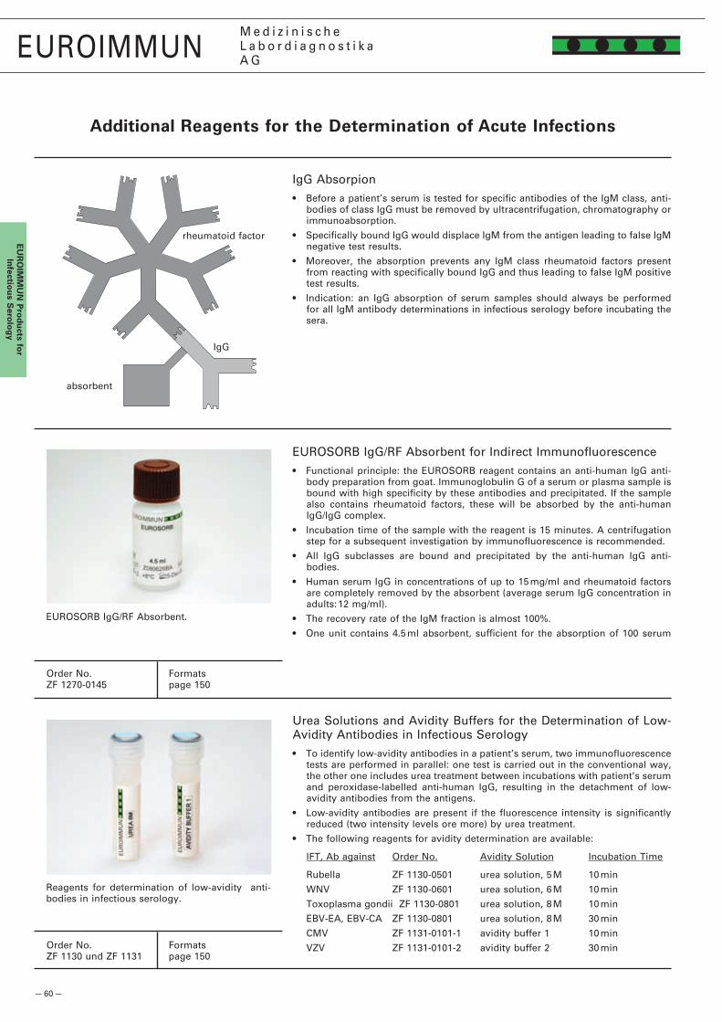
— 60 —
EUROIMMUNM e d i z i n i s c h eL a b o r d i a g n o s t i k aA G
EU
RO
IMM
UN
Pro
du
cts for
Infectio
us S
erolo
gy
Additional Reagents for the determination of Acute Infections
IgG Absorpion• Before a patient’s serum is tested for specific antibodies of the IgM class, anti-
bodies of class IgG must be removed by ultracentrifugation, chromatography or immunoabsorption.
• Specifically bound IgG would displace IgM from the antigen leading to false IgM negative test results.
• Moreover, the absorption prevents any IgM class rheumatoid factors present from reacting with specifically bound IgG and thus leading to false IgM positive test results.
• Indication: an IgG absorption of serum samples should always be performed for all IgM antibody determinations in infectious serology before incubating the sera.
EUROSORB IgG/RF Absorbent for Indirect Immunofluorescence• Functional principle: the EUROSORB reagent contains an anti-human IgG anti-
body preparation from goat. Immunoglobulin G of a serum or plasma sample is bound with high specificity by these antibodies and precipitated. If the sample also contains rheumatoid factors, these will be absorbed by the anti-human IgG/IgG complex.
• Incubation time of the sample with the reagent is 15 minutes. A centrifugation step for a subsequent investigation by immunofluorescence is recommended.
• All IgG subclasses are bound and precipitated by the anti-human IgG anti-bodies.
• Human serum IgG in concentrations of up to 15 mg/ml and rheumatoid factors are completely removed by the absorbent (average serum IgG concentration in adults: 1� mg/ml).
• The recovery rate of the IgM fraction is almost 100%.
• One unit contains 4.5 ml absorbent, sufficient for the absorption of 100 serum
EUROSORB IgG/RF Absorbent.
rheumatoid factor
IgG
absorbent
Order No. FormatsZF 1�70-0145 page 150
Order No. FormatsZF 1130 und ZF 1131 page 150
Reagents for determination of low-avidity anti-bodies in infectious serology.
Urea Solutions and Avidity Buffers for the Determination of Low-Avidity Antibodies in Infectious Serology• To identify low-avidity antibodies in a patient’s serum, two immunofluorescence
tests are performed in parallel: one test is carried out in the conventional way, the other one includes urea treatment between incubations with patient’s serum and peroxidase-labelled anti-human IgG, resulting in the detachment of low-avidity antibodies from the antigens.
• Low-avidity antibodies are present if the fluorescence intensity is significantly reduced (two intensity levels ore more) by urea treatment.
• The following reagents for avidity determination are available:
IFT, Ab against Order No. Avidity Solution Incubation Time
Rubella ZF 1130-0501 urea solution, 5 M 10 min
WNV ZF 1130-0601 urea solution, 6 M 10 min
Toxoplasma gondii ZF 1130-0801 urea solution, 8 M 10 min
EBV-EA, EBV-CA ZF 1130-0801 urea solution, 8 M 30 min
CMV ZF 1131-0101-1 avidity buffer 1 10 min
VZV ZF 1131-0101-� avidity buffer � 30 min

— 61 —
EUROIMMUNM e d i z i n i s c h eL a b o r d i a g n o s t i k aA G
EU
RO
IMM
UN
Pro
du
cts
for
Alle
rgo
log
y
EUROIMMUN PROdUCTS fOR ALLERgOLOgY
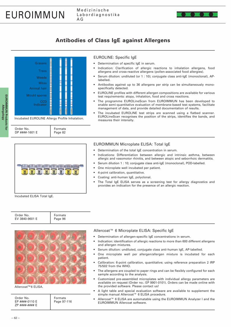
— 6� —
EUROIMMUNM e d i z i n i s c h eL a b o r d i a g n o s t i k aA G
EU
RO
IMM
UN
Pro
du
cts for
Allerg
olo
gy
EUROLINE: Specific IgE• Determination of specific IgE in serum.
• Indication: Clarification of allergic reactions to inhalation allergens, food allergens and cross-reactive allergens (pollen-associated food allergies).
• Serum dilution: undiluted (or 1 : 10); conjugate class anti-IgE (monoclonal), AP-labelled.
• Antibodies against up to 36 allergens per strip can be simultaneously mono-specifically detected.
• EUROLINE profiles with different allergen compositions are available for various test requirements: atopy, inhalation, food and cross reactions.
• The programme EUROLineScan from EUROIMMUN has been developed to enable semi-quantitative evaluation of membrane-based test systems, facilitate management of data, and provide detailed documentation of results.
• The incubated EUROLINE test strips are scanned using a flatbed scanner. EUROLineScan recognises the position of the strips, identifies the bands, and measures their intensity.Incubated EUROLINE Allergy Profile Inhalation.
EUROIMMUN Microplate ELISA: Total IgE• Determination of the total IgE concentration in serum.
• Indications: Differentiation between allergic and intrinsic asthma, between allergic and vasomotor rhinitis, and between atopic and seborrhoic dermatitis.
• Serum dilution 1 : 10; conjugate class anti-IgE (monoclonal), POD-labelled.
• One microplate well incubated per patient.
• 4-point calibration, quantitative.
• Coating: anti-human IgE, polyclonal.
• The Total IgE ELISA serves as a screening test for allergy diagnostics and provides an indication for the presence of an allergic reaction.
Incubated ELISA Total IgE.
Grasses
Animal hair
Trees
Mould spores
Weeds
CCD
Mites
Antibodies of Class IgE against Allergens
Order No. FormatsDP ####-1601 E Page 8�
Order No. FormatsEV 3840-9601 E Page 96
Order No. FormatsEP ####-0110 E Page 97-116ZP ####-#### E
Allercoat™ 6 Microplate ELISA: Specific IgE• Determination of allergen-specific IgE concentrations in serum.
• Indication: identification of allergic reactions to more than 600 different allergens and allergen mixtures.
• Serum dilution: undiluted, conjugate class anti-human IgE, AP-labelled.
• One microplate well per allergen/allergen mixture is incubated for each patient.
• Calibration: 6-point calibration, quantitative; using reference preparation � IRP 75/50� from the WHO.
• The allergens are coupled to paper rings and can be flexibly configured for each sample according to the analysis.
• Customized pre-assembled microplates with individual allergy parameters are available on request (Order no.: EP 9901-0101). Orders can be made online with the provided software. Please contact us!
• A light table and special evaluation software are available to supplement the simple manual Allercoat™ 6 ELISA procedure.
• Allercoat™ 6 ELISA are automatable using the EUROIMMUN Analyzer I and the EUROIMMUN Allercoat software.
Indicator
AllercoatTM 6 ELISA.

— 63 —
AllercoatTM Software.
EUROIMMUNM e d i z i n i s c h eL a b o r d i a g n o s t i k aA G
EU
RO
IMM
UN
Pro
du
cts
for
Alle
rgo
log
y
Antibodies of Class IgE against Allergens
Customized Laboratory Software for Flexible Automation of Allercoat™ System• Convenient online ordering of individual pre-prepared Allercoat™ microtiter
plates.
• Integrated light table for manual preparation of ELISA plates and sample distribution.
• Direct control of photometer, automated evaluation via calibration curve and compilation of results.
• Fully automated incubation of samples and evaluation of results via connection to the EUROIMMUN Analyzer I.
• Fully automated administration, documentation and archiving of all data.
• Simple operation using graphic user commands and clear presentation of the most important information at a glance.
• Connection to commonly used laboratory systems for convenient transfer of requests and results.
• Additional security through user administration with individual access rights and confirmation step before results editing.
Fitting of allergen rings into microplate wells
Incubate: 60 min, 37 °C
Wash: 300 µl wash buffer per well
allergen rings
undiluted samples
enzymeconjugate
chromogen-/substrate sol.
Pipette: 50 µl per well
Pipette: 50 µl per well
Incubate: 60 min, 37 °C
Evaluate: photometric measurement at a wavelength of 405 nm
Wash: 300 µl wash buffer per well
Pipette: 100 µl per well
Incubate: 30 min, 37 °C
Pipette: 100 µl per well
stop solution
Individual equipment with allergen rings

— 151 —
EUROIMMUNM e d i z i n i s c h eL a b o r d i a g n o s t i k aA G
Products from EUROIMMUN AG will be delivered according to the following conditions, as long as no other conditions have been agreed in writing. On issuing an order, the buyer acknowledges the delivery conditions. EUROIMMUN does not accept the sales conditions of the buyer, even when EUROIMMUN has not expressly contradicted them. Contract terms are valid for all future business connection even if they have not been expressly agreed upon again.
Orders: Orders to EUROIMMUN can be made in writing (including electronic communication) or verbally. Verbally issued orders first become legally binding for EUROIMMUN with a written confirmation from the orderer or with the delivery of goods.
Delivery: EUROIMMUN generally delivers to end users within 7 days of order receipt and to distributors within 14 days, but is not tied to any definite delivery period. If a delivery is not possible through unforseeable circumstances (e.g., factory disruptions, raw material delivery delays, transport difficulties, strikes), the delivery obligation does not apply. EUROIMMUN reserves the right to deliver in part. The delivery obligation of EUROIMMUN AG ceases as long as the customer is in arrears. EUROIMMUN chooses the packing and dispatching method at its own discretion, according to the respective requirements.
Product characteristics: The delivered products comply with specifications given in the product catalogue, on the product itself, or in the supplied information sheets. If specifications are inconsistent, the labels on the product itself and the details in the information sheet provided with the product apply. After the given expiration date has passed, the test reagents must not be used.
EUROIMMUN products must only be used for the intended purpose. It is the buyer’s responsibility to check the functional capability immediately on receipt of the product as well as on every day of usage. In particular, the functioning of test reagents can be perturbed through causes outside the direct influence of the maunfacturer, for example, through suboptimal transportation, incorrect storage, or incorrect usage. When test reagents delivered from EUROIMMUN are used, the user must supervise the analysis with appropriate proficiency.
Warranty and liability: If nothing else has been agreed upon in writing, all risks pass to the buyer as soon as the goods have been delivered to the carrier or has left EUROIMMUN AG premises for dispatch. With notification of dispatch by EUROIMMUN the risk passes to the buyer if the delivery is postponed or delayed by request of the buyer. On receipt of the goods, the buyer has one working day to check if they are in accordance with the nature and quantity of the agreement and if the goods are free from visible defects. If any complaints arise from this inspection, they should be communicated to EUROIMMUN AG in writing within � working days. Hidden defects or functional faults that were not identifiable with the initial inspection and are later discovered should be communicated to EUROIMMUN AG in writing within 14 days of receipt of the goods. If no complaints are received within the stipulated period, it is assumed that the goods are of appropriate quality and quantity for the customer. Complaints do not release the buyer from payment obligation.
With prompt and reasonable complaints on the grounds of product defects or the delivery of something other than the ordered goods, EUROIMMUN is obliged to exchange or amend the goods within 14 days or to withdraw or reimbourse the payment, as it chooses. If the defect is not corrected in spite of delivery of a replacement or an amended product, the buyer can demand that the sale is cancelled.
If defects are promptly reclaimed, EUROIMMUN has the choice between a further delivery or an appropriate credit note. Further compensatory claims from the buyer are excluded, as long as EUROIMMUN has not violated its contractual or legal obligations through outright negligence or by intention. In such cases EUROIMMUN provides compensation of up to a maximum of �0 times the price of one packet (test kit) of the defective product. EUROIMMUN takes no responsibility for any damage resulting from faulty goods.
Prices: Prices from the official EUROIMMUN pricelist in the country of the buyer are applicable. Invoices are provided in the agreed currency. Prices are “ex works”. Expenses for packing and freight as well as for cooling during transport are added on, including costs for the disposal of packaging material by the customer. Statutory value added tax is not included in the list prices.
Payment terms: Payment obligations in countries of export must be settled within 30 days after recieving the goods. No cash discounts will be given unless agreed in writing. Payments by cash transfer or check are valid from the timepoint that the invoice amount is credited to a EUROIMMUN AG bank account. In cases of payment arrears, EUROIMMUN reserves the right to charge compensatory interest of 10% p.a., applicable from the settlement date, without any further notice. In special cases EUROIMMUN can arrange alternative payment periods or demand advance payments. EUROIMMUN’s demands resulting from an order cannot be offset by the customer through counter demands.
Ownership rights: The delivered goods remain the property of EUROIMMUN AG until full payment. Selling on of EUROIMMUN products is only permitted for companys who are authorized in writing from EUROIMMUN to do so. Software received from EUROIMMUN should not be passed on to third parties without written permission from EUROIMMUN AG.
Consultation: Advice from EUROIMMUN AG, although provided to the best knowledge, is nevertheless not binding. No liability claims can ensue from erroneous advice.
Applicable law, place of jurisdiction, ineffective regulations: The contract conditions are subject to the laws of the Federal Republic of Germany. The United Nations Conventions on Contracts for the International Sale of Goods does not apply. The place of jurisdiction and fulfilment is Luebeck. If any provision of these general delivery conditions will be held invalid or unenforcable, this general delivery condition will not be rendered invalid as a whole, and the provisions will be changed and interpreted so as best to accomplish the objective
general delivery Conditions
— 15� —
EUROIMMUNM e d i z i n i s c h eL a b o r d i a g n o s t i k aA G
IndexAutoantibody Diagnosticsacetylcholine receptor (ACHR) ...................................................................................................................1�, 17, 89actin ...........................................................................................................................16, 17, 3�, 38, 47, 68, 1��, 1�9adrenal cortex ............................................................................................................................... 13, 17, 45, 66, 117agglutination test ................................................................................................................................................... 89AGNA ...................................................................................................................................................................... 17alpha-amylase ........................................................................................................................................................ 96AMA ...............................................................6, 13, 16-17, 33, 37-40, 45, 47, 68, 78, 80, 88, 117, 1�3, 1�7, 1�9-131AMPA receptor .................................................................................................................................... 1�, 17, 43, 119amphiphysin ..................................................................................................................................17, 4�, 80, 118-119ANA ....................................................................... 14, 16-17, 3�-34, 37-38, 4�, 46-47, 68, 80, 87, 119-1�5, 1�7-131ANCA ..................................................................................................1�, 14, 16-17, 3�, 46-47, 66, 86, 1�0-1��, 1�4aquaporin ...................................................................................................................................... 1�, 17, 4�, 66, 119aquaporin-4 ................................................................................................................................... 1�, 17, 4�, 66, 119ASMA ................................................................................................................................................1�, 16-17, 38, 68Autoantibody Profile � .........................................................................................................................................1�8basement membrane ............................................................................1�, 14, 16-17, 41, 67, 80, 86, 1�1-1��, 1�5�-glycoprotein ................................................................................................................................................ 16, 88bile ducts ...........................................................................................................................................................17, 3�BP180 .................................................................................................................................1�, 17, 41, 67, 87, 1�5-1�6BP�30 .................................................................................................................................1�, 17, 41, 67, 87, 1�5-1�6BPI ........................................................................................................................................................... 17, 46-47, 86brain .................................................................................................................................................... 1�, 66, 117-118C1q ...............................................................................................................................................................16, 87, 88cANCA..................................................................................................... 1�, 16-17, 3�, 46-47, 66, 86, 1�0-1��, 1�4cardiolipin (AMA M1) ............................................................................................................................16-17, 84, 88cartilage .......................................................................................................................................................... 16, 1�4Caspr� ..........................................................................................................................................................17, 43, 67cathepsin G ............................................................................................................................................ 17, 46-47, 86cell nuclei (ANA global test) .................................14, 16-17, 3�-34, 37-38, 4�, 46-47, 68, 80, 87, 119-1�4, 1�7-131centromere .................................................................................................................... 14, 16, 3�, 34, 47, 68, 87-88cerebellum .......................................................................................................................... 1�-14, 4�-44, 83, 117-119circulating immune complexes (CIC) ............................................................................................................. 16, 88collagen VII ............................................................................................................................................................. 41cortisol .................................................................................................................................................................... 96CUZD1 (rPAg1) .................................................................................................................................... 1�, 17, 67, 1�4CV� .................................................................................................................................................17, 4�, 80, 118-119cyclic citrullinated peptide (CCP) ............................................................................................................. 16, 36, 87cyclin I (PCNA) .............................................................................................................. 1�, 16, 3�-33, 47, 68, 80, 87cyclin II (mitosin) ........................................................................................................................................1�, 47, 68cytoplasm of granulocytes ............................................................ 1�-14, 16-17, 3�, 46-47, 66, 86, 1�0-1��, 1�4desmoglein ...............................................................................................................................1�, 17, 41, 67, 87, 1�5desmosomes ................................................................................................................................. 1�, 17, 41, 67, 1�5DHEA ....................................................................................................................................................................... 96dsDNA .............................................................................. 1�, 16-17, 3�-35, 38, 4�, 47, 68, 78, 80, 87, 89, 1�8, 149elastase ................................................................................................................................................... 17, 46-47, 86elastin .....................................................................................................................................................1�, 16-17, 69ENA ..........................................................................................................................................16, 3�, 34, 78, 80, 87endomysium.................................................................................................... 1�, 17, 3�, 48, 69, 88, 1�5, 131-133endothelial cells .......................................................................................................................1�, 16-17, 3�, 69, 134envoplakin ...........................................................................................................................................17, 41, 86, 1�5epidermis ................................................................................................................................. 1�-13, 17, 41, 67, 1�5EUROArray ............................................................................................................................................................ 89EUROPLUS ....................................................................................... 7, 1�, 3�, 37-38, 40-41, 46-48, 50, 5�, 117-147eye antigens ..........................................................................................................................................................1�0F-actin...........................................................................................................................................17, 38, 68, 1�3, 1�9FT3 ...............................................................................................................................................................17, 40, 90FT4 ...............................................................................................................................................................17, 40, 90GABAB receptor ..................................................................................................................... 1�, 17, 43, 66, 119-1�0GAD ..............................................................................................................................1�, 17, 4�, 44, 66, 86, 89, 117gangliosides ......................................................................................................................................................17, 80gliadin .............................................................................................. 1�, 17, 48, 69, 75, 96, 10�, 1�5, 131-133, 149glomerular basement membrane (GBM)................................................................. 1�, 17, 46, 67, 80, 86, 1�1-1��glutamate receptor ........................................................................................................................ 1�, 17, 43, 66,119glutamic acid decarboxylase ............................................................................................ 1�, 17, 4�, 44, 66, 86, 89glycine receptor ................................................................................................................................................17, 66Golgi apparatus ..............................................................................................................................1�, 16, 3�, 47, 68goblet cells .......................................................................................................................................... 1�, 17, 67, 1�4GP� (rPAg�) ......................................................................................................................................... 1�, 17, 67, 1�4granulocyte cytoplasm .......................................................................1�-14, 16-17, 3�, 46-47, 66, 86, 1�0-1��, 1�4heart muscle ...................................................................................................................................... 13, 17, 1�4-1�5HEp-�-cells ............................................................................1�-14, 3�, 37-38, 4�, 46-47, 83, 119-1�3, 1�5, 1�7-131histones ......................................................................................................................... 13, 16, 3�-34, 47, 78, 80, 87Hu ........................................................................................................................... 13, 17, 4�-43, 66, 80, 83, 117-119HUVEC ...................................................................................................................................................................134hypothalamus antigens ................................................................................................................................. 13, 118IA-� .........................................................................................................................................................17, 44, 86, 89insulin ..............................................................................................................................................14, 17, 44, 89, 97intestinal goblet cells ......................................................................................................................... 1�, 17, 67, 1�4intrinsic factor ........................................................................................................................ 13, 17, 45, 67, 86, 1�3Jo-1 ..................................................................................................... 13, 16, 3�-34, 47, 68, 78, 80, 87-88, 1�7-1�8keratin ...........................................................................................................................1�, 13, 16-17, 36, 41, 67, 1�6Ku .............................................................................................................................................. 13, 16, 33, 47, 68, 80lactoferrin ...................................................................................................................1�, 17, 46-47, 66, 86, 1��, 1�4latex agglutination test ........................................................................................................................................ 89LC-1 ...................................................................................................................................... 17, 3�, 38-39, 78, 80, 86Leydig cells ....................................................................................................................................17, 45, 66, 117-118Lgi1 ...................................................................................................................................................... 17, 43, 67, 1�5liver-kidney microsomes (LKM) ........................................ 13, 17, 3�, 37-39, 67, 78, 80, 86, 117, 1�3, 1�7, 1�9-131liver-specific antigens ........................................................................................................13, 17, 3�, 38-39, 67, 1��LKM-1 ........................................................................................................................................ 17, 3�, 39, 78, 80, 86lung alveoli ...................................................................................................................................................... 17, 1��lymphocyte antigens ............................................................................................................................... 13, 17, 1��M� antigen (AMA-M�) ................................................................................................. 17, 33, 37-39, 68, 78, 80, 88M4 antigen .............................................................................................................................. 13, 17, 3�, 37, 78, 109M9 antigen .............................................................................................................................. 13, 17, 3�, 37, 78, 109Ma1/Ma� ................................................................................................................................................................. 17Mab ...................................................................................................................................................... 17, 40, 66, 117MAG .......................................................................................................................................................13, 17, 4�, 66Mi-� ..................................................................................................................................................13, 16, 33, 47, 80mitochondria (AMA) .............................................. 13, 16-17, 33, 37-40, 45, 47, 68, 78, 80, 117, 1�3, 1�7, 1�9-131mitosin (cyclin II) ........................................................................................................................................1�, 47, 68myelin ...............................................................................................................................................1�-13, 17, 4�, 66myeloperoxidase (MPO) ................................................................................ 13, 17, 46-47, 66, 78, 80, 86, 1�1-1��nDNA................................................................. 1�, 14, 16-17, 3�-35, 38-39, 41-4�, 47, 68, 78, 80, 87, 89, 1�8, 149nerves .................................................................................................................................... 1�-14, 17, 4�, 118-119neuronal autoantibodies .................................................................................................................17, 4�-43, 80, 83NMDA receptor ............................................................................................................................. 1�, 17, 43, 66, 119NMO ............................................................................................................................................................17, 4�, 119nRNP/Sm ...............................................................................................................16, 3�-34, 47, 78, 80, 87, 1�7-1�8nuclear membrane................................................................................................................................ 17, 3�, 47, 68nucleosomes ................................................................................................................. 16, 3�-33, 35, 47, 78, 80, 87oesophagus ............................................................................................................ 1�-13, 41, 48, 1�5-1�6, 131-13�ovarial antigens ..................................................................................................................................45, 86, 89, 117P/Q calcium channel (VGCC) ............................................................................................................................17, 89pANCA ......................................................................................................14, 16-17, 3�, 46-47, 66, 86, 1�0-1��, 1�4pancreas islets ........................................................................................................................14, 44-45, 66, 117, 118parathyroid gland ..................................................................................................................................... 13, 17, 117parietal cells (PCA) .......................................................................................................... 1�, 17, 45, 67, 86, 117, 1�3parotid gland excretory ducts and acini ................................................................................................ 14, 17, 1�4PCA-� ..................................................................................................................................................................... 17PCNA (cyclin I) .............................................................................................................. 1�, 16, 3�-33, 47, 68, 80, 87 phosphatidylserine .......................................................................................................................................... 16, 88phospholipase-A� receptor (PLA�R) ...............................................................................................................17, 67phospholipids ......................................................................................................................................................... 16pituitary gland antigens ..................................................................................................................... 13, 17, 66, 118PL-1� .......................................................................................................................................................16, 33, 47, 80
PL-7 .........................................................................................................................................................16, 33, 47, 80 placenta .............................................................................................................................................. 14, 17, 117-118 PM-Scl .................................................................................................................................. 16, 3�-34, 47, 78, 80, 87 PNMA� (Ma�/Ta) .........................................................................................................................................17, 4�, 80proteinase 3 (PR3) ......................................................................................... 14, 17, 46-47, 66, 78, 80, 86, 1�1-1��proteinase 3 (capture test) .............................................................................................................................. 46, 86 renal glomeruli (GBM)............................................................................................... 1�, 17, 46, 67, 80, 86, 1�1-1�� recoverin ................................................................................................................................................................. 17reticulin .......................................................................................................................................14, 17, 1�5, 131-133 retina .....................................................................................................................................................13-14, 17, 1�0rheumatoid factors ..............................................................................................................................16, 36, 60, 88Ri .............................................................................................................................14, 17, 4�-43, 66, 80, 83, 117-119 RIA ...................................................................................................................................... 16, 30, 35, 40, 44, 89-90 ribosomal P-proteins......................................................................................................................14, 16, 3�, 68, 88RNA-Polymerase .................................................................................................................................. 14, 16, 3�, 47 Ro-5� ................................................................................................................... 16-17, 3�-34, 39, 47, 78, 80, 83, 87 Sa ................................................................................................................................................................ 16, 36, 87Saccharomyces cerevisiae ................................................................................................. 15, 17, 75, 96, 10�, 1�4 saliva ....................................................................................................................................................................... 96Scl-70 .................................................................................................. 14, 16, 3�-34, 47, 68, 78, 80, 87-88, 1�7-1�8 skeletal muscle...................................................................................................................... 1�-14, 17, 38, 1�3-1�4 SLA/LP.................................................................................................................................. 17, 3�, 38-39, 78, 80, 86Sm ............................................................................................................. 14, 16, 3�-34, 47, 68, 78, 80, 87, 1�7-1�8 smooth muscles (ASMA) ..........................................................................................1�, 16-17, 37-38, 1�3, 1�9-131 Sp100 ...............................................................................................................................................17, 38-39, 68, 80 spermatozoa ......................................................................................................................14, 17, 66, 86, 89, 117-118 spindle fibers.........................................................................................................................................16, 3�, 47, 68 SS-A ................................................................................................ 14, 16-17, 3�-34, 47, 68, 78, 80, 83, 87, 1�7-1�8SS-B ................................................................................................14, 16-17, 3�-34, 47, 68, 78, 80, 87-88, 1�7-1�8 ssDNA ......................................................................................................................................... 14, 16-17, 34, 47, 87striated muscles ..................................................................................................................................................... 67TAb ....................................................................................................................................................... 17, 40, 66, 117 testis ...................................................................................................................................14, 17, 40, 45, 66, 117-118 testosterone............................................................................................................................................................ 96thrombocyte antigens .............................................................................................................................. 14, 17, 1��thyroglobulin (TG) .......................................................................................... 14, 17, 40, 45, 66, 78, 86, 89-90, 117 thyroid gland .................................................................................................. 13, 14, 17, 40, 45, 66, 78, 86, 89, 117thyroid peroxidase (TPO) ...............................................................................................................17, 40, 78, 86, 89 titin .......................................................................................................................................................................... 17Tr ............................................................................................................................................................................. 17TRAb ......................................................................................................................................................17, 40, 86, 89transglutaminase ........................................................................................................................................17, 48, 88U1-nRNP ............................................................................................................................................... 14, 16, 68, 87 vascular endothelium .................................................................................................................................16, 17, 69 VGKC ....................................................................................................................................................14, 17, 43, 1�5vimentin .................................................................................................................................................14, 16, 47, 68 Yo ............................................................................................................................14, 17, 4�-43, 66, 80, 83, 117-119zink transporter 8 ................................................................................................................................................... 17zona pellucida .......................................................................................................................................17, 66, 86, 89
Infectious serologyAdenovirus ........................................................................................................................15, 18, 58-59, 74, 94, 143Afipia felis...........................................................................................................................................................15,18Bartonella ....................................................................................................................................15, 18, 58, 71, 138 Bordetella .........................................................................................................................15, 18, 58, 70, 81, 91, 135 Borrelia ................................................................................................. 15-18, 50-51, 58-59, 70, 76, 81, 84, 91, 136Brucella abortus ..................................................................................................................................................... 9� Campylobacter .............................................................................................................................15, 18, 59, 91, 135 Candida .................................................................................................................................. 15, 18, 58-59, 109, 148 Chikungunya virus .......................................................................................................................15, 57, 75, 148-149Chlamydia ........................................................................................................15-16, 18, 56, 58-59, 71, 9�, 137-138 Coxsackie virus ................................................................................................................ 15, 18, 58-59, 74, 144-145 Crimean Congo fever virus ................................................................................................................15, 57, 75, 147Cytomegalovirus (CMV) .............................................................................. 15, �1, 58-60, 7�, 81, 85, 93, 141, 149 Dengue virus ..................................................................................................................15, 57, 73, 94, 14�-143, 148EBNA ................................................................................................. 15, 18, 5�-53, 75, 77, 81, 85, 95, 146-147, 150 EBV-CA ............................................................................. 15, 18, �1, 5�-53, 58-60, 75, 77, 81, 85, 95, 146-147, 150 EBV-EA ......................................................................................... 18, 5�-53, 58-60, 75, 77, 81, 85, 95, 146-147, 150 Echinococcus granulosus ....................................................................................15, 18, 59, 71-7�, 84, 9�, 111, 140 Echo virus ................................................................................................................................ 15, 18, 58-59, 74, 145 Epstein-Barr virus (EBV) ................................................ 15, 18, �1, 5�-53, 58-60, 75, 77, 81, 85, 95, 146-147, 150EUROPLUS ....................................................................................... 7, 1�, 3�, 37-38, 40-41, 46-48, 50, 5�, 117-147EUROSORB IgG/RF absorbens ..................................................................................................................... 60, 150 Haemophilus influenzae ........................................................................................................................... 15, 18, 58Hantavirus ...............................................................................................................................15, 57, 74, 81, 95, 145Helicobacter pylori .............................................................................................. 15, 18, 54, 59, 70, 76, 84, 91, 135Herpes simplex (HSV) ........................................................... 15, 18, �1, 55, 58-59, 7�, 77, 81, 85, 9�-93, 140-141 HHV-6 ............................................................................................................................................ 15, 18, 58, 7�, 141 HIV ....................................................................................................................................................15, 18, 36, 58-59 Influenza virus ..................................................................................................................15, 58-59, 74, 95, 143-144 Japanese encephalitis virus ................................................................................................15, 57, 73, 14�-143, 148Klebsiella pneumoniae ......................................................................................................................... 15, 18, 58-59Legionella ..............................................................................................................15, 18, 58, 70-71, 91-9�, 136-137Leishmania ....................................................................................................................................15, 18, 57, 71, 140Listeria ............................................................................................................................................... 15, 18, 58, 136 Measles virus ..............................................................................................................15, 18, �1, 58-59, 7�, 93, 14� Mumps virus ......................................................................................................…15, 18, �1, 58-59, 73, 93-94, 14� Mycoplasma ......................................................................................................................15, 18, 58-59, 71, 9�, 139 Parainfluenza virus ...........................................................................................................15, 18, 58-59, 74, 95, 144 Parvovirus ......................................................................................................................................................... 36, 93Plasmodium ................................................................................................................................15, 18, 57, 140, 148Respiratory syncytial virus (RSV) ....................................................................................15, 18, 58, 73-74, 94, 143 Rift valley fever virus.............................................................................................................................. 57, 145, 147Rubella virus .................................................................................... 15, 18, �1, 58-60, 7�, 81, 85, 93, 96, 140, 14� Sandfly fever virus..............................................................................................................................15, 57, 74, 145SARS coronavirus .....................................................................................................................................15, 57, 14� TBE virus .................................................................................................................... 15, 18, �1, 50, 57, 73, 94, 14�Toxoplasma gondii .............................................................................................. 15, 18, �1, 58-60, 7�, 81, 9�, 140Treponema pallidum .........................................................................................................15, 18, 58, 70, 76, 84, 91 Ureaplasma urealyticum ................................................................................................................... 15, 18, 71, 139 Varicella zoster virus (VZV) ........................................................................................15, 18, �1, 58-60, 73, 94, 144 VlsE ............................................................................................................................. 15, 50-51, 70, 81, 84, 91, 136 West Nile virus ................................................................................................................ 15, �1, 57, 73, 94, 14�-143 Yellow fever virus ........................................................................................................................15, 57, 73, 14�-143Yersinia enterocolitica ................................................................................................15-16, 18, 59, 78, 84, 9�, 137
Allergologyanimal allergens ....................................................................................................................................... 19, 99-100 environmental allergens ...........................................................................................................................19,113-114 food ................................................................................................................................... 19, 6�, 79, 8�-83, 101-107 further allergens.................................................................................................................................................... 111 grasses .......................................................................................................................................................19, 6�, 108 herbal and flower pollen ........................................................................................................... 19, 6�, 103, 115-116 house dust .......................................................................................................................................................19, 109 insects ........................................................................................................................................................19, 83, 109 mites ................................................................................................................................................... 19, 6�, 99, 109moulds ...................................................................................................................................................... 19, 109-111 parasites .......................................................................................................................................................... 19, 111 pharmaceutical drugs ..................................................................................................................................19, 97-98 trees ...........................................................................................................................................................19, 111-11�
AutomationEUROBlotMaster ..........................................................................................................................................�6-�7, �9EUROIMMUN Analyzer ............................................................................................................................... ��, 6�-63EUROLineScan ..................................................................................................................................4, �6-�9, 51, 6�EUROStar.................................................................................................................................................................. 8instruments .................................................................................................................................................. 8, ��, �7
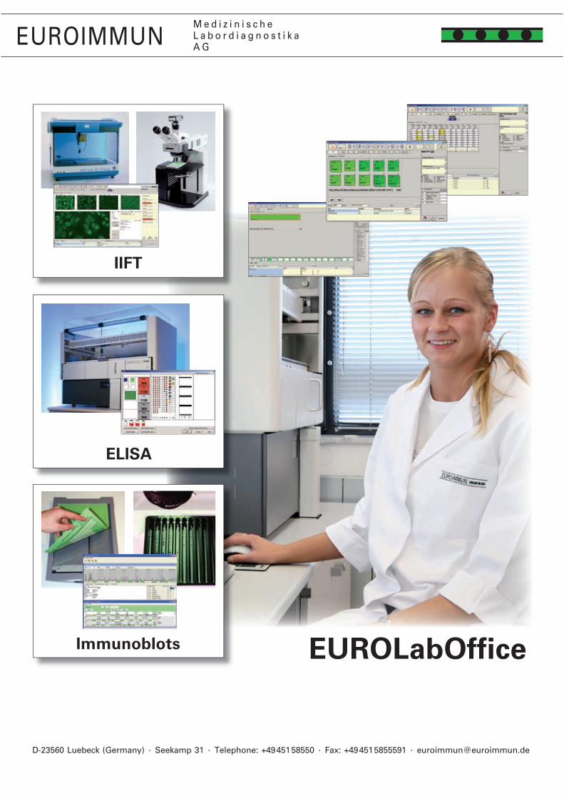
— 154 —
EUROIMMUNM e d i z i n i s c h eL a b o r d i a g n o s t i k aA G
D-�3560 Luebeck (Germany) · Seekamp 31 · Telephone: +49 451 58550 · Fax: +49 451 5855591 · [email protected]
EUROLabOffice
EUROIMMUNM e d i z i n i s c h eL a b o r d i a g n o s t i k aA G
IIFT
ELISA
Immunoblots
One laboratory management soft-ware for all automated systems
YG_0250_R_UK_B03, 10/2010
EUROIMMUN AG · D-23560 Luebeck (Germany) · Seekamp 31 · Tel +49 451 58550 · Fax 5855591 · E-mail [email protected]