Embed Size (px)
Citation preview

August 2011- Under Review
PROCEDURES FOR THE
VISION SCREENING PROGRAM
FOR
PENNSYLVANIA’S SCHOOL-AGE
POPULATION

TABLE OF CONTENTS
Page
ACKNOWLEDGEMENTS ..................................................................................... i
PREFACE ............................................................................................................... iii
I. INTRODUCTION ...................................................................................................................... 1
II. LEGAL BASIS FOR THE SCHOOL VISION SCREENING PROGRAM .......... 2
III. TESTING SCHEDULE PRIORITIES .................................................................... 3
IV. PREPARATION OF FACILITIES. ........................................................................ 4
V. PREPARATION OF THE STUDENT ................................................................... 4
VI. OBSERVATIONS OF THE STUDENT ................................................................. 5
VII. SCREENING PROCEDURE .................................................................................. 7
VIII. SCREENING TESTS. ............................................................................................. 8
Far Visual Acuity Test .......................................................................................... 8
Near Visual Acuity Test ..................................................................................... 11
Convex Lens Test – (Plus Lens) ......................................................................... 13
Color Vision – Color Discrimination Test ........................................................ 15
Stereo/Depth Perception Test ............................................................................. 18
IX. FOLLOW-UP AND CASE MANAGEMENT ..................................................... 19
X. REPORTING ......................................................................................................... 19
XI. CONTACT LENSES ............................................................................................. 20
XII. VISION SCREENING TESTS FOR STUDENTS WITH SPECIAL HEALTH
CARE NEEDS ....................................................................................................... 23
XIII. REFERENCES ...................................................................................................... 27
APPENDICES ................................................................................................................... 29
Appendix A Eye Language ......................................................................... 30
Appendix B1 Anatomy and Physiology of the Eye ..................................... 31
Appendix B2 Refraction of the Eye ............................................................. 32
Appendix C Eye Glossary .......................................................................... 33
Appendix D Common Eye Disorders in Children ...................................... 35
Appendix E Resources for Information and Equipment ............................ 37
Appendix F1 Parent/Guardian Notification Letter....................................... 40
Appendix F2 Vision Screening Referral ...................................................... 41
Appendix F3 Eye Specialist Report ............................................................. 42
Appendix G Near Point of Convergence Information ................................ 43
Appendix H Nursing Diagnoses in the Treatment of Ophthalmic
Conditions .............................................................................. 45
Appendix I First Aid for Eye Emergencies ............................................... 48

i
ACKNOWLEDGEMENTS
The Division of School Health, Pennsylvania Department of Health gratefully acknowledges the
assistance of the members of the 2000-01 Advisory Task Force on School Vision Screening in
the revision of this manual.
Maggie Beall, MA, BS, RN,C, School Nurse, Moniteau School District; President, School Nurse
Section, Department of Pupil Services, Pennsylvania State Education Association
Deborah S. Blanchard, Pennsylvania Optometric Association, Director of Communications/Staff
Contact
Mark B. Boas, OD, MS, Pennsylvania Optometric Association, Pediatric Vision Care Task Force
Elise B. Ciner, OD, FAAO, Pennsylvania Optometric Association, Pediatric Vision Care Task
Force
Lorraine Crum, BSN, RN, School Nurse, Conneaut School District
Jon Dale, MS, Director, Division of School Health, Pennsylvania Department of Health
Sharon Daly, RN,C, School Health Consultant, Southeast District, Pennsylvania Department of
Health
Linda D. Deeter, BSN, RN, School Health Consultant, Northwest District, Pennsylvania
Department of Health
Michelle Ficca, DNSc, RN, Assistant Professor, Department of Nursing, Bloomsburg University
Deborah Fontaine, BSN, RN, School Health Consultant, Northcentral District, Pennsylvania
Department of Health
Judith Gardner, CRNP, BSN, School Health Consultant, Southcentral District, Pennsylvania
Department of Health
Debra Gilbert, MSN, BSN, RN, School Nurse, Bloomsburg School District
Douglas J. Goepfert, OD, Pennsylvania Optometric Association, Pediatric Vision Care Task
Force
Judith Lavrich, MD, Total Eye Care Centers
Letitia Leitzel, MA, BSN, RN,C, School Health Consultant, Central Office, Pennsylvania
Department of Health
Robert Lloyd, OD, Optometrist, Bradford, Pennsylvania
Linda McGrath, BSN, RN, School Nurse, Grove City School District
James W. McManaway III, M.D., Pennsylvania Academy of Ophthalmology
David R. McPhillips, OD, FAAO, President, Pennsylvania Optometric Association, Pediatric
Vision Care Task Force
Patricia Montalbano, RN,C, School Health Consultant, Northeast District, Pennsylvania
Department of Health
Marla L. Moon, OD, FAAO, Pennsylvania Optometric Association, Pediatric Vision Care Task
Force
Ann Murray, BSN, RN, School Nurse, Midd-West School District
George William Orren, III, OD, Orren Eye Associates
Rita Schmitt, BSN, RN, School Nurse Coordinator, City of Erie School District
James S. Spangler, OD, Pennsylvania Optometric Association; Chairman, Pediatric Vision Care
Task Force
Karolyn Stone, BSN, RN, MEd., Retired School Nurse
Charles V. Stuckey, Jr., OD, FAAO, Executive Director, Pennsylvania Optometric Association
Cynthia Thomas, BSN, RN, School Health Consultant, Southwest District, Pennsylvania
Department of Health
Rita Verma, OD, Betz Ophthalmology Associates, Lewisburg, Pennsylvania
Denise T. Wilcox, OD, Pennsylvania Optometric Association, Pediatric Vision Care Task Force

ii
Special acknowledgement is given to the following: Mark Boas, OD, MS, Pennsylvania
Optometric Association for contributing many hours to the clinical review of this manual and the
writing of Appendix G, Near Point of Convergence; Doris Luckenbill, MSN, CSNP for the
original writing of the Students with Special Health Care Needs section; and Charles J. Stuckey,
Jr., OD, FAAO, Pennsylvania Optometric Association for his contribution to the Contact Lens
section.
Review and comment of the manual prior to publication was also provided by the following
individuals or organizations:
Robert S. Muscalus, D.O., Physician General of Pennsylvania
Pennsylvania Academy of Ophthalmology
Pennsylvania Association of School Nurses and Practitioners
Pennsylvania Optometric Association
The previous document, Guidelines for the School Vision Screening Program for Pennsylvania’s
School-Age Children and Adolescents, was developed by members of the Vision Screening Task
Force and the School Nurse Advisory Committee in 1989.

iii
PREFACE
The purpose of a school vision screening program is to identify students with visual
impairments. “Vision problems affect one in 20 preschoolers and one in four school-age
children” (Prevent Blindness America, 2000). Visual problems can and do affect the
educational, social and emotional development of children. Early detection of vision problems
assures the child of the opportunity of taking the best advantage of his/her educational
opportunities.
Ninety percent of all information is transferred to the brain via the eyes. Most vision problems
are correctable, at least to some degree. Impaired vision is most damaging in primary grades
because it is at these grade levels that the foundations for learning are taught. Those children
with vision loss severe enough to require special educational opportunities must be identified
early if they are to be helped.
It is routine for infants to have their ocular health screened at birth, and vision authorities agree
that children should have a more thorough eye examination very early in life. The American
Optometric Association (AOA) recommends an eye examination by six months of age, at three
years of age, before first grade, and every two years thereafter. According to the American
Academy of Ophthalmology (1996), “Two to four percent of America’s children develop
strabismus and/or amblyopia. Early detection and treatment of these disorders during childhood
are essential for preventing permanent vision loss.”
Although it is recommended that every child have an eye examination very early in life, vision
screenings continue to provide an important tool in the early detection of vision disorders in the
pediatric population. However, the opportunity for vision screenings is not always afforded to
every child in the early years of life. As attendance at school is mandated for all children in
Pennsylvania, the school setting provides an accessible place where children may have their
vision screened. It is possible for children in Pennsylvania as young as four to have their vision
screened if they attend kindergarten.
Recognizing the above statements, vision screening has been rightly mandated for Pennsylvania
school age children since 1957. The purpose of this procedure manual is to provide standards for
the school vision screening program throughout the Commonwealth of Pennsylvania. This
manual replaces the “Guidelines for the School Vision Screening Program for Pennsylvania’s
School-Aged Children and Adolescents”, H514.032P, revised 9/89.
Disclaimer: Any reference to trade names or products does not represent an endorsement by the
Pennsylvania Department of Health. Resources and references do not represent an all inclusive
listing.

1
PROCEDURES FOR THE VISION SCREENING PROGRAM FOR
PENNSYLVANIA’S SCHOOL-AGE POPULATION
I. INTRODUCTION
“Vision screening is not diagnostic, but is a practical approach to identifying
children needing professional eye services. It is an efficient, economical, and
efficacious manner of detecting possible vision problems in the pre-school and
school age populations. By definition, screening is the process by which a large
number of persons are tested by a fast, efficient method in order to separate them
into different groups. The purpose of the vision screening test is to separate those
children who probably have no vision problems from those who should be
examined by an eye doctor for potential problems and possible treatment”
(National Association of School Nurses, 1995).
According to the American Academy of Pediatrics Policy Statement (1996),
“Vision screening and eye examination are vital for the detection of conditions that
distort or suppress the normal visual image, which may lead to inadequate school
performance or, at worst, blindness in children”. “A screening program of early
identification, diagnosis and correction of children’s vision disorders is an essential
part of all child health programs. The early detection and treatment of vision
disorders gives children a better opportunity to develop educationally, socially,
emotionally and physically” (Ohio Vision Manual, Rev. 1998).
The frequency of vision disorders increases with age, therefore it is important to
have a clear understanding of critical periods in human visual development. For
example, studies show that the greatest proportions of these are errors in refraction.
Approximately five percent of pupils in the first grade may have errors of
refraction, while “nearly 20% of the pediatric population require the use of
eyeglasses for refractive errors before the late teenage years” (American Academy
of Pediatrics, 1996).
Because vision screening is not diagnostic, children who fail the test must be
referred to an eye specialist for a diagnostic examination. Screening will not
identify every child who needs eye care, nor will every child who is referred
require treatment. But the criteria for referral have been set to keep both the over-
referrals (those with no problem on examination) and the under-referrals (those
who are missed) at a minimum.
In addition to detecting vision problems, vision screening programs are valuable in
raising the awareness of parents, teachers and the community to the importance of
eye care. Another screening benefit is the identification of children who may need
special education services because of a visual impairment.
The most important aspect of the screening program is referral with follow-up. The
child who fails the screening should receive a comprehensive eye examination by
an eye care specialist. If the child does not receive attention by an eye care
specialist, then the screening program has not accomplished its goal.

2
II. LEGAL BASIS FOR THE SCHOOL VISION SCREENING PROGRAM
The Public School Code of 1949, Section 1402(a) (Act 404 of 1957) requires that
“Each child of school age shall be given by methods established by the Advisory
Health Board, a vision test...”. The vision test shall be administered by a certified
school nurse, medical technician (health room aide) or teacher. 28 Pa. Code § 23.4
(Regulations) of the Department of Health requires vision screening tests to be given
annually. The regulations specify that the Snellen chart or other screening devices
approved by the Department of Health shall be utilized for vision screening.
Test Requirements Condition Grade Levels
1. Far Visual Acuity Test Amblyopia, Astigmatism,
Myopia
All students yearly
2. Near Visual Acuity Test Astigmatism, Focusing
Problems, Hyperopia
All students yearly
3. Convex Lens Test (Plus
Lens)
Excessive Hyperopia 1st grade students meeting
criteria (See p. 14), new
students not previously
screened
4. Color Vision Test Color Discrimination 1st or 2
nd, new students not
previously screened
5. Stereo/Depth Perception
Test
Binocularity, Strabismus 1st or 2
nd, new students not
previously screened

3
III. TESTING SCHEDULE PRIORITIES
The certified school nurse/practitioner is responsible for the vision screening
program in Pennsylvania’s schools. At the beginning of the school year, the school
nurse/practitioner will establish a testing schedule for all students, (K4, K–12th
).
The following priorities should be considered when scheduling students for
screening.
A. All students enrolled in Kindergarten classes.
B. All students identified as belonging to high-risk groups, such as the
following:
Students known to have severe or progressive eye conditions.
Students who have medical conditions that may affect vision; have a
significant medical, surgical or familial history; have had recent eye
injuries or are taking medications that may affect vision.
Students having emotional or behavioral problems.
Students being evaluated for special class placement.
Students identified as showing poor progress in learning to read.
Students scheduled for driver education classes.
C. All students suspected of having a vision problem who are self-referred or
referred by teachers, parents or physicians.
D. All new entrants to school who have no record of a previous vision test or
have previous tests that indicate other-than-normal acuity.
E. All students in Grades 1 through 3 and special education classes.
F. All students in Grades 4 through 12.

4
IV. PREPARATION OF FACILITIES
A. When planning for vision screening it is imperative to select a room or area
that is quiet and free from interruptions (ideally, the health office). Be sure
that other students can not see the chart.
B. If a wall mounted visual acuity chart is used, the room should be large enough
to allow for a physical distance of 10 ft for a 10 ft chart or 20 ft for a 20 ft
chart from the student being tested.
C. If a wall mounted visual acuity chart is used, it should be placed against a
background, the brightness of which should not be in great contrast with the
chart itself. The background should be free of distractions (posters, toys,
pictures and other children).
D. The room should be well lighted and free from glare. If there is insufficient
light on the chart, position gooseneck lamps on the floor in front of the chart.
The illumination in the room should be the same or slightly less than the
illumination of the chart.
E. The chart should be hung so that the chart’s “20 feet” line is level with the
student’s eyes.
V. PREPARATION OF THE STUDENT
All students should be educated so that they understand the purpose of vision
screening and their role in the activity.
A. Education of the student should emphasize the following:
1. The value of early and periodic screening tests;
2. The relationship of correct health and safety practices to the prevention of
eye diseases;
3. The prompt medical treatment of eye injuries and preservation of sight.
B. The individual who will be doing the screening should plan time with the
Primary grade teacher to demonstrate the screening procedures to the
students:
1. When the tumbling “E” chart is to be used, a large letter “E” can be turned
in various positions to show the students how to use their arms to indicate
the direction of the shafts. Other names can be substituted for the term
“vision test”, such as “E game” or “Table leg game” since the word “test”
may imply to the student the “need to pass.”
2. Related health education appropriate to the student’s grade level and
maturity should be offered concurrently. Health education should
emphasize: (1) the importance of early and periodic screening tests; (2) the
relationship of correct health and safety practices to the prevention of eye
diseases, eye injuries and preservation of sight; (3) prompt medical
treatment of correctable and/or reversible eye health conditions, and (4)
environmental factors which are conducive to the maintenance of eye health
and safety. If well planned, the screening procedure will then become a
laboratory experience in health, which enriches the instructional program
for the child.

5
VI. OBSERVATIONS OF THE STUDENT
When a student is scheduled for screening, whether based on self-referral or by
class schedule, teacher observations of visual behavior should be gathered and
reviewed. The review of information may be conducted prior to or subsequent to
the screening procedures. No decision on referral (i.e., to refer or not to refer)
should be made without a review of observations. If in doubt, the school nurse may
choose to observe the student performing a variety of visual tasks. Sharing this
information with an eye care specialist may be much more valuable than the test
score results.
Document complaints, concerns and observations reported by teachers,
parents and student.
Observe and record any problems detected, which may or may not include the
following:
Signs of Possible Eye Trouble in Children – “ABC Checklist for Vision”
A. Appearance of External Eye
1. Eyelids – any edema, ptosis, swelling, redness, discharge, excess
tearing, condition of the eyelashes, and lack of or excessive blinking of
the eye.
2. Conjunctiva – any discharge, hemorrhage, allergic signs, or scars.
3. Eyeball – size, shape, alignment.
4. Cornea – any cloudiness, bulging, abrasions or ulcers.
5. Sclera – any inflammation or unusual color, such as the bluish tint found
in osteogenesis imperfecta, or the yellow of jaundice.
6. Iris – color, any irregularities, cuts, or spots.
7. Pupil – dilatation or constriction, anisocoria (difference in pupil size).
8. Lens – clear or cloudy, any opacity.
B. Behavior of Child:
1. Has difficulty reading or doing work requiring close vision. Skips
words or lines, loses place, re-reads, or reads too slowly. Is inattentive
during chalkboard, wall chart, or map lessons. Tends to reverse words
or syllables, or confuses the following letters in reading or spelling: a
and o; e and c; n and m; h, n and r; and f and t.
2. Frowns, blinks excessively, scowls, squints or uses other facial
distortions when reading.
3. Holds books and objects either too close or too far, or avoids close work
whenever possible. Makes frequent change in distance at which book is
held.

6
4. Rubs eyes frequently, or attempts to brush away blur.
5. Shuts or covers one eye, tilts or thrusts head forward when looking at
near or distant objects. Has poor hand-eye coordination.
6. Has general fatigue or drowsiness while reading or doing close work.
Has poor work performance. Loses place and/or attention while
reading. Appears awkward or excessively daydreams.
7. Stumbles or trips over small objects.
8. Does not do well in games requiring distant vision.
9. May be unduly sensitive to light, and poor in color detection.
C. Complaints – Child’s Statements
1. Cannot see well. Letters or lines “run together” or “jump.”
2. Headaches, dizziness, or even nausea following close eye work.
3. Double vision.
4. Fatigue and listlessness after close eye work. “Eyes hurt.”
Note: It must be recognized that many of these signs and symptoms occur transiently
during colds and other illnesses, but any persistence of these complaints indicate
the need for further evaluation (Prevent Blindness America, 2000). Some
students may have difficulty with reading and comprehension despite having
normal outcomes on the required eye screenings. These students may benefit from
additional screening, such as Near Point of Convergence (NPC). NPC testing in
the school vision screening program can be a valuable tool to help understand the
cause of a student’s visual complaints. NPC testing may be utilized by the school
nurse if a student passes the school screenings but the parents, teachers, school
nurse, etc. note performance problems. (See Appendix G for NPC information
and screening procedure).

7
VII. SCREENING PROCEDURE
A. The purpose of the history and external observations is to detect any history
of eye problems or obvious ocular pathology or abnormalities. (See VI,
“ABC Checklist for Vision") Any obvious deviations from the usual or
expected should be noted so that if referral for professional examination is
indicated, these may be included. An up-to-date record of eye health status
should be maintained as part of the child’s cumulative health record.
1. Review each student’s health record for existing problems or
predisposing factors:
a. Familial tendencies, such as glaucoma, congenital cataracts, retinal
problems, strabismus;
b. Prior injuries, infections or eye conditions.
2. Interview student (and parent, if present) for subjective data:
a. Record student’s statements, specific complaints relative to eye
health;
b. Note last professional eye examination, if known.
3. Visual inspection and observations of behaviors:
a. Eyelids, lashes, surrounding tissues, conjunctiva and pupils;
b. Redness, congestion, inflammation, crusting, or flaking of eyelids;
secretions, lumps, masses, swellings;
c. Do eyes look normal; turn in or out, drooping lids, haziness or
clouding;
d. Tilts head to read; covers or closes one eye for critical seeing;
points with finger to read; excessive stumbling; awkwardness; day
dreaming; holds printed materials in an unusual position.
4. Affirmative answers to any of the above should be pursued by further
questioning of the student and/or parent/guardian.
5. After Steps 1-4 have been completed, the selected screening test(s)
should be administered.

8
VIII. SCREENING TESTS
Visual Acuity
Visual acuity is acuteness or sharpness of vision. It is measured by the smallest object
that can be seen at a certain distance. The test for visual acuity is used to determine
whether a person has light sense and can perceive the shape and form of objects. Visual
acuity is a function of not only the refractive apparatus of the eye, but also the retina,
nerve paths, and central nervous system. Poor visual acuity may indicate more than the
need for lenses to correct a refractive error; it can be indicative of disease or anomaly of
other parts of the seeing mechanism.
HISTORY: The Dutch ophthalmologist, Herman Snellen, was the first person to
introduce the scientific standardization of visual acuity. His test, which is based on that
standardization, is given at 20 feet because a minimum of accommodation is required at
that distance. The test types are graduated in size and numbered to indicate the standard
distance at which a person with normal visual acuity should be able to distinguish them.
When giving the test, the subject is placed at a physical distance of 20 feet from a chart
and is asked to indicate the smallest letter they can read. The acuity is recorded by using
two numbers, the first of which indicates the distance from the chart; the second
designates distance for the smallest line the individual is able to read, e.g.; 20/20, 20/40,
or 20/70. A visual acuity of 20/40 means that the person can read at 20 feet, what the
average eye can read at 40 feet. The larger the second number, the poorer the vision. The
score does not represent a fraction; it is merely a short method of recording two facts.
Far Visual Acuity Test
Purpose: To test clearness of vision at a distance; to detect amblyopia, astigmatism,
myopia. (See Appendices C, D)
Equipment: (Choose one)
1. AUTOMATED VISION SCREENER (Titmus, Optec, etc.)
2a. VISUAL ACUITY CHARTS (Wall mount or illumination cabinet): These charts
are used for monocular acuity for far distance (10 or 20 ft chart; 20 ft chart
preferred). Commonly used visual acuity charts may include:
Snellen Chart
Symbol “E” chart or Tumbling “E”, for children who are not familiar with the
letter form.
HOTV letter and LEA symbol (circle, square, apple, house) charts
Tumbling Hand
2b. OCCLUDER: An object used to cover one eye comfortably while screening the
other eye (Use with Visual Acuity Charts).
Plastic eye patch type occluders (hand-held occluders preferred over those with
attached elastic bands). Plastic occluders should be wiped thoroughly with
alcohol after each student is tested.

9
Small pieces of colored construction paper such as 3 x 5 filing cards, small
paper cups (to be used by older children), or plastic spoons. If paper occluders
are used with a young child, the screener should hold the occluder. Each
disposable occluder should be discarded after individual use.
If a child exhibits “peeking” behavior, a disposable adhesive patch is highly
recommended. It is very common for students with amblyopia to use the better
seeing eye by peeking around any of the above-mentioned occluders.
Grades to be screened: ALL students yearly.
Procedure: (Use corresponding procedure for chosen equipment)
AUTOMATED VISION SCREENER
1. The student should be seated comfortably facing the person screening.
2. Instruct the student to keep glasses or contact lenses on for testing.
Exceptions: Glasses for DISTANCE ONLY should be worn for FAR test only.
Glasses for READING ONLY should be worn for NEAR test only.
3. Follow manufacturer’s instructions for screening procedure.
4. Instruct the student to keep both eyes open (even the eye which is occluded) and
proceed with the screening.
5. The visual acuity for each eye is recorded in the appropriate space on the
student’s health record. Visual acuity is recorded in the form of a fraction. The
figure above the line represents the distance from the chart; the figure below the
line indicates the smallest line read successfully.
Referral Criteria:
1. Any student enrolled in Kindergarten or Grade 1 whose visual acuity in either eye
is less than 20/40 or if a two line difference exists between the eyes (ie. 20/25 and
20/40) should be re-tested. If re-testing results in visual acuity less than 20/40 in
either eye or a two line difference between the eyes, referral is indicated.
Any student in Grade 2 or above whose visual acuity in either eye is less than
20/30 or if a two line difference between the eyes (ie. 20/25 and 20/40) should be
re-tested. If re-testing results in visual acuity less than 20/30 in either eye or a
two line difference between the eyes, referral is indicated.
2. Unless the student is showing signs of impending illness, re-testing should be
scheduled without undue delay.
3. The results should be recorded on the student’s health record in the appropriate
space, giving the date, name of test, the fractional reading for each eye and
“passed” or “failed”.
4. Parent/Guardian Notification (Appendix F1) should be sent home if the student
has PASSED the screening test. Vision Screening Referral (Appendix F2) and
Eye Specialist Report (Appendix F3) should be sent home if the student has
FAILED the screening test.
10
5. Follow-up (See page 20).
OR
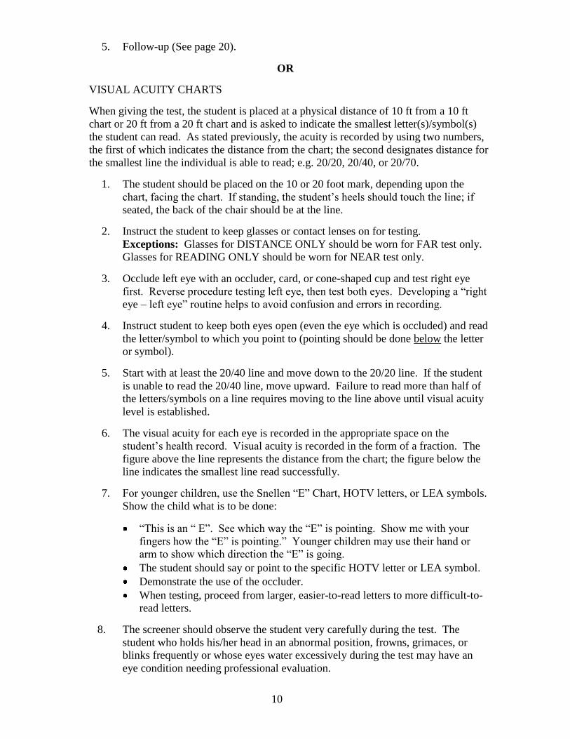
VISUAL ACUITY CHARTS
When giving the test, the student is placed at a physical distance of 10 ft from a 10 ft
chart or 20 ft from a 20 ft chart and is asked to indicate the smallest letter(s)/symbol(s)
the student can read. As stated previously, the acuity is recorded by using two numbers,
the first of which indicates the distance from the chart; the second designates distance for
the smallest line the individual is able to read; e.g. 20/20, 20/40, or 20/70.
1. The student should be placed on the 10 or 20 foot mark, depending upon the
chart, facing the chart. If standing, the student’s heels should touch the line; if
seated, the back of the chair should be at the line.
2. Instruct the student to keep glasses or contact lenses on for testing.
Exceptions: Glasses for DISTANCE ONLY should be worn for FAR test only.
Glasses for READING ONLY should be worn for NEAR test only.
3. Occlude left eye with an occluder, card, or cone-shaped cup and test right eye
first. Reverse procedure testing left eye, then test both eyes. Developing a “right
eye – left eye” routine helps to avoid confusion and errors in recording.
4. Instruct student to keep both eyes open (even the eye which is occluded) and read
the letter/symbol to which you point to (pointing should be done below the letter
or symbol).
5. Start with at least the 20/40 line and move down to the 20/20 line. If the student
is unable to read the 20/40 line, move upward. Failure to read more than half of
the letters/symbols on a line requires moving to the line above until visual acuity
level is established.
6. The visual acuity for each eye is recorded in the appropriate space on the
student’s health record. Visual acuity is recorded in the form of a fraction. The
figure above the line represents the distance from the chart; the figure below the
line indicates the smallest line read successfully.
7. For younger children, use the Snellen “E” Chart, HOTV letters, or LEA symbols.
Show the child what is to be done:
“This is an “ E”. See which way the “E” is pointing. Show me with your
fingers how the “E” is pointing.” Younger children may use their hand or
arm to show which direction the “E” is going.
The student should say or point to the specific HOTV letter or LEA symbol.
Demonstrate the use of the occluder.
When testing, proceed from larger, easier-to-read letters to more difficult-to-
read letters.
8. The screener should observe the student very carefully during the test. The
student who holds his/her head in an abnormal position, frowns, grimaces, or
blinks frequently or whose eyes water excessively during the test may have an
eye condition needing professional evaluation.

11
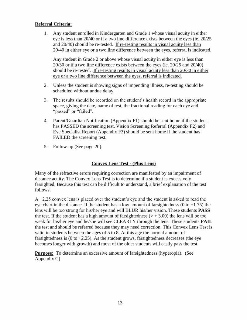
Referral Criteria:
1. Any student enrolled in Kindergarten and Grade 1 whose visual acuity in either
eye is less than 20/40 or if a two line difference exists between the eyes (ie. 20/25
and 20/40) should be re-tested. If re-testing results in visual acuity less than
20/40 in either eye or a two line difference between the eyes, referral is indicated.
Any student in grade 2 or above whose visual acuity in either eye is less than
20/30 or if a two line difference exists between the eyes (ie. 20/25 and 20/40)
should be re-tested. If re-testing results in visual acuity less than 20/30 in either
eye or a two line difference between the eyes, referral is indicated.
2. Unless the student is showing signs of impending illness, re-testing should be
scheduled without undue delay.
3. The results should be recorded on the student’s health record in the appropriate
space, giving the date, name of test, the fractional reading for each eye and
“passed” or “failed”.
4. Parent/Guardian Notification (Appendix F1) should be sent home if the student
has PASSED the screening test. Vision Screening Referral (Appendix F2) and
Eye Specialist Report (Appendix F3) should be sent home if the student has
FAILED the screening test.
5. Follow-up (See page 20).
Near Visual Acuity Test
Purpose: To test clearness of close vision; to detect astigmatism, focusing problems, and
hyperopia (See Appendices C, D).
Equipment: (Choose one)
1. AUTOMATED VISION SCREENER (Titmus, Optec, etc.)
2. NEAR VISUAL ACUITY CARDS (Snellen, HOTV letters and LEA Symbols) with
OCCLUDER
Grades to be screened: ALL students yearly.
Procedure: (Use corresponding procedure for chosen equipment)
AUTOMATED VISION SCREENER
1. The student should be seated comfortably facing the person screening.
2. Instruct the student to keep glasses or contact lenses on for testing.
Exceptions: Glasses for DISTANCE ONLY should be worn for FAR test only.
Glasses for READING ONLY should be worn for NEAR test only.
3. Follow manufacturer’s instructions for screening procedure.
4. Instruct the student to keep both eyes open (even the eye which is occluded) and
proceed with the screening.

12
5. The visual acuity for each eye is recorded in the appropriate space on the
student’s health record. Visual acuity is recorded in the form of a fraction. The
figure above the line represents the distance from the chart; the figure below the
line indicates the smallest line read successfully.
Referral Criteria:
1. Any student enrolled in Kindergarten and Grade 1 whose visual acuity in either
eye is less than 20/40 or if a two line difference exists between the eyes (ie. 20/25
and 20/40) should be re-tested. If re-testing results in visual acuity less than
20/40 in either eye or a two line difference between the eyes, referral is indicated.
Any student in Grade 2 or above whose visual acuity in either eye is less than
20/30 or if a two line difference exists between the eyes (ie. 20/25 and 20/40)
should be re-tested. If re-testing results in visual acuity less than 20/30 in either
eye or a two line difference between the eyes, referral is indicated.
2. Unless the student is showing signs of impending illness, re-testing should be
scheduled without undue delay.
3. The results should be recorded on the student’s health record in the appropriate
space, giving the date, name of test, the fractional reading for each eye and
“passed” or “failed”.
4. Parent/Guardian Notification (Appendix F1) should be sent home if the student
has PASSED the screening test. Vision Screening Referral (Appendix F2) and
Eye Specialist Report (Appendix F3) should be sent home if the student has
FAILED the screening test.
5. Follow-up (See page 20).
OR
NEAR VISUAL ACUITY CARDS
1. Prepare the testing area, making sure that it is as quiet as possible and free from
distraction. Be sure that the lighting is adequate and that the near cards are
sufficiently illuminated for easy viewing.
2. Instruct the student to keep glasses or contact lenses on for testing.
Exceptions: Glasses for DISTANCE ONLY should be worn for FAR test only.
Glasses for READING ONLY should be worn for NEAR test only.
3. Occlude the left eye and test the right eye first.
4. Have the student hold the near card at the distance specified in the manufacturer’s
instructions (approximately 13-16 in, [33-40 cm]) and ask the student to identify
the smallest letter(s)/symbol(s) possible.
5. Record Near visual acuities similar to the method used for Far visual acuities.
Follow the appropriate scoring protocol for the HOTV letter and LEA symbol
tests of Near Visual Acuity.
6. Switch occluder to the right eye and repeat the Near Visual Acuity test on the left
eye.
13
Referral Criteria:
1. Any student enrolled in Kindergarten and Grade 1 whose visual acuity in either
eye is less than 20/40 or if a two line difference exists between the eyes (ie. 20/25
and 20/40) should be re-tested. If re-testing results in visual acuity less than
20/40 in either eye or a two line difference between the eyes, referral is indicated.
Any student in Grade 2 or above whose visual acuity in either eye is less than
20/30 or if a two line difference exists between the eyes (ie. 20/25 and 20/40)
should be re-tested. If re-testing results in visual acuity less than 20/30 in either
eye or a two line difference between the eyes, referral is indicated.
2. Unless the student is showing signs of impending illness, re-testing should be
scheduled without undue delay.
3. The results should be recorded on the student’s health record in the appropriate
space, giving the date, name of test, the fractional reading for each eye and
“passed” or “failed”.
4. Parent/Guardian Notification (Appendix F1) should be sent home if the student
has PASSED the screening test. Vision Screening Referral (Appendix F2) and
Eye Specialist Report (Appendix F3) should be sent home if the student has
FAILED the screening test.
5. Follow-up (See page 20).
Convex Lens Test - (Plus Lens)
Many of the refractive errors requiring correction are manifested by an impairment of
distance acuity. The Convex Lens Test is to determine if a student is excessively
farsighted. Because this test can be difficult to understand, a brief explanation of the test
follows.
A +2.25 convex lens is placed over the student’s eye and the student is asked to read the
eye chart in the distance. If the student has a low amount of farsightedness (0 to +1.75) the
lens will be too strong for his/her eye and will BLUR his/her vision. These students PASS
the test. If the student has a high amount of farsightedness (> + 3.00) the lens will be too
weak for his/her eye and he/she will see CLEARLY through the lens. These students FAIL
the test and should be referred because they may need correction. This Convex Lens Test is
valid in students between the ages of 5 to 8. At this age the normal amount of
farsightedness is (0 to +2.25). As the student grows, farsightedness decreases (the eye
becomes longer with growth) and most of the older students will easily pass the test.
Purpose: To determine an excessive amount of farsightedness (hyperopia). (See
Appendix C)

14
Equipment: (Choose one using a +2.25 lens)
1. AUTOMATED VISION SCREENER
2. VISUAL ACUITY CHARTS
A PAIR OF +2.25 SPHERICAL LENS (Note: This lens power is appropriate for ages
5 through 8 years; a 1.75 lens should be used for students over 8 years of age to test for
hyperopia).
Grades/Students to be Screened: First grade students who have passed the Far Visual
Acuity Test. All new students not previously screened for this test and who have passed
the Far Visual Acuity Test.
The test should not be given to those students who are already wearing glasses.
Procedure: (Use corresponding procedure for chosen equipment)
AUTOMATEDVISION SCREENER
1. The student should be seated comfortably facing the person screening.
2. Follow the manufacturer’s instructions for screening procedure.
3. Instruct the student to read the letter(s)/symbol(s) corresponding to the 20/40 line
while keeping both eyes open and proceed with the screening. Each eye should be
screened individually.
Referral Criteria:
1. If the student is unable to read the letters/symbols with either eye while wearing
the glasses, the student has passed the test.
If the student is able to read the letters/symbols with either eye while wearing the
glasses, the student has failed the test. The student should then be referred.
2. The results should be recorded on the student’s health record in the appropriate
space, giving the date, name of test and “passed” or “failed.”
3. Parent/Guardian Notification (Appendix F1) should be sent home if the student
has PASSED the screening test. Vision Screening Referral (Appendix F2) and
Eye Specialist Report (Appendix F3) should be sent home if the student has
FAILED the screening test.
4. Follow-up (See page 20).
OR
VISUAL ACUITY CHARTS
The student remains at the 20/20 line, after being screened for Far Visual Acuity.
While wearing the Plus lens glasses, the student should be instructed to occlude the
left eye and to read the letter(s)/symbol(s)corresponding to the 20/40 line on the
chart with his/her right eye. Then instruct the student to occlude the right eye and
read the chart with the left eye. Each eye should be screened individually.
15
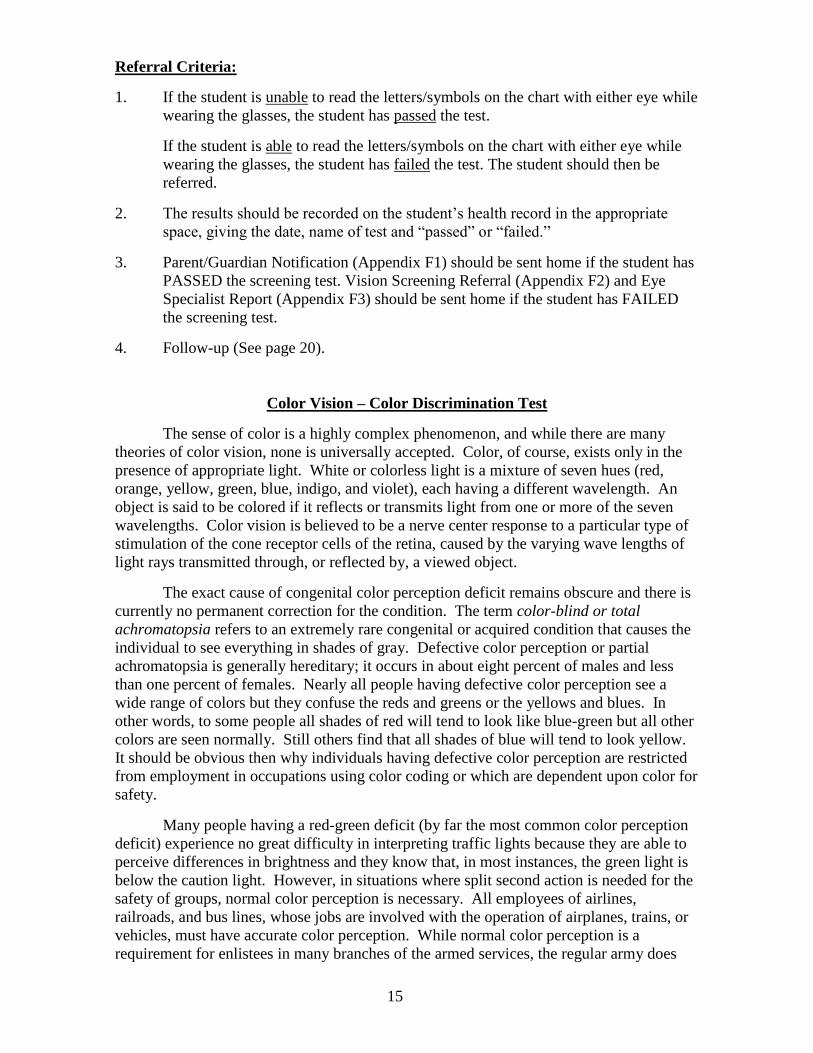
Referral Criteria:
1. If the student is unable to read the letters/symbols on the chart with either eye while
wearing the glasses, the student has passed the test.
If the student is able to read the letters/symbols on the chart with either eye while
wearing the glasses, the student has failed the test. The student should then be
referred.
2. The results should be recorded on the student’s health record in the appropriate
space, giving the date, name of test and “passed” or “failed.”
3. Parent/Guardian Notification (Appendix F1) should be sent home if the student has
PASSED the screening test. Vision Screening Referral (Appendix F2) and Eye
Specialist Report (Appendix F3) should be sent home if the student has FAILED
the screening test.
4. Follow-up (See page 20).
Color Vision – Color Discrimination Test
The sense of color is a highly complex phenomenon, and while there are many
theories of color vision, none is universally accepted. Color, of course, exists only in the
presence of appropriate light. White or colorless light is a mixture of seven hues (red,
orange, yellow, green, blue, indigo, and violet), each having a different wavelength. An
object is said to be colored if it reflects or transmits light from one or more of the seven
wavelengths. Color vision is believed to be a nerve center response to a particular type of
stimulation of the cone receptor cells of the retina, caused by the varying wave lengths of
light rays transmitted through, or reflected by, a viewed object.
The exact cause of congenital color perception deficit remains obscure and there is
currently no permanent correction for the condition. The term color-blind or total
achromatopsia refers to an extremely rare congenital or acquired condition that causes the
individual to see everything in shades of gray. Defective color perception or partial
achromatopsia is generally hereditary; it occurs in about eight percent of males and less
than one percent of females. Nearly all people having defective color perception see a
wide range of colors but they confuse the reds and greens or the yellows and blues. In
other words, to some people all shades of red will tend to look like blue-green but all other
colors are seen normally. Still others find that all shades of blue will tend to look yellow.
It should be obvious then why individuals having defective color perception are restricted
from employment in occupations using color coding or which are dependent upon color for
safety.
Many people having a red-green deficit (by far the most common color perception
deficit) experience no great difficulty in interpreting traffic lights because they are able to
perceive differences in brightness and they know that, in most instances, the green light is
below the caution light. However, in situations where split second action is needed for the
safety of groups, normal color perception is necessary. All employees of airlines,
railroads, and bus lines, whose jobs are involved with the operation of airplanes, trains, or
vehicles, must have accurate color perception. While normal color perception is a
requirement for enlistees in many branches of the armed services, the regular army does

16
accept men and women having color perception deficits. A high degree of color
discrimination is required of many of the employees of the telephone company and
electronics industry where color codes are used for wiring. All law enforcement officers
must, of course, be able to identify all colors with accuracy.
Knowledge of a student’s ability to perceive colors should be of significance to
classroom teachers. In the early elementary grades many teaching materials, visual aids
and teaching systems are color-coded. The student with an unidentified color perception
deficit may thus be at a disadvantage in a color-coded learning situation. Additionally,
vocational guidance, to be appropriate, will require awareness of the student’s ability to
perceive color. Therefore, it is recommended that a color perception test be administered
to each student as early as practical in his or her school career. The test should be
administered to all girls as well as boys, despite the fact that relatively few females have
color perception deficits.
Purpose: To identify any discrepancy in the ability to recognize color. (See Appendix D)
Equipment: (Choose one)
1. PSEUDO-ISOCHROMATIC TEST PLATES (e.g. Ishihara)
2. AUTOMATED VISION SCREENER
3. COLOR VISION TESTING MADE EASY®.
Grades/Students to be Screened: First or second grade and new students who have not
been previously screened.
Procedure: (Use corresponding procedure for chosen equipment)
PSEUDO-ISOCHROMATIC TEST PLATES (preferred)
Holding the test cards approximately 30 in (75 cm) away, instruct the student to
identify the number, symbol or trail seen on each of the designated cards, allowing
for 3 seconds per card.
Referral Criteria:
1. If the student does not correctly identify the manufacturer’s suggested number of
cards, he/she has failed the test.
2. The results should be recorded on the student’s health record, giving the date, name
of test and “passed” or “failed.”
3. Parent/Guardian Notification (Appendix F1) should be sent home if the student has
PASSED the screening test. A Vision Screening Referral (Appendix F2) and Eye
Specialist Report (Appendix F3) should be sent home if the student has FAILED
the screening test. However, a referral to an eye care specialist is not usually
necessary since there is no remedial treatment or cure for color deficiency.
4. Results should be shared with the teacher, guidance counselor and primary care
provider.
5. Education and counseling of the student and parent is recommended.
OR

17
AUTOMATED VISION SCREENER
1. The student should be seated comfortably facing the person screening.
2. Follow the manufacturer’s instructions for screening procedure.
3. Instruct the student to keep both eyes open and proceed with the screening.
Referral Criteria:
1. Follow the manufacturer’s instructions to determine if the student has passed or
failed.
2. The results should be recorded on the student’s health record, giving the date,
name of test and “passed” or “failed.”
3. Parent/Guardian Notification (Appendix F1) should be sent home if the student
has PASSED the screening test. A Vision Screening Referral (Appendix F2) and
Eye Specialist Report (Appendix F3) should be sent home if the student has
FAILED the screening test. However, a referral to an eye care specialist is not
usually necessary since there is no remedial treatment or cure for color deficiency.
4. Results should be shared with the teacher, guidance counselor and primary care
provider.
5. Education and counseling of the student and parent is recommended.
OR
COLOR VISION TESTING MADE EASY® (ages 3-6)
1. Holding the test cards approximately 20-30 in (50-75 cm) away, instruct the
student to identify or trace the object that he/she sees on each of the 9 cards,
allowing for 3 seconds per card.
2. If using the three cards “Quick” test: Instruct the student to identify the objects
(balloon, boat, dog) on the three cards.
Referral Criteria:
1. Refer if the student does not correctly identify 8 of the 9 cards or all 3 “Quick” test
cards.
2. The results should be recorded on the student’s health record, giving the date, name
of test and “passed” or “failed”.
3. Parent/Guardian Notification (Appendix F1) should be sent home if the student
has PASSED the screening test. A Vision Screening Referral (Appendix F2) and
Eye Specialist Report (Appendix F3) should be sent home if the student has
FAILED the screening test. However, a referral to an eye care specialist is not
usually necessary since there is no remedial treatment or cure for color deficiency.
4. Results should be shared with the teacher, guidance counselor and primary care
provider.
5. Education and counseling of the student and parent is recommended.

18
Stereo/Depth Perception Test
Purpose: To test for binocularity; to detect amblyopia, strabismus and poor ocular
alignment. (See Appendix C, D)
Equipment:
RANDOM DOT “E”
Grades to be Screened: First or second grade, new students not previously screened.
Procedure:
RANDOM DOT “E”
1. Have the student put on the pair of polarized glasses. If the student wears glasses,
have the student put the polarized glasses over them.
2. Show the student the raised “E” figure on the demonstration card at 16 in (40 cm).
Tell the student that the raised figure is popping off the card. Show the student the
raised “E” paired with the blank card. Ask the student to point to the raised “E”.
Repeat this process until you are certain that the student understands and can
correctly identify the raised “E”. Once the student understands, start the screening
test.
3. Present the cards six times at 16 in (40 cm) and ask the student to point to the “E”
on each presentation (shuffle the cards behind your back between each
presentation).
Referral Criteria:
1. Refer if the student cannot identify the “E” correctly in four of six attempts.
2. The results should be recorded on the student’s health record, giving the date, name
of test and “passed” or “failed”.
3. Parent/Guardian Notification (Appendix F1) should be sent home if the student has
PASSED the screening test. Vision Screening Referral (Appendix F2) and Eye
Specialist Report (Appendix F3) should be sent home if the student has FAILED
the screening test.
4. Follow-up (See page 20).
19
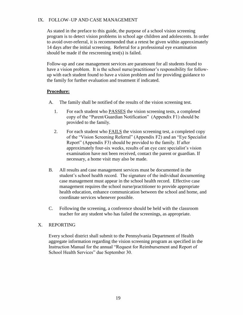
IX. FOLLOW–UP AND CASE MANAGEMENT
As stated in the preface to this guide, the purpose of a school vision screening
program is to detect vision problems in school age children and adolescents. In order
to avoid over-referral, it is recommended that a retest be given within approximately
14 days after the initial screening. Referral for a professional eye examination
should be made if the rescreening test(s) is failed.
Follow-up and case management services are paramount for all students found to
have a vision problem. It is the school nurse/practitioner’s responsibility for follow-
up with each student found to have a vision problem and for providing guidance to
the family for further evaluation and treatment if indicated.
Procedure:
A. The family shall be notified of the results of the vision screening test.
1. For each student who PASSES the vision screening tests, a completed
copy of the “Parent/Guardian Notification” (Appendix F1) should be
provided to the family.
2. For each student who FAILS the vision screening test, a completed copy
of the “Vision Screening Referral” (Appendix F2) and an “Eye Specialist
Report” (Appendix F3) should be provided to the family. If after
approximately four-six weeks, results of an eye care specialist’s vision
examination have not been received, contact the parent or guardian. If
necessary, a home visit may also be made.
B. All results and case management services must be documented in the
student’s school health record. The signature of the individual documenting
case management must appear in the school health record. Effective case
management requires the school nurse/practitioner to provide appropriate
health education, enhance communication between the school and home, and
coordinate services whenever possible.
C. Following the screening, a conference should be held with the classroom
teacher for any student who has failed the screenings, as appropriate.
X. REPORTING
Every school district shall submit to the Pennsylvania Department of Health
aggregate information regarding the vision screening program as specified in the
Instruction Manual for the annual “Request for Reimbursement and Report of
School Health Services” due September 30.

20
XI. CONTACT LENSES
Contact Lens Modalities
There are two types of contact lenses: rigid and soft. Rigid contact lenses,
previously known as hard contact lenses, are not flexible. Gas permeable materials
are now used to make rigid lenses, which allow oxygen transmission through the
lens itself. While some rigid lenses are FDA approved for use as extended,
overnight wear, most are designed for conventional daily wear. Soft contact lenses
are larger, flexible lenses. Like rigid lenses, most soft contact lenses are designed
for daily wear while a few are designed for extended wear. There is a trend toward
disposability of soft contact lenses, allowing patients to replace their lenses
quarterly, monthly or even daily.
Solutions
While it is not important to know the specifics of the types of solutions, it is
important to know that certain solutions which students might use in their regimen
can cause an uncomfortable “red eye,” burning or tearing. For example, some daily
cleaners and disinfecting solutions can cause redness and discomfort if they come in
direct contact with the eye or are not neutralized properly. Saline solutions that
contain preservatives can cause ocular irritation as well. Therefore, if a student
complains of burning, tearing or has red eyes, ask what type of solution he/she used
– a daily cleaner, disinfecting solution or saline solution with preservative may be
the culprit. If this is the case, remove the lenses and rinse with a multipurpose or
saline solution. If irritation continues upon reinsertion of the lenses, store the lenses
in a case for the student to take home.
School health offices may stock multipurpose cleaning solution and saline, as well
as a supply of clean contact lens storage cases. It is advisable to discard any cases
used by students because they can pass on infections to other students who use the
cases later.
Solutions used specifically for rigid lenses should not be used with soft contact
lenses. Tap water is not recommended for rinsing or storing lenses, especially soft
lenses due to potential permanent visual loss secondary to contamination from
microbes in the water. Saliva should never be used on contact lenses.
Common Complaints
In addition to irritation caused by solutions, the most common problems that will
bring a student to a school health office include a speck of dirt beneath the contact
lens, a decentered lens, a lens that is present but believed to have been removed or
fallen out, a lens that has fallen out but believed to be present, and a lens that is
cracked, chipped, or torn.
Handling of Contact Lenses
If the student is unable to remove the lens himself/herself, the nurse may need to
remove the lens for the student. Rinse the lens and store it in a case with saline

21
solution or a multipurpose disinfecting solution appropriate for soft or gas permeable
lenses. It is extremely important to first make sure the lens is on the eye before an
attempt is made to remove it – make sure the lens can be seen and moved. Never try
to remove what “might be” a lens or a lens that appears to be stuck.
Rigid Lens Removal
The two-handed method is used to remove a rigid lens. With the student looking
straight ahead and the eyes opened widely, place the index finger to the outer canthus
and apply gentle pressure outward and slightly upward. Cup a hand under the
student’s eye and have the patient blink. The lens will be pinched by the pressure
exerted by the eyelid and will pop out into the palm of the hand. Remember, it is
physically impossible for the contact lens to become lost behind the eye. If previous
attempts fail, fill a sink or pan with water and have the student place his/her face in
the water, open his/her eyes wide and roll his/her eyes around. The lens should float
off his/her eye. An eyecup will serve the same purpose.
Soft Lens Removal
The preferred technique is to place a fingertip on the lens with the student looking
straight ahead while gently pulling the student’s lower lid down; have the student
look up; slide the lens onto the lower white part of the eye, and simply pinch the lens
between the thumb and index finger to remove. Inspect the lens to make sure the
entire lens has been removed. Rinse and store the lens in saline solution.
On occasion, soft contact lenses may be rolled up or folded beneath the upper lid.
These lenses are very difficult to find with the naked eye. Try pulling the lower lid
down and have the student look up, down and sideways. Repeat while pulling the
upper lid up. Most likely, the nurse will need to refer the student to an eye care
specialist who will use a biomicroscope, or fluorescein if necessary, to locate the
lens. Remember, it is anatomically impossible for the lens to become lost behind the
eyeball.
Referrals
Obviously, the first choice when making a referral is to the doctor who fit the student
for his/her contact lenses because that doctor will have a complete record. If the
student’s doctor cannot be reached, school health offices should have a list of local
eye doctors for referral, consultation or general vision questions. Calls should be
made any time a nurse has questions, if the nurse has gone through the
troubleshooting sequence and pain persists, or if the contact lens problem surpasses
the nurse’s comfort zone.
Be prepared to provide the following information: age and sex; type of contact
lenses; types of solutions; nature of problem; and level of irritation and blurriness.

22
Contact Lens Troubleshooting Checklist Pennsylvania Optometric Association
_____ Are you wearing contact lenses?
_____ What type are they?
_____ How long have you been wearing this pair?
_____ Did you sleep or nap with contact lenses on recently?
_____ How long has this pair been in your eyes continuously?
_____ What type of solution do you use?
_____ Describe your pain: foreign body sensation, dull ache, sharp pain.
_____ Does it itch or burn?
_____ How long has it been red and/or painful?
_____ Do you have sensitivity to light?
_____ Is there discharge? What type?
_____ Has this ever happened before?
_____ Can you see? Is it blurry?
_____ Why did you come to see me?
_____ Location of contact lens.
_____ Disposition of contact lens.
_____ Does it still hurt after removing the lens?
_____ Referral.

23
XII. VISION SCREENING TEST FOR STUDENTS WITH SPECIAL HEALTH
CARE NEEDS
STUDENTS WITH SPECIAL HEALTH CARE NEEDS
Vision screening for students with developmental and/or disabling conditions
can be difficult, but there are many tools available to aid in the testing. The student
can be screened according to his/her level of functioning, and the series of
assessment steps can be used. All of the steps can be used in vision screening,
although the type of vision test used may vary according to the student’s functional
level.
A. Health History
Review Each Student – The complete health history will assist the nurse in
determining whether the parent/guardian has had any suspicion of vision
problems in the student. The family history should be carefully documented
for any known diabetes, glaucoma, blindness, or other diseases which may
lead to problems for the student.
B. Assessment of Vision
1. Record observations noted by teachers or other staff regarding functional
level of the student. Can the student name, match colors or identify
pictures such as:
Apple
House
Umbrella
Symbols
Letters
2. Document clues as to whether the student turns his/her head to one side,
holds objects close to the face, closes one eye while working, or seems to
reach beyond objects when attempting to pick them up.
3. Collaborate with the teacher to determine which test may be appropriate
for use with the individual student. Evaluate the student’s attention span,
and the need for an assistant during testing.
C. Teaching/Conditioning of the Student
It may be appropriate to have the teacher include as goals on the Individual
Education Plan (I.E.P.) some teaching of letters, symbols, or pictures
commonly used for vision testing. Some students can also be conditioned to
respond to certain stimuli in appropriate ways. For example, the non-verbal,
low-functioning student may be taught to point to a picture of an object when
the object is held some distance away, and rewarded with a Cheerio, M & M,
or raisin. Applause, or verbal strokes (“O.K.!” or “Good Job!”) may be
reward enough for some students.

24
D. Assessment of External Eye
The examination of the external eye should include:
Eyelids – any edema, ptosis, swelling, redness, discharge, excess
tearing, condition of the eyelashes, and lack of or excessive blinking
of the eye.
Conjunctiva – any discharge, hemorrhage, allergic signs/symptoms or
scars.
Eyeball – size, shape and alignment.
Cornea – any cloudiness, bulging, abrasions or ulcers.
Sclera – any inflammation or unusual color, such as the bluish tint
found in osteogenesis imperfecta, or the yellow of jaundice.
Iris – color, any irregularities, cuts, or spots.
Pupil – dilatation or constriction, any opacity.
Lens – clear or cloudy.
E. Tools Available for Vision Screening
1. Optokinetic Drum
This instrument will give a very gross assessment of visual function.
The drum is held 24 in (61 cm) from the student, who is seated. The
drum is rotated slowly, then more rapidly. The student’s eyes are
closely observed. If nystagmus appears as the drum is rotated, there is
at least some functional vision.
2. Blocks
Use 1 in (2.54 cm) colored blocks against a contrasting background and
scatter them on the floor at various distances from the student. Record
the distance at which the student appears to see the blocks, or makes an
effort to pick them up or move toward them. If the student makes an
effort to obtain blocks which are four to five feet away, there is
probably adequate vision for classroom function.
3. Raisins/Cheerios
Place either raisins or cheerios against a contrasting background at a
distance of approximately 12 to 14 in (30 to 36 cm) from the student’s
eyes. (On the tray of a triangle chair or wheelchair, or on a tabletop, or
on a mat on the floor if the student cannot sit up). If the student
sees/picks up the objects, he/she can see well enough for educational
purposes.

25
4. Miniature Toys Test
The test is for use with verbal students, or those who can point to
pictures of the toys used. It may require the assistance of a second
person. Use several small toys, such as cars, planes, chairs, dolls,
knives, forks, and spoons, and a piece of black cloth at least 24 in (61
cm) square. The toys should measure either 2 in (6 cm) or 3 ¼ in (8 cm)
long. Allow the student to play with the toys until you are certain that
he/she can identify them in some way, either by name (or some
recognizable sound) or by pointing to a picture (matching).
Move 10 ft away from the student. Place a black cloth on your lap,
across your knees. Place each toy on the cloth and have the student
identify it. If the student can identify the 2 in (6 cm) objects at 10 feet,
vision is approximately 20/20. If the student can identify the 3 ¼ in (8
cm) objects at 10 feet, vision is approximately 20/30. With an assistant,
you can test each eye separately, by occluding one eye at a time, to
determine differences between the two eyes.
5. Far and Near Visual Acuity Tools
There are several different Far and Near Visual Acuity tools available
that are useful in testing students with special health needs. For Far
Visual Acuity, it is recommended that Far Visual Acuity Charts be used
at a distance of 10 ft, because you are closer to the student and it is
easier to maintain his/her attention. An assistant may be necessary to
cover the student’s eye during testing, and to help the student focus
attention on the task at hand.
The following are four suggested tools for Visual Acuity testing:
Apple/House/Umbrella – Most students can be taught to identify
(name them or make some appropriate sound, point or sign) these
pictures. The use of the beginning sound of each word is sufficient
for the screener to recognize which picture the student is
identifying.
HOTV – Some higher-functioning students in this group can learn
to recognize these four letters. Some will be able to name them
after appropriate classroom teaching, others will be able to match
them to a card kept in front of the student.
Tumbling “E” or Hand – These may be appropriate for the higher-
functioning students in this group.
Broken Wheel – This test is excellent for students ages 2-5 years
who are visually and mentally disabled, have multiple disabilities
or are deaf.

26
6. Goodlite Dot Test
This is a lighted device which is used at a distance of 18 inches from the
student, which makes it appropriate if you do not have an assistant for
your testing. It can also be used by non-verbal students who have the
ability to point, either with a finger, headstick, or light stick. While
used in close proximity to the student, it is not a test of near vision.
The Dot Test is a circular screen, lighted from below, with a series of
graduated black dots on it. One dot at a time is uncovered in a three
inch opening, and the student points to the dot. Visual acuity is read
directly from the center of the screen, according to which dots the
student can find.
7. PhotoScreener
This is a portable, instant vision screening device. It provides a means
to screen for potential eye problems in difficult-to-screen students as
well as developmentally challenged students.
8. Welch Allyn Suresight Vision Screener
This is a lightweight, portable, handheld machine that addresses
compliance problems during eye chart acuity screenings. It is ideal for
young students, and those with special needs and/or language barriers.
F. Other Testing
Color Vision
An estimate of the student’s color vision may be documented from his/her
ability to name or match colored blocks or toys. Ishihara plates are also
available for “unlettered persons.” These plates use symbols such as squares
and circles instead of letters and numbers. The student must have enough
understanding and hand control to be able to follow the shapes with a
pointer, unless he/she is verbal enough to identify the shapes.
G. Documentation/Case Management
Record, on the student’s health record, the appearance of the external eye,
the teacher assessment of vision, and the results of your screening test.
Include the method that you used to obtain the best results for each student.
Document any problems found, and referrals for further care, as well as
follow-up and case management according to Section IX.

27
XIII. REFERENCES
1. American Academy of Ophthalmology. (1996). Don’t wait until it’s too late:
Understanding strabismus and amblyopia. Retrieved September 1, 2000 from
the World Wide Web:
http://www.kidsource.com/kidsource/content2/news2/strabismus.html
2. American Academy of Pediatrics. (1996). Eye examination and vision screening
in infants, children, and young adults. Retrieved October 10, 2000 from the
World Wide Web: http://www.aap.org/policy/0461.html
3. American Optometric Association. (1999). School nurses’ guide to vision
screening and ocular emergencies. St. Louis, MO: Author.
4. Arnold, M. J. & Silkworth, C. K. (1999). The school nurse’s source book of
individualized healthcare plans, Vol. II, (pp. 276-278). North Branch, MN:
Sunrise River Press.
5. Bates, B. (1995) A guide to physical examination, (6th
ed.). Philadelphia: J. B.
Lippincott
6. Carpenito, L. J. (1997) Nursing diagnosis: Application to clinical practice, (7th
ed.). Philadelphia, PA: Lippincott-Raven Publishers.
7. Colorado Department of Health, Community Nursing Section. (1991).
Guidelines for school vision screening programs. Denver, CO: Author.
8. Division of Family Health Services, Vision and Hearing Section. (n.d.).
Instructions for the Massachusetts vision test. Boston, MA : Author
9. Garzia, R. (1994). The relationship between visual efficiency problems and
learning. In Scheiman, M. M., & Rouse, M. W.: Optometric Management of
Learning Related Vision Problems, St. Louis: Mosby.
10. Granet, D. B. (2000). Connection possible between eye disorder in children and
ADHD. Proceedings of the American Academy of Pediatric Ophthalmology
and Strabismus, San Diego.
11. Guyton, A.C. (1971). Textbook of medical physiology, (pp. 604-15 and 695).
Philadelphia: W.B. Saunders Co.
12. Hayes, G. J., Cohen, B. E., Rouse, M.W., & De Land, P.N. (1998). Normative
values the near point of convergence of elementary school children. Optom Vis
Sci, 75, 506-512.
13. Institute of Medicine, National Academy of Science. (1973). A strategy for
evaluating health services: Visual disorders in health status. (pp. 67-95).
Washington D.C: Author.

28
14. MTI Photoscreener, Medical Technology & Innovations, Inc. (n.d.). MTI
PhotoScreener, innovative vision screening for kids. Lancaster, PA: Author.
15. National Association of School Nurses, Inc. (1995). Vision screening guidelines
for school nurses. (Rev.ed.). Scarborough, Maine: Author.
16. North American Nursing Diagnosis Association. (1999-2001). About NANDA.
Retrieved March 15, 2001 from the World Wide Web:
http://www.nanda.org/html/about.htm1
17. Ohio Department of Health. (1998). Vision conservation program policies for
children: Requirements and recommendations. (Rev. ed). Ohio: Author.
18. Pennsylvania Public School Code of 1949, § 1402(a)(1).
19. 28 Pa. Code § 23.4 (School Health Regulations).
20. Prevent Blindness America. (2000). Children’s eye problems. Retrieved
September 1, 2000 from the World Wide Web:
http://www.preventblindness.org/children/ch-eye-problems.html
21. Prevent Blindness America. (2000). First aid for eye emergencies. Retrieved
September 1, 2000 from World Wide Web:
http://www.preventblindness.org/safety/firstaid.html
22. Prevent Blindness America. (2000). Signs of Possible Eye Trouble in Children.
Retrieved November 20, 2001 from the World Wide Web:
http://www.preventblindness.org/children/trouble_signs.html
23. Taber, C.W. (1997). Taber’s cyclopedic medical dictionary edition, (18th
ed.).
Philadelphia: Davis Company.
24. The New York State Education Department, Bureau of School Health Education
Services. (1982). Vision screening program guidelines, (pp. 1-8 and 18).
Albany, NY: Author.
25. Wisconsin Children’s Vision Check Program. (1997-1998 pilot). Manual for eye
protection in Wisconsin schools. Wisconsin: Author.
29
Page
APPENDICES
Appendix A – Eye Language ................................................................................ 30
Appendix B1 – Anatomy and Physiology of the Eye ............................................ 31
Appendix B2 – Refraction of the Eye ................................................................... 32
Appendix C – Eye Glossary .................................................................................. 33
Appendix D – Common Eye Disorders in Children .............................................. 35
Appendix E – Resources for Information and Equipment .................................... 37
Appendix F1 – Parent/Guardian Notification Letter ............................................. 40
Appendix F2 – Vision Screening Referral ............................................................ 41
Appendix F3 – Eye Specialist Report ................................................................... 42
Appendix G – Near Point of Convergence Information ........................................ 43
Appendix H – Nursing Diagnoses in the Treatment of Ophthalmic Conditions ... 45
Appendix I – First Aid for Eye Emergencies ........................................................ 48

30
Appendix A
Anterior Chamber Space in front portion of eye between the cornea and iris and filled with aqueous humour.
Aqueous Humour A clear, watery fluid that fills the anterior of the posterior chamber, that part between the cornea and
the lens.
Canal of Schlemm Circular channel at the junction of the sclera and cornea through which the aqueous humour leaves
the eye.
Choroid The middle covering of the eyeball containing veins and arteries which furnish nourishment to the other parts of
the eyes, especially the outer portions of the retina.
Ciliary Body A ring of tissue, between the iris and the choroid consisting of muscles and blood vessels, that changes the
shape of the lens and manufactures aqueous humour.
Conjunctiva Mucous membrane which lines the eyelids and covers the front part of the eyeball.
Cornea Clear transparent curved portion of the outer coat of the front of the eyeball.
Fovea (centralis) Small depression in the central retina adapted for acute vision. Contains only cones and is the most
light-sensitive part of the eye. Responsible for central vision (visual acuity) and color vision.
Iris Colored, muscular, circular part of the eye that is suspended from the Ciliary Body in front of the lens. A circular
opening in its center, the pupil, controls the amount of light entering the eye.
Lens (crystalline) Transparent colorless disk, suspended in the middle of the eye behind the iris, which brings rays of
light to focus on the retina.
Macula Lutea Small area that surrounds the fovea.
Optic Disk Head of optic nerve; formed by the meeting of all retinal nerve fibers at the retina.
Optic Nerve Special nerve which carries messages from the rods and cones of the retina to the brain, beginning in the
retina as the optic disk.
Orbit Bony structure surrounding and protecting the eye.
Posterior Chamber Space between the back of the crystalline lens and the retina.
Pupil Circular opening in the iris that controls the amount of light that reaches the retina by dilating or contracting.
Retina Innermost coat of the eye containing nerve cells and fibers; connecting with the brain through the optic nerve.
Rods and Cones Light-sensitive visual cells distributed throughout the retina. Cones, which are concentrated in the
macula area, work best in daylight allowing one to perceive details, shapes and especially color. Rods, more numerous in
the periphery of the retina, work best in darkness to provide greater sensitivity for perception of motion.
Sclera The white part of the eye – a tough covering which, with the cornea, forms the external, protective coat of the eye.
Suspensory Ligament A complex structure of multiple bands of tissue which hold the crystalline lens in place.
Vitreous Body Transparent, colorless mass of soft, gelatinous material that fills the posterior chamber.

31
Appendix B1
ANATOMY AND PHYSIOLOGY OF THE EYE
Seeing is a complex function of both the eye and the brain. The eye is made up of
protective parts of the eye and the eyeball. The eyeball is responsible for receiving and carrying
impulses to the brain for interpretation. Seeing is not possible without light.
The protective parts of the eye are the bony orbits, the eyelids and the lacrimal apparatus.
The bony orbits provide a hard area around the exposed part of the eyeball which prevent injury.
The eyelids and their accessory structures protect the exposed portion of the eyeball and keep the
cornea moist. The lacrimal apparatus produces and secretes tears. Tears flush foreign bodies
from the eye and have bacteriocidal properties which aid in preventing eye infections. Since the
eyes are frequently invaded by foreign objects and bacteria, these qualities are very important.
The eyeball consists of three layers; the outer layer, the middle (vascular) layer and the
inner (nervous) layer. The outer layer is made up of the sclera and the cornea. The white opaque
sclera covers the posterior 5/6 of the eyeball continuing back to the dural sheath surrounding the
optic nerve. The cornea is the transparent, anterior part of the fibrous coat which contains many
sensitive fibers. The middle vascular layer is composed of the choroid, ciliary body and the iris.
The choroid lines the recessed part of the eyeball and contains part of the vascular system that
nourishes the retina and sends blood to other parts of the eye. The ciliary body is the anterior
continuation of the choroid which consists of muscle that suspends the lens and facilitates
accommodation. The ciliary body contains ciliary processes that produce aqueous humor. The
iris is the colored part of the eye and contains muscle fibers that control the size of the pupil as it
responds to light. The lens is a transparent bi-convex structure that rests in a depression in the
vitreous body and anteriorly contacts the border of the iris near the pupil. The inner nervous
layer is the retina. The function of the retina is to receive visual stimuli, analyze them and send
the information to the brain. The optic nerve is formed by cells of one of the layers of the retina
and exits the retina at the optic disc.
The interior of the eyeball has two chambers. The anterior chamber is delineated by the
posterior surface of the cornea and the anterior surface of the crystalline lens and is filled with
aqueous humor. The posterior chamber is the entire space behind the lens and extending to the
retina. The posterior chamber is filled with a gel-like fluid called the vitreous.
The lens and cornea are the principal structures of the eye that refract or bend light rays
to focus the rays precisely, so that a clear image may be seen. The shape of the cornea and lens,
plus the length of the eyeball determine where the light rays converge on the retina. The lens
bulges to focus on close objects. This is called accommodation.
Six extra-ocular muscles control eyeball movement within the socket or bony orbit. The
eyeball moves in relation to three axes. These axes are called vertical, transverse and anterior-
posterior. The muscles of one eye work together, either supplementing or limiting one another’s
movements. The muscles of one eye work together with the same muscles of the other eye to
create simultaneous movement.
The school nurse/practitioner must understand the normal anatomy and physiology of the
eye in order to determine when abnormal conditions are present. Understanding normal eye
functioning enables the nurse to interpret abnormal conditions to the teacher so that the teacher
can better understand the problems of a student with visual problems.

32
Appendix B2

33
Appendix C
EYE GLOSSARY
Acuity – Sharpness of vision.
Amblyopia (a.k.a. lazy eye) – Vision is reduced in one eye and is not able to be corrected with
glasses or contact lenses. Vision therapy may help if begun early enough.
Astigmatism – An irregularity in the shape of the cornea, lens, or in rare conditions, retina.
Light rays entering the eye fail to focus at a single point but rather focus at two distinct retinal
points. Both far and near objects appear blurry.
Bifocal – A spectacle lens with two areas, one of which has more dioptic power than the other.
Binocular – The term used to describe simultaneous use of the two eyes in the act of vision.
Cataract – Opacification in various degrees of the crystalline lens substance or capsule which
can diminish visual acuity from normal to bare light perception. It may be congenital or caused
by metabolic derangement or by trauma. Vision is re-established by the surgical removal of the
lens and capsule.
Cone – One of the two types of nerve endings of the retina. In the macular area they are tightly
bunched to produce maximum visual acuity. They are sensitive to color, much less sensitive to
motion.
Contact Lens – A thin plastic shell shaped like the front of the eyeball which is held in position
by the suction of a thin layer of tears. This lens is used to correct refractive errors including:
irregularly shaped cornea, as a cosmetic lens to change iris color or to cover unsightly scars.
Diopter – Unit of focal power in ophthalmic optics.
Diplopia – Double vision either horizontal or vertical.
Esotropia – One eye pointing nasally while the other eye fixates straight ahead.
Exophthalmos – An increase in the volume of the orbital content, causing a protusion of the
globes forward.
Exotropia – One eye pointing temporally while the other eye fixates straight ahead.
Hyperopia (farsightedness) – Light rays focus after hitting the retina causing blurred near vision
or better far sight. Inability to clearly see close objects.
Iris – The colored part of the eye that helps control the amount of light entering the eye by
regulating the diameter of the pupil.
Lacrimal Gland – Located in the temporal region of the superior eyelid and produces tears that
moisten the eye. Tears flow over the cornea and drain via the lacrimal sac into the nasal meatus.
Myopia (nearsightedness) – Light rays focus before hitting the retina causing blurred distance
vision or better near sight. Inability to clearly see distant objects.
Oculomotor III Cranial Nerve – A peripheral motor nerve responsible for the raising of the
eyelids and most extraocular movements.

34
Optic Nerve – The neural connection between the eye and the brain.
Ophthalmologist – A medical doctor specializing in treatment and surgery of the eye.
Optic II Cranial Nerve – A peripheral sensory nerve responsible for visual acuity and visual
fields.
Optician – A specialist in fabricating and dispensing eyeglasses and sometimes contact lenses.
Optometrist – State licensed healthcare professionals specifically trained to examine, detect,
diagnose, treat and manage diseases and disorders of the visual system, the eye and associated
structures, as well as to diagnose related systemic conditions.
Phoria –A deviation in which there is failure of the visual axes to remain parallel.
Retinoblastoma – A congenital malignant tumor arising from the retina.
Rods – One of the two principal nerve endings of the retina which are highly sensitive to low
variations in illumination but relatively insensitive to color differences.
Stereopsis – Depth perception.
Strabismus (crossed-eyes) – A condition whereby one or both eyes turn in, out, up, or down.
Trochlear IV Cranial Nerve – A peripheral motor nerve responsible for inward eye movement.
Trigeminal V Cranial Nerve – A peripheral motor and sensory nerve responsible for sensation
to the cornea, iris, lacrimal glands, conjunctiva, eyelids, forehead, nose, ear, facial skin, tongue,
teeth, nasal and mouth mucosa. Its motor functions include: jaw opening and clenching,
chewing and mastication.
35
Appendix D
COMMON EYE DISORDERS IN CHILDREN
Eye disorders most frequently seen in children and detected in the vision screening program are
described below.
Amblyopia (lazy eye)
A condition of concern to Eye Care Specialists because if it is not discovered and treated before
the age of six or seven, it usually leads to permanent reduction of vision in the affected eye. An
eye with amblyopia has dimness of vision without any apparent disease of the eye. It is often
caused when one eye turns in or out while the other sees straight (strabismus) so that a double
image is sent to the brain. It may also be associated with a marked difference in the refractive
error of each eye (anisometropia) resulting in two images. The brain solves this confusion by
ignoring the message from one eye, which gradually weakens through disuse. The usual
treatment is patching the good eye in order to force the use of the weaker one. Sometimes this is
combined with glasses, surgery (for strabismus) or eye exercises.
Anisometropia
A condition in which there is an unequal refraction of the two eyes. Both may have myopia or
hyperopia but of different degrees in each eye; or one may be myopic and the other hyperopic.
Marked anisometropia is a common cause of amblyopia because the eye with the greater
refractive error is ignored.
Astigmatism
An eye refractive error problem in which there is blurred vision because of the irregular or
defective curvature of the cornea or the lens, or both. This results in a distorted image because
light rays cannot focus on a single point of the retina. If the astigmatic person looks at a figure
consisting of straight lines radiating out from a center, the lines pointing in only one direction
may be seen clearly while the lines radiating out in another direction are blurred. Astigmatism
affects the vision at all distances. It may be associated with myopia or hyperopia. Most cases of
astigmatism can be corrected with glasses or contact lenses.
Color Deficiency
An inherited vision defect, not a disease, characterized by the inability to recognize certain
colors – primarily red or green, rarely blue or yellow. Deficiency in this visual function is not
correctable, but it is important for students, parents and teachers to be aware of any such
condition. An estimated 5% of the population have defective color vision; 8% of the males and
0.5% of the females.
Myopia (nearsightedness)
A refractive error in which the light rays are bent and focused in front of the retina, either
because the eyeball is too long, or because the lens is too thick and curved so that it bends the
rays too much. As a result, the myopic child can see objects at near distances clearly, but cannot
focus on objects at far distances. Myopia is usually first seen in children around 6-8 years of
age. It can be corrected with glasses or contact lenses.

36
Strabismus
The term used to describe eyes that are not straight or properly aligned due to a muscle
imbalance. One eye, or sometimes both, may turn in (crossed eyes – esotropia), turn out (wall
eyes – exotropia), turn up or turn down (hypertropia). Sometimes more than one of the turns is
present. The deviation may be constant or it may come and go. It may be present at birth or it
may become apparent at a later age spontaneously, or it may occur after an illness or accident.
Strabismus may be due to birth injuries, heredity, faulty muscle attachments, and illness with
fever. It cannot be outgrown, nor will it improve by itself. An eye deviation which persists
without treatment may result in permanent visual impairment because the vision in one eye is
suppressed causing amblyopia. Treatment directed toward straightening the eyes can involve
glasses, patching, eye drops, surgery or eye exercises, singly or in combination.

37
Appendix E
Resources for Information and Equipment
1. American Academy of Ophthalmology
P.O. Box 7424
San Francisco, CA 94120
Phone: (415) 561-8500
Website: www.eyenet.org
Pennsylvania Academy of Ophthalmology
777 East Park Drive
P.O. Box 8820
Harrisburg, PA 17105-8820
Phone: (717) 558-7750
Website: www.paeyemds.org
2. American Optometric Association
243 North Lindbergh Boulevard
St. Louis, MO 63141
Voice: (314) 991-4100
Fax: (314) 991-4101
Website: www.aoanet.org
Pennsylvania Optometric Association
218 North Street
P.O. Box 3312
Harrisburg, PA 17105
Phone: (717) 233-6455
Fax: (717) 233-6833
E-mail: [email protected]
Website: www.poaeyes.org
3. Bernell Corporation
4016 North Home Street
Mishawaka, IN 46545
Toll Free: (800) 348-2225
Phone: (219) 259-2070
Fax: (219) 259-2103
E-mail: [email protected]
Website: www.bernell.com
4. Good-Lite
c/o School Health Corporation
865 Muirfield Drive
Hanover Park, IL 60103
Phone: (630) 529-9720
Fax: (630) 529-9293
Website: www.good-lite.com

38
5. MacGill Discount School Nurse Catalog
720 Annoreno Drive
P.O. Box 369
Addison, IL 60101
Toll Free: (800) 323-2841
Fax: (800) 727-3433
Website: www.macgill.com
6. Micro Bio-Medics, Inc.
846 Pelham Parkway
Pelham Manor, NY 10803
Toll Free: (800) 431-2743
Phone: (914) 738-8400
Fax: (914) 738-8999
Website: www.microbiomedics.com
7. National Eye Institute
2020 Vision Place
Bethesda, MD 20892-3655
Phone: (301) 496-5248
Website: www.nei.nih.gov
8. The PhotoScreener
Medical Technology, Inc.
3125 Nolt Road
Lancaster, PA 17601
Toll Free: (800) 277-1710, ext. 103 – Technical Support Department
Phone: (717) 892-6770
Fax: (717) 892-6788
Website: www.photoscreener.com
9. School Health Corporation
865 Muirfield Drive
Hanover Park, IL 60103
Toll Free: (800) 323-1305
Fax: (800) 235-1305
Email: [email protected]
Website: www.healthgiant.com
10. Stereo Optical Inc.
3539 North Kenton Avenue
Chicago, IL 60641
Toll Free: (800) 344-9500
Fax: (773) 777-4985
Website: www.stereooptical.com

39
11. Titmus Optical, Inc.
3811 Corporate Drive
Petersburg, VA 238053
Toll Free: (800) 446-1802
Phone: (804) 732-6121
Fax: (804) 862-3734
Website: www.titmus.com
12. Welch Allyn Suresight
Welch Allyn Medical Products
4341 State Street Road
P.O. Box 220
Skaneateles Falls, NY 13153-0220
Toll Free: (800) 535-6663
Phone: (315)-685-4100
Fax: (315)-685-3361
Website: www.welchallyn.com
13. Western Ophthalmics Corporation
Colonial Park Business Center
19019 36th
Avenue West, Suite G
Lynnwood, WA 98036
Toll Free: (800) 426-9938
Fax: (800) 423-4284
Website: www.western-ophthalmics.com

40
Appendix F1
SCHOOL HEALTH PROGRAM
PARENT/GUARDIAN NOTIFICATION
Date: _______________________
Dear Parent/Guardian:
I have completed the vision screening service provided as a part of the School Health
Program for your child, ________________________________. I am informing you that your
child has PASSED the school vision screening. This test is not a substitute for a comprehensive
eye examination by an eye care specialist. The American Optometric Association recommends
eye examinations every two years during the school age years. Please feel free to contact me at
the school if you have any questions.
Sincerely,
______________________________
School Nurse/Practitioner
______________________________
Telephone Number

41
Appendix F2
SCHOOL HEALTH PROGRAM
VISION SCREENING REFERRAL
Name _________________________________________________________ Age _____ Sex ____
Address_________________________________________________________________________
School ____________________________________ Grade _____ Teacher____________________
Dear Parent/Guardian:
We have completed the vision screening service provided as part of the School Health Program.
Results of your child’s vision test indicate the need for an eye examination by an Eye Care Specialist.
*Please note: Failure of the Color Vision Test does not require an eye examination. The findings of the
school vision screening test are recorded below:
FINDINGS: SCHOOL VISION SCREENING TESTS
Date _____________________
1. Visual Acuity: FAR NEAR
2. Convex Lens (excessive farsightedness): Passed ____ Failed ____ Not Tested ____
3. Color Vision: Passed ____ Failed ____ Not Tested ____
*Eye exam not required.
4. Stereo/Depth Perception: Passed ____ Failed ____ Not Tested ____
Comments:___________________________________________________________________________________
_____________________________________________________________________________________________
_____________________________________________________________________________________________
Since uncorrected vision disorders can affect learning potential, it is important to have your child’s
Eye Care Specialist complete the form on the back of this letter and return it to the school.
Thank you for your cooperation. If you have any questions or I can be of assistance, please contact
me.
__________________________________________
School Nurse/Practitioner
__________________________________________
Telephone Number
Right / Left Right / Left
With glasses: ___ ___ Passed ___ Failed ____ ____ ____ Passed ____ Failed ____
Without glasses: ___ ___ Passed ___ Failed ____ ____ ____ Passed ____ Failed ____

42
Appendix F3
SCHOOL HEALTH PROGRAM
EYE SPECIALIST REPORT
Student’s Name ___________________________________ Date: _________________
Visual Acuity: FAR NEAR
Right / Left Right / Left
Without correction: ____ ____ ____ ____
With correction: ____ ____ ____ ____
Diagnosis or explanation of eye condition:
_____________________________________________________________________________________________
_____________________________________________________________________________________________
_____________________________________________________________________________________________
Plan of Treatment:
Glasses Prescribed Yes _____ No _____
Constant Wear Yes _____ No _____
Near Work Only Yes _____ No _____
Distance Work Only Yes _____ No _____
Contact(s) Prescribed Yes _____ No _____
Recommendation for school:
_____________________________________________________________________________________________
_____________________________________________________________________________________________
_____________________________________________________________________________________________
Return visit: __________________________________________________________________
_________________________________________
Print Name of Eye Care Specialist
(Return report to School Nurse) _________________________________________
Signature of Eye Care Specialist
_________________________________________
Telephone

43
Appendix G
Near Point of Convergence (NPC)
The ability to “see” clearly and comfortably at near (within arms length range) is extremely
important for students of all ages. Reading is obviously one of the major modes of learning and
reading disability is the most frequently encountered learning disability (Garzia, 1994). One of
the primary visual skills necessary for clear, comfortable, binocular vision at near is
convergence, defined as the ability to turn the eyes inward so that the visual axes intersect on the
object of regard, e.g., text on a page, display on a computer monitor, etc. This “turning in” of the
eyes allows each eye to view the same object and permits the cortical fusion of these two inputs
into a single, binocular image. There is great variation among students in the ability to converge
the eyes, and those students with poor convergence abilities often struggle to work for any length
of time at the near point. A student’s ability to efficiently and comfortable converge the eyes on
the task at hand for the necessary period of time has been shown to be correlated with
performance at near. In fact, recent ophthalmologic research has demonstrated a relationship
between the inability to converge the eyes (defined as a convergence insufficiency) and
Attention Deficit Hyperactivity Disorder (Granet, 2000).
One method to determine a student’s convergence ability is to measure the “NEAR POINT OF
CONVERGENCE” or NPC. The NPC is defined as the point of intersection of the visual axes
when the eyes are in the position of maximum convergence. The eyes are unable to converge
any further than the NPC and thus, objects held closer than the NPC appear doubled (diplopic).
The student may report that they see two targets (subjective diplopia) or the observer notices that
one of the eyes is no longer directed at the target (objective diplopia). The result of this rather
simple test is often the first sign to an eye health professional that a near point vision problem
exists. NPC testing in the school screening setting can be a valuable tool to help understand the
cause of a student’s near point complaints (or the observation of a student’s difficulty when
working at near) in the absence of any other abnormal findings in the required elements of the
school screening. NPC testing may be indicated if a student passes the school screenings but the
parents, teachers, school nurse, etc. note performance problems.
Following is information for conducting a Near Point of Convergence Test. This test is currently
not a requirement of the School Vision Screening Program.
Equipment:
NEAR POINT FIXATION TARGET – (e.g. fixation stick, “Optistick”)
TAPE MEASURE
Grades to be Screened: ALL students yearly.
Procedure:
1. Have the student sit comfortably in a chair. If the student wears glasses for reading, the
glasses should be worn for the screening test. A target is presented at the student’s
midline at a distance of 16-20 in (40-51 cm) from the student’s eyes. A single 20/30
letter is a recommended target.

44
2. Ask the student to “Follow (concentrate on) the target with your eyes and tell me if it
ever doubles or splits into two. The target will probably appear blurred before it doubles.
That’s normal.”
3. While observing the student’s eyes, slowly move the target in, towards the bridge of the
nose, along the midline, approximately 20 degrees below the eye level. Note the distance
from the child’s bridge when he/she reports diplopia (subjective break) and/or you see
misalignment of the eyes (objective break).
4. Next, determine the “recovery” distance. With the target inside the NPC, instruct the
student to “Keep watching the target and tell me when the target is single again.”
5. Move the target slowly away from the student. Note the distance from the bridge of the
nose when the student that the target is single (subjective recovery) and/or you see the
eyes realign or regain fixation (objective recovery).
Referral Criteria:
1. BREAK
Refer the student if the NPC break is equal to or greater than 5 in (13 cm).
2. RECOVERY
Refer the student if the NPC recovery distance is equal to or greater than 7 in (18 cm).
3. The results should be recorded on the student’s health record in the appropriate space,
giving the date, name of the test and “passed” or “failed”.
4. Parent/Guardian Notification (Appendix F1) should be sent home if the student has
passed the screening test. Vision Screening Referral (Appendix F2) and Eye Specialist
Report (Appendix F3) should be sent home if the student has FAILED the screening test.
5. Follow-up (See page 20).
45
Appendix H
Nursing Diagnoses in the Treatment of Ophthalmic Conditions
“North American Nursing Diagnosis Association (NANDA) is recognized as a major contributor
to nursing knowledge development through the identification and use of concepts that are the
building blocks of nursing science. NANDA is recognized as the leader in development and
classification of nursing diagnoses. Nursing Dx is seen as an essential component of any
professional nursing/client interaction” (NANDA, 1999-2001).
NURSING DIAGNOSES (N.D.)
N.D. 1 Impaired tissue integrity (corneal, eyelid, etc.) (NANDA 1.6.2.1) related to:
Mechanical injury
Chemical injury
Infection
N.D. 2 Alteration in sensory perception (visual) (NANDA 7.2) related to:
Eye injury
Eye infection
N.D. 3 Pain (NANDA 9.1.1) related to:
Corneal abrasion
Eye, lid laceration
Eye injury
Conjunctival inflammation
N.D. 4 Noncompliance with medication or treatment plan (NANDA) 5.2.1.1) related to:
Individual issues (wearing eye patch, remembering to use medication, etc.)
Family issues (financial, transportation for access to care, etc.)
N.D. 5 Alterations in sensory perception (depth perception) (NANDA 7.2) related to:
Eye being covered with a patch
Loss of vision in one eye (temporary or permanent)
N.D. 6 Risk for injury (NANDA 1.6.1) related to alteration in depth perception due to need for
eye patch
N.D. 7 Knowledge deficit (NANDA 8.1.1) regarding:
Need for and use of eye protection glasses or goggles
Healing process of the eye tissue

46
GOALS
The student will:
Participate in measures to prevent eye infections. (ND 1,2)
Participate in measures to prevent reinfection of his/her eye(s). (ND 1)
Participate in measures that promote healing of the eye injury. (ND 3,4,7)
Attain/maintain preinjury visual acuity. (ND 2,5)
Be compliant with the prescribed treatment and medication plan. (ND 4)
Access and utilize healthcare providers for eye problems, treatment, and follow-up
care. (ND 1,2,3,4)
Safely move about and participate in activities in the school environment. (ND 6)
Increase knowledge regarding eye injuries and the healing process of the eye. (ND 7)
Increase knowledge of eye injury prevention. (ND 7)
Maintain eye safety by participating in measures to prevent eye injuries at school, at
home, and in sports activities. (ND 5,6,7)
NURSING INTERVENTIONS
Promote and provide information to school staff and students regarding maintaining eye
safety in the classroom, in sports activities, and in the school environment. (ND 1,7)
Provide health reminders in all restroom facilities and in the health office educating
students about the importance of frequent hand washing to reduce the risk of all types of
infections, including eye infections. (ND 1)
Assess all eye complaints for signs of infection or trauma. (ND 1-3)
Assist parents with referral to an ophthalmologist/healthcare provider/emergency room, as
needed. (ND 1-5)
Inform teachers of student’s possible visual needs: (ND 1,2,4,6)
Preferential seating in the front of the classroom
Possible problems with depth perception if one eye is patched
Sensitivity to light (indoor and sunlight)
Need for time to take medication during school hours
Need for assignment modifications (due to eye pain/discomfort, fatigue)
Administer (or assist the student to administer) eye medications as directed by the
physician. (ND 1,3)
Replace eye patch as needed or prescribed, note any: (ND 1,2,3,4)
Discharge
New or increased pain
Increased sensitivity to light

47
Explain the importance of administering medication according to the physician’s
instructions, and the importance of wearing the eye patch and completing the follow-up
care that was prescribed. (ND 1,4)
Assist the parents to obtain follow-up care as prescribed or as indicated by the physician.
(ND 4)
Explain the role of healthcare providers (primary care physician, ophthalmologist, and
school nurse) in treating the symptoms of an eye infection or injury. (ND 1,2,3,4,7)
Consult with the physical education teachers concerning physical activity limitations:
(ND 1,2,5,6)
Visual acuity issues
Depth perception issues (catching and hitting activities)
Risk for reinjury of the eye (contact sports)
Sensitivity to light issues (need to wear sunglasses for outdoor activities)
Safety glasses for sports activity
Discuss with the student the necessity to report if photophobia or headaches accompany an
injury or infection. (ND 1,2,3,6)
EXPECTED STUDENT OUTCOMES
The student will:
Remain free of infection. (ND 1,2)
Participate in classroom activities, with modifications made as needed. (ND 2,5)
Participate in physical education activities, with modifications made as needed. (ND
2,5.7)
Access and utilize health/ophthalmologic services and consultation for initial and
follow-up care of eye injuries/infections. (ND 1,2,3,4)
Complete the full treatment plan including a medication plan and follow-up care. (ND
1,2,3,4,5)
Report incidence of increased or continuing pain/discomfort to the school nurse/
parents/healthcare provider. (ND 3)
Describe his/her eye injury and the healing process that is or has been occurring.
(ND 7)
Participate in the treatment plan for his or her eye injury/infection. (ND 1,4)
Successfully and safely move from class to class and up and down stairs in the school
setting. (ND 6)
Wear safety glasses/goggles in classes/activities, as directed and as appropriate. (ND
1,7)
Describe the importance of wearing safety glasses or goggles in a particular class or
activity. (ND 1,7)
(Arnold and Silkworth, 1999, p. 276-278)
.

48
Appendix I
FIRST AID FOR EYE EMERGENCIES
Foreign object
1. DO wash your hands before examining the eye.
2. DO NOT rub the eye.
3. DO lift the upper eyelid outward and down over the lower lid. This action will cause tearing
which usually will flush the object from the eye. If you can see the object gently wash it out
with water.
4. DO NOT try to remove the object if it does not wash out or is embedded. Cover the eye
lightly with a sterile pad or clean cloth and see a doctor.
Chemical injury
1. DO immediately flush the injured eye with large amounts of water for at least 15 to 30
minutes. You may have to assist holding the eyelids open for optimum flushing.
2. DO seek medical care immediately!
Blow to the eye
1. DO gently apply a cool compress to injured eye for at least 15 minutes.
2. DO NOT apply pressure to control bleeding. Cover the eye with a sterile dressing or clean
cloth and seek medical care immediately!
Cut, puncture or penetrating injury
1. DO NOT flush the eye with water.
2. DO NOT remove any objects that may be stuck in the eye. Cover the eye gently and lightly
with a sterile bandage or clean cloth. If the object is large, place a cone or paper cup over the
object and secure with tape.
3. DO seek medical care immediately!
Key points to remember when treating an eye injury
DO always wash hands.
DO be careful and gentle.
DO NOT ever remove an object embedded in the eye!
DO seek medical care for eye pain that is persistent, blurred vision and swelling in or around
the eye

Spring 2002
ROBERT S. ZIMMERMAN, JR., M.P.H.
SECRETARY OF HEALTH
HELEN K. BURNS, R.N., Ph.D.
DEPUTY SECRETARY FOR HEALTH PLANNING AND ASSESSMENT
BUREAU OF COMMUNITY HEALTH SYSTEMS
Alice Gray, R.N., Director
Division of School Health
Jon W. Dale, M.S., Director
Rosemary Moyer, B.S.N., M.A.
Stephanie Weigle
Valerie Morgan, Clerk-Typist
2000-01 Advisory Task Force on School Vision Screening
Debbie Fontaine, Co-chair
Linda Deeter, Co-chair