Embed Size (px)
Citation preview

Procedures for bactrerial isolation and identificationDr. Yuting Deng
Pearl River Fisheries Research Institute (PRFRI), Chinese Academy of Fishery Sciences (CAFS), China

OUTLINE
General procedure of bacterial isolation and identification5.2.1
Procedures for isolation and identification of Aeromonas5.2.2
Procedures for isolation and identification of Vibrio5.2.3
Procedures for isolation and identification of E.coli5.2.4

General procedure of bacterial isolation and identification5.2.1

uGeneral procedure of bacterial isolation and identification
In AMR monitoring an surveillance,
we should obtain sufficient growth of target bacteria to test antimicrobial susceptibility.
• As all CLSI ECVs published so far for fish pathogens have been bacterial species-specific,
it is recommended that all isolates be identified to the species level.

WHAT IS PURE CULTURE ?
• In theory, each colony (~107) bacteria arises from a single bacterium
deposited on the surface of the Petri dish.
• Each colony consist of a pure clone of cells
• Best obtained on solid media; less sure in liquid media
-- Inna Vostrikova / Alamy Stock Vector, Image ID: 2ATW1TP, 2019

HOW TO ISOLATE ?
In microbiology, the term isolation refers to the separation of a strain from a natural,
mixed population of living microbes.
Inoculate the sample onto certain solid agar plates with the streak plate method or
into liquid culture medium, depending what the objective of the isolation is.
ü If one wants to isolate only a particular group of bacteria, one can use a
selective or differential medium that will suppress the growth of
concomitant bacteria expected in the mix.
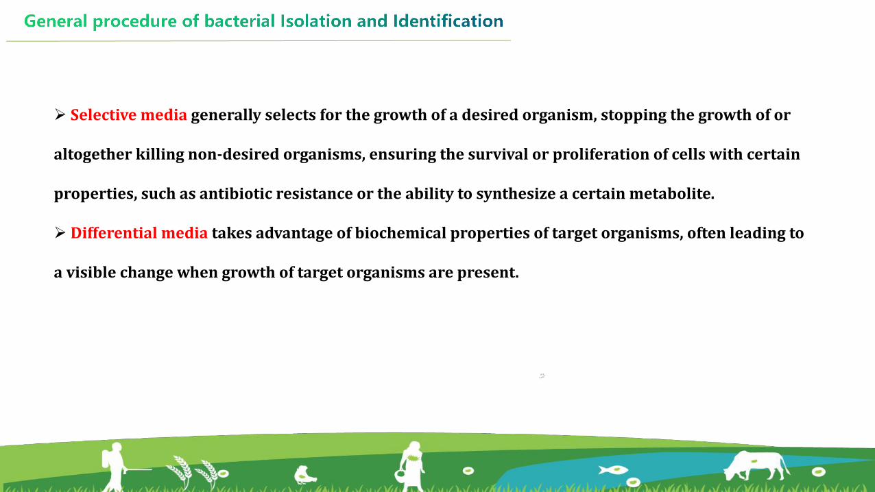
Ø Selective media generally selects for the growth of a desired organism, stopping the growth of or
altogether killing non-desired organisms, ensuring the survival or proliferation of cells with certain
properties, such as antibiotic resistance or the ability to synthesize a certain metabolite.
Ø Differential media takes advantage of biochemical properties of target organisms, often leading to
a visible change when growth of target organisms are present.

Some differential media
MacConky agar or EMB agar for E.coliTCBS agar for VibrioR-S agar for Aeromonas
--Cipriano RC, Fish Disease Leaflet 68, 2001 --Supono et al, AACL Bioflux, 2019 --Dhaval Patel, Junior Research Fellow, St. Xavier’s College, Ahmedabad

Streak Plate Method
Streak plate technique is used for the isolation into a pure culture of the
organisms (mostly bacteria), from a mixed population.
The inoculum is streaked over the agar surface in such a way that it “thins out”
the bacteria. Some individual bacterial cells are separated and well-spaced from
each other.
HOW TO PURIFY ?
https://microbeonline.com/streak-plate-method-principle-purpose-procedure-results/
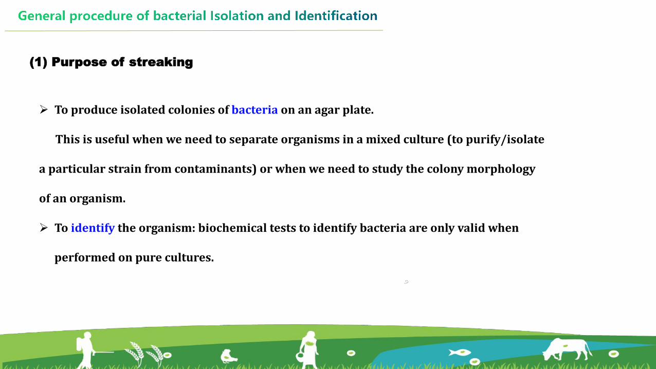
(1) Purpose of streaking
Ø To produce isolated colonies of bacteria on an agar plate.
This is useful when we need to separate organisms in a mixed culture (to purify/isolate
a particular strain from contaminants) or when we need to study the colony morphology
of an organism.
Ø To identify the organism: biochemical tests to identify bacteria are only valid when
performed on pure cultures.

Ø The sample/inoculum is diluted by streaking it across the surface of the agar plate.
Ø While streaking in successive areas of the plate, the inoculum is diluted to the point where there
is only one bacterial cell deposited every few millimeters on the surface of the agar plate.
Ø When these lone bacterial cells divide and give rise to thousands and thousands of new bacterial
cells, an isolated colony is formed.
Ø Pure cultures can be obtained by picking well-isolated colonies and re-streaking these on fresh
agar plates.
(2) Principle of Streaking

(3) Materials required
Ø A source of bacteria (stock culture, previously streaked agar plate or any other
inoculum)
Ø Inoculation loop
Ø A striker/lighter
Ø Bunsen burner
Ø Fresh agar plate (nutrient agar or any other agar medium)
Ø Incubator
Ø Marker pen

(4) Procedure
1) Flame the inoculating loop until it is red hot, and then cool it.
https://slidetodoc.com/media-preparation-and-sterilization-aseptic-technique-pure-culture/
-- photo by Deng YT

2) Pick an isolated colony from the agar plate culture
with the sterilized loop.
3) Immediately streak the loop very gently over a
quarter of the plate using a back and forth motion.
4) Flame the loop again and allow it to cool. Going
back to the edge of area that you just streaked, extend
the streaks into the second quarter of the plate.
5) Repeat the procedure, and streak the remaining
areas.
6) Label the bottom of the plate with the strain name
and the date.
https://microbeonline.com/streak-plate-method-principle-purpose-procedure-results/
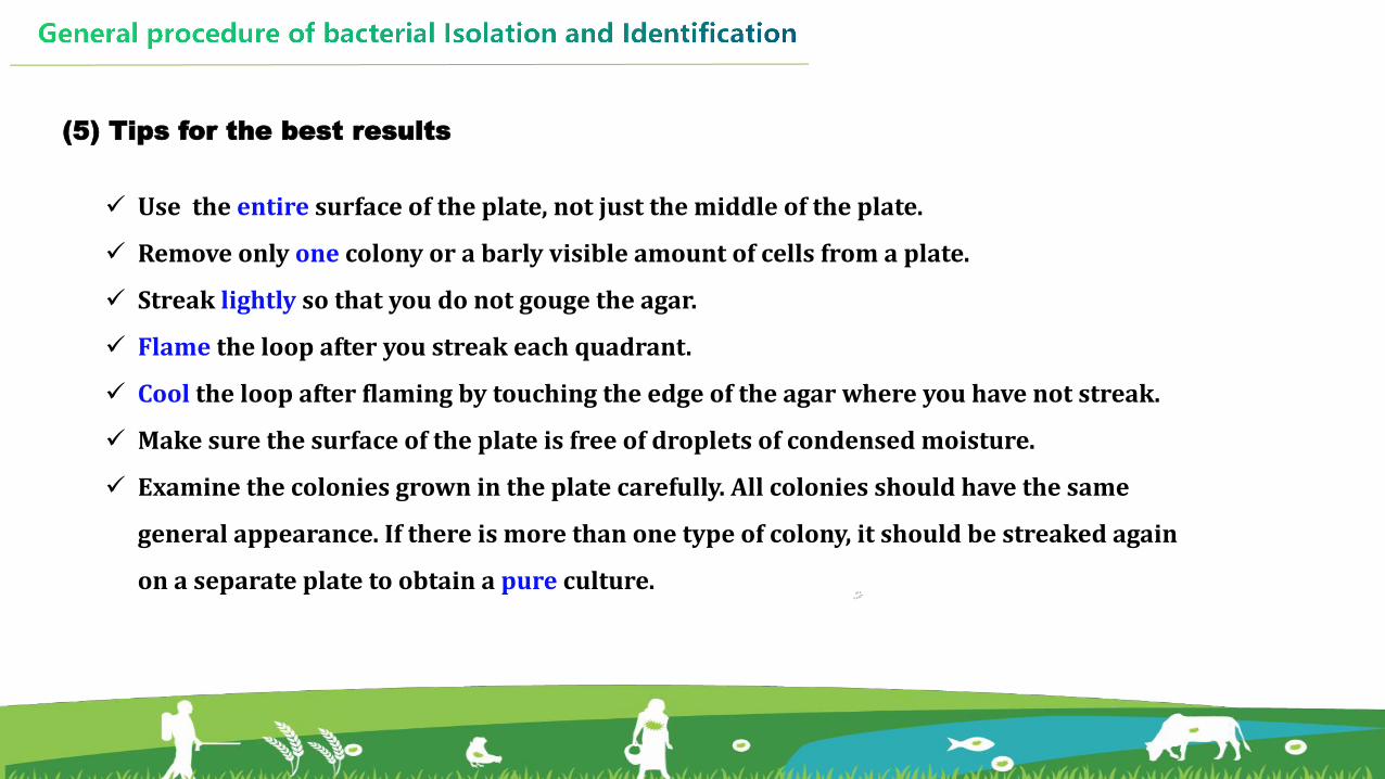
(5) Tips for the best results
ü Use the entire surface of the plate, not just the middle of the plate.
ü Remove only one colony or a barly visible amount of cells from a plate.
ü Streak lightly so that you do not gouge the agar.
ü Flame the loop after you streak each quadrant.
ü Cool the loop after flaming by touching the edge of the agar where you have not streak.
ü Make sure the surface of the plate is free of droplets of condensed moisture.
ü Examine the colonies grown in the plate carefully. All colonies should have the same
general appearance. If there is more than one type of colony, it should be streaked again
on a separate plate to obtain a pure culture.

Molecular IdentificationØ PCR amplification (16S rRNA
gene,housekeeping gene)
Ø Genotyping Methods
Ø MALDI-TOF MS
Bacterial Identification
Phenotypic IdentificationØ Gram stain
Ø Biochemical tests
Ø Commercial identification
systems
HOW TO IDENTIFY ?

1. Phenotypic Identification
• Phenotypic identification is made by physiological, morphological, and
biochemical characteristics.
• Despite that, identification to the species level using this approach is difficult
due to the variable behavior of the strains.

One of the most useful staining reactions for bacteria is called the Gram stain,
developed in 1884 by the Danish physician Hans Christian Gram.
(1) Gram stain
Bacteria in suspension are fixed to a glass slide by brief heating and then exposed
to two dyes that combine to form a large blue dye complex within each cell.
https://medicallabscientist.org/gram-staining-procedure-results-relationship/

• When the slide is flushed with an alcohol solution, gram-
positive bacteria retain the blue colour and gram-negative
bacteria lose the blue colour.
• The slide is then stained with a weaker pink dye that
causes the gram-negative bacteria to become pink,
whereas the gram-positive bacteria remain blue.
• The Gram stain reacts to differences in the structure of the
bacterial cell surface, differences that are apparent when
the cells are viewed under an electron microscope.
Gram stained streptococcus 100X magnified --Maria A Carrillo-Marquez et al., 2016
Aeromonas spp. --CDC/ Dr. W.A. Clark - http://phil.cdc.gov/ ID# 1255

(2) Biochemical tests
• Biochemical tests are used to identify bacterial species by
differentiating them on the basis of biochemical activities.
• Routine biochemical tests include tests for carbohydrate
fermentation , methyl red, citric acid utilization, hydrogen
sulfide production, and oxidase test.
https://microbeonline.com/carbohydrate-fermentation-test-uses-principle-procedure-results/
https://onlinesciencenotes.com/oxidase-test-by-kovacs-method-principle-procedure-result-interpretation-and-precautions/

(3) Bacterial Biochemical Identification Kits
The most representative biochemical test kits
ü Minitek identification system using paper substrates,
ü API-20A system using dry powder substrates,
ü PIZYMAN-IDENT rapid enzyme activity assay system using primary materials,
ü RaPID-ANA systems, and fully automated microbial identification systems.
It offers biochemical identification panels, discs and strips for a broad range of bacterial species.
The panels coated with dehydrated chemical reagents utilize a series of conventional and
chromogenic biochemical tests for rapid identification of different organisms, which may be read
manually or using a reader.

(4) Automated microbial identification system
Ø It has been developed for the detection, enumeration and identification of bacteria in clinical specimens.
Ø It is an instrument used for automatic computer-assisted identification of bacteria
Ø It mainly involves staining, motility test, cultural characteristics, a series of biochemical tests.
Ø The automatic bacteria identification system automatically identifies the bacteria in very short time.

2. Molecular Identification
• Microorganisms can be characterised by Gram's staining and biochemical tests to
decipher their physiological characteristics.
• However, these techniques are time consuming and have limitations with erroneous
identification at species level.
• Therefore, the advanced techniques like sequence based, gel based and protein based
systems have become advantageous due to their fast reactions, high specificity and less
chance of error.

(1) Sequence based techniques for bacteria
Ø Different genetic targets for microbial identifification are 16S rRNA and house keeping genes
(gyrB, rpoA, rpoB, rpoC, rpoD, genes), etc. These genes are conserved, variable and
hypervariable regions.
Ø Many other genes which are used as molecular markers to study the diversity of different
groups of microorganisms, e.g., gapA for Escherichia coli, pyrH for classifying Vibrio
vulnifificus and Vibrio spp., etc.
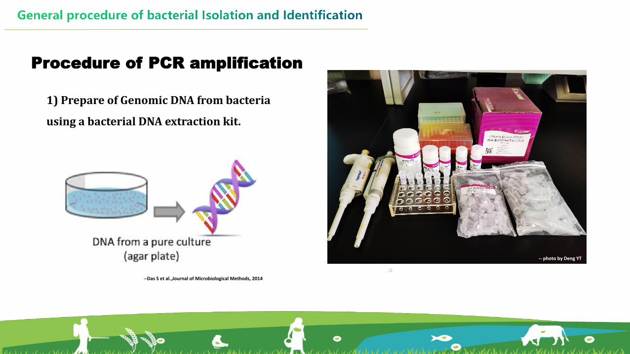
1) Prepare of Genomic DNA from bacteria
using a bacterial DNA extraction kit.
Procedure of PCR amplification
--Das S et al.,Journal of Microbiological Methods, 2014
-- photo by Deng YT

2) Pepare the PCR reation components based on the
instruction of manufacturer. PCR conditions for each
primer combination were optimized in pilot
experiments.
--Das S et al.,Journal of Microbiological Methods, 2014
-- photo by Deng YT

3) The PCR products were analyzed by 1~2% agarose gel electrophoresis in
TAE buffer and were visualized under UV transilluminator.

3) Amplified products were sent for direct sequencing at the sequencing company.
Identification of target gene sequences to the GenBank database was performed using
nucleotide BLAST.
--Das S et al.,Journal of Microbiological Methods, 2014

(2) Genotyping Methods
Different molecular methods have been employed to trace whether two isolates belong,
or not, to the same clone and therefore share an epidemiological relationship.
ü Enterobacterial repetitive intergenic consensus-PCR (ERIC-PCR)
ü Randomly amplified polymorphic DNA-PCR (RAPD-PCR)
ü Amplified fragment length polymorphism (AFLP)
ü Pulsed-field gel electrophoresis (PFGE)
ü Multilocus sequence typing (MLST)
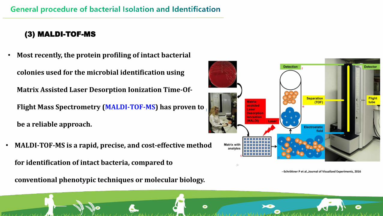
(3) MALDI-TOF-MS
• Most recently, the protein profiling of intact bacterial
colonies used for the microbial identification using
Matrix Assisted Laser Desorption Ionization Time-Of-
Flight Mass Spectrometry (MALDI-TOF-MS) has proven to
be a reliable approach.
• MALDI-TOF-MS is a rapid, precise, and cost-effective method
for identification of intact bacteria, compared to
conventional phenotypic techniques or molecular biology. --Schröttner P et al.,Journal of Visualized Experiments, 2016

Procedures for isolation and identification of Aeromonas5.2.2

u Introduction of Aeromonas
l Aeromonas organisms are Gram-negative,
facultatively anaerobic bacilli with a single, polar
flagella.
l The genus Aeromonas, belongs to the class of
Gammaproteobacterias,order Aeromonadales,
and family Aeromonadaceae.
-- CDC/ Dr. W.A. Clark - http://phil.cdc.gov/ ID# 1255

l This genus comprises 36 species that are considered autochthonous of aquatic environments.
l They are also isolated from foods, animals, and various infectious processes in humans.
l Aeromonas spp. are an important fish pathogen, some species, like A. veronii and A. hydrophila, which
particularly affect freshwater fish, causing ulcers, hemorrhages, furunculosis and septicemias.
Typical symptoms of motile Aeromonas septicemia (MAS) caussed by A. hydrophila. ---Zhang D et al., Aquaculture Reports, 2016

l There are currently no international standards to selectively isolate Aeromonas spp.
l Many commonly used methods are available, and laboratories can use any acceptable
methods for isolation and identification.
l If required, the isolation media, temperatures and length of incubation can also be
adjusted according to the purpose of the study.

u PROCEDURE
01 03
0402
05
Iso lat ion
Pur i f icat ion
Oxidase test
PCR ampl i f icat ion
Sequencing
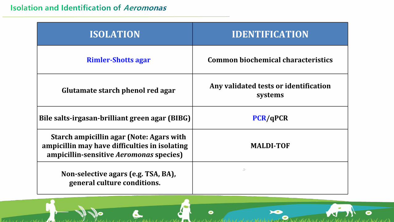
ISOLATION IDENTIFICATION
Rimler-Shotts agar Common biochemical characteristics
Glutamate starch phenol red agar Any validated tests or identification systems
Bile salts-irgasan-brilliant green agar (BIBG) PCR/qPCR
Starch ampicillin agar (Note: Agars with ampicillin may have difficulties in isolating
ampicillin-sensitive Aeromonas species)MALDI-TOF
Non-selective agars (e.g. TSA, BA), general culture conditions.

1. Isolation and purification of Aeromonas
Diseased fish
Healthy fish
Sample collection Inculation
Streak plate method
Spread plate method
Isolation Purification

Morphological type of Aeromonas on RS agar
On the RS agar plate, the suspet colonies of Aeromonas spp. are yellow
or/ and with a black center.
NOTE:
Ø For mesophilic Aeromonas, the plate was incubated at 28℃ for
20~24 h.
Ø The plate should be observed after the 20th hours but no later than
24th hours.
Ø The colour of the colonies would show a reversion of yellow toward
green after 26 hours of incubation.
-- photo by Deng YT
-- photo by Deng YT

Tips:ü To avoid the same colone, no more than 3 strains of the same morphological
type should be isolated from each samples.
ü Transfer individual colony with the inoculating loop onto the RS agar plates to
obtain the pure cultures.
-- photo by Deng YT-- photo by Deng YT
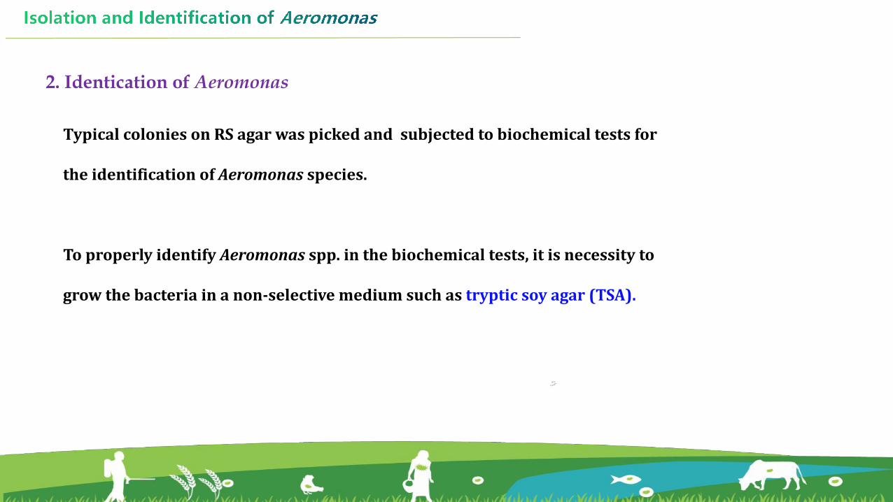
2. Identication of Aeromonas
Typical colonies on RS agar was picked and subjected to biochemical tests for
the identification of Aeromonas species.
To properly identify Aeromonas spp. in the biochemical tests, it is necessity to
grow the bacteria in a non-selective medium such as tryptic soy agar (TSA).

(1) Biochemical tests
The suspected colony can be characterized by certain biochemical tests, like oxidase,
glucose fermentation, etc.
Despite that, conventional biochemical tests, as well as automated systems, are of
limited utility in the identifification of some Aeromonas species.

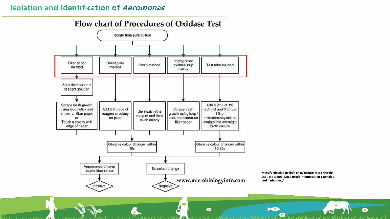
(1) Oxidase test
The oxidase test is a additional method for
differentiating Aeromonas spp. from Citrobacter spp.
which was also with yellow color on the RS plate.
Aeromonas spp. oxidase positve Citrobacter spp. oxidase negative
-- Shoits EB and Rimler R, Applied Microbiology, 1973
https://microbiologyinfo.com/oxidase-test-principle-uses-procedure-types-result-interpretation-examples-and-limitations/

Filter Paper Method
1) Add a drop of oxidase reagent (ρ-phenylenediamine) to a clean filter paper.
-- photo by Deng YT

2) Pick a single purified colony and smeared over the moist area.
-- photo by Deng YT-- photo by Deng YT

3) Observe the color change of the area coated with the bacteria in 10 seconds.
If the purplish-blue color appears at the area, the oxidase activity is positive.
If not, the oxidase activity is negative.
The test strains with oxidase-positive are
presumed to be Aeromonas spp. and
should be confirmed by molecular
identification.
https://onlinesciencenotes.com/oxidase-test-by-kovacs-method-principle-procedure-result-interpretation-and-precautions/
-- photo by Deng YT
(2) PCR amplification
Molecular techniques are still the best option for identification and taxonomic
classification of the genus Aeromonas.
The most common technique involves the 16S rRNA gene. However, accuracy of this
method is limited when analyzing strains whose sequences are very similar.
As the low accuracy of 16S rRNA sequencing is due to the high similarity among the
sequences, the amplification of so-called housekeeping genes is presented as the best way
to do taxonomic classification.

Specifically, gyrB and rpoD are the most widely used housekeeping genes in taxonomic
studies and allow greater reliability in the phylogenetic classification in Aeromonas.
--Soler L et al., International Journal of Systematic and Evolutionary Microbiology, 2004

--Das S et al., Journal of Microbiological Methods, 2014
Ø Extract the genomic DNA from the
oxidase-positive bacteria.
Ø Amplify the gyrB gene by PCR method.
Ø Analyze the PCR products by agarose
gel electrophoresis.
Ø Visualize the gel under UV
transilluminator.
Ø Amplified products are directly
sequenced to determine the species.
Procedure of PCR amplification

Procedures for isolation and identification of Vibrio5.2.3

u Introduction of Vibrio
• The vibrios are Gram-negative rod-shaped bacteria that
are fermentative, catalase and oxidase positive, motile
by polar flagella, are usually sensitive to the vibriostatic
agent O/129, and mostly have a requirement for
sodium chloride.
• The genus Vibrio, belongs to the class of
Gammaproteobacteria,order Vibrionales, and family
Vibrionaceae.
---BSIP SA / Alamy Stock Photo, Image ID: E0CJ58
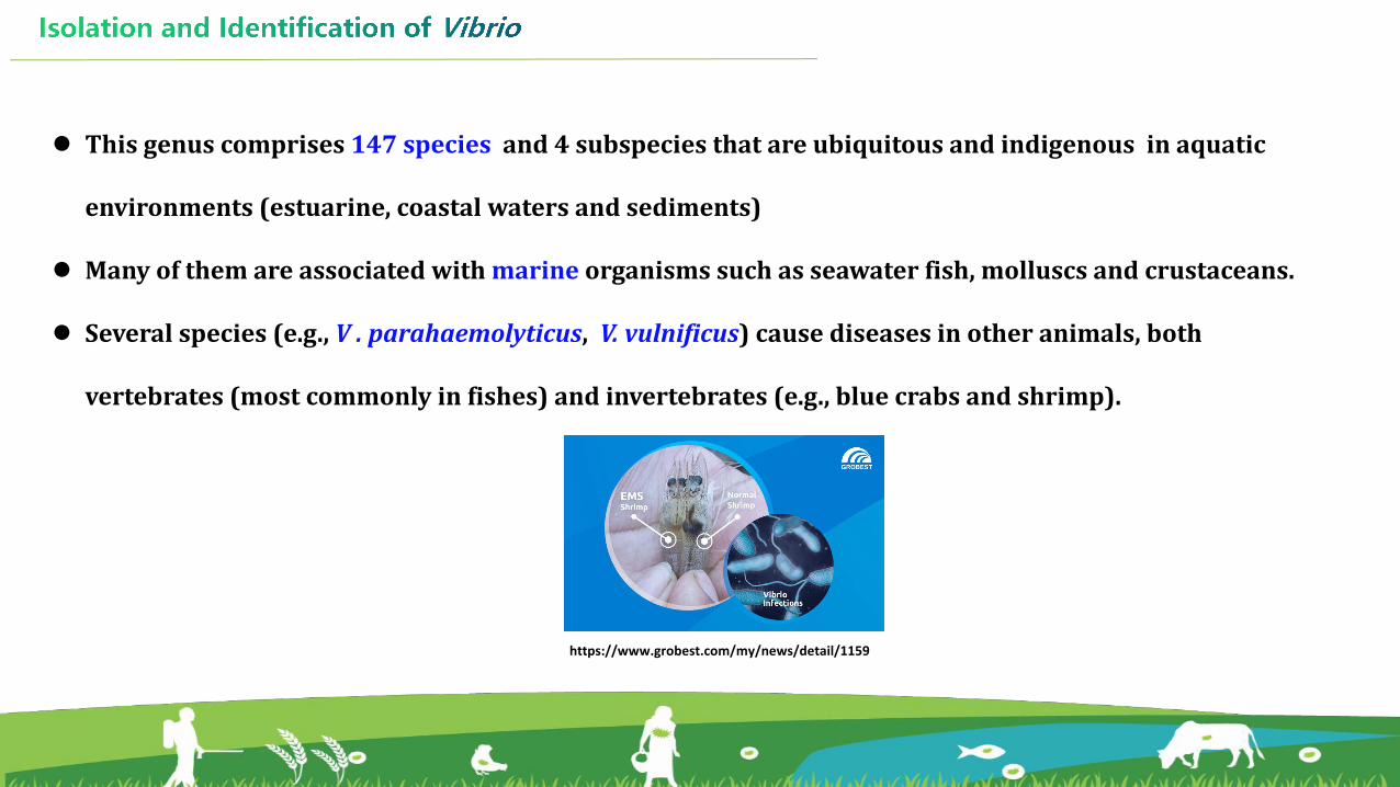
l This genus comprises 147 species and 4 subspecies that are ubiquitous and indigenous in aquatic
environments (estuarine, coastal waters and sediments)
l Many of them are associated with marine organisms such as seawater fish, molluscs and crustaceans.
l Several species (e.g., V . parahaemolyticus, V. vulnificus) cause diseases in other animals, both
vertebrates (most commonly in fishes) and invertebrates (e.g., blue crabs and shrimp).
https://www.grobest.com/my/news/detail/1159

u PROCEDURE
01 03
0402Iso lat ion
Pur i f icat ion
PCR ampl i f icat ion
Sequencing

ISOLATION IDENTIFICATION
Thiosulfate citrate bile salts sucrose (TCBS) agar Common biochemical characteristics
CHROMagar™ Vibrio or ChromIDTM Vibrio Any validated tests or identification systems
Non-selective agars (e.g. TSA), general culture conditions. PCR/qPCR
MALDI-TOF

1. Isolation and purification of Vibrio
Diseased shrimp
Healthy shrimp
Sample collection Inculation
Streak plate method
Spread plate method
Isolation Purification

Morphological type of Vibrio on TCBS agar
• On TCBS plate, Vibrio spp. produce either yellow or green
colonies depending on whether they are able to ferment
sucrose.
• On the TCBS medium, Vibrio parahaemolyticus was yellow,
while Vibrio harvey and Vibrio alginolyticus were blu-
green.
• NOTE: Cultures grown on TCBS plate should be examined
immediately after removal from the incubator as yellow
colonies of Vibrio spp. may revert to a green color when
left at room temperature.
-- photo by Deng YT
-- photo by Deng YT
-- photo by Deng YT
-- photo by Deng YT

Ø Biochemical identification includes:
When perfroming gram staining, oxidase, motility, or other tests, the strains should be cultured in
borth with increased salt concentrations (0, 2, 6, 8, 10% NaCl) at 28 or 37℃ for 24-28 hours.
(1) Biochemical tests
2. Identication of Vibrio
Ø Species differentiation within the Vibrio genus remains difficult by biochemical tests.
Therefore, a molecular step is usually required to confirm identification of the Vibrio species.
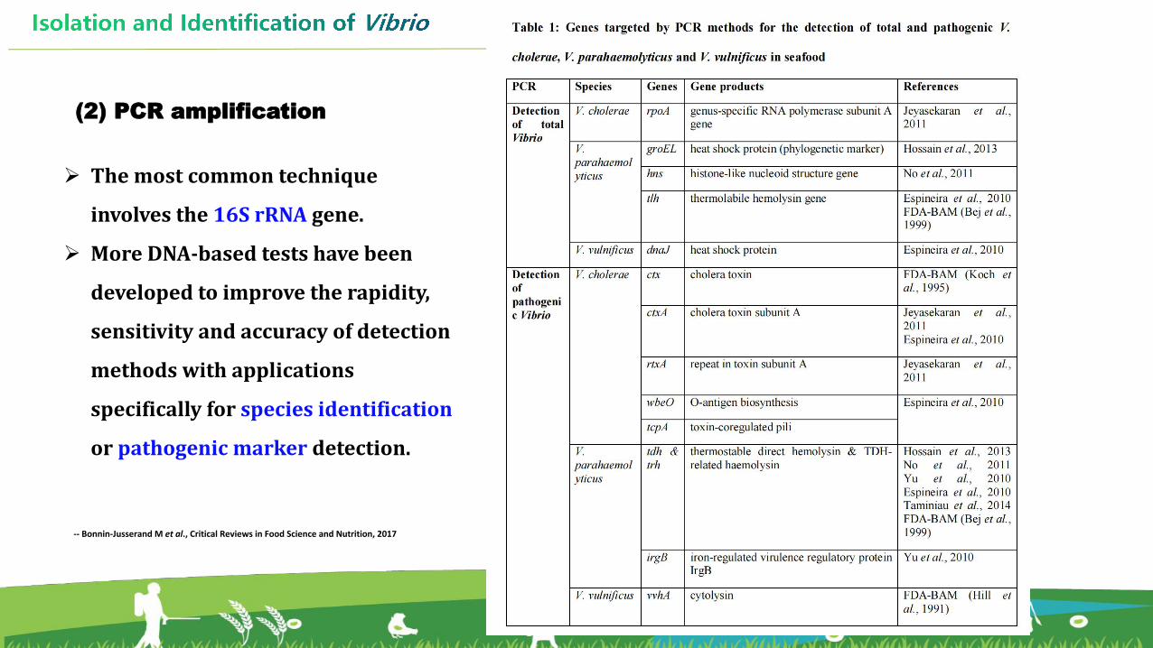
(2) PCR amplification
-- Bonnin-Jusserand M et al., Critical Reviews in Food Science and Nutrition, 2017
Ø The most common technique
involves the 16S rRNA gene.
Ø More DNA-based tests have been
developed to improve the rapidity,
sensitivity and accuracy of detection
methods with applications
specifically for species identification
or pathogenic marker detection.

Procedures for isolation and identification of E.coli5.2.4

• Escherichia coli are Gram-negative, facultatively anaerobic rod.
• E.coli, belongs to the class of Gammaproteobacteria,order
Enterobacteriales, family Enterobacteriaceae, and genus Escherichia.
u Introduction of E.coli
Scanning electron micrograph of E. coli.Image by NIAID
---Kabir M and Seel SK, Bangl. J. Vet. Med. , 2016

• E. coli are bacteria found in the environment,
foods, and intestines of people and animals.
• E.coli is often used as an indicator bacteria for
One Health analysis in aquatic AMR surveillance
programs.
• Drug-resistant E. coli have been found in
multiple species, which could be a result of
environmental spread of human and animal
antibiotics.---Harbarth S et al., 5Antimicrobial Resistance & Infection Control, 2015

u PROCEDURE
01 03
0402Iso lat ion
Pur i f icat ion
PCR ampl i f icat ion
Sequencing
ISOLATION IDENTIFICATION
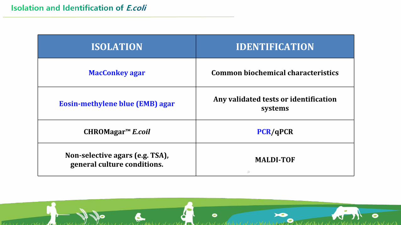
MacConkey agar Common biochemical characteristics
Eosin-methylene blue (EMB) agar Any validated tests or identification systems
CHROMagar™ E.coil PCR/qPCR
Non-selective agars (e.g. TSA), general culture conditions. MALDI-TOF

1. Isolation and purification of E.coli
Sample collection
Enrichment
Spread plate
Purification
Streak plate
Isolation
Tips for isolation:
Ø If the number of E. coli in a sample is expected to be low, such as when sampling aquatic food and
aquatic envrionment, enrichment should be performed.
One gram of sample is mixed with Buffered Peptone Water in 1 in 10 (w/v) and incubated at 35 ℃
± 2 ℃ for 18 h to 22 h. Then, one loop of the enrichment may be streaked on the diferential media.
Ø If the expected number of E. coli in a sample (e.g. intestines content) is high. The processed sample
may be directly streaked on the diferential media or the dilution of processed sample may be
spread on the media.

Morphological type of E.coli on MacConkey agar
On the MacConkey agar , the suspected
colonies of E.coli are pink to red, or some
sourrounded by zones of precipitated bile.
-- photo by Deng YT
-- photo by Deng YT

Eosin-methylene blue (EMB) agar is an additional media
selective for differentiating E.coli from Enterobacter aerogene.
Morphological type of E.coli on EMB agar
On the EMB agar , the suspected colonies of E.coli
are nucleated dark-centre, green metallic sheen. -- photo by Deng YT
-- photo by Deng YT -- photo by Deng YT

2. Identication of E.coli
Ø Typical colonies are selected for biochemical testing to identify colonies to species level.
Ø Indole (minimum test), ONPG and other tests may be performed as deemed appropriate.
(1) Biochemical tests

(2) PCR amplification
PCR has been used for the rapid and reliable detection of E.coli from foods and the
envrionmental samples.
Subsequent analyses of nucleotide sequences of 16S rRNA and housekeeping genes
can be applied for species identification.

-- Devane M et al., Water Research, 2020

REFERENCES
Isolation and identification of E.coli:Devane ML, et al. Fecal indicator bacteria from environmental sources; strategies for identification to improve water quality monitoring. Water Res. 2020, 15;185:116204.
Isolation and identification of Vibrio spp.: SBonnin-Jusserand M, et al. Vibrio species involved in seafood-borne outbreaks (Vibrio cholerae, V. parahaemolyticus and V. vulnificus): Review of microbiological versus recent molecular detection methods in seafood products. Crit Rev Food Sci Nutr. 2019;59(4):597-610.
Streak plate method: https://microbeonline.com/streak-plate-method-principle-purpose-procedure-results/
Molecular identification of bacteria:Das S, et al. Understanding molecular identification and polyphasic taxonomic approaches for genetic relatedness and phylogenetic relationships of microorganisms. J Microbiol Methods. 2014, 103:80-100.
Isolation and identification of Aeromonas spp.: Gonçalves P, et al. The genus Aeromonas: A general approach. Microb Pathog. 2019, 130:81-94.

Pearl River Fisheries Research Institute (PRFRI),
Chinese Academy of Fishery Sciences (CAFS), China