Embed Size (px)
Citation preview

Probing the lithium-response pathway in hiPSCsimplicates the phosphoregulatory set-point for acytoskeletal modulator in bipolar pathogenesisBrian T. D. Tobea,b,c,1, Andrew M. Craina,b,1, Alicia M. Winquista,b, Barbara Calabreseb,d, Hiroko Makiharae, Wen-ning Zhaof,g,Jasmin Lalondef,g, Haruko Nakamurae, Glenn Konopaskeh,i,j, Michelle Sidork, Cameron D. Perniaa,b, Naoya Yamashitae,Moyuka Wadae, Yuuka Inouee, Fumio Nakamurae, Steven D. Sheridanf,g, Ryan W. Logank, Michael Brandela,b, Dongmei Wua,Joshua Hunsbergerl, Laurel Dorsetta,b, Cordulla Duerra,b, Ranor C. B. Basam, Michael J. McCarthyc,n, Namrata D. Udeshio,PhilippMertinso, Steven A. Carro, Guy A. Rouleaup, LinaMastrangeloa,b, Jianxue Liq, Gustavo J. Gutierreza,r, LaurenceM. Brilla,2,Nikolaos Venizeloss, Guang Chent, Jeffrey S. Nyet,3, Husseini Manjit, Jeffrey H. Pricea,m, Colleen A. McClungk, Hagop S. Akiskaln,Martin Aldap, De-MawM. Chuangl, Joseph T. Coyleh,i, Yang Liua, Yang D. Tengu,v, Toshio Ohshimaw,x, KatsuhikoMikoshibaw,x,Richard L. Sidmanq,4,5, Shelley Halpainb,d,4, Stephen J. Haggartyf,g,4,5, Yoshio Goshimae,4,5, and Evan Y. Snydera,b,y,4,5
aSanford Burnham Prebys Medical Discovery Institute, La Jolla, CA 92037; bSanford Consortium for Regenerative Medicine, La Jolla, CA 92037; cDepartment ofPsychiatry, Veterans Administration Medical Center, La Jolla, CA 92037; dSection of Neurobiology, Division of Biological Sciences, University of California, SanDiego, La Jolla, CA 92037; eDepartment of Molecular Pharmacology & Neurobiology, Yokohama City University Graduate School of Medicine, Yokohama236-0004, Japan; fChemical Neurobiology Laboratory, Center for Genomic Medicine, Department of Neurology, Massachusetts General Hospital, Harvard MedicalSchool, Boston, MA 02114, gDepartment of Psychiatry, Massachusetts General Hospital, Harvard Medical School, Boston, MA 02114; hMailman Research Center,McLean Hospital, Belmont, MA 02478; iDepartment of Psychiatry, Harvard Medical School, Boston, MA 02115; jDepartment of Psychiatry, University ofConnecticut School of Medicine, Farmington, CT 06030; kDepartment of Psychiatry, University of Pittsburgh Medical Center, Pittsburgh, PA 15219; lMolecularNeurobiology Section, National Institute of Mental Health, Bethesda, MD 20892-1363; mVala Sciences, Inc., San Diego, CA 92121; nDepartment of Psychiatry,University of California, San Diego, La Jolla, CA 92093-0737; oBroad Institute of MIT and Harvard University, Cambridge, MA 02142; pDepartment of Psychiatry,Dalhousie University, Halifax, NS, Canada B3H 2E2; qDepartment of Neurology, Beth Israel-Deaconess Medical Center, Boston, MA 02215; rDepartment of Biology,Vrije Universiteit Brussels, 1050 Brussels, Belgium; sDepartment of Cinical Medicine, Örebro University, Örebro SE-701 82, Sweden; tJanssen Research &Development Labs, La Jolla, CA 92037; uDepartment of Physical Medicine and Rehabilitation, Harvard Medical School, Boston, MA 02115; vDepartment ofNeurosurgery, Harvard Medical School, Boston, MA 02115; wDepartment of Life Science and Medical Bio-Science, Waseda University, Shinjuku-ku, Tokyo169-8555, Japan; xLaboratory for Developmental Neurobiology, Brain Science Institute, RIKEN, Wako 351-0198, Japan; and yDepartment of Pediatrics, Universityof California, San Diego, La Jolla, CA 92037
Contributed by Richard L. Sidman, March 13, 2017 (sent for review November 19, 2016; reviewed by Milos Pekny and D. Eugene Redmond Jr.)
The molecular pathogenesis of bipolar disorder (BPD) is poorlyunderstood. Using human-induced pluripotent stem cells (hiPSCs) tounravel suchmechanisms in polygenic diseases is generally challenging.However, hiPSCs from BPD patients responsive to lithium offeredunique opportunities to discern lithium’s target and hence gain mo-lecular insight into BPD. By profiling the proteomics of BDP–hiPSC-derived neurons, we found that lithium alters the phosphorylationstate of collapsin response mediator protein-2 (CRMP2). Active non-phosphorylated CRMP2, which binds cytoskeleton, is present through-out the neuron; inactive phosphorylated CRMP2, which dissociatesfrom cytoskeleton, exits dendritic spines. CRMP2 elimination yields ab-errant dendritogenesis with diminished spine density and lost lithiumresponsiveness (LiR). The “set-point” for the ratio of pCRMP2:CRMP2 iselevated uniquely in hiPSC-derived neurons from LiR BPD patients, butnot with other psychiatric (including lithium-nonresponsive BPD) andneurological disorders. Lithium (and other pathway modulators) lowerspCRMP2, increasing spine area and density. Human BPD brains showsimilarly elevated ratios and diminished spine densities; lithium therapynormalizes the ratios and spines. Consistent with such “spine-opathies,”human LiR BPD neurons with abnormal ratios evince abnormally steepslopes for calcium flux; lithium normalizes both. Behaviorally, transgenicmice that reproduce lithium’s postulated site-of-action in dephosphory-lating CRMP2 emulate LiR in BPD. These data suggest that the “lithiumresponse pathway” in BPD governs CRMP2’s phosphorylation, whichregulates cytoskeletal organization, particularly in spines, modulatingneural networks. Aberrations in the posttranslational regulation ofthis developmentally critical molecule may underlie LiR BPD pathogen-esis. Instructively, examining the proteomic profile in hiPSCs of a func-tional agent—even one whose mechanism-of-action is unknown—might reveal otherwise inscrutable intracellular pathogenic pathways.
posttranslational modification | proteomics | psychiatric disease modeling |CRMP2 | dendrites
Although human induced pluripotent stem cells (hiPSCs)have proven valuable for studying the molecular pathology
of monogenic diseases, one of the technique’s greatest challengeshas been to offer similar insights into the molecular pathogenesisof polygenic, multifactorial disorders for which the underlyingpathophysiology is unknown. The struggle has been to go beyondphenotypic description to discerning underlying molecular mech-anisms. Neuropsychiatric illnesses are a prototype for such com-plex conditions (1–3). They are difficult to model not only becauseof the likelihood of polygenic influences, but also because of thesubjectivity with which these diseases must often be diagnosed, theempirical fashion with which drugs are prescribed, and the het-erogeneity of patient response. Of such maladies, bipolar disorder
Author contributions: B.T.D.T., A.M.C., H. Makihara, F.N., G.J.G., L.M.B., J.S.N., H. Manji,J.H.P., C.A.M., H.S.A., M.A., D.-M.M.C., Y.L., Y.D.T., R.L.S., S.H., S.J.H., Y.G., and E.Y.S. designedresearch; B.T.D.T., A.M.C., A.M.W., B.C., W.-n.Z., J. Lalonde, H.N., G.K., M.S., C.D.P., N.Y.,M.W., Y.I., S.D.S., R.W.L., M.B., D.W., J.H., L.D., C.D., R.C.B.B., M.J.M., N.D.U., P.M., S.A.C.,G.A.R., L.M., J. Li, G.J.G., and J.T.C. performed research; B.T.D.T., A.M.C., A.M.W., M.B., D.W.,J.H., J. Li, N.V., G.C., M.A., Y.L., T.O., and K.M. contributed new reagents/analytic tools;B.T.D.T., A.M.C., C.D.P., M.B., M.J.M., R.L.S., S.H., S.J.H., Y.G., and E.Y.S. analyzed data; B.T.D.T.,R.L.S., and E.Y.S. wrote the paper; and B.T.D.T., A.M.W., B.C., M.B., S.J.H., Y.G., and E.Y.S.generated figures.
Reviewers: M.P., Sahlgrenska Academy at University of Gothenburg; and D.E.R., YaleUniversity School of Medicine.
Conflict of interest statement: R.C.B.B., L.M.B., G.C., J.S.N., H.M., and J.H.P. are employeesof private companies. Their role in the study was solely as researchers with no financial orproprietary involvement.
Freely available online through the PNAS open access option.
Data deposition: All MS data are publicly accessible; for SILAC data, the mass spectra maybe downloaded from MassIVE, massive.ucsd.edu (accession no. MSV000080975); the dataare directly accessible via ftp://massive.ucsd.edu/MSV000080975.1B.T.D.T. and A.M.C. contributed equally to this work.2Present address: Intertek Pharmaceutical Services, San Diego, CA 92121.3Deceased March 6, 2017.4R.L.S., S.H., S.J.H., Y.G., and E.Y.S. contributed equally to this work.5To whom correspondence may be addressed. Email: [email protected], [email protected], [email protected], or [email protected].
This article contains supporting information online at www.pnas.org/lookup/suppl/doi:10.1073/pnas.1700111114/-/DCSupplemental.
E4462–E4471 | PNAS | Published online May 12, 2017 www.pnas.org/cgi/doi/10.1073/pnas.1700111114
(BPD) type 1, a chronic illness of episodic mania with interveningperiods of depression for which the interplay between genetic andenvironmental factors is poorly understood, is unique in that, forunclear reasons, ∼35% of patients respond to monotherapy withlithium salts (4–7); indeed, lithium responsiveness (LiR) is oftenregarded as pathognomic of BPD. However, despite our knowl-edge of lithium’s ubiquitous multisystemic influences and pleio-tropic actions (4), the molecular mechanism underlying this drugresponsiveness specifically in BPD, as well as BPD’s molecularpathogenesis, are poorly understood. The former, however, couldlend insight into the latter. For example, although lithium maysuppress hyperexcitability of a subset of neurons in culture (2) [manymechanisms have been proffered (4)], clinical trials have shown thatdrugs that simply suppress neuronal activity, such as calcium channelblockers, are ineffective in BPD (7). Furthermore, of the one-third ofpatients who are LiR, many become noncompliant because of fre-quent adverse side effects (e.g., weight gain, hypothyroidism, tremor,kidney dysfunction, dermatologic reactions, teratogenicity). Suchpleiotropic effects underscore our ignorance with regard to lithium’saction specifically for BPD. Additionally, the safety index of lithiumis narrow (5, 8). In view of its prevalence (the sixth leading cause ofdisability worldwide), suboptimal treatment options, and absence ofbiomarkers for onset and progression, neuropsychiatric disordersin general—and BPD in particular—represent a pressing unmetmedical need (1 in 250 sufferers die from complications of BPD).Two obstacles to developing safer, more effective mood stabilizershave been a lack of known clinically relevant molecular drug targetsand of drug-screening assays that are rooted in the molecularpathogenesis and pathophysiology of the disorder. Although heri-tability of BPD is ∼80%, few disease-specific gene associations havebeen identified with sufficient consistency and statistical significanceto guide further studies (9, 10); multiple loci are more likely tocontribute to LiR than any single reliable genetic marker, making itchallenging for hiPSC disease-modeling technology. The approachpresented here might help address these challenges.Because most of lithium’s actions have been linked to post-
translational regulation rather than to transcription (4), we electedto start with an unbiased differential proteomic approach. Thus,whereas lithium’s action as a modifier of kinase signaling has beendescribed for numerous substrates (4), precisely how phosphory-lation plays a role, and what the substrate of that phosphorylationmight be that is relevant to BPD, are not understood. Here wedescribe inroads into probing, mapping, and understanding theregulation of the molecular “lithium-response pathway” in BPDinitially using proteomic profiling (by two independent techniques)of patient-derived hiPSCs to identify putative lithium targets,followed by bioinformatic pathway analyses to determine the hi-erarchy and convergence of these candidates. We validated our
conclusions in: (i) biochemical analyses comparing hiPSC-derivedneurons from LiR, lithium-nonresponsive (LiNR), and unaffectedindividuals (as well as those with other psychiatric and neurologicalconditions); (ii) assays of neuronal function; (iii) neurocytologicaland behavioral analyses of transgenic mice in which the pathway’sputative central node is eliminated or lithium’s putative site-of-action is reproduced; and (iv) biochemical and histological as-sessment of primary human patient brain specimens. Extrapolatingfrom the mediators of LiR to conclusions regarding the molecularunderpinnings of BPD, our data implicate not a gene defect perse, but rather aberrant posttranslational modification of a de-velopmentally critical molecule: an abnormally high phosphor-egulatory set-point for the central cytoskeletal modulator CollapsinResponse Mediator Protein-2 (CRMP2) (11–16) which, by determiningCRMP2’s active state, in turn influences dendritic form and functionand hence, presumably, neural network development and activity.
ResultsWe generated hiPSCs from cohorts of LiR and LiNR BPD, andunaffected patients, which included two sets of first-degree rela-tives, each set with an LiR patient and an unaffected familymember and one set with a family member with the diagnosis ofunipolar/major depression (MD) (SI Appendix, Fig. S1). Addi-tionally, we generated hiPSCs from a patient with Parkinson’sdisease (PD) as a neurologically affected nonpsychiatric control(SI Appendix, Fig. S1). hiPSC clones (typically duplicates for eachpatient sample) (SI Appendix, Fig. S1) were validated to confirmthat they: (i) retained a SNP pattern identical to their somaticcell-of-origin; (ii) were immunopositive for OCT4, NANOG,SSEA4, and Tra-1-81 (SI Appendix, Fig. S2A); (iii) showed gene-expression profiles consistent with the pluripotent state (SI Ap-pendix, Fig. S2B); and (iv) were capable of forming embryoidbodies or teratomas containing derivatives of the three primitivegerm layers (SI Appendix, Figs. S2 C and D). We observed nodifferences in neuronal induction efficiency and yield amongLiR, LiNR, or unaffected patient-derived hiPSCs, all of whichshowed similar expression of neuronal markers [Tau, βIII-tubulin(Tuj1), MAP2, vGLUT, GABA] and produced neural progenitorcells (NPCs) and electrophysiologically active (17) glutamatergic(vGLUT+) and GABAergic (GABA+) neurons that initiallyexpressed markers consistent with a dorsal anterior forebrain corticalphenotype (SI Appendix, Figs. S2 E–L and S3 A–J) (17–20). For ourin vitro studies, we elected to preserve the distinction made by cli-nicians between LiR and LiNR patients (5–8) and to probe lithium’sprotein targets within LiR BPD neurons (SI Appendix, Figs. S1).We began unbiased differential proteomic analysis of a single
clone of BPD hiPSC-derived neurons by comparing lithium treatedto untreated neurons. By 2D differential gel electrophoresis (2D-DIGE), we identified 26 differentially represented protein spotsyielding 15 distinct proteins identified by mass spectrometry (Fig. 1Aand SI Appendix, Fig. S4A). The genes corresponding to the 15 pro-teins were queried against a publicly available human gene-expressiondatabase constructed from the dorsal lateral prefrontal cortex of30 BPD patient brains compared with 31 control patient brains(https://www.ncbi.nlm.nih.gov/sites/GDSbrowser?acc=GDS2190):three genes, whose products changed significantly in response tolithium in the 2D-DIGE dataset (and hence were candidate lithiumtargets), were also differentially expressed in BPD compared withcontrol brains (P < 0.03): CRMP2, mitochondrial ribosomal proteinS22 (MRPS22), and cystatin B (CSTB). Interrogation of the SullivanLab Evidence Project (SLEP) was performed to indicate proteinswhose encoding genes might already be linked to psychiatric dis-orders by genome-wide association studies or genetic linkages. In-terrogation of SLEP (<50 kb for linkage, P < 1 × 10⁻³ for genome-wide association studies, logarithm of the odds score of 3) showedmicrosatellite enrichment of 4 of the 15 protein-encoding genes—CRMP2, WD repeat and FYVE domain containing protein-1(WDFY1), and reticulocalbin-1 (RCN1)—and SNP enrichment oftwo: nuclear transport factor-2 (NUTF2) and RCN1. Finally, be-cause, 8p21 has been implicated as a susceptibility locus forschizophrenia (21–23), genes linked to that region have also been
Significance
One-third of bipolar disorder (BPD) patients are lithium-responsive(LiR) for unknown reasons. Were lithium’s target to be identified,then BPD’s pathogenesis might be unraveled. We identified andmapped the “lithium-response pathway,” which governs thephosphorylation of CRMP2, a cytoskeleton regulator, particularlyfor dendritic spines: hence, a neural network modulator. Although“toggling” between inactive (phosphorylated) and active (non-phosphorylated) CRMP2 is physiologic, the “set-point” in LiR BPDis abnormal. Lithium (and other pathway-modulators) normalizethat set-point. Hence, BPD is a disorder not of a gene but of theposttranslational regulation of a developmentally critical mole-cule. Such knowledge should enable better mechanistically basedtreatments and bioassays. Instructively, lithium was our “molec-ular can-opener” for “prying” intracellularly to reveal otherwiseinscrutable pathophysiology in this complex polygenic disorder.
Tobe et al. PNAS | Published online May 12, 2017 | E4463
MED
ICALSC
IENCE
SPS
YCHOLO
GICALAND
COGNITIVESC
IENCE
SPN
ASPL
US

viewed as risk factors for psychiatric disease more broadly, althoughthe mechanism by which they might predispose to a mental disorderis unknown (21–23). Among the 2D-DIGE candidate proteins, onlyCRMP2 is encoded within chromosomal region 8p22-21.The 2D-DIGE candidates were then subjected to Ingenuity
Pathway Analysis (IPA), as well as modeling with the STRINGNetwork tool (string-db.org) to generate a canonical pathwaydendrogram that might indicate the functional relationships be-
tween gene products using gene ontology terms (SI Appendix, Fig.S4B). This analysis revealed that lithium treatment of human neuronsappears to modulate four pathways significantly: semaphorin (SEMA)signaling in neurons, uracil degradation II (reductive), thymine deg-radation, and axonal guidance signaling. We noted that CRMP2 was aconstituent of each of the four pathways. Additionally, axonal guid-ance signaling appeared most centralized; its function connected with75% of the pathways identified in the IPA analysis. Of the 15 candi-date protein targets, the one most pivotal to axonal guidance andcytoskeletal dynamics was CRMP2, originally discovered as the me-diator of Sema3A’s (initially named “collapsin”) guidance of neuriteextension and axonal growth cone development (5–11).Given the converging proteomic and bioinformatics results, we
focused on CRMP2 (a central node in cytoskeletal dynamics). Itis known that phosphorylation of CRMP2 at threonine (T) 514(CRMP2-p-T514) causes its dissociation from cytoskeletal pro-teins (e.g., tubulin heterodimers) (24–26). However, phosphory-lation of CRMP2-T514 (as well as of CRMP2-S518 and CRMP2-T509) must first be primed by phosphorylation at CRMP2-S522.Phosphorylation of CRMP2 by kinases is balanced by its de-phosphorylation by phosphatases (24–26) (Fig. 1B). (Note: Althoughlithium is known to have numerous targets, these 2D-DIGE experi-ments were designed to highlight ideally only those that differedspecifically in BPD in response to lithium; those targets that were notspecifically relevant to BPD would cancel each other out.)IPA of the 2D-DIGE results showed the lithium-response
pathway in BPD neurons overlaps significantly with the glycogensynthase kinase 3β (GSK3β) signaling axis, reinforcing presump-tions that GSK3β is a major node upstream of CRMP2 (15), andsuggesting that phosphorylation of CRMP2 at T514 is significantlyinfluenced, although not necessarily exclusively, by GSK3β, a knownsubstrate of lithium-mediated inhibition (but not the paralogGSK3α) (27). Therefore, we next questioned whether CRMP2 isthe primary GSK3β substrate in BPD hiPSC neurons, or if addi-tional substrates downstream of GSK3β may be involved. Sur-prisingly, bioinformatic analysis revealed that none of the other15 candidates from our 2D-DIGE analysis contained GSK3βphosphorylation sites, suggesting that CRMP2 may be the majordirect downstream effector of GSK3β in the specific context ofhuman NPCs and neurons, particularly from BPD patients.To further ascertain the extent of the molecular consequences
of GSK3β inhibition within neural cells, we used “stable isotopelabeling by amino acids in cell culture” (SILAC)-labeled (28) hiPSC-derived neural cells with a highly specific GSK3β inhibitor,CHIR99021, and compared proteolytic digests of treated and un-treated cells using high-resolution liquid chromatography coupled withtandem mass spectrometry (LC-MS/MS) to identify differentiallyenriched peptides and proteins (SI Appendix, Fig. S5). As expected,CRMP2-p-T514 peptide was substantially reduced by CHIR99021 (SIAppendix, Fig. S5A). Of putative GSK3β substrates, CRMP2 wasamong the most robust in terms of overall peptide coverage and dose-dependency. Overall, these data suggested that CRMP2 is a primarymediator of GSK3β-dependent lithium action in human neurons.We next monitored the time course of CRMP2 and CRMP2-p-
T514 levels during differentiation of hiPSCs to neurons. Interestingly,CRMP2-p-T514 is largely restricted to NPCs and neurons, and mostabundant in the latter (SI Appendix, Fig. S6A). Note that CRMP2-p-T514 in these neural lineages evolves into three isoforms of slightlydifferent molecular weight, known to result from transcription initi-ation from alternate start codons, although all transcripts maintainthe coding region for the phospho-motif containing T514. [Onlyisoform 2 shows any detectable phosphorylation in hiPSCs, likelyreflecting the microtubule-dependence of the reprogramming process(29).] Isoform 3 (the top, heaviest band in SI Appendix, Fig. S6A)appears most pertinent to neural lineages, does not appear until theNPC stage, increases with maturation, and appears in our hands to bethe one most influenced by lithium and CHIR99021 treatment;hence, we focused on the phospho-state of that isoform.These findings prompted us to study in greater detail the
phosphoregulation of CRMP2 in hiPSC-derived neurons, with aneye toward discerning how lithium interposes itself upon that
Fig. 1. CRMP2 appears to be the central node in the lithium-response pathwayin hiPSC-derived neurons from LiR BPD patients. (A) Differential proteomicanalysis by 2D-DIGE of BPD patient hiPSC-derived neurons identified CRMP2 as atarget of lithium. Patient-derived dermal fibroblasts were reprogrammed intohiPSCs and differentiated to cortical interneurons (17). (Scale bar: 400 μm, Top;200 μm, Bottom.) Neurons from a single clone were treated or left untreatedwith LiCl [red (Cy3) and green (Cy5), respectively], and then subjected to 2D-DIGE,separating proteins based on their molecular weight, charge, and degree ofenrichment under each condition. In an overlay of gels, proteins unchanged inresponse to LiCl fluoresce both green and red, and hence, look yellow; proteinsaltered by LiCl fluoresce solely red or solely green. Differential protein spots(encircled white) were picked and identified by MS (SI Appendix, Fig. S2), withspots #2 and #5 (enlarged) identified as CRMP2. (B) Proposed model of thelithium-response pathway in BPD, regulating CRMP2’s phosphorylation-stateand, hence, its association with cytoskeletal elements. The proposed action oflithium in this context—asmediated by its presumptive direct and indirect targets(black lines radiating from lithium working in concert) (Figs. 2 and 3 and SI Ap-pendix, Figs. S6–S10)—is to promote dephosphorylation of CRMP2 at T514: thatis, to reduce the CRMP2-p-T514:CRMP2 ratio, the set-point for which, we pro-pose, is excessively high in LiR BPD (Figs. 2, 3D and F, and 5 and SI Appendix, Figs.S10 and S12). It does so through both GSK3β-dependent and independent routes.Putative components: phosphorylation of CRMP2 at T514 inactivates CRMP2, suchthat it dissociates from cytoskeletal molecules (e.g., tubulin heterodimers). Thatphosphorylation, as well as at S518, T509, and T555 (suggested by IPA analysis to beby GSK3β), must first be “primed” by phosphorylation of S522 (believed to bemediated by CDK5). Phosphorylation of CRMP2 by kinases is balanced by its de-phosphorylation by phosphatases (e.g., PP2A, which must be released by i2PP2Aand complexed with β-arrestin-2 to function). Lithium can inactivate GSK3β bypromoting its phosphorylation at S9 or by increasing p-AKT that can also phospho-inactivate GSK3β. Lithium can also block phosphorylation of S522 or increasephosphatase (PP2A) action. [Not schematized: The influence of PP2A/β-arrestin-2 complexes on AKT in the GSK3β-dependent portion of the schematic (30).]
E4464 | www.pnas.org/cgi/doi/10.1073/pnas.1700111114 Tobe et al.

regulation (Fig. 1B). We therefore began our delineation of thelithium-response pathway to and from CRMP2 by applying lithiumto neurons derived from LiR BPD hiPSCs compared with LiNRBPD and unaffected patients, as well compared with other psy-chiatric and nonpsychiatric neurological conditions (e.g., MD and PD,respectively). (The patients used in the specific experiments describedare specified in the appropriate figure legends, in SI Appendix, Sup-plementary Methods and in the table in SI Appendix, Fig. S1).We found by immunostaining (Fig. 2 and SI Appendix, Fig. S6B)
and Western blotting (Fig. 3 and SI Appendix, Fig. S6C) thatlithium significantly decreased CRMP-p-T514 in all hiPSC-derivedNPCs (Fig. 3B) and neurons, but without significant effect onnonphosphorylated or the total level of CRMP2 protein (Figs. 2Aand 3 A and C–E, and SI Appendix, Figs. S3K, S6 B and C, S10, andS11). The effect of LiCl on CRMP-p-T514 was not emulatedby other chloride salts: for example, NaCl or MgCl2 (SI Appendix,Fig. S6C). We reasoned that, if lithium lowers CRMP2-p-T514, atleast in part, by inhibiting downstream GSK3β, then direct appli-
cation of a GSK3β inhibitor (e.g., CHIR99021) should similarlylower CRMP2-p-T514 abundance, reducing the CRMP2-p-T514:CRMP2 ratio. This was found to be the case by SILAC (SI Ap-pendix, Fig. S5), immunocytochemical (Fig. 2 and SI Appendix, Fig.S7), and Western blot analysis (Fig. 3), starting as early as the NPCstage (Fig. 3B) and persisting into the more mature neuronal stage(Figs. 2 and 3 A and C–E, and SI Appendix, Figs. S5A and S7).[Suppression of isoform 3 at the developmentally earlier NPCstage (Fig. 3B) appears more complete than in neurons becauseβIII-tubulin is less abundant in NPCs (Fig. 3A).] LiCl increased theinactive form of GSK3β (GSK3β-p-S9) (30) (within 1 h of expo-sure) (Fig. 3A), and both LiCl and direct GSK3β inhibition de-creased CRMP2-p-T514 in a dose-dependent manner, but nottotal CRMP2 levels (Figs. 3 A–C and SI Appendix, Figs. S5A andS10C). All inhibitors of GSK3β tested (including CHIR99021,SB216763, and SB415286) lowered CRMP2-p-T514; CHIR99021had the greatest effect and is the most selective of the threecompounds (Fig. 3F). As per the model in Fig. 1B, the reduction ofCRMP2-p-T514 (by lithium or GSK3β inhibition) restores theassociation of CRMP2 with tubulin in BPD hiPSC-derived neurons(without affecting total tubulin) based on coimmunoprecipitationpull-down experiments (Fig. 3C and SI Appendix, Fig. S8).Finally, further fleshing out lithium’s putative interposition
upon the phosphoregulation of CRMP2 in hiPSC neural deriv-atives, we observed that LiCl increased the active AKT phospho-form, AKT-p-T308 (SI Appendix, Fig. S9A), for which GSK3β-S9 is a known substrate, hence indirectly decreasing GSK3β’sactivity because pf elevation of GSK3β-p-S9 levels.Although inhibition of GSK3β is a prominent node in the
lithium-response pathway’s reduction of CRMP2-p-T514, we ob-served LiCl treatment also resulted in a reduction of phosphory-lation at CRMP2-S522 (and subsequent decrease in CRMP2-p-T514) (SI Appendix, Fig. S9B), implicating other parallel (oradditive) GSK3β-independent lithium interactions (Fig. 1B). In-terestingly, inhibitors of CDK5 (cyclin-dependent kinase-5), thepresumed S522 kinase that primes other CRMP2 sites for subsequentGSK3β phosphorylation (25), did not decrease CRMP2-p-T514,suggesting that lithium decreases CRMP2-p-S522 independently ofCDK5 in human neurons (SI Appendix, Fig. S9C).Protein phosphatase 2A (PP2A) (26, 27) dephosphorylates
CRMP2 as a counterpoise to the kinases (Fig. 1B), Among the15 putative lithium candidates revealed by 2D-DIGE, i2PP2A, aninhibitor of PP2A, was decreased 1.5-fold in response to lithium(SI Appendix, Fig. S4A), suggesting that lithium relieves inhibitionof PP2A, thereby decreasing CRMP2-p-T514. Following its dis-sociation from i2PP2A, PP2A requires binding to β-arrestin-2 forit to be active (26, 27); lithium increased β-arrestin-2 levels (SIAppendix, Fig. S9D). LiCl does not alter the phosphorylation ofCRMP2-p-T555, the kinase for which is Rho-associated proteinkinase. Finally, we also assessed the possibility that lithium mayaffect other upstream interactors of CRMP2. However, neitherlithium nor CHIR99021 altered protein levels of SEMA3A(CRMP2’s most prominent upstream partner) (SI Appendix, Fig.S9E) nor the tyrosine kinase YES1.We next determined whether these actions on CRMP2 regu-
lation (i.e., decreasing CRMP2-p-T514 in hiPSC-derived neurons)was specific to lithium among mood-stabilizing medications. Incontrast to lithium, other psychotropic agents, including thoseroutinely used clinically in LiNR BPD patients and other psychiatricdisorders (e.g., haloperidol, risperidone, clozapine, valproic acid),did not similarly reduce CRMP2-p-T514 (Fig. 3G). Furthermore, ofthese drugs, only lithium appeared to increase phosphorylation ofGSK3β-S9, thereby inhibiting GSK3β action (Fig. 3G).“Toggling” between active and inactive CRMP2 is a normal phys-
iologic process speculated to facilitate corticogenic lamination duringdevelopment and to inhibit abnormal sprouting following acute CNStrauma (12, 13, 16, 24). Accordingly, it was not unexpected thatlithium reduced CRMP2-p-T514 (and hence the CRMP2-p-T514:CRMP2 ratio) in all hiPSC-derived NPCs and neurons regardless ofthe donor patient’s diagnosis, including unaffected individuals with nopsychiatric or neurologic disease (SI Appendix, Fig. S1). Strikingly, the
A
B
unaffected BPD
Fig. 2. Immunocytochemical analysis showing that the baseline intracellular levelof CRMP2-p-T514 is higher in LiR BPD than in unaffected neurons but is reduced tonormal levels (i.e., those in untreated unaffected neurons) by LiCl or GSK3β in-hibition. (A) Image captures of hiPSC-derived MAP2+ (green) neurons from anunaffected individual (i) compared with those from an untreated BPD patient (ii),both immunostained for CRMP2-p-T514 (red). The BPD neurons were then treatedwith LiCl (iii) or with the GSK3β inhibitor CHIR99021 (iv). With either treatment, thehigh initial CRMP2-p-T514 immunofluorescence in the BPDneuronswas returned tothe level of the unaffected neurons (i). (Scale bar: 20 μm.) (B) Quantification ofimages inA: mean± SEM of CRMP2-p-T514 pixel intensity in the cell body of hiPSC-derived MAP2+ neurons in each of three conditions: untreated, LiCl-treated,CHIR99021-treated. Two-tailed t test confirmed that CRMP2-p-T514 is significantlymore abundant at baseline in BPD neurons than in unaffected neurons. One-wayANOVA revealed a significant effect of CHIR99021 and lithium on lowering CRMP2-p-T514 levels in BPD (F2, 63 = 44.59, *P < 0.0001) as well as in unaffected (F2, 114 =44.59, *P< 0.0001) neurons (Tukey’s HSD post hoc test). Shown are Pt-UC-6 (clone 1)and Pt-LiR-7 (clone 1). Methods specific for this figure are in SI Appendix.
Tobe et al. PNAS | Published online May 12, 2017 | E4465
MED
ICALSC
IENCE
SPS
YCHOLO
GICALAND
COGNITIVESC
IENCE
SPN
ASPL
US

baseline level of CRMP2-p-T514 was significantly higher in hiPSC-derived neurons from LiR BPD patients compared with those fromother individuals, including those with LiNR BPD. Even at the singlehuman neuron level, quantitative analysis of the intracellular immu-nofluorescence signal demonstrated a significantly higher baseline levelof CRMP2-p-T514 immunoreactivity within BPD compared with un-affected neurons (Fig. 2). Intriguingly, lithium treatment, as well asGSK3β inhibition, of BPD neurons reduced elevated CRMP2-p-T514 levels to that of normal neurons (Fig. 2B).To test the generalizability of this observation in hiPSC-derived
neurons across many patient samples, we extended our study tosemiquantitative Western blot analysis of the CRMP2-p-T514:CRMP2 ratios across a spectrum of conditions and patients (Fig.3 and SI Appendix, Figs. S1, S10, and S11): again, CRMP2-p-T514 was abnormally high in LiR BPD neurons in contrast toneurons from unaffected patients, patients with other neuropsy-chiatric disorders (e.g., MD, including first-degree relatives ofLiR BPD patients), other neurologic diseases (e.g., PD), andstrikingly, even LiNR BPD. Again, although lithium (and GSK3βinhibition) decreased CRMP2-p-T514 in hiPSC-derived neuronsfrom all patients (without altering CRMP2), abnormally highCRMP2-p-T514 levels in LiR BPD were reduced to a level ap-proximating the baseline level of unaffected individuals, which, inturn, was not significantly different from the baseline levels in
patients with LiNR BPD, MD, and Parkinsonism. Hence, an ab-normally high CRMP2-p-T514:CRMP2 ratio appeared to be disease-specific, prompting the hypothesis that the set-point for the CRMP2-p-T514:CRMP2 ratio may be abnormally high in LiR BPD and, atleast with respect to hiPSC-based analysis, a molecular hallmark ofLiR BPD (Fig. 3D). Interestingly, we did not observe decreasedbaseline levels of GSK3β-p-S9 in LiR BPD cells. Given thatCRMP2 is a GSK3β substrate, one explanation for an abnormallyhigh inactive CRMP2-p-T514 might be that the inactive inhibitoryform of GSK3β (i.e., GSK3β-p-S9) is simply too low, allowing GSK3βlevels to rise, and hence rendering the observation of an elevatedCRMP2-p-T514 as just the manifestation of a GSK3β problem.However, in not finding decreased baseline levels of GSK3β-p-S9 inLiR BPD cells, the abnormal regulation of CRMP2 (a central actor inits own right) must be attributable to additional converging upstreampathways independent of GSK3β regulation (Fig. 3E).Given these observations from hiPSCs in vitro, we next needed
to (i) validate their relevance to actual primary patient specimens(Fig. 4), as well as (ii) determine what physiological relevanceCRMP2 might have to BPD (Figs. 5–7). These investigations werepursued in parallel, the latter providing guidance as to whatmetrics should be assessed in the former.Because determining CRMP2’s role in neurons in situ entails
experimental manipulations in living systems not feasible in humans
Fig. 3. Western analyses show that the baselineCRMP2-p-T514:CRMP2 ratio is higher in LiR BPD than inunaffected or LiNR BPD neurons (although GSK3β-p-S9:GSK3β is not reduced), and is normalized by LiCl (anaction replicated by direct GSK3β inhibition but not byother psychotropic drugs) and, in so doing, promotesreassociation of CRMP2 with cytoskeletal elements. (A)LiCl reduces CRMP2-p-T514 (but not CRMP2) in maturehiPSC-derived BPD neurons in part by inhibiting (phos-phorylating) GSK3β-S9 (orange arrow). Decreased T514-CRMP2 phosphorylation is most prominent in isoform 3(Top band; red arrow), the isoform most pertinent toneural lineages. (Data are shown for patient Pt-LiR-1,although similar results were observed for all clonesfrom at least eight patients examined in this manner:Pt-LiR-2, Pt-LiR-3, Pt-LiR4, Pt-UC-1, Pt-UC-2, Pt-UC-3, Pt-UC-4; six biological replicates, each with two technicalreplicates. See SI Appendix, Fig. S1 for patient infor-mation.) Neuronal protein was normalized to neuron-specific enolase (NSE; which also served as the loadingcontrol) to eliminate confounding protein contami-nants. (B) LiCl’s suppression of CRMP2 phosphoryla-tion starts as early as the NPC stage shown here, anaction matched by direct chemical inhibition of GSK3βwith CHIR99021. Only isoform 3 is shown, suppressionof which, at this younger stage, is more completebecause βIII-tubulin is less abundant than in neurons. (C) Reduction of CRMP2-p-T514 (red arrow) increases CRMP2’s association with tubulin in BPD hiPSC-derived neurons,as illustrated here by increased coimmunoprecipitation of CRMP2 and tubulin (green arrow) in a manner not seen under high CRMP2-p-T514 conditions. (Performed on allclones from Pt-LiR-1, Pt-LiR-3, Pt-UC-2, Pt-UC-1, Pt-UC-6; two technical replicates each.) See also SI Appendix, Fig. S8. (D) Across multiple patients, the baseline CRMP2-p-T514:CRMP2 ratio is higher in LiR BPD neurons (red arrow) than in neurons fromunaffected or LiNR BPD patients (the latter approximating each other). Lithium reduced CRMP2-p-T514 in all patient groups (see SI Appendix, Figs. S10 and S11 for expanded data), and hence, by shifting the balance away from phosphorylated CRMP2, established alower CRMP2-p-T514:CRMP2 ratio in LiR BPD neurons now comparable to the baseline of untreated unaffected control neurons (green arrow). (Western blot in F isrepresentative of the primary data fromwhich pixel intensity was measured, normalized to NSE, and ratios calculated.Western blots in F are representative of the primarydata from which pixel intensity was measured, normalized to NSE, and ratios calculated. Statistics based on Pt-UC-2, Pt-UC-3, Pt-UC-4, Pt-LiR-2, Pt-LiR-3, Pt-LiR-4, Pt-LiR-5,Pt-LiNR-1, Pt-LiNR-2, Pt-LiNR-3, each with three technical replicates; t test; *P < 0.0001; ns, not significant in D and E.) (E) Inhibitory S9 phosphorylation of GSK3β wasincreased in all neurons treated with LiCl. However, surprisingly, across the same patients as in D, baseline GSK3β-p-S9:GSK3β ratios did not differ. Given that CRMP2 is aGSK3β substrate, one explanation for the abnormally high baseline inactive CRMP2-p-T514 seen inDmight have been that baseline GSK3β-p-S9 is too low, allowing GSK3βlevels to rise, and hence elevating CRMP2-p-T514 in LiR BPD neurons: that is, a manifestation solely of GSK3β dysregulation. However, in the absence of decreased baselineGSK3β-p-S9 in those neurons, their abnormal regulation of CRMP2 must be attributable to upstream pathways independent of GSK3β regulation (Fig. 1B; see also SIAppendix, Fig. S10). (F) Representative Western blot on which the statistical analyses in D were performed, suggesting that the baseline CRMP2-p-T514 level in LiR BPDpatients is higher than normal (red arrow), but that lithium exposure or GSK3β inhibition (shown here) reduces it to a level as low (or lower than) that in untreatedunaffected patients (green arrows). (The patients shown here—Pt-LiR-1 and Pt-UC-1—are first-degree relatives.) High baseline CRMP2-p-T514 is not seen in LiNR BPD (D), inother psychiatric disorders (e.g., MD; another first-degree relative with MD is shown in SI Appendix, Fig. S11), or in other neurologic disorder [e.g., PD (SI Appendix, Fig.S11)], which have levels no higher than in unaffected patients, suggesting that elevated ratios have disease-specificity. All GSK3β inhibitors tested (CHIR99021, SB216763,SB415286) lowered CRMP2-p-T514; CHIR99021, the most specific, had the greatest impact. (G) Western analyses of hiPSC-derived neurons suggest thatCRMP2-p-T514 reduction (without altering CRMP2) and GSK3β inhibition by increased S9 phosphorylation are drug-specific actions of lithium (red box), but not of othermood-stabilizing agents often prescribed for LiNR BPD (risperidone and haloperidol shown). (Shown are representative patients Pt-UC-1 and Pt-LiR-1, each with twotechnical replicates, but applies also to all clones from Pt-LiR-2, Pt-LiR-3, Pt-LiR4, Pt-UC-2, Pt-UC-3, Pt-UC-4. See SI Appendix, Fig. S1 for patient information.)
E4466 | www.pnas.org/cgi/doi/10.1073/pnas.1700111114 Tobe et al.

or human postmortem material and beyond the state-of-the-art forhiPSCs (e.g., high-resolution dendrite and dendritic spine mor-phometrics), we turned to rodents for a series of studies to provideguidance. We performed an immunohistochemical survey of theintact adult mouse brain and determined that CRMP2 is highlyexpressed in neurons of the hippocampus (e.g., CA1 pyramidalneurons) (Fig. 6A), cerebral cortex, olfactory bulb, and cerebellum(Purkinje neurons), regions postulated to be involved in BPD (31).We verified that, as was seen in BPD hiPSC-derived neurons invitro, lithium administration to mice increases the inactive form ofGSK3β (GSK3β-p-S9) and lowers CRMP2-p-T514 in vivo (Fig.6B). We next eliminated CRMP2 function systemically by gener-ating a constitutive CRMP2-KO mouse strain (recognizing thatCRMP2 is also expressed in nonneural systems and that lithium is asystemically administered drug). In contrast to a brain-specificCRMP2-deleted mouse generated contemporaneously (32), these
animals had grossly normal bodies and brains (as per actual BPDpatients), except for a unique dendrite aberrancy: The brains of adultCRMP2-KO mice were characterized by a fivefold increase in bi-furcation of apical dendrites proximally (creating increased dendriticbranching points at the expense of main trunk dendrites) (33) (Fig. 6C and D). Prominent as well was the loss of dendritic spine density(25% fewer spines in CRMP2-KO mice compared with WT) (Fig. 6E and F). These perturbations in dendritogenesis prompted us toexamine the subcellular localization of CRMP2 in neurons, partic-ularly in relation to its phosphorylation state as modified by lithium.(Examining the dendritic arbor of primary hippocampal neurons insitu across the stratum radiatum is regarded as the most valid andreproducible system for rigorously evaluating anatomical dendriticspine parameters and cannot yet be reproduced in cell culture.)An antibody that detects CRMP2 independent of phosphoryla-tion state showed that CRMP2 was expressed throughout theneuron, including the dendritic shaft, branches, and spines (Fig.6G). In contrast, inactive CRMP2-p-T514, which dissociates fromtubulin, was not detectable in dendritic spines (Fig. 6G), suggestingthat, when CRMP2 becomes phosphorylated (i.e., inactivated), itexits or is excluded from the spines. LiCl administration, however,which, as noted above, decreases the proportion of phosphorylatedinactive CRMP2 in vitro and in vivo (hence increasing the amountof active CRMP2), induced a 60% increase in dendritic spine area(Figs. 6 H and I) and a 36% increase in spine density. This lithiumresponse is lost in CRMP2-null neurons (an informative loss-of-function observation) (Fig. 6J). (Interestingly, CRMP2-KO miceare hyperactive and overanxious in stressful environments and showbriefer social interactions compared with WT littermates).Given that subcellular localization of CRMP2 (based on its
lithium-modifiable phosphorylation state) might influence den-dritic spine regulation, we next sought validation in actual humanBPD patients (Fig. 4 and SI Appendix, Fig. S12). Our predictionwas that, in BPD patients, levels of inactive CRMP2-p-T514 wouldbe abnormally high and that, accordingly (as in the CRMP-KOmouse), dendritic spine densities would be diminished (comparedwith unaffected patients). First we performed Western blot anal-ysis on protein preparations from primary postmortem brains ofBPD patients. Indeed, CRMP2-p-T514:CRMP2 ratios were ele-vated in samples from unmedicated BPD compared with un-affected patients, suggesting that CRMP2 might be aberrantlyregulated (Fig. 4B and SI Appendix, Fig. S12). Next we observedcytoarchitecturally that BPD patients, compared with unaffectedpatients, had diminished dendritic spine densities (Fig. 4A), apicture reminiscent of that seen in the CRMP-KOmouse (Fig. 6 Eand F). As presented in Fig. 4 B–F, we next determined that therewas a moderately strong coefficient-of-correlation (0.61) betweenabnormally elevated CRMP2-p-T514:CRMP2 ratios in BPD pa-tient brains (compared with brains of unaffected patients withlower ratios) and diminished dendritic spine density and decreaseddendritic length [a metric of excess proximal branching, as per theCRMP2-KO mice (Fig. 6 C and D)] in those brains. As furthershown in Fig. 4 E and F, LiR BPD patients placed on lithium had adecrease in their levels of CRMP2-p-T514 toward normal ratios, aswell as an improvement in dendritic spine density such that it was nolonger significantly different from unaffected individuals, again with amoderately strong correlation, suggesting (within the limitations ofassessing archived postmortem material) that lithium treatmentmight have a normalizing influence in LiR BPD patients on bothCRMP2 ratios and dendritic spine abnormalities that appear linked.At a minimum, these data support the hypothesis that the set-pointfor CRMP2-p-T514 is higher than normal in LiR BPD patients.Whereas actual primary patient specimens provide the ultimate
histopathologic and biochemical validation, because one cannot gleanfunctionality from postmortem material, we turned again to livinghuman neurons derived from BPD patient hiPSCs. Diminisheddendritic spine density, as seen in primary human neurons in situ inBPD patient pathological specimens, predicts that there should bealtered regulation of intracellular Cai
2+ transients within individualneurons (i.e., more rapid calcium flux) (34, 35). Altered calcium dy-namics has long been suspected in BPD (2, 36), although calcium
A
C DB
FE
# Sp
ines
/Den
drite
Ra�o
Fig. 4. Excessively elevated CRMP2-p-T514:CRMP2 ratios are associated with di-minished dendritic spine morphology in primary human postmortem specimens,both of which are improved by lithium. Shown are data from the dorsolateralprefrontal cortex of BPD patients (n = 9 patients) in which CRMP2-p-T514 ratios(by Western blot) were observed to correlate with dendritic spine morphologies(based on Golgi stain analysis). (A) Golgi-stained dorsolateral prefrontal cortexfrom a representative BPD and unaffected patient showing diminished spinedensity in the former. (Compare with similar data from the CRMP2-KO mouse inFig. 6 E and F.) (Scale bar: 0.3 μm, Upper and 0.15 μm, Lower.) The averageCRMP2-p-T514:CRMP2 ratio was significantly (*) increased (P = 0.01) (B) and thedendritic length (P = 0.011) (C) and spine density (P = 0.003) (D) were significantly(*) reduced in BPD patients not treated with lithium compared with unaffectedindividuals (n = 5 and n = 16, respectively). Diminished dendritic spine density hada moderately strong coefficient-of-correlation of 0.61 with the elevated ratio.Treatment of BPD patients with lithium (n = 4 patients) provided a normalizingeffect for both decreasing the ratio (E) and increasing spine density (F), such thatthey were no longer significantly different from unaffected patients (“ns”). Notethat spine density was confirmed by two separate measurements which were inagreement: “number of spines per dendrite” and “number of spines per 100-μmdendrite length.” (See also biochemical data in SI Appendix, Fig. S12.)
Tobe et al. PNAS | Published online May 12, 2017 | E4467
MED
ICALSC
IENCE
SPS
YCHOLO
GICALAND
COGNITIVESC
IENCE
SPN
ASPL
US
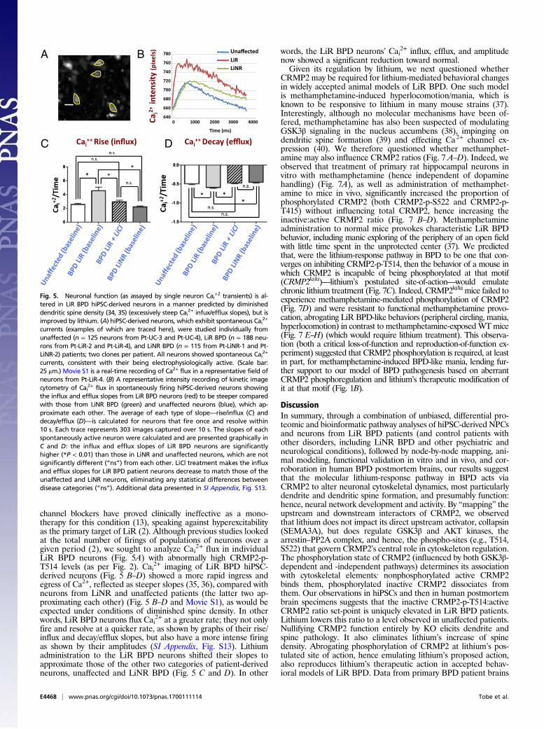
channel blockers have proved clinically ineffective as a mono-therapy for this condition (13), speaking against hyperexcitabilityas the primary target of LiR (2). Although previous studies lookedat the total number of firings of populations of neurons over agiven period (2), we sought to analyze Cai
2+ flux in individualLiR BPD neurons (Fig. 5A) with abnormally high CRMP2-p-T514 levels (as per Fig. 2). Cai
2+ imaging of LiR BPD hiPSC-derived neurons (Fig. 5 B–D) showed a more rapid ingress andegress of Ca2+, reflected as steeper slopes (35, 36), compared withneurons from LiNR and unaffected patients (the latter two ap-proximating each other) (Fig. 5 B–D and Movie S1), as would beexpected under conditions of diminished spine density. In otherwords, LiR BPD neurons flux Cai
2+ at a greater rate; they not onlyfire and resolve at a quicker rate, as shown by graphs of their rise/influx and decay/efflux slopes, but also have a more intense firingas shown by their amplitudes (SI Appendix, Fig. S13). Lithiumadministration to the LiR BPD neurons shifted their slopes toapproximate those of the other two categories of patient-derivedneurons, unaffected and LiNR BPD (Fig. 5 C and D). In other
words, the LiR BPD neurons’ Cai2+ influx, efflux, and amplitude
now showed a significant reduction toward normal.Given its regulation by lithium, we next questioned whether
CRMP2 may be required for lithium-mediated behavioral changesin widely accepted animal models of LiR BPD. One such modelis methamphetamine-induced hyperlocomotion/mania, which isknown to be responsive to lithium in many mouse strains (37).Interestingly, although no molecular mechanisms have been of-fered, methamphetamine has also been suspected of modulatingGSK3β signaling in the nucleus accumbens (38), impinging ondendritic spine formation (39) and effecting Ca2+ channel ex-pression (40). We therefore questioned whether methamphet-amine may also influence CRMP2 ratios (Fig. 7 A–D). Indeed, weobserved that treatment of primary rat hippocampal neurons invitro with methamphetamine (hence independent of dopaminehandling) (Fig. 7A), as well as administration of methamphet-amine to mice in vivo, significantly increased the proportion ofphosphorylated CRMP2 (both CRMP2-p-S522 and CRMP2-p-T415) without influencing total CRMP2, hence increasing theinactive:active CRMP2 ratio (Fig. 7 B–D). Methamphetamineadministration to normal mice provokes characteristic LiR BPDbehavior, including manic exploring of the periphery of an open fieldwith little time spent in the unprotected center (37). We predictedthat, were the lithium-response pathway in BPD to be one that con-verges on inhibiting CRMP2-p-T514, then the behavior of a mouse inwhich CRMP2 is incapable of being phosphorylated at that motif(CRMP2ki/ki)—lithium’s postulated site-of-action—would emulatechronic lithium treatment (Fig. 7C). Indeed, CRMP2ki/ki mice failed toexperience methamphetamine-mediated phosphorylation of CRMP2(Fig. 7D) and were resistant to functional methamphetamine provo-cation, abrogating LiR BPD-like behaviors (peripheral circling, mania,hyperlocomotion) in contrast to methamphetamine-exposedWTmice(Fig. 7 E–H) (which would require lithium treatment). This observa-tion (both a critical loss-of-function and reproduction-of-function ex-periment) suggested that CRMP2 phosphorylation is required, at leastin part, for methamphetamine-induced BPD-like mania, lending fur-ther support to our model of BPD pathogenesis based on aberrantCRMP2 phosphoregulation and lithium’s therapeutic modification ofit at that motif (Fig. 1B).
DiscussionIn summary, through a combination of unbiased, differential pro-teomic and bioinformatic pathway analyses of hiPSC-derived NPCsand neurons from LiR BPD patients (and control patients withother disorders, including LiNR BPD and other psychiatric andneurological conditions), followed by node-by-node mapping, ani-mal modeling, functional validation in vitro and in vivo, and cor-roboration in human BPD postmortem brains, our results suggestthat the molecular lithium-response pathway in BPD acts viaCRMP2 to alter neuronal cytoskeletal dynamics, most particularlydendrite and dendritic spine formation, and presumably function:hence, neural network development and activity. By “mapping” theupstream and downstream interactors of CRMP2, we observedthat lithium does not impact its direct upstream activator, collapsin(SEMA3A), but does regulate GSK3β and AKT kinases, thearrestin–PP2A complex, and hence, the phospho-sites (e.g., T514,S522) that govern CRMP2’s central role in cytoskeleton regulation.The phosphorylation state of CRMP2 (influenced by both GSK3β-dependent and -independent pathways) determines its associationwith cytoskeletal elements: nonphosphorylated active CRMP2binds them, phosphorylated inactive CRMP2 dissociates fromthem. Our observations in hiPSCs and then in human postmortembrain specimens suggests that the inactive CRMP2-p-T514:activeCRMP2 ratio set-point is uniquely elevated in LiR BPD patients.Lithium lowers this ratio to a level observed in unaffected patients.Nullifying CRMP2 function entirely by KO elicits dendrite andspine pathology. It also eliminates lithium’s increase of spinedensity. Abrogating phosphorylation of CRMP2 at lithium’s pos-tulated site of action, hence emulating lithium’s proposed action,also reproduces lithium’s therapeutic action in accepted behav-ioral models of LiR BPD. Data from primary BPD patient brains
A
C DCai++ Rise (influx) Cai
++ Decay (efflux)Time (ms)
B UnaffectedLiRLiNR
780
760
740
720
700
680
660
640Cai2+
inte
nsity
(pix
els)
0 1000 2000 3000 4000
Fig. 5. Neuronal function (as assayed by single neuron Cai+2 transients) is al-
tered in LiR BPD hiPSC-derived neurons in a manner predicted by diminisheddendritic spine density (34, 35) (excessively steep Cai
2+ infux/efflux slopes), but isimproved by lithium. (A) hiPSC-derived neurons, which exhibit spontaneous Cai
2+
currents (examples of which are traced here), were studied individually fromunaffected (n = 125 neurons from Pt-UC-3 and Pt-UC-4), LiR BPD (n = 188 neu-rons from Pt-LiR-2 and Pt-LiR-4), and LiNR BPD (n = 115 from Pt-LiNR-1 and Pt-LiNR-2) patients; two clones per patient. All neurons showed spontaneous Cai
2+
currents, consistent with their being electrophysiologically active. (Scale bar:25 μm.) Movie S1 is a real-time recording of Ca2+ flux in a representative field ofneurons from Pt-LiR-4. (B) A representative intensity recording of kinetic imagecytometry of Cai
2+ flux in spontaneously firing hiPSC-derived neurons showingthe influx and efflux slopes from LiR BPD neurons (red) to be steeper comparedwith those from LiNR BPD (green) and unaffected neurons (blue), which ap-proximate each other. The average of each type of slope—rise/influx (C) anddecay/efflux (D)—is calculated for neurons that fire once and resolve within10 s. Each trace represents 303 images captured over 10 s. The slopes of eachspontaneously active neuron were calculated and are presented graphically inC and D: the influx and efflux slopes of LiR BPD neurons are significantlyhigher (*P < 0.01) than those in LiNR and unaffected neurons, which are notsignificantly different (“ns”) from each other. LiCl treatment makes the influxand efflux slopes for LiR BPD patient neurons decrease to match those of theunaffected and LiNR neurons, eliminating any statistical differences betweendisease categories (“ns”). Additional data presented in SI Appendix, Fig. S13.
E4468 | www.pnas.org/cgi/doi/10.1073/pnas.1700111114 Tobe et al.

further confirm the predicted link between abnormally elevatedCRMP2-p-T514 and dendritic spine abnormalities, as well as ev-idence that lithium treatment of patients acts to normalize bothCRMP2 ratios and dendritic spine density and length.We emphasize that these actions of lithium in BPD do not rule
out potential roles for that cation’s multiple other actions (4–7),which may function additively or synergistically in this condition.We now simply identify a promising regulatable molecular path-way upon which to focus etiologically and pharmacologically.Elevated baseline CRMP2-p-T514 might be associated with LiR
as a clinical classification of BPD (potentially a biomarker). (Pro-spective studies in large cohorts of living patients will inevitablybe required to help define what the critical clinical thresholdfor a CRMP2-p-T514:CRMP2 ratio should be.) Our data cannot
yet determine whether the CRMP2-p-T514:CRMP2 set-point ischronically high in LiR BPD patients or rather that the response ofLiR BPD patients to stimuli that increase CRMP2-p-T514 is morepronounced, prolonged, or of earlier onset than in unaffected pa-tients, or a combination of these. Nevertheless, these results mayprovide impetus for biomarker assay development, for examplemeasuring CRMP2-p-T514:CRMP2 ratios in reprogrammedpatient-derived cells (including those obtained from the peripheralblood, like some of ours) as a diagnostic aid to predict drugresponsiveness. A qualitative, not just quantitative, distinction be-tween LiR and LiNR BPD based on an abnormally high set-point foran otherwise physiologic posttranslational modification of a cytoskeletalregulator (uniquely in LiR BPD) invites speculation that LiNR BPD isactually a separate disease that “pheno-copies” BPD but is unrelated
G
D
H
I
J
F
E
A CBFig. 6. CRMP2 function is pivotal for proper dendriticbranching and spine organization in vivo. (A) Representativephotomicrographs from an immunohistochemical survey of theadult mouse brain determining the regions and cell typesexpressing phosphorylated CRMP2 in situ. Shown are sectionsthrough the CA1 region of the hippocampus (Upper) and thecerebellum (Lower), costained with an antibody againstCRMP2-p-T514 (yellow) and DAPI (blue). Regions in the redboxes are magnified (Left) to visualize the pyramidal and Pur-kinje cell layers, respectively. Other regions in which neuronsshowed expression were cerebral cortex, olfactory bulb, andstriatum. (Scale bar: 250 μm, Right, and 100 μm, Left.) (B) LiCladministration to mice increases levels of inactive (phosphory-lated) GSK3β (red arrow) and lowers levels of inactive CRMP2-p-T514 (red arrow) compared with water (H2O) (based on quan-tification ofWestern blot analysis of hippocampal protein). (n =7, H2O-treated; n = 7, LiCl-treated, *P = 0.05.) (C–F) ConstitutiveCrmp2-KO mice have grossly normal bodies and brains but arecharacterized at adulthood by defects in dendritic morphology(compared with WT) in the regions where CRMP2 is expressed[e.g., in CA1 of the hippocampus, as shown here in Golgi stainsexamined along the stratum radiatum (33), but also seen instriatum and cortex]. (C and D) CRMP2-KOmice show a fivefoldincrease in bifurcation of apical dendrites proximally (creatingincreased dendritic branching points at the expense of maintrunk dendrites). The representative pyramidal neurons in-dicated by red arrows in C are each magnified in the respectiveInsets below the overview and are quantified in D. (Scale bar,100 μm.) (Data shown are mean ± SEM from 49 to 76 neuronsfrom three mice of each genotype; Student t test: *P <0.001 compared with WT.) (E and F) The dendrites themselvesare characterized by a decreased density of spines (i.e., averagenumber of spines per micrometer). The red blocked areas in Eare magnified in the Insets and the data are quantified in F.(Scale bar: 10 μm; 2.5 μm in Insets.) (Data shown aremean ± SEMfrom >20 dendrites from each of threeWT and three CRMP2-KOmice; **P = 0.0006 compared withWT.) This diminished dendriticspine density and length in the CRMP2-KO mouse is strikinglysimilar to that seen in the primary human postmortem brainspecimens from LiR BPD patients (Fig. 4 A, C, and D) and wouldbe consistent with the functional consequences seen in LiRBPD neurons (Fig. 5). (G) Differential localization of non-phosphorylated and phosphorylated CRMP2, in and out ofspines, respectively. A representative field of neurons examinedin situ along the stratum radiatum in the rat hippocampus. Cyanboxes (first row) indicate the dendritic regions magnified in the second row and stained, respectively, for filamentous actin (F-actin, white), the neuronal marker MAP2(blue), CRMP2 (red), and CRMP2-p-T514 (green); these same regions, in their respective columns in the third row, are costainedwith combinations of the othermarkers andthe images merged. Arrowheads indicate phalloidin-stained dendritic spines; they contain nonphosphorylated CRMP2 (red) but do not stain for CRMP2-p-T514 (green).Magnified images from the white boxed areas in the third row are magnified in the fourth row and again show that nonphosphorylated CRMP2 (red) is expressedthroughout the neuron, including the dendritic shaft, branches, and spines; CRMP2-p-T514 (green) is not expressed in the spines, suggesting that, when CRMP2 becomesphosphorylated, it leaves or is excluded from the spines. In the fourth row, a white arrow points to the same representative dendritic spines in both Left and Right panelsnicely showing that, although nonphosphorylated CRMP2 fills the spines, phosphorylated CRMP2 is absent. (Scale bar: 90 μm, first row; 40 μm, second and third rows;15 μm, fourth row.) (H–J) LiCl increases dendritic spine volume and density, an action abrogated by KO of CRMP2. Rat hippocampal neurons (MAP2+, green) show anincrease in F-actin staining (red) after 7 d of incubationwith LiCl (3mM) (H), presented quantitatively in I. (Scale bar: 15 μm, Top; 7 μm, Bottom.) F-actin area correlates withdendritic spine volume (44, 45). LiCl induced a 60% increase in the area of F-actin puncta associated with dendritic spines and increased spine density by 36% [11 spines/10 μm (untreated) vs. 15 spines/10 μm (LiCl)]. (Data expressed as mean ± SEM; ***P < 0.001 unpaired Student’s t test; n = 23 neurons per group; 3,255 actin spinesmeasured in control neurons, 3,812 spines in LiCl-treated neurons.) (J) In contrast, hippocampal neurons from the CRMP2-KO mouse evince no increase in spine densityfrom their baseline when similarly treatedwith LiCl, in contrast toWT littermates. (KO untreated, n= 13; KO+LiCl, n= 11;WT+LiCl, n= 9. One-way ANOVA, **P= 0.0013.)
Tobe et al. PNAS | Published online May 12, 2017 | E4469
MED
ICALSC
IENCE
SPS
YCHOLO
GICALAND
COGNITIVESC
IENCE
SPN
ASPL
US
pathophysiologically to the lithium-response pathway. (See SI Appen-dix, Figs. S9 and S12 for further discussion of this possibility based onbiochemical and functional data, respectively.) Although CRMP2 mayplay a role in other neuropsychiatric diseases—for example, totalCRMP2 levels may be abnormal in postmortem brains of schizo-phrenics—aberrantly elevated inactive:active CRMP2 ratios with ex-cessively high CRMP2-p-T514 seems specific to LiR BPD.The lithium-response pathway impinging on CRMP2 likely has
additional modes of input that go beyond those illustrated in Fig.1B. For example, brain-derived neurotrophic factor (BDNF) alsoreduced CRMP2-p-T514 in a manner similar to lithium (SI Ap-pendix, Fig. S13A), suggesting that systems downstream of tyrosinereceptor kinase B (TRKB, the BDNF receptor) might be in-tegrated into the pathway (SI Appendix, Fig. S13B). Moreover,CRMP2 has been associated with tauopathies via its interactionswith microtubule-associated protein tau and presenillin 1. Indeed,CRMP2 interacts with amyloid precursor protein, and has been notedto be a component of neurofibrillary tangles in Alzheimer’s disease(41). Hence, not only might this axis be implicated in diseases other
than BPD (cytoskeletal dynamics coming to be recognized as centralto a growing number of neuropathological processes), but also drugsthat lower CRMP2-p-T514 may potentially be more widely thera-peutic (particularly in disorders characterized by deposition of cyto-skeletal elements such as tau either as a cause or a biomarker).If our premise is correct that identifying a therapeutic pathway
also implicates that pathway as central to that disease’s patho-genesis, then certain broader ideas warrant mentioning. First,whereas BPD has heritable features and developmental under-pinnings, it would appear to be a disorder not of a defective geneper se but rather of dysregulated posttranslational modulation of anormally produced gene product that, although developmentallycritical, plays a physiologic role in all individuals throughout life.Second, attributing pathophysiology to aberrant cytoskeletal dy-namics (dendritic disorganization representing one consequence)would appear to implicate not merely defective neurons but alsodysregulated interneuronal networks. Whether these observationsare particular to LiR BPD or are applicable more widely to otherneuropsychiatric disorders warrants study.
A
C
F
E Time in Center Test
G
In Vitro
D
CRM
P2-p
-T51
4/CR
MP2
In VivoB
Tota
l tra
vel d
istan
ce(x
104
cm)
5
4
3
2
1
0
A�erBefore(Meth or Saline administra�on)
H
WT
Saline MethCrmp2 KI/KI
MethSaline
Ctrl Meth
CRM
P2-p
-T51
4/CR
MP2
CRM
P2-p
-514
/ß-a
c�n
CRM
P2-p
-522
/ß-a
c�n
Phosphoryla�on Sites CRMP2 C-Terminus
1st2nd
522518514509
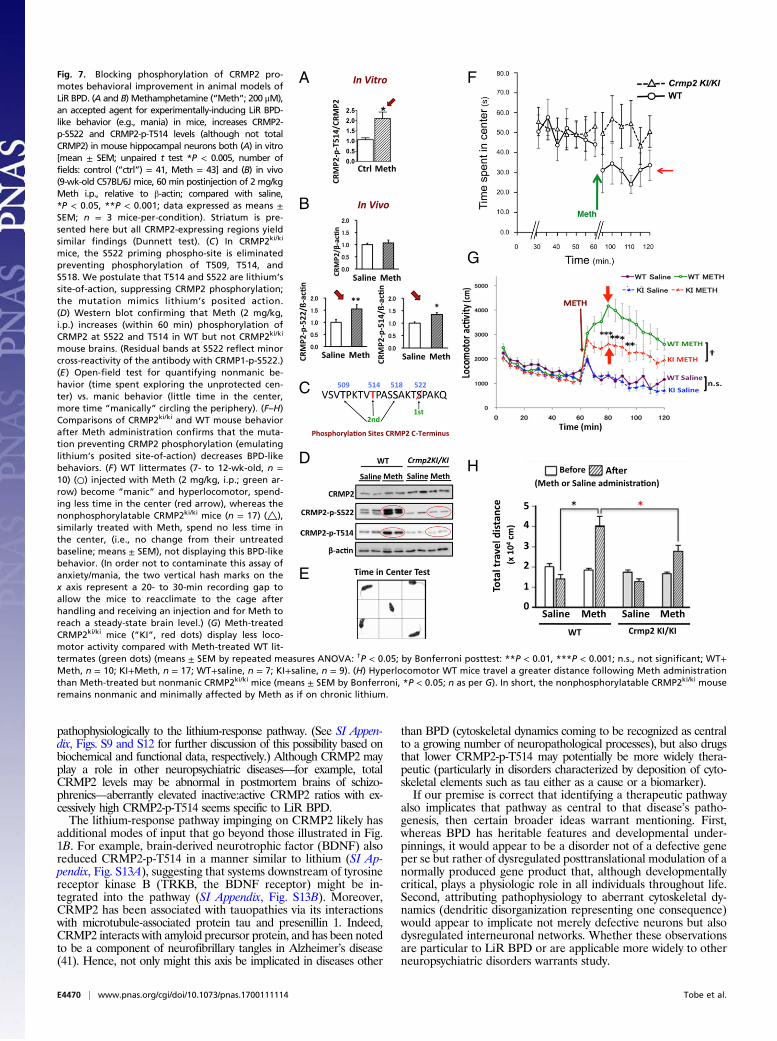
Fig. 7. Blocking phosphorylation of CRMP2 pro-motes behavioral improvement in animal models ofLiR BPD. (A and B) Methamphetamine (“Meth”; 200 μM),an accepted agent for experimentally-inducing LiR BPD-like behavior (e.g., mania) in mice, increases CRMP2-p-S522 and CRMP2-p-T514 levels (although not totalCRMP2) in mouse hippocampal neurons both (A) in vitro[mean ± SEM; unpaired t test *P < 0.005, number offields: control (“ctrl”) = 41, Meth = 43] and (B) in vivo(9-wk-old C57BL/6J mice, 60 min postinjection of 2 mg/kgMeth i.p., relative to β-actin; compared with saline,*P < 0.05, **P < 0.001; data expressed as means ±SEM; n = 3 mice-per-condition). Striatum is pre-sented here but all CRMP2-expressing regions yieldsimilar findings (Dunnett test). (C) In CRMP2ki/ki
mice, the S522 priming phospho-site is eliminatedpreventing phosphorylation of T509, T514, andS518. We postulate that T514 and S522 are lithium’ssite-of-action, suppressing CRMP2 phosphorylation;the mutation mimics lithium’s posited action.(D) Western blot confirming that Meth (2 mg/kg,i.p.) increases (within 60 min) phosphorylation ofCRMP2 at S522 and T514 in WT but not CRMP2ki/ki
mouse brains. (Residual bands at S522 reflect minorcross-reactivity of the antibody with CRMP1-p-S522.)(E ) Open-field test for quantifying nonmanic be-havior (time spent exploring the unprotected cen-ter) vs. manic behavior (little time in the center,more time “manically” circling the periphery). (F–H)Comparisons of CRMP2ki/ki and WT mouse behaviorafter Meth administration confirms that the muta-tion preventing CRMP2 phosphorylation (emulatinglithium’s posited site-of-action) decreases BPD-likebehaviors. (F) WT littermates (7- to 12-wk-old, n =10) (○) injected with Meth (2 mg/kg, i.p.; green ar-row) become “manic” and hyperlocomotor, spend-ing less time in the center (red arrow), whereas thenonphosphorylatable CRMP2ki/ki mice (n = 17) (△),similarly treated with Meth, spend no less time inthe center, (i.e., no change from their untreatedbaseline; means ± SEM), not displaying this BPD-likebehavior. (In order not to contaminate this assay ofanxiety/mania, the two vertical hash marks on thex axis represent a 20- to 30-min recording gap toallow the mice to reacclimate to the cage afterhandling and receiving an injection and for Meth toreach a steady-state brain level.) (G) Meth-treatedCRMP2ki/ki mice (“KI”, red dots) display less loco-motor activity compared with Meth-treated WT lit-termates (green dots) (means ± SEM by repeated measures ANOVA: †P < 0.05; by Bonferroni posttest: **P < 0.01, ***P < 0.001; n.s., not significant; WT+Meth, n = 10; KI+Meth, n = 17; WT+saline, n = 7; KI+saline, n = 9). (H) Hyperlocomotor WT mice travel a greater distance following Meth administrationthan Meth-treated but nonmanic CRMP2ki/ki mice (means ± SEM by Bonferroni, *P < 0.05; n as per G). In short, the nonphosphorylatable CRMP2ki/ki mouseremains nonmanic and minimally affected by Meth as if on chronic lithium.
E4470 | www.pnas.org/cgi/doi/10.1073/pnas.1700111114 Tobe et al.

With regard to disease modeling in general, this study suggests astrategy for merging hiPSC technology with proteomics to discernunderlying pathophysiological mechanisms in complex, polygenic,multifactorial diseases in which causative genes, cells, proteins,and pathways are not well-understood. If there exists an agent that isknown to be functionally impactful even if its molecular mechanism-of-action is uncertain (like lithium in BPD), such an agent may allowan investigator to probe otherwise inscrutable intracellular signaling byidentifying its target and then reconstructing the regulatory molecularroutes upstream and downstream of that node with an eye towardmapping underlying pathogenic pathways and identifying more spe-cific drug targets for the development of safer, cheaper, or more ef-fective pharmacotherapeutics. In this way, hiPSCs may be used in themost challenging diseases not only to reflect phenomenology and aphenotype, but also to identify underlying molecular mechanisms.
Materials and MethodsHuman fibroblasts or lymphoblasts from multiple well-sourced patients (SI Ap-pendix, Fig. S1) were reprogrammed to hiPSCs via nonintegrating episomal-mediated (42), lentivirus-mediated (1), or retrovirus-mediated (19) gene transfer,characterized (43), and differentiated to NPCs and cortical interneurons, as per ourroutine and as previously described (17–20). Protein isolation, 2D-DIGE and SILAC,Western blotting, and coimmunoprecipitation were performed as described pre-viously (17, 28). Immunofluorescence was quantified by pixel number captured viaimage analysis software using unbiased stereology. All MS data are publicly ac-cessible; for SILAC data, the mass spectra may be downloaded from MassIVE(massive.ucsd.edu) using accession no. MSV000080975; the data are directly ac-cessible via ftp://massive.ucsd.edu (MSV000080975). Creation and analysis ofCRMP2 knockout and knockin mice used standard transgenic techniques. Analysis
of primary cultures of rodent hippocampal neurons also followed standard tech-niques (44). Cai
2+ transients and flux were measured via kinetic imaging analysis(34, 35). Animal modeling of BPD behavior (to determine the response to lithium-response pathway manipulation) was as described previously (37–39). All animaluse was conducted in accordance with the NIH guidelines and approved by theYokohama City University Institutional Animal Care and Use Committee. Humanpostmortemmaterial was obtained from the University of Pittsburgh and from theMcLean Hospital. Patient samples were obtained following informed consent; allpatient identification was removed and the material was processed according toIRB approvals including from Nova Scotia Health Authority Research Ethics Board,National Institutes of Mental Health, and the Sanford-Burnham Prebys MedicalDiscovery Institute. Dendrite and dendritic spine morphology were assessed as perroutine procedures with Golgi stains and image-assisted quantification (44, 45).Bioinformatic analysis was performed as per the Sullivan Lab Evidence Project,ProtKIN, and IPA. See SI Appendix, Supplemental Methods for details.
ACKNOWLEDGMENTS. We thank M. Niepel, N. Moerke, C. Shamu, and P. Sorgerfor kinase perturbagen screens; J. Wong and V. Chen for help with Cai
2+ imaging;S. Ghose and the Harvard Brain Tissue Resource Center for access to postmortembrains. This study was supported by Grant RC2MH090011 (to E.Y.S.); NIH’s Libraryof Integrated Network-based Cellular Signatures Program (E.Y.S.); the ViterbiFoundation Neuroscience Initiative (E.Y.S.); Stanley Medical Research InstituteGrants R21MH093958, R33MH087896, and R01MH095088 (to S.J.H.); the TauConsortium (S.J.H.); NIH Grant R01MH087823 (to S.H.); California Institute of Re-generative Medicine training grants (to B.T.D.T., L.D., and C.D.); a University ofCalifornia, San Diego T32 training grant in psychiatry (to B.T.D.T.); the CaliforniaBipolar Foundation; the International Bipolar Foundation; and Creation ofInnovation Centers for Advanced Interdisciplinary Research Areas Program inthe Project for Developing Innovation Systems from the Ministry of Education,Science, Sports and Culture in Japan (Grant 42890001) (Y.G.). Dedicated to thememory of Dr. Jeffrey Nye and his contributions to neuropsychopharmacology.
1. Tobe BT, Snyder EY, Nye JS (2011) Modeling complex neuropsychiatric disorders withhuman induced pluripotent stem cells. Curr Opin Pharmacol 11:521–527.
2. Mertens J, et al.; Pharmacogenomics of Bipolar Disorder Study (2015) Differential responsesto lithium in hyperexcitable neurons from patients with bipolar disorder.Nature 527:95–99.
3. Brennand KJ, et al. (2011) Modelling schizophrenia using human induced pluripotentstem cells. Nature 473:221–225.
4. Malhi GS, Tanious M, Das P, Coulston CM, Berk M (2013) Potential mechanisms ofaction of lithium in bipolar disorder. Current understanding. CNS Drugs 27:135–153.
5. Gershon S, Chengappa KN, Malhi GS (2009) Lithium specificity in bipolar illness: Aclassic agent for the classic disorder. Bipolar Disord 11:34–44.
6. Miller C, Bauer MS (2014) Excess mortality in bipolar disorders. Curr Psychiatry Rep 16:499.7. Levy NA, Janicak PG (2000) Calcium channel antagonists for the treatment of bipolar
disorder. Bipolar Disord 2:108–119.8. Baldessarini RJ, et al. (2006) Decreased risk of suicides and attempts during long-term
lithium treatment: A meta-analytic review. Bipolar Disord 8:625–639.9. Sullivan PF, Daly MJ, O’Donovan M (2012) Genetic architectures of psychiatric disor-
ders: The emerging picture and its implications. Nat Rev Genet 13:537–551.10. Charney AW, et al. (2017) Evidence for genetic heterogeneity between clinical sub-
types of bipolar disorder. Transl Psychiatry 7:e993.11. Goshima Y, Nakamura F, Strittmatter P, Strittmatter SM (1995) Collapsin-induced growth
cone collapsemediated by an intracellular protein related to UNC-33.Nature 376:509–514.12. Ip JP, et al. (2011) α2-chimaerin controls neuronal migration and functioning of the
cerebral cortex through CRMP-2. Nat Neurosci 15:39–47.13. Marques JM, et al. (2013) CRMP2 tethers kainate receptor activity to cytoskeleton
dynamics during neuronal maturation. J Neurosci 33:18298–18310.14. Uchida Y, et al. (2005) Semaphorin3A signalling is mediated via sequential Cdk5 and
GSK3beta phosphorylation of CRMP2: Implication of common phosphorylating mecha-nism underlying axon guidance and Alzheimer’s disease. Genes Cells 10:165–179.
15. Kaneko S, et al. (2006) A selective Sema3A inhibitor enhances regenerative responsesand functional recovery of the injured spinal cord. Nat Med 12:1380–1389.
16. Wilson SM, et al. (2012) Prevention of posttraumatic axon sprouting by blockingcollapsin response mediator protein 2-mediated neurite outgrowth and tubulin po-lymerization. Neuroscience 210:451–466.
17. Singec I, et al. (2016) Quantitative analysis of human pluripotency & neural specifi-cation by in-depth (phospho)proteomic profiling. Stem Cell Rep 7:527–542.
18. Li W, et al. (2011) Rapid induction and long-term self-renewal of primitive neuralprecursors from human embryonic stem cells by small molecule inhibitors. Proc NatlAcad Sci USA 108:8299–8304.
19. Madison JM, et al. (2015) Characterization of bipolar disorder patient-specific inducedpluripotent stem cells from a family reveals neurodevelopmental and mRNA ex-pression abnormalities. Mol Psychiatry 20:703–717.
20. Maroof AM, et al. (2013) Directed differentiation and functional maturation of cor-tical interneurons from human embryonic stem cells. Cell Stem Cell 12:559–572.
21. Blouin JL, et al. (1998) Schizophrenia susceptibility loci on chromosomes 13q32 and8p21. Nat Genet 20:70–73.
22. Fallin MD, et al. (2005) Bipolar I disorder and schizophrenia: A 440-single-nucleotidepolymorphism screen of 64 candidate genes among Ashkenazi Jewish case-parenttrios. Am J Hum Genet 77:918–936.
23. Fallin MD, et al. (2011) Linkage and association on 8p21.2-p21.1 in schizophrenia. AmJ Med Genet B Neuropsychiatr Genet 156:188–197.
24. Yoshimura T, et al. (2005) GSK-3β regulates phosphorylation of CRMP-2 and neuronalpolarity. Cell 120:137–149.
25. Cole AR, et al. (2006) Distinct priming kinases contribute to differential regulation ofcollapsin response mediator proteins by glycogen synthase kinase-3 in vivo. J BiolChem 281:16591–16598.
26. Zhu LQ, et al. (2010) Protein phosphatase 2A facilitates axonogenesis by dephos-phorylating CRMP2. J Neurosci 30:3839–3848.
27. Soutar MP, et al. (2010) Evidence that glycogen synthase kinase-3 isoforms havedistinct substrate preference in the brain. J Neurochem 115:974–983.
28. Hoedt E, Zhang G, Neubert TA (2014) Stable isotope labeling by amino acids in cellculture (SILAC) for quantitative proteomics. Adv Exp Med Biol 806:93–106.
29. Sakurai K, et al. (2014) Kinome-wide functional analysis highlights the role of cyto-skeletal remodeling in somatic cell reprogramming. Cell Stem Cell 14:523–534.
30. Beaulieu JM, et al. (2008) A beta-arrestin 2 signaling complex mediates lithium actionon behavior. Cell 132:125–136.
31. Strakowski SM, et al. (2012) The functional neuroanatomy of bipolar disorder: Aconsensus model. Bipolar Disord 14:313–325.
32. Zhang H, et al. (2016) Brain-specific Crmp2 deletion leads to neuronal developmentdeficits and behavioural impairments in mice. Nat Commun 7:11773.
33. Nakamura F, et al. (2009) Increased proximal bifurcation of CA1 pyramidal apicaldendrites in sema3A mutant mice. J Comp Neurol 516:360–375.
34. Sabatini BL, Oertner TG, Svoboda K (2002) The life cycle of Ca(2+) ions in dendriticspines. Neuron 33:439–452.
35. Merriam EB, et al. (2013) Synaptic regulation of microtubule dynamics in dendriticspines by calcium, F-actin, and drebrin. J Neurosci 33:16471–16482.
36. Chen HM, et al. (2014) Transcripts involved in calcium signaling and telencephalicneuronal fate are altered in induced pluripotent stem cells from bipolar disorderpatients. Transl Psychiatry 4:e375.
37. Gould TD, O’Donnell KC, Picchini AM, Manji HK (2007) Strain differences in lithiumattenuation of d-amphetamine-induced hyperlocomotion: A mouse model for thegenetics of clinical response to lithium. Neuropsychopharmacology 32:1321–1333.
38. Xu CM, et al. (2011) Glycogen synthase kinase 3β in the nucleus accumbens core is criticalfor methamphetamine-induced behavioral sensitization. J Neurochem 118:126–139.
39. Young EJ, et al. (2014) Selective, retrieval-independent disruption of methamphetamine-associated memory by actin depolymerization. Biol Psychiatry 75:96–104.
40. Shibasaki M, Kurokawa K, Ohkuma S (2010) Upregulation of L-type Ca(v)1 channels inthe development of psychological dependence. Synapse 64:440–444.
41. Yoshida H, Watanabe A, Ihara Y (1998) Collapsin response mediator protein-2 is associatedwith neurofibrillary tangles in Alzheimer’s disease. J Biol Chem 273:9761–9768.
42. Okita K, et al. (2011) A more efficient method to generate integration-free human iPScells. Nat Methods 8:409–412.
43. Müller FJ, et al. (2011) A bioinformatic assay for pluripotency in human cells. NatMethods 8:315–317.
44. Calabrese B, Halpain S (2005) Essential role for the PKC target MARCKS in maintainingdendritic spine morphology. Neuron 48:77–90.
45. Hotulainen P, Hoogenraad CC (2010) Actin in dendritic spines: Connecting dynamicsto function. J Cell Biol 189:619–629.
Tobe et al. PNAS | Published online May 12, 2017 | E4471
MED
ICALSC
IENCE
SPS
YCHOLO
GICALAND
COGNITIVESC
IENCE
SPN
ASPL
US