Embed Size (px)
Citation preview
Principles of Medical Ultrasound
Zahra Kavehvash
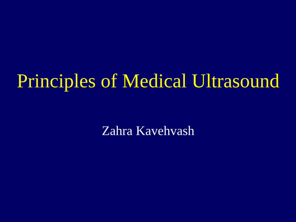
Medical Ultrasound Course (25-636)
• Introduction • Acoustic wave propagation • Attenuation, scattering and speckle • Transducers
– Generation and detection of ultrasound – Equivalent circuit – Piezoelectric materials
• Beam forming and Diffraction – Array beam forming
• Image formation • Doppler modes
– Ultrasound flow imaging
• Contrast and resolution • Ultrasound bioeffects and safety • Emerging technologies and trends (If time
permits) • Acousto-optic (Photo-Acoustic) imaging (If
time permits)
Medical Ultrasound Course (25-636)
• Reference: – Thomas L. Szabo , Diagnostic Ultrasound Imaging: Inside Out , Elsevier
, Academic Press Series in BME , 2004. – Haim Azhari, Basics of Biomedical Ultrasound for Engineers, IEEE ,
Wiley, 2010. – K. Kirk Shung and Gray A. Thieme, Ultrasonic Scattering in Biological
Tissues, CRC Press, 1993. – B. D. Steinberg, Principles of Aperture and Array System Design, John
Wiley and Sons, 1976. – D. H. Evans, W. N. McDicken, R. Skidmore, and J. P. Woodcock,
Doppler Ultrasound, John Wiley and Sons, 1989. – J.W. Goodman, Introduction to Fourier Optics, McGraw-Hill, 1968.
Medical Ultrasound Course (25-636)
• Journals: – IEEE Transactions on Ultrasonics, Ferroelectrics and Frequency
control. – IEEE Transactions on Medical Imaging – Journal of Acoustical Society of America
Medical Ultrasound Course (25-636)
• Grading: – Homework: 20 % – Project: 10 % – Mid Term: 30 % – Final: 40 %

History of Ultrasound Who are the smart guys?
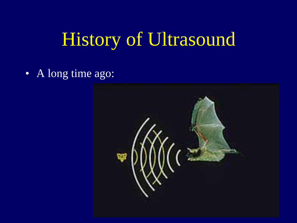
History of Ultrasound • A long time ago:
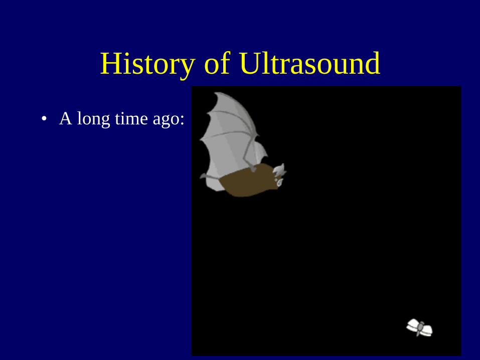
History of Ultrasound • A long time ago:
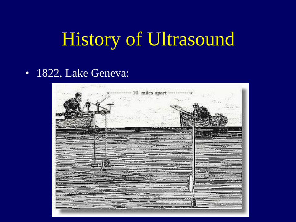
History of Ultrasound • 1822, Lake Geneva:
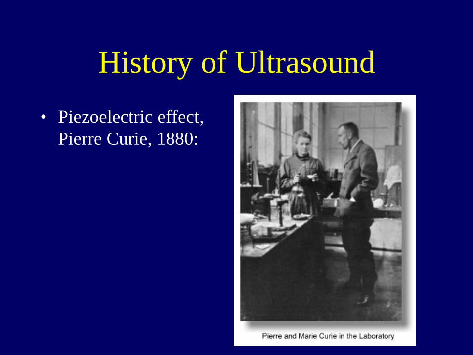
History of Ultrasound • Piezoelectric effect,
Pierre Curie, 1880:

History of Ultrasound • 1954-1957:
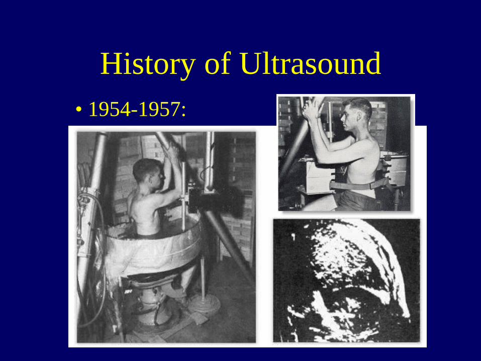
History of Ultrasound • 1954-1957:
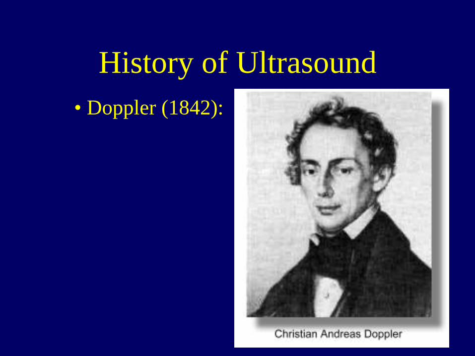
History of Ultrasound • Doppler (1842):
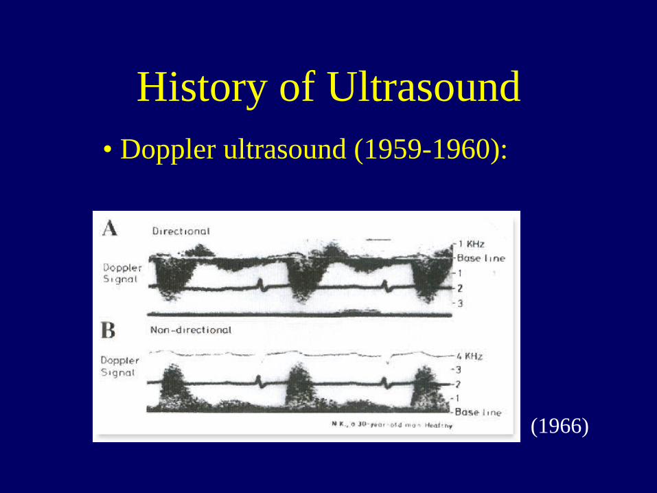
History of Ultrasound • Doppler ultrasound (1959-1960):
(1966)
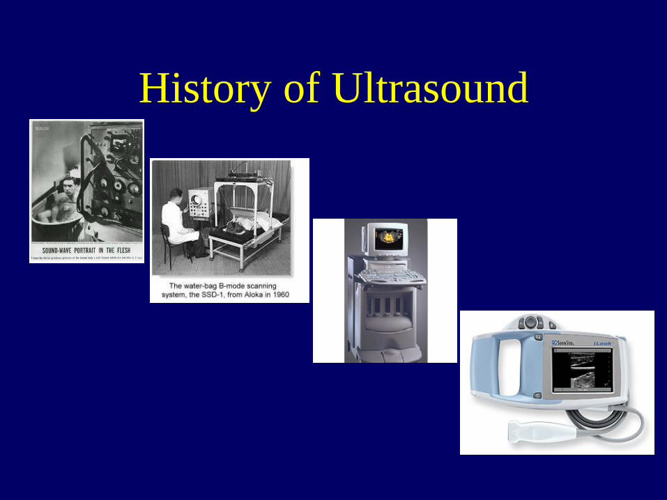
History of Ultrasound
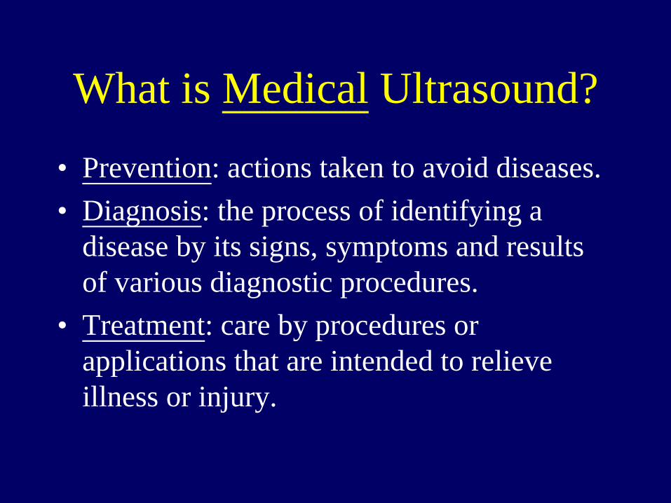
What is Medical Ultrasound?
• Prevention: actions taken to avoid diseases. • Diagnosis: the process of identifying a
disease by its signs, symptoms and results of various diagnostic procedures.
• Treatment: care by procedures or applications that are intended to relieve illness or injury.
Diagnosis
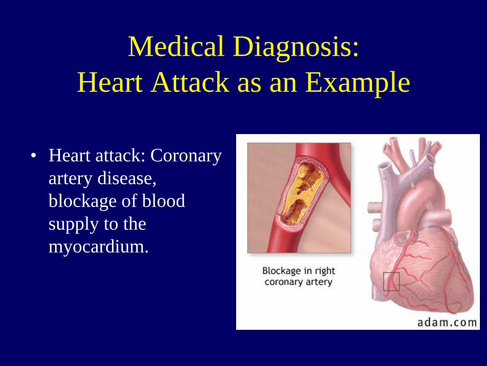
Medical Diagnosis: Heart Attack as an Example
• Heart attack: Coronary artery disease, blockage of blood supply to the myocardium.
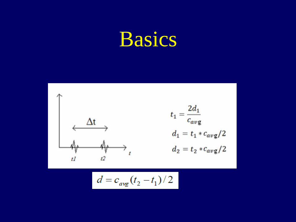
Basics

Basics
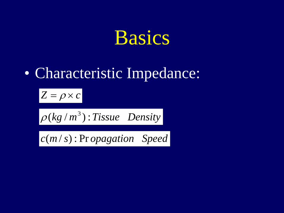
Basics • Characteristic Impedance:
cZ ×= ρ
DensityTissuemkg :)/( 3ρ
Speedopagationsmc Pr:)/(

Basics
in
ref
PP
ZZZZRF =+−= )/()( 1212
Amplitude of Acoustic Wave Pressure

Scan Modes • A-mode (A-scan, 1D). • B-mode (Gray scale, 2D). • M-mode (motion) • Color Doppler (2D, blood flow). • Spectral Doppler (localized, blood flow). • Audio Doppler.

A-Scan (Amplitude, 1D)
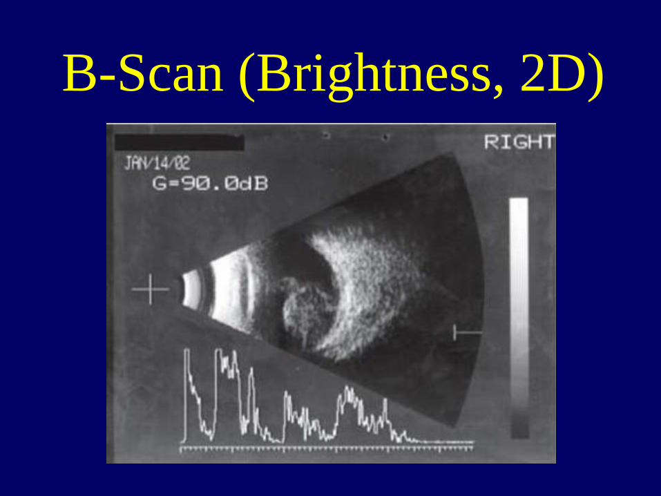
B-Scan (Brightness, 2D)

M-Mode (Brightness, 2D) • A-Mode data in time
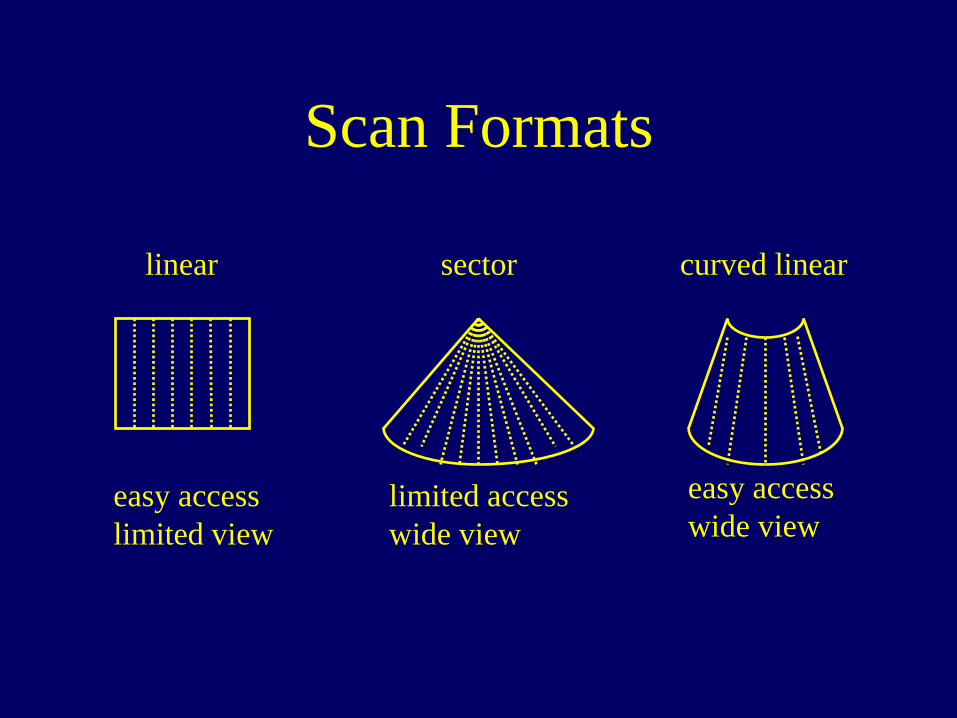
Scan Formats
linear sector curved linear
easy access limited view
limited access wide view
easy access wide view
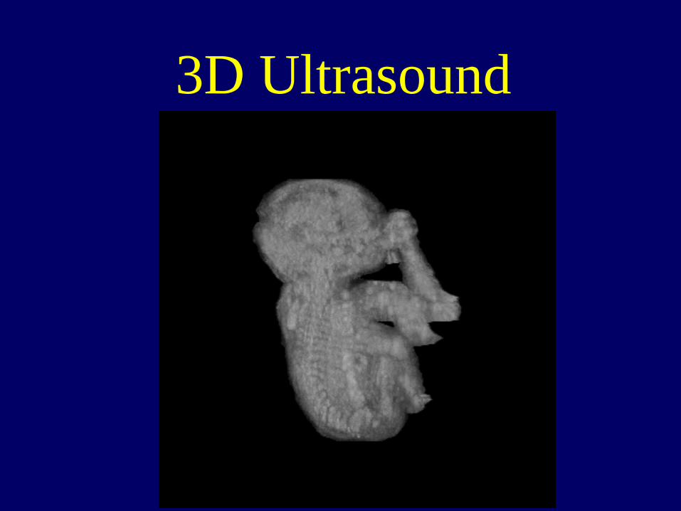
3D Ultrasound
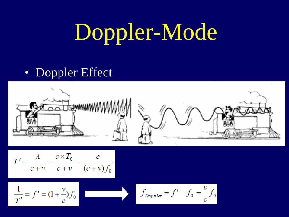
Doppler-Mode • Doppler Effect
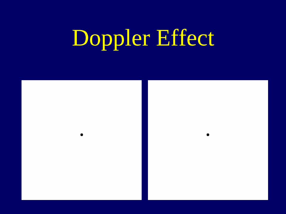
Doppler Effect
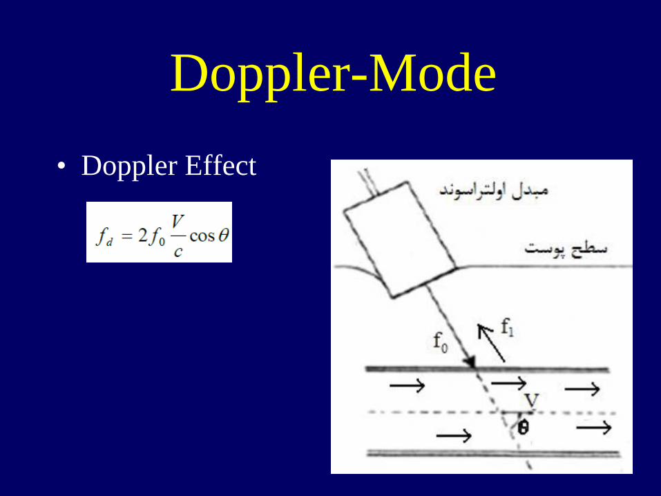
Doppler-Mode • Doppler Effect
Doppler-Mode • Doppler Systems:
– Continuous Doppler – Pulse Doppler
Doppler-Mode • Continuous Doppler:
– Just records the change in frequency – Display the changes in color or sound
• Pulse Doppler: – Records the reflection depth along with the
Doppler frequency

Ultrasound vs. Other Imaging Systems
Ultrasound Developements • Harmonic imaging • 2D Transducers for 3D imaging • Increasing the Number of
Transducer Elements • Intra Vascular Ultrasound

Ultrasound Developements • Contrast improvement:
– Contrast agents (containing bobbles for THI and Endothelial Dysfunction measurement)
Ultrasound Developements • Miniaturization:
– Decreasing the weight to a few hundred grams.
– Leads to more invasive applications

Ultrasound Developements • Immunity: • Appropriate power dose for
different diagnostic and therapeutic applications
Bio-Effects • Heating • Cavitation
Cavitation
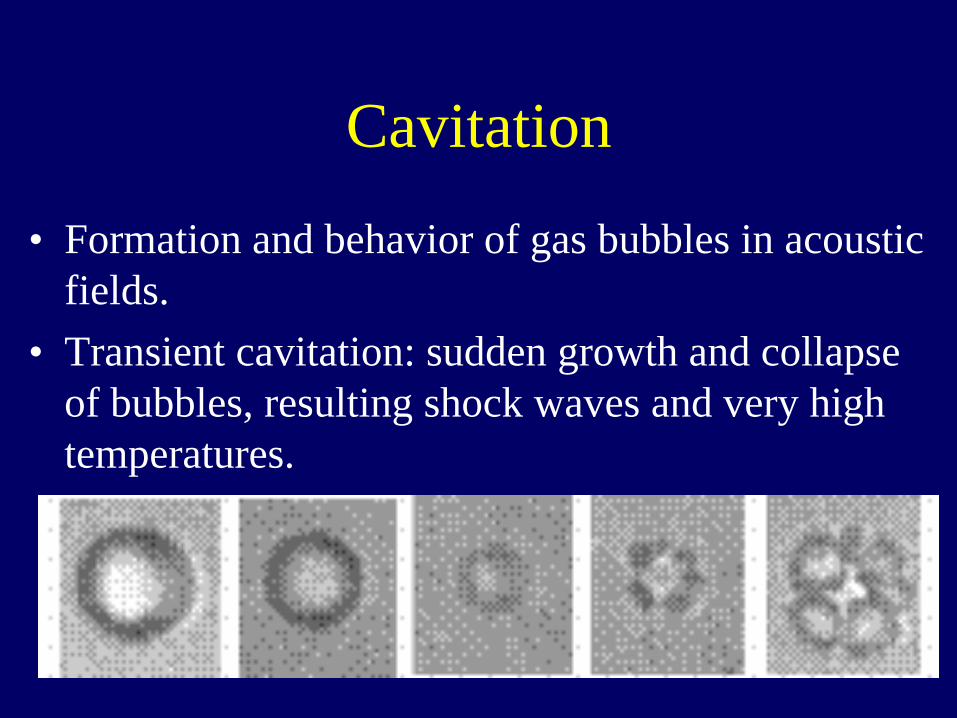
• Formation and behavior of gas bubbles in acoustic fields.
• Transient cavitation: sudden growth and collapse of bubbles, resulting shock waves and very high temperatures.
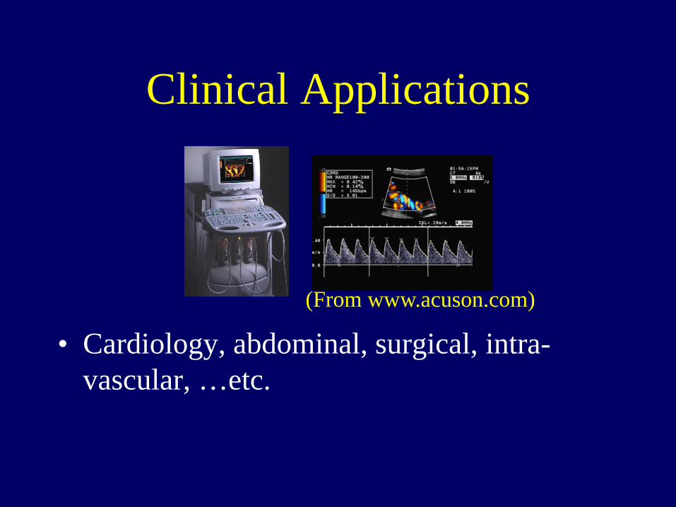
Clinical Applications
• Cardiology, abdominal, surgical, intra-vascular, …etc.
(From www.acuson.com)
Characteristics of Diagnostic Ultrasound
• Non-invasive. • Safe (under regulations). • Real-time. • Reflection mode (similar to RADAR). • Blood flow imaging. • Portable. • Body type dependent.
Characteristics of Diagnostic Ultrasound
• Spatial resolution: – Lateral and elevational: diffraction limited. – Axial: transducer and system bandwidth, pulse energy.
• Contrast resolution: spatial resolution and speckle brightness variations.
• Temporal resolution: speed of sound in tissue.
Ultrasonic Transducers
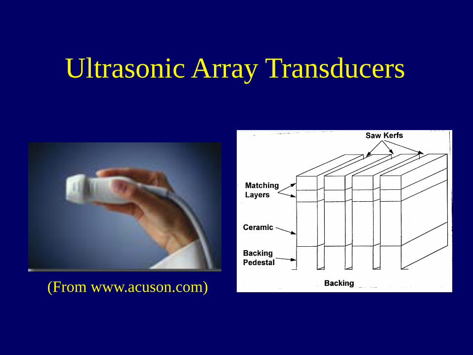
Ultrasonic Array Transducers
(From www.acuson.com)

Generic Ultrasonic Imaging System
• Receiver: – Programmable apodization, delay control and
frequency control. – Arbitrary receive direction.
• Image processing: – Pre-detection filtering. – Post-detection filtering.
• Scan converter: various scan format.
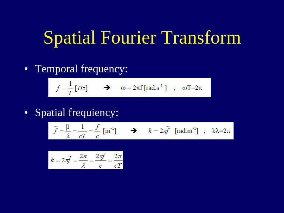
Spatial Fourier Transform • Temporal frequency:
• Spatial frequiency:
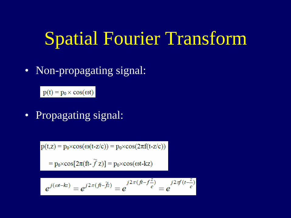
Spatial Fourier Transform • Non-propagating signal:
• Propagating signal:
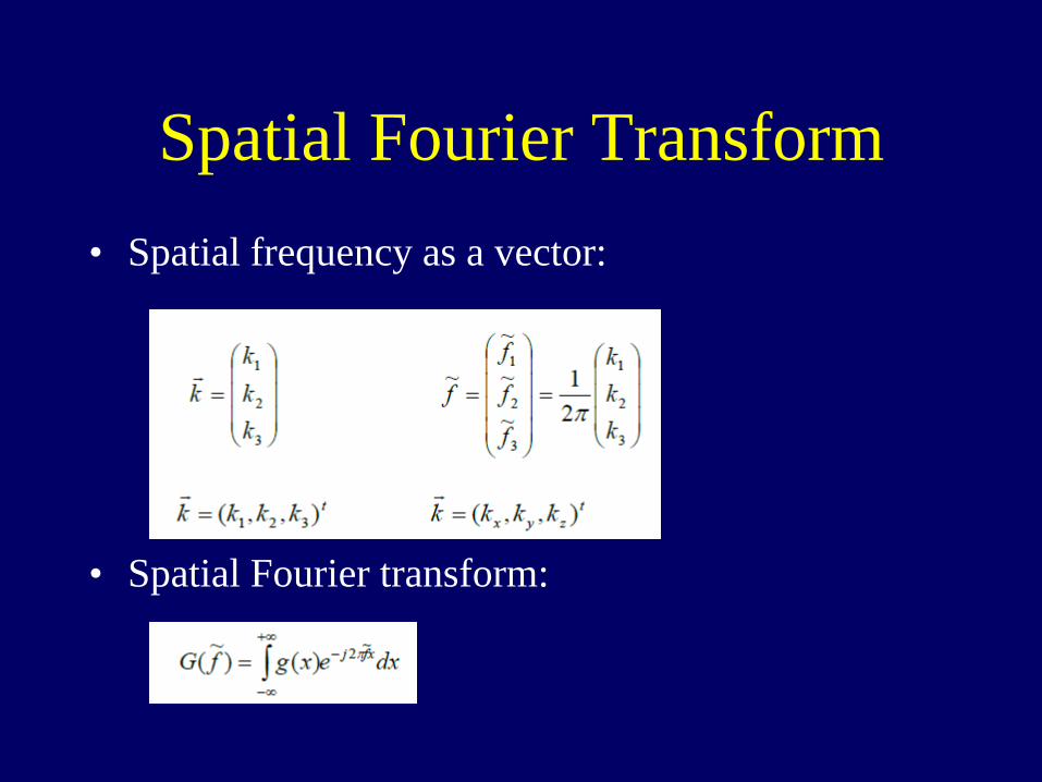
Spatial Fourier Transform • Spatial frequency as a vector:
• Spatial Fourier transform:
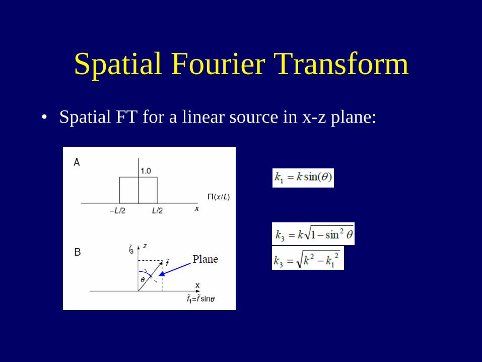
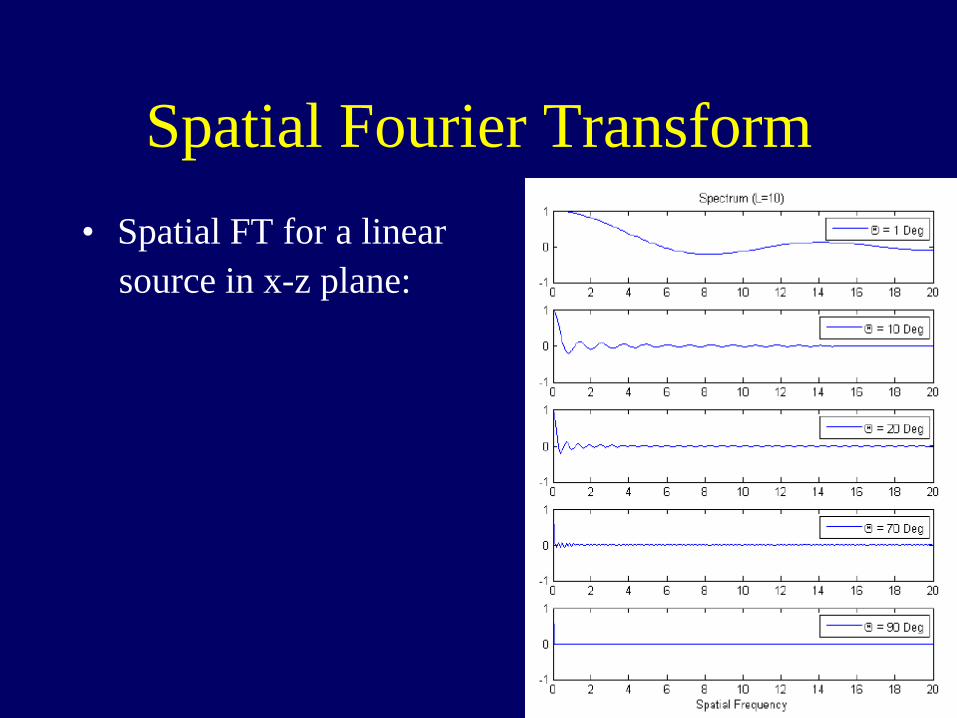
Spatial Fourier Transform • Spatial FT for a linear source in x-z plane:
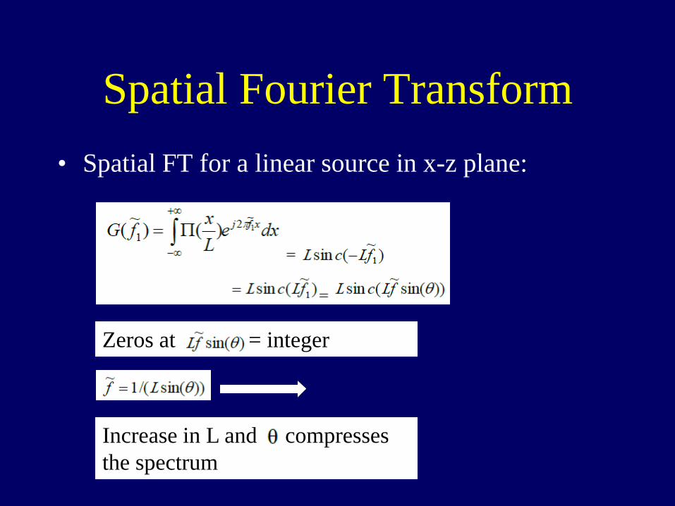
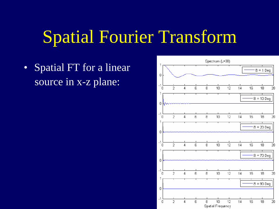
Spatial Fourier Transform • Spatial FT for a linear source in x-z plane:
Zeros at = integer
Increase in L and compresses the spectrum
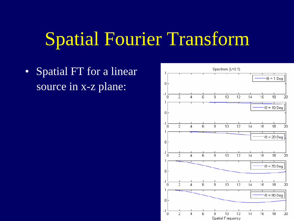
Spatial Fourier Transform • Spatial FT for a linear source in x-z plane:
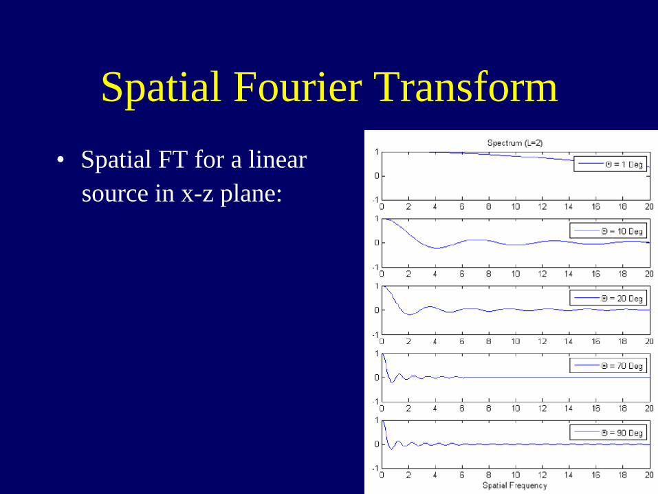
Spatial Fourier Transform • Spatial FT for a linear source in x-z plane:

Spatial Fourier Transform • Spatial FT for a linear source in x-z plane:
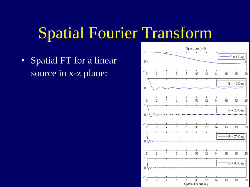
Spatial Fourier Transform • Spatial FT for a linear source in x-z plane:
Spatial Fourier Transform • Spatial FT for a linear source in x-z plane:
Spatial Fourier Transform • Spatial FT for a linear source in x-z plane: