Embed Size (px)
Citation preview
296 Med J Malaysia Vol 71 No 5 October 2016
SUMMARYPrimary cutaneous T-cell lymphoma gamma-delta subtype isan extremely rare entity of all the cutaneous T-celllymphomas. Our case provides an insight on clinicalbehavior and treatment response with feasible effectivecombination chemotherapy. We believe this will be of greatinterest to clinicians when facing this difficult clinical entity.We present a case of a 66-year-old Malay man with a three-week history of rapidly growing skin nodules and plaqueswhich spread throughout his body. He was commenced oncombination chemotherapy gemcitabine, etoposide, andcarboplatin with near complete remission on completion ofsecond cycle but he defaulted. He relapsed within a monthand he progressed despite treatment with the same regime.He was salvaged with fludarabine, cytarabine, andvinblastine combination chemotherapy but progressed withbrain metastasis and died. However, more investigationsand studies need to be done in this relatively unknown rareentity. A rare lymphoma registry might be of help to betterunderstand and treat similar conditions.
KEY WORDS:Cutaneous lymphoma, Gamma delta subtype, T cell lymphoma
INTRODUCTIONPrimary cutaneous gamma-delta (γδ) subtype is alymphoma composed of a clonal proliferation of mature,activated γδ T-cells with a cytotoxic phenotype. It isextremely rare and represents approximately 1% of allcutaneous T-cell lymphomas.1 In our own cohort, allcutaneous T-cell lymphoma comprises only 2% of our totalNon Hodgkin Lymphoma population. We report a case ofprimary cutaneous T cell lymphoma (gamma delta subtype)which is probably the first reported case in Malaysia.
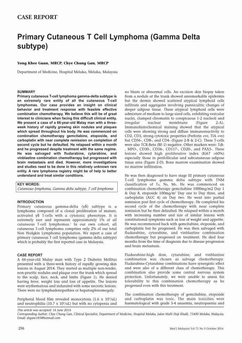
CASE REPORTA 66-year-old Malay man with Type 2 Diabetes Mellituspresented with a three-week history of rapidly growing skinlesions in August 2014. They started as multiple non-tender,non-pruritic nodules and plaque over the trunk which spreadto the scalp, face, neck, and limbs (Figure 1). He deniedhaving fever, weight loss and loss of appetite. The lesionswere erythematous and indurated with some necrotic lesions.There were no lymphadenopathies or hepatosplenomegaly.
Peripheral blood film revealed monocytosis (1.6 x 10³/uL)and neutrophilia (10.7 x 10³/uL) but with no cytopenia and
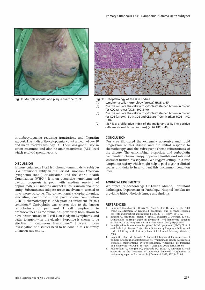
no blasts or abnormal cells. An excision skin biopsy takenfrom a nodule at the trunk showed unremarkable epidermisbut the dermis showed scattered atypical lymphoid cellsinfiltrate and aggregates involving panniculitic changes ofdeeper adipose tissue. These atypical lymphoid cells wereadmixture of medium to large sized cells, exhibiting vesicularnuclei, clumped chromatin to conspicuous 1-2 nucleoli andirregular nuclear membrane (Figure 2-A).Immunohistochemical staining showed that the atypicalcells were showing strong and diffuse immunoreactivity toCD2, CD3, strong cytotoxic properties (Perforin +ve, TIA +ve)but CD56-, CD8-, and CD4- (Figure 2-B & 2-C). These T-cellswere also TCR-Beta (BF-1) negative. Other markers were: Tdt-, MPO-, CD30-, CD34-, CD117-, CD20-, and PAX5-. Theselesions showed high proliferative index (Ki67 >60%)especially those in perifollicular and subcutaneous adiposetissue area (Figure 2-D). Bone marrow examination showedno marrow infiltration.
He was then diagnosed to have stage III primary cutaneousT-cell lymphoma gamma delta subtype with TNMclassification of T4, N0, M0. He was commenced oncombination chemotherapy gemcitabine 1000mg/m2 Day 1& Day 8, etoposide 100mg/m2 Day one to Day three, andcarboplatin (AUC 4) on Day two. He went into partialremission post first cycle of chemotherapy. He completed hissecond cycle of the chemotherapy with near completeremission but he then defaulted. He relapsed within a monthwith increasing number and size of similar lesions withconstitutional symptoms such as loss of weight and appetite.He was recommenced back with gemcitabine, etoposide, andcarboplatin but he progressed. He was then salvaged withfludarabine, cytarabine, and vinblastine combinationchemotherapy but progressed on treatment. He died fourmonths from the time of diagnosis due to disease progressionand brain metastasis.
Fludarabine-high dose, cytarabine, and vinblastinecombination was chosen as salvage chemotherapy.Fludarabine-Cytarabine combinations have synergistic effectand were also of a different class of chemotherapy. Thiscombination also provide some central nervous systemprotection. Unfortunately, we were unable to assess histolerability to this combination chemotherapy as heprogressed even with this treatment.
The combination chemotherapy of gemcitabine, etoposideand carboplatin was toxic. The main toxicities werehaematological with grade 3-4 anaemia, neutropaenia and
Primary Cutaneous T Cell Lymphoma (Gamma Deltasubtype)
Yong Khee Guan, MRCP, Chye Chung Gan, MRCP
Department of Medicine, Hospital Melaka, Melaka, Malaysia
CASE REPORT
This article was accepted: 16 June 2016Corresponding Author: Chye Chung Gan, Clinical Specialist, Department of Medicine, Hospital Melaka, Jalan Mufti Haji Khalil, 75400 Melaka, Malaysia. Email: [email protected]
16-Primary Cutaneous00012_3-PRIMARY.qxd 1/4/17 2:00 PM Page 296
Primary Cutaneous T Cell Lymphoma (Gamma Delta subtype)
Med J Malaysia Vol 71 No 5 October 2016 297
thrombocytopaenia requiring transfusions and filgrastimsupport. The nadir of the cytopaenia was at a mean of day 10and mean recovery was day 14. There was grade 1 rise inserum creatinine and alanine aminotransferase (ALT) levelwhich resolved spontaneously.
DISCUSSIONPrimary cutaneous T cell lymphoma (gamma delta subtype)is a provisional entity in the Revised European AmericanLymphoma (REAL) classification and the World HealthOrganization (WHO).1 It is an aggressive lymphoma andoverall prognosis is poor with median survival ofapproximately 15 months1 and not much is known about theentity. Subcutaneous adipose tissue involvement seemed tohave worse outcome. The conventional cyclophosphamide,vincristine, doxorubicin, and prednisolone combination(CHOP) chemotherapy is inadequate as treatment for thiscondition.2,3 Carboplatin was chosen due to the knownrefractoriness of peripheral T cell lymphoma toanthracyclines.3 Gemcitabine has previously been shown tohave better efficacy in T cell Non Hodgkin Lymphoma andbetter tolerability in the elderly.2 Etoposide is known to beeffective in cutaneous lymphoma.4,5 However, moreinvestigation and studies need to be done in this relativelyunknown rare entity.
CONCLUSIONOur case illustrated the extremely aggressive and rapidprogression of this disease and the initial response tochemotherapy and the subsequent chemo-refractoriness ofthe disease. The gemcitabine, etoposide, and carboplatincombination chemotherapy appeared feasible and safe andwarrants further investigation. We suggest setting up a rarelymphoma registry which might help to pool together clinicalcourse and data to help to treat this uncommon conditionlater.
ACKNOWLEDGEMENTSWe gratefully acknowledge Dr Faizah Ahmad, ConsultantPathologist, Department of Pathology, Hospital Melaka forproviding histopathologic image and findings.
REFERENCES1. Campo E, Swerdlow SH, Harris NL, Pileri S, Stein H, Jaffe ES. The 2008
WHO classification of lymphoid neoplasms and beyond: evolvingconcepts and practical applications. Blood. 2011; 117(19): 5019-32.
2. Zinzani PL, Venturini F, Stefoni V, Fina M, Pellegrini C, Derenzini E, et al.Gemcitabine as single agent in pretreated T-cell lymphoma patients:evaluation of the long-term outcome. Ann Oncol. 2010; 21(4): 860-3.
3. Vose JM, editor International Peripheral T-Cell Lymphoma (PTCL) Clinicaland Pathologic Review Project: Poor Outcome by Prognostic Indices andLack of Efficacy with Anthracyclines. ASH Annual Meeting Abstracts;2005.
4. Isogai R, Fukao M, Kawada A. Successful treatment for recurrence ofprimary cutaneous anaplastic large-cell lymphoma in elderly patient withetoposide, mitoxantrone, cyclophosphamide, vincristine, prednisoloneand bleomycin (VNCOP-B) therapy. J Dermatol. 2007; 34(8): 556-60.
5. Rijlaarsdam JU, Huijgens PC, Beljaards RC, Bakels V, Willemze R. Oraletoposide in the treatment of cutaneous large-cell lymphomas. Apreliminary report of four cases. Br J Dermatol. 1992; 127(5): 524-8.
Fig. 1: Multiple nodules and plaque over the trunk. Fig. 1: Histopathology of the skin nodule.(A) Lymphoma cells morphology (arrows) (H&E, x 60)(B) Positive cells are the cells with cytoplasm stained brown in colour
for CD2 (arrows) (CD2+ IHC, x 40) (C) Positive cells are the cells with cytoplasm stained brown in colour
for CD3 (arrows). Both CD2 and CD3 are T Cell Markers (CD3+ IHC,x 40)
(D) Ki67 is a proliferative index of the malignant cells. The positivecells are stained brown (arrows) (Ki 67 IHC, x 40)
16-Primary Cutaneous00012_3-PRIMARY.qxd 1/4/17 2:00 PM Page 297