Embed Size (px)
Citation preview
ORIGINAL RESEARCH
Cutaneous CD8+ Cytotoxic T-Cell LymphomaInfiltrates: Clinicopathological Correlationand Outcome of 35 Cases
Marion Wobser . Theresa Reinartz . Sabine Roth . Matthias Goebeler .
Andreas Rosenwald . Eva Geissinger
Received: May 9, 2016 / Published online: August 18, 2016� The Author(s) 2016. This article is published with open access at Springerlink.com
ABSTRACT
Introduction: Cytotoxic CD8? T-cell
lymphomas are only rarely encountered and
thus remain only poorly characterized. Our aim
was to collect and correlate clinical and
histological data of CD8? skin lymphoma
infiltrates to obtain a proper subtype
assignment of CD8? skin lymphoma
infiltrates and to derive putative prognostic
markers thereof.
Methods: Formalin-fixed and
paraffin-embedded (FFPE) tissue of 35 patients
with CD8? cytotoxic cutaneous T-cell
lymphoma infiltrates was retrieved from the
archives of the Institute of Pathology and the
Department of Dermatology, University
Hospital Wuerzburg, dating back from 1998
until 2015. Cytological, histological,
immunohistochemical and molecular genetic
features were assessed and correlated with
respective clinical data.
Results: The identified cases of CD8? cytotoxic
atypical lymphoproliferative infiltrates of the
skin (n = 35) comprised 13 cases of mycosis
fungoides (MF)/Sezary syndrome (SS), 4 cases of
subcutaneous panniculitis-like T-cell
lymphoma (SPTCL), 5 cases of primary
cutaneous acral CD8? lymphoma [formerly
indolent CD8? lymphoid proliferation (ILP)]
and 1 case of aggressive epidermotropic primary
cutaneous T-cell lymphoma (AECTCL).
Moreover, nine cases were classified as primary
cutaneous peripheral T-cell lymphoma, not
otherwise specified (PTCL-NOS) and three
cases as systemic PTCL-NOS. Multiple skin
lesions, a high proliferative index and
especially a final subtype attribution to
AECTCL or systemic PTCL-NOS were
associated with a worse survival. Coexpression
of CD68 by tumor cells was exclusively observed
in indolent acral CD8? T-cell lymphoma and
thus indicated an invariably benign clinical
course. No further distinctive markers could be
derived from our analysis.
Enhanced content To view enhanced content for thisarticle go to http://www.medengine.com/Redeem/A4E4F0600276E635.
M. Wobser (&) � T. Reinartz � M. GoebelerDepartment of Dermatology, University HospitalWurzburg, Wurzburg, Germanye-mail: [email protected]
S. Roth � A. Rosenwald � E. GeissingerInstitute of Pathology and Comprehensive CancerCenter Mainfranken, University of Wurzburg,Wurzburg, Germany
Oncol Ther (2016) 4:199–210
DOI 10.1007/s40487-016-0026-y
Conclusion: Cutaneous infiltrates of CD8?
cytotoxic T-cell lymphoma comprise clinically
and histologically heterogeneous entities of
either primary cutaneous T-cell lymphomas or
secondary infiltrates of otherwise systemic
peripheral T-cell lymphomas. A thorough
clinicopathological correlation with respective
staging examinations remains the mainstay for
correct subtype assignment and proper
prognostication as long as no better markers
have been defined.
Keywords: Cutaneous lymphomas; Cytotoxic;
Histology; Prognosis
INTRODUCTION
Primary cutaneous T-cell lymphomas comprise
heterogeneous entities with diverse histological,
phenotypic and molecular genetic features
dependent on the respective cell of origin [1].
Most T-cell lymphomas of the skin exhibit a
skin-homing CD4? T-helper cell phenotype.
CD8? cutaneous lymphomas usually represent
rare CD8? variants of otherwise common and
well-characterized CD4? lymphomas, such as
CD8? variants of mycosis fungoides (MF) and
Sezary syndrome (SS), CD30?
lymphoproliferative disorders, subcutaneous
panniculitis-like T-cell lymphoma (SPTCL) or
peripheral T-cell lymphoma, not otherwise
specified (PTCL-NOS) [2]. In contrast, an
exclusive CD8? phenotype is present in the
eponymous CD8? acral T-cell lymphoma [3]
formally known as CD8? indolent lymphoid
proliferation (ILP) [4] as well as in aggressive
epidermotropic cutaneous T-cell lymphoma
(AECTCL) [5].
Such rare CD8? cytotoxic cutaneous
lymphomas often confront the
dermatopathologist with intricate diagnostic
workup and, moreover, may represent a
therapeutic dilemma for the treating
dermatologist. We therefore systematically
collected all appropriately recorded CD8?
cytotoxic lymphoma infiltrates of the skin
being encountered at our institution, spanning
a time period of more than 15 years. Our
intention was to better characterize such CD8?
lymphoma infiltrates based on histological,
immunophenotypical and clinical grounds, and
ultimately we tried to provide a better subtype
attribution and to delineate putative diagnostic
and/or prognostic markers to guide the patient
management of this rare lymphoma variant.
METHODS
Formalin-fixed and paraffin-embedded (FFPE)
tissue of patients with EBV-negative cutaneous
CD8? cytotoxic cutaneous T-cell lymphoma
infiltrates was retrieved from the archives of the
Institute of Pathology, University of Wuerzburg,
and the Department of Dermatology, University
Hospital Wuerzburg, dating back from 1998
until 2015. Only cases with sufficient analyzable
FFPE tissue and corresponding clinical data were
included for further analysis. The final diagnosis
of lymphoma with subtype assignment was
based on a compatible histomorphology,
immunophenotype and clonal T-cell receptor
gene rearrangement in close correlation with
the medical history and clinical presentation
according to the current World Health
Organization (WHO)/European Organization
for Research and Treatment of Cancer
(EORTC) classification [1] taking account of
the revised WHO proposal [3].
Immunohistochemical studies were performed
on FFPE tissue sections using the
avidin–biotin-peroxidase complex method and
stained in an autostainer in the case of the
200 Oncol Ther (2016) 4:199–210
routine antibody panel and manually for
selected remaining antibodies, such as PIM1,
VEGFR2 and PDGFRa. Polymerase chain
reaction (PCR) analysis of the TCR-c gene was
performed on DNA extracted from FFPE tissue
according to the previously published protocol
of the Biomed-2 guidelines. Statistical analysis
of histological and clinical data was performed
with the SPSS software version 22 (IBM GmbH,
Germany).
All procedures followed were in accordance
with the ethical standards of the responsible
committee on human experimentation
(institutional and national) and with the
Helsinki Declaration of 1964, as revised in
2013. Informed consent was obtained from all
patients for being included in the study and for
publication of the patient photographs.
RESULTS
During a 17-year time period, 35 cases could be
identified that fulfilled our inclusion criteria;
more than 30 cases had to be excluded because
of lack of adequate tissue and/or clinical data.
Clinicopathological correlation yielded the
diagnosis of MF/SS in 13 cases, SPTCL in 4
cases, CD8? acral lymphoma in 5 cases and
AECTCL in 1 case. Moreover, 9 out of 35 cases
were classified as primary cutaneous PTCL-NOS,
while secondary lymphoma infiltrates of the
skin due to underlying systemic PTCL-NOS were
encountered in 3 cases. Detailed clinical patient
characteristics are summarized in Table 1 and
histological and immunophenotypic findings
in Tables 2 and 3, respectively. Survival data are
shown in Fig. 1a.
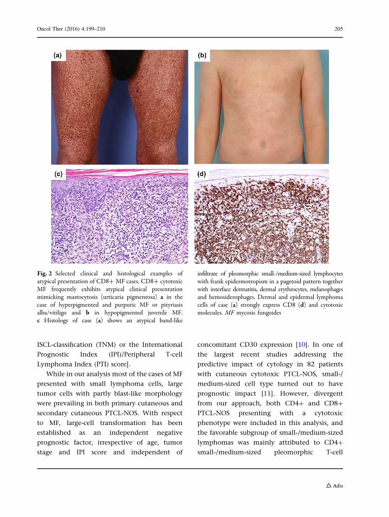
With respect to CD8? MF, atypical clinical
features mimicking cutaneous mastocytosis or
purpura pigmentosa (Fig. 2a) and pityriasis alba
(Fig. 2b) commonly diverted the clinician from
a prompt initial diagnosis. The corresponding
histological pattern was otherwise rather similar
to classical CD4? MF (Fig. 2c,d). Mean overall
survival of patients with CD8? MF was
56 months with a corresponding wide range of
12 to 159 months depending on the lymphoma
stage. The patients with CD8? MF showed a
clinical course quite similar to otherwise
encountered CD4? variants; one of these
patients died because of progressive lymphoma
after 12 months.
CD8? lymphomas (n = 21) with deep
(dermal or subcutaneous) infiltrates mainly
comprised rare and hitherto provisional
entities designated as lymphoma subtypes.
Five of these cases belonged to the provisional
entity of acral CD8? T-cell lymphoma (ILP) and
invariably showed an indolent course with
unrestricted survival. The remaining 16 cases
exhibited a significantly worse survival and
were diagnosed as either primary (n = 9) or
secondary (n = 3) cutaneous PTCL-NOS or as
SPTCL (n = 4). These patients exhibited a mean
overall survival of 51 ± 66, 24 ± 17 and
27 ± 43 months, respectively. Of this subgroup
5/16 patients died of lymphoma (among these
all three patients with secondary PTCL-NOS),
whereas an additional 4/16 patients died of
other unrelated causes. All 16 of these patients
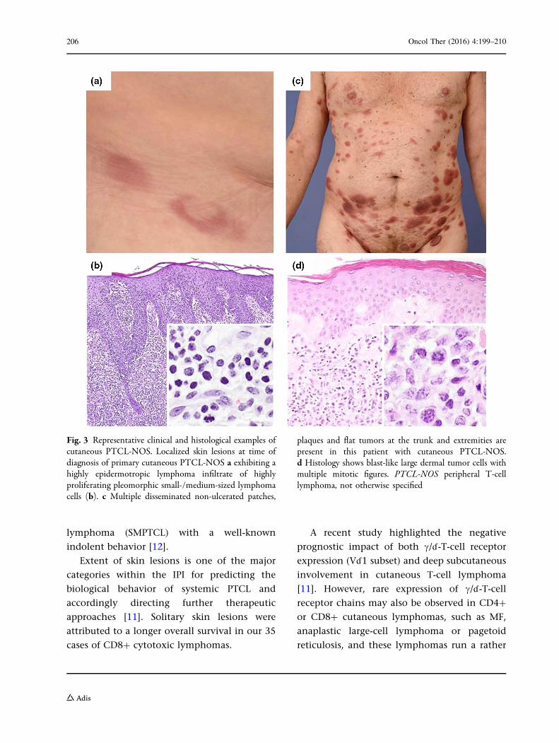
presented with either solitary/regional (n = 8;
Fig. 3a) or multiple (n = 8) tumors and/or
infiltrated plaques (Fig. 3c). Those patients
with a solitary skin lesion of their PTCL-NOS
or SPTCL had a trend to better mean overall
survival than patients with multiple skin
manifestations (61 ± 65 vs. 21 ± 23 months;
P = 0.1), albeit exhibiting a wide variation of
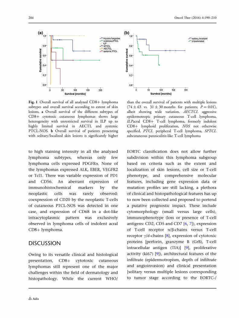
survival time (Fig. 1b). Solitary manifestation at
an acral site (face and finger) represented an
independent positive prognostic factor within
the patient subgroup of PTCL-NOS with an
average overall survival of 112 months. Better
Oncol Ther (2016) 4:199–210 201
survival for patients with solitary skin
manifestation at the time of initial
presentation was not only observed for this
subgroup of deep dermal/subcutanous CD8?
lymphoma infiltrates, but was also true for all
studied cases of CD8? lymphomas: overall
survival was 74 ± 63 months for patients with
localized disease manifestation as compared to
31 ± 30 months for patients exhibiting multiple
lesions (P = 0.01).
In addition to clinical features, we were also
interested in thorough histological and
immunohistological characterization of our
selected CD8? lymphomas. Histological
features and further immunophenotypic
characterization are depicted in Tables 2 and 3.
Whereas most of the MF cases showed small-
and medium-sized neoplastic cells with rare
large-cell transformation and systemic
PTCL-NOS showed large neoplastic cells, cell
size was otherwise rather evenly distributed
among the different entities (Fig. 3b, d).
Hence, survival rates were not significantly
different between lymphomas with
predominantly small- or medium-sized cells
and lymphomas with large-cell morphology
(P = 0.6). Moreover, ulceration, angiocentricity
and adnexotropism were a common feature of
PTCL-NOS but not of indolent acral CD8?
lymphoma.
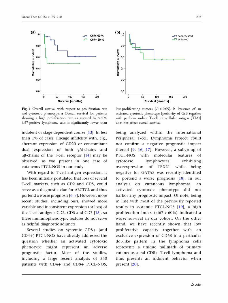
With regard to immunohistochemical
features, a high proliferation index
(ki67[60%) implied a worse mean overall
survival (P\0.05; Fig. 4a). Loss of one or
several of the T-cell antigens (CD5 and CD7),
overexpression of PIM1, PDGFRa or an activated
cytotoxic phenotype (positivity of GrB in
conjunction with variable expression of T-cell
intracellular antigen-1 [TIA] or perforin; Fig. 4b)
were not associated with a more aggressive
subtype or a worse clinical course. To note,
PIM1 expression was observed with a moderate
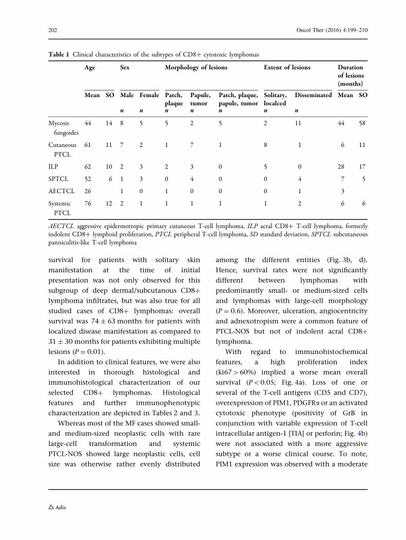
Table 1 Clinical characteristics of the subtypes of CD8? cytotoxic lymphomas
Age Sex Morphology of lesions Extent of lesions Durationof lesions(months)
Mean SO Male Female Patch,plaque
Papule,tumor
Patch, plaque,papule, tumor
Solitary,localced
Disseminated Mean SO
n n n n n n n
Mycosis
fungoides
44 14 8 5 5 2 5 2 11 44 58
Cutaneous
PTCL
61 11 7 2 1 7 1 8 1 6 11
ILP 62 10 2 3 2 3 0 5 0 28 17
SPTCL 52 6 1 3 0 4 0 0 4 7 5
AECTCL 26 1 0 1 0 0 0 1 3
Systemic
PTCL
76 12 2 1 1 1 1 1 2 6 6
AECTCL aggressive epidermotropic primary cutaneous T-cell lymphoma, ILP acral CD8? T-cell lymphoma, formerlyindolent CD8? lymphoid proliferation, PTCL peripheral T-cell lymphoma, SD standard deviation, SPTCL subcutaneouspanniculitis-like T-cell lymphoma
202 Oncol Ther (2016) 4:199–210
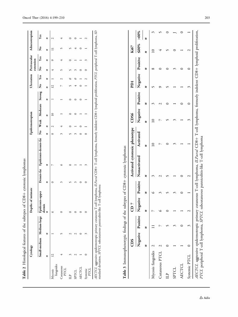
Table2
Histologicalfeatures
ofthesubtypes
ofCD8?
cytotoxiclymphom
as
Cytology
Depthsof
intimate
Epiderm
otropism
Ulceration
Perivascular
extension
Adn
exotropism
Small-medium
Medium-large
Epiderm
is-upp
erderm
isDetmis-fat
Epiderm
is-dermis-fat
No
Weak
Mod
erate
Strong
No
Yes
No
Yes
No
Yes
nn
nn
nn
nn
nn
nn
nn
n
Mycosis
fungoides
121
60
70
210
l12
14
911
2
Cutaneous
PTCL
45
03
63
41
17
25
45
4
ILP
32
05
05
00
05
05
05
0
SPTCL
22
03
13
10
04
03
14
0
AECTCL
10
00
10
01
00
10
10
1
System
icPT
CL
03
00
30
I1
11
20
31
2
AECTCLaggressive
epidermotropicprim
arycutaneousT-celllymphom
a,ILPa
cral
CD8?
T-celllymphom
a,form
erlyindolent
CD8?
lymphoidproliferation,P
TCLperipheralT-celllymphom
a,SD
standard
deviation,
SPTCLsubcutaneous
pann
iculitis-like
T-celllymphom
a
Table3
Immun
ophenotypicfin
dingsof
thesubtypes
ofCD8?
cytotoxiclymphom
as
CDS
CD
7Activated
cytotoxicph
enotype
CD56
PD1
Ki67
Negative
Positive
Negative
Positive
Non
activated
Activated
Negative
Positive
Negative
Positive
£60%
>60%
nn
nn
nn
nn
nn
nn
Mycosisfungoides
211
76
310
103
58
103
Cutaneous
PTCL
27
63
27
72
90
45
ILP
05
05
50
50
50
50
SPTCL
13
13
13
31
13
0J
AECTCL
10
10
01
10
10
10
System
icPT
CL
03
21
21
30
30
21
AECTCLaggressive
epidermotropicprim
arycutaneousT-celllymphom
a,ILPa
cral
CD8?
T-celllymphom
a,form
erly
indolent
CD8?
lymphoidproliferation
,PT
CLperipheralT-celllymphom
a,SP
TCLsubcutaneous
pann
iculitis-like
T-celllymphom
a
Oncol Ther (2016) 4:199–210 203
to high staining intensity in all the analyaed
lymphoma subtypes, whereas only few
lymphoma cells expressed PDGFRa. None of
the lymphomas expressed ALK, EBER, VEGFR2
or Tcl1. There was variable expression of PD1
and CD56. An aberrant expression of
immunohistochemical markers by the
neoplastic cells was rarely observed:
coexpression of CD20 by the neoplastic T-cells
of cutaneous PTCL-NOS was detected in one
case, and expression of CD68 in a dot-like
intracytoplasmic pattern was exclusively
observed in lymphoma cells of indolent acral
CD8? lymphoma.
DISCUSSION
Owing to its versatile clinical and histological
presentation, CD8? cytotoxic cutaneous
lymphomas still represent one of the major
challenges within the field of dermatology and
histopathology. While the current WHO/
EORTC classification does not allow further
subdivision within this lymphoma subgroup
based on criteria such as the extent and
localization of skin lesions, cell size or T-cell
phenotype, and comprehensive molecular
features, including gene expression data or
mutation profiles are still lacking, a plethora
of clinical and histopathological features has up
to now been collected and proposed to portend
a putative prognostic impact. These include
cytomorphology (small versus large cells),
immunophenotype (loss or presence of T-cell
antigens: CD2, CD5 and CD7 [6, 7]), expression
of T-cell receptor a/b-chains versus T-cell
receptor c/ë-chains [8], expression of cytotoxic
proteins [perforin, granzyme B (GrB), T-cell
intracellular antigen (TIA)] [9], proliferative
activity (ki67) [9]), architectural features of the
infiltrate (epidermotropism, depth of infiltrate
and angioinvasion) and clinical presentation
[solitary versus multiple lesions corresponding
to tumor stage according to the EORTC-/
Fig. 1 Overall survival of all analyzed CD8? lymphomasubtypes and overall survival according to extent of skinlesions. a Overall survival of the different subtypes ofCD8? cytotoxic cutaneous lymphomas shows largeheterogeneity with unrestricted survival in ILP up tohighly limited survival in AECTL and systemicPTCL-NOS. b Overall survival of patients presentingwith solitary/localized skin lesions is significantly higher
than the overall survival of patients with multiple lesions(74 ± 63 vs. 31 ± 30 months for patients; P = 0.01),albeit showing wide variation. AECTCL aggressiveepidermotropic primary cutaneous T-cell lymphoma,ILPacral CD8? T-cell lymphoma, formerly indolentCD8? lymphoid proliferation, NOS not otherwisespecified, PTCL peripheral T-cell lymphoma, SPTCLsubcutaneous panniculitis-like T-cell lymphoma
204 Oncol Ther (2016) 4:199–210
ISCL-classification (TNM) or the International
Prognostic Index (IPI)/Peripheral T-cell
Lymphoma Index (PTI) score].
While in our analysis most of the cases of MF
presented with small lymphoma cells, large
tumor cells with partly blast-like morphology
were prevailing in both primary cutaneous and
secondary cutaneous PTCL-NOS. With respect
to MF, large-cell transformation has been
established as an independent negative
prognostic factor, irrespective of age, tumor
stage and IPI score and independent of
concomitant CD30 expression [10]. In one of
the largest recent studies addressing the
predictive impact of cytology in 82 patients
with cutaneous cytotoxic PTCL-NOS, small-/
medium-sized cell type turned out to have
prognostic impact [11]. However, divergent
from our approach, both CD4? and CD8?
PTCL-NOS presenting with a cytotoxic
phenotype were included in this analysis, and
the favorable subgroup of small-/medium-sized
lymphomas was mainly attributed to CD4?
small-/medium-sized pleomorphic T-cell
Fig. 2 Selected clinical and histological examples ofatypical presentation of CD8?MF cases. CD8? cytotoxicMF frequently exhibits atypical clinical presentationmimicking mastocytosis (urticaria pigmentosa) a in thecase of hyperpigmented and purpuric MF or pityriasisalba/vitiligo and b in hypopigmented juvenile MF.c Histology of case (a) shows an atypical band-like
infiltrate of pleomorphic small-/medium-sized lymphocyteswith frank epidermotropism in a pagetoid pattern togetherwith interface dermatitis, dermal erythrocytes, melanophagesand hemosiderophages. Dermal and epidermal lymphomacells of case (a) strongly express CD8 (d) and cytotoxicmolecules. MF mycosis fungoides
Oncol Ther (2016) 4:199–210 205
lymphoma (SMPTCL) with a well-known
indolent behavior [12].
Extent of skin lesions is one of the major
categories within the IPI for predicting the
biological behavior of systemic PTCL and
accordingly directing further therapeutic
approaches [11]. Solitary skin lesions were
attributed to a longer overall survival in our 35
cases of CD8? cytotoxic lymphomas.
A recent study highlighted the negative
prognostic impact of both c/ë-T-cell receptor
expression (Vë1 subset) and deep subcutaneous
involvement in cutaneous T-cell lymphoma
[11]. However, rare expression of c/ë-T-cell
receptor chains may also be observed in CD4?
or CD8? cutaneous lymphomas, such as MF,
anaplastic large-cell lymphoma or pagetoid
reticulosis, and these lymphomas run a rather
Fig. 3 Representative clinical and histological examples ofcutaneous PTCL-NOS. Localized skin lesions at time ofdiagnosis of primary cutaneous PTCL-NOS a exhibiting ahighly epidermotropic lymphoma infiltrate of highlyproliferating pleomorphic small-/medium-sized lymphomacells (b). c Multiple disseminated non-ulcerated patches,
plaques and flat tumors at the trunk and extremities arepresent in this patient with cutaneous PTCL-NOS.d Histology shows blast-like large dermal tumor cells withmultiple mitotic figures. PTCL-NOS peripheral T-celllymphoma, not otherwise specified
206 Oncol Ther (2016) 4:199–210
indolent or stage-dependent course [13]. In less
than 1% of cases, lineage infidelity with, e.g.,
aberrant expression of CD20 or concomitant
dual expression of both c/ë-chains and
ab-chains of the T-cell receptor [14] may be
observed, as was present in one case of
cutaneous PTCL-NOS in our study.
With regard to T-cell antigen expression, it
has been initially postulated that loss of several
T-cell markers, such as CD2 and CD5, could
serve as a diagnostic clue for AECTCL and thus
portend a worse prognosis [6, 7]. However, more
recent studies, including ours, showed more
variable and inconsistent expression (or loss) of
the T-cell antigens CD2, CD5 and CD7 [15], so
these immunophenotypic features do not serve
as helpful diagnostic adjuncts.
Several studies on systemic CD8? (and
CD4?) PTCL-NOS have already addressed the
question whether an activated cytotoxic
phenotype might represent an adverse
prognostic factor. Most of the studies,
including a large recent analysis of 340
patients with CD4? and CD8? PTCL-NOS,
being analyzed within the International
Peripheral T-cell Lymphoma Project could
not confirm a negative prognostic impact
thereof [9, 16, 17]. However, a subgroup of
PTCL-NOS with molecular features of
cytotoxic lymphocytes exhibiting
overexpression of TBX21 while being
negative for GATA3 was recently identified
to portend a worse prognosis [18]. In our
analysis on cutaneous lymphomas, an
activated cytotoxic phenotype did not
harbor any prognostic impact. Of note, being
in line with most of the previously reported
results in systemic PTCL-NOS [19], a high
proliferation index (ki67[60%) indicated a
worse survival in our cohort. On the other
hand, we have recently shown that low
proliferative capacity together with an
exclusive expression of CD68 in a particular
dot-like pattern in the lymphoma cells
represents a unique hallmark of primary
cutaneous acral CD8? T-cell lymphoma and
thus presents an indolent behavior when
present [20].
Fig. 4 Overall survival with respect to proliferation rateand cytotoxic phenotype. a Overall survival for patientsshowing a high proliferation rate as assessed by [60%ki67-positive lymphoma cells is significantly lower than
low-proliferating tumors (P\0.05). b Presence of anactivated cytotoxic phenotype [positivity of GrB togetherwith perforin and/or T-cell intracellular antigen (TIA)]does not affect overall survival
Oncol Ther (2016) 4:199–210 207
With the aim to gain further insight into the
pathogenesis and the biological behavior of rare
cutaneous lymphomas, such as CD8? cytotoxic
lymphomas, much can be learned and
transferred from recent work on molecular
profiling of systemic PTCL-NOS, including
gene expression and deep sequencing analysis
[19, 21, 22]. Gene expression profiling has once
again confirmed that within the generic term of
systemic PTCL-NOS, there is extensive
molecular heterogeneity as already evidenced
by divergent clinical, histological and
immunophenotypical data [18, 23, 24].
Moreover, taking the limited therapeutic
opportunities and the often dismal prognosis
of PTCL-NOS into consideration, the progress in
deciphering the mutational landscape and the
subtype-specific gene expression profiles has
revealed several novel therapeutic options [25].
Putative targets include the network of
epigenetic modifiers [26] as well as the NFjB,
STAT and JAK pathways [27], PIM kinases [27]
and downstream signaling of tyrosine receptors,
such as PDGFRa [28] and VEGFR [29]. In the
lymphoma cases presented here, the expression
pattern of PIM1 (which was actually present in
almost all cases), VEGFR or PDGFRa (which
were almost altogether absent in our cohort) did
not serve as putative prognostic markers, so that
these data could not be recapitulated in our
CD8? cutaneous lymphoma cohort.
CONCLUSIONS
Our retrospective analysis once again underlines
that when dealing with CD8? cutaneous
lymphoma, the crucial approach still remains to
unify the histological and clinical data to make a
correct diagnosis with prognostically relevant
subtype attribution as long as better
immunophenotypical and especially genetic
data are still lacking. In the future, molecular
profiling of such rare lymphoma variants will
hopefully contribute to ameliorate treatment
strategies as a result of more precise subtype
definition and to develop more individualized
treatment strategies, including targeted
therapies. As cutaneous CD8? lymphomas
represent rare entities and our study data are
therefore limited in its conclusions, a broader
multi-institutional approach for these lymphoma
entities is urgently warranted in the future.
ACKNOWLEDGMENTS
No funding or sponsorship was received for this
study or publication of this article. All named
authors meet the International Committee of
Medical Journal Editors (ICMJE) criteria for
authorship for this manuscript, take
responsibility for the integrity of the work as a
whole and have given final approval for the
version to be published.
Disclosures. Marion Wobser, Theresa
Reinartz, Sabine Roth, Matthias Goebeler,
Andreas Rosenwald and Eva Geissinger have
nothing to disclose.
Compliance with Ethics Guidelines. All
procedures followed were in accordance with
the ethical standards of the responsible
committee on human experimentation
(institutional and national) and with the
Helsinki Declaration of 1964, as revised in
2013. Informed consent was obtained from all
patients for being included in the study and for
publication of the patient photographs.
Open Access. This article is distributed
under the terms of the Creative Commons
Attribution-NonCommercial 4.0 International
208 Oncol Ther (2016) 4:199–210
License (http://creativecommons.org/licenses/
by-nc/4.0/), which permits any
noncommercial use, distribution, and
reproduction in any medium, provided you
give appropriate credit to the original
author(s) and the source, provide a link to the
Creative Commons license, and indicate if
changes were made.
REFERENCES
1. Willemze R, Jaffe ES, Burg G, Cerroni L, Berti E,Swerdlow SH, et al. WHO-EORTC classification forcutaneous lymphomas. Blood. 2005;105:3768–85.
2. Lu D, Patel KA, Duvic M, Jones D. Clinical andpathological spectrum of CD8-positive cutaneousT-cell lymphomas. J Cutan Pathol. 2002;29:465–72.
3. Swerdlow SH, Campo E, Pileri SA, Harris NL, SteinH, Siebert R, et al. The 2016 revision of the WorldHealth Organization classification of lymphoidneoplasms. Blood. 2016;127:2375–90.
4. Petrella T, Maubec E, Cornillet-Lefebvre P,Willemze R, Pluot M, Durlach A, et al. IndolentCD8-positive lymphoid proliferation of the ear: adistinct primary cutaneous T-cell lymphoma? Am JSurg Pathol. 2007;31:1887–92.
5. Gormley RH, Hess SD, Anand D, Junkins-Hopkins J,Rook AH, Kim EJ. Primary cutaneous aggressiveepidermotropic CD8? T-cell lymphoma. J Am AcadDermatol. 2010;62:300–7.
6. Berti E, Tomasini D, Vermeer MH, Meijer CJ, AlessiE, Willemze R. Primary cutaneous CD8-positiveepidermotropic cytotoxic T cell lymphomas. Adistinct clinicopathological entity with anaggressive clinical behavior. Am J Pathol.1999;155:483–92.
7. Agnarsson BA, Vonderheid EC, Kadin ME.Cutaneous T cell lymphoma withsuppressor/cytotoxic (CD8) phenotype:identification of rapidly progressive and chronicsubtypes. J Am Acad Dermatol. 1990;22:569–77.
8. Toro JR, Liewehr DJ, Pabby N, Sorbara L, Raffeld M,Steinberg SM, et al. Gamma-delta T-cell phenotypeis associated with significantly decreased survivalin cutaneous T-cell lymphoma. Blood.2003;101:3407–12.
9. Weisenburger DD, Savage KJ, Harris NL, GascoyneRD, Jaffe ES, MacLennan KA, et al. Peripheral T-celllymphoma, not otherwise specified: a report of 340cases from the International Peripheral T-cellLymphoma Project. Blood. 2011;117:3402–8.
10. Scarisbrick JJ, Prince HM, Vermeer MH, Quaglino P,Horwitz S, Porcu P, et al. cutaneous lymphomainternational consortium study of outcome inadvanced stages of mycosis Fungoides and SezarySyndrome: effect of specific prognostic markers onsurvival and development of a prognostic model.Oncol: J Clin Oncol Off J Am Soc Clin; 2015.
11. Bekkenk MW, Vermeer MH, Jansen PM, van MarionAMW, Canninga-van Dijk MR, Kluin PM, et al.Peripheral T-cell lymphomas unspecifiedpresenting in the skin: analysis of prognosticfactors in a group of 82 patients. Blood.2003;102:2213–9.
12. Beltraminelli H, Leinweber B, Kerl H, Cerroni L.Primary cutaneous CD4? small-/medium-sizedpleomorphic T-cell lymphoma: a cutaneousnodular proliferation of pleomorphic Tlymphocytes of undetermined significance? Astudy of 136 cases. Am J Dermatopathol.2009;31:317–22.
13. Rodrıguez-Pinilla SM, Ortiz-Romero PL, MonsalvezV, Tomas IE, Almagro M, Sevilla A, et al. TCR-cexpression in primary cutaneous T-cell lymphomas.Am J Surg Pathol. 2013;37:375–84.
14. Tomasini D, Niccoli A, Crivelli F. Pagetoidreticulosis tumor cells with double expression ofTCRcd and TCRab: an off-target phenomenon orgenuine expression? J Cutan Pathol.2015;42:427–34.
15. Robson A, Assaf C, Bagot M, Burg G, Calonje J,Castillo C, et al. Aggressive epidermotropiccutaneous CD8? Lymphoma: a cutaneouslymphoma with distinct clinical and pathologicalfeatures Report of an EORTC CutaneousLymphoma Task Force Workshop. Histopathology.2014.
16. Went P, Agostinelli C, Gallamini A, Piccaluga PP,Ascani S, Sabattini E, et al. Marker expression inperipheral T-cell lymphoma: a proposedclinical-pathologic prognostic score. J Clin OncolOff J Am Soc Clin Oncol. 2006;24:2472–9.
17. Geissinger E, Odenwald T, Lee S-S, Bonzheim I,Roth S, Reimer P, et al. Nodal peripheral T-celllymphomas and in particular, theirlymphoepithelioid (Lennert’s) variant are oftenderived from CD8(?) cytotoxic T-cells. VirchowsArch Int J Pathol. 2004;445:334–43.
Oncol Ther (2016) 4:199–210 209
18. Iqbal J, Wright G, Wang C, Rosenwald A, GascoyneRD, Weisenburger DD, et al. Gene expressionsignatures delineate biological and prognosticsubgroups in peripheral T-cell lymphoma. Blood.2014;123:2915–23.
19. Costello R, Sanchez C, Le Treut T, Rihet P, Imbert J,Sebahoun G. Peripheral T-cell lymphoma geneexpression profiling and potential therapeuticexploitations. Br J Haematol. 2010;150:21–7.
20. Wobser M, Roth S, Reinartz T, Rosenwald A,Goebeler M, Geissinger E. CD68 expression is adiscriminative feature of indolent cutaneousCD8-positive lymphoid proliferation anddistinguishes this lymphoma subtype from otherCD8-positive cutaneous lymphomas. Br J Dermatol.2015;172:1573–80.
21. Gaulard P, de Leval L. Pathology of peripheral T-celllymphomas: where do we stand? Semin Hematol.2014;51:5–16.
22. Vose J, Armitage J, Weisenburger D. InternationalT-Cell lymphoma project. International peripheralT-cell and natural killer/T-cell lymphoma study:pathology findings and clinical outcomes. J ClinOncol Off J Am Soc Clin Oncol. 2008;26:4124–30.
23. Couronne L, Bastard C, Gaulard P, Hermine O,Bernard O. Molecular pathogenesis of peripheralT-cell lymphoma (1): angioimmunoblastic T-celllymphoma, peripheral T-cell lymphoma, nototherwise specified and anaplastic large celllymphoma. Medecine Sci MS. 2015;31:841–52.
24. Gaulard P, de Leval LVIII. New markers inperipheral T-cell lymphomas: more entities or
more confusion? Hematol Oncol. 2013;31(Suppl1):51–6.
25. Iqbal J, Wilcox R, Naushad H, Rohr J, Heavican TB,Wang C, et al. Genomic signatures in T-celllymphoma: How can these improve precision indiagnosis and inform prognosis? Blood Rev. 2015.
26. Lemonnier F, Couronne L, Parrens M, Jaıs J-P,Travert M, Lamant L, et al. Recurrent TET2mutations in peripheral T-cell lymphomascorrelate with TFH-like features and adverseclinical parameters. Blood. 2012;120:1466–9.
27. Martinez-Delgado B, Melendez B, Cuadros M,Alvarez J, Castrillo JM, Ruiz De La Parte A, et al.Expression profiling of T-cell lymphomasdifferentiates peripheral and lymphoblasticlymphomas and defines survival related genes.Clin Cancer Res Off J Am Assoc Cancer Res.2004;10:4971–82.
28. Piccaluga PP, Agostinelli C, Califano A, Rossi M,Basso K, Zupo S, et al. Gene expression analysis ofperipheral T cell lymphoma, unspecified, revealsdistinct profiles and new potential therapeutictargets. J Clin Invest. 2007;117:823–34.
29. Jørgensen JM, Sørensen FB, Bendix K, Nielsen JL,Funder A, Karkkainen MJ, et al. Expression level,tissue distribution pattern, and prognostic impactof vascular endothelial growth factors VEGF andVEGF-C and their receptors Flt-1, KDR, and Flt-4 indifferent subtypes of non-Hodgkin lymphomas.Leuk Lymphoma. 2009;50:1647–60.
210 Oncol Ther (2016) 4:199–210