Embed Size (px)
Citation preview

Vol.:(0123456789)1 3
Lung (2018) 196:231–238 https://doi.org/10.1007/s00408-018-0086-x
RESPIRATORY PHYSIOLOGY
Primary Ciliary Dyskinesia Due to Microtubular Defects is Associated with Worse Lung Clearance Index
S. Irving1,2 · M. Dixon1 · M. R. Fassad3,4 · E. Frost1,2 · J. Hayward3,5 · K. Kilpin2 · S. Ollosson2 · A. Onoufriadis6 · M. P. Patel3 · J. Scully3 · S. B. Carr2 · H. M. Mitchison3 · M. R. Loebinger2 · C. Hogg2 · A. Shoemark1,2,7 · A. Bush1,2
Received: 7 September 2017 / Accepted: 4 January 2018 / Published online: 24 January 2018 © The Author(s) 2018. This article is an open access publication
AbstractPurpose Primary ciliary dyskinesia (PCD) is characterised by repeated upper and lower respiratory tract infections, neutro-philic airway inflammation and obstructive airway disease. Different ultrastructural ciliary defects may affect lung function decline to different degrees. Lung clearance index (LCI) is a marker of ventilation inhomogeneity that is raised in some but not all patients with PCD. We hypothesised that PCD patients with microtubular defects would have worse (higher) LCI than other PCD patients.Methods Spirometry and LCI were measured in 69 stable patients with PCD. Age at testing, age at diagnosis, ethnicity, ciliary ultrastructure, genetic screening result and any growth of Pseudomonas aeruginosa was recorded.Results Lung clearance index was more abnormal in PCD patients with microtubular defects (median 10.24) than those with dynein arm defects (median 8.3, p = 0.004) or normal ultrastructure (median 7.63, p = 0.0004). Age is correlated with LCI, with older patients having worse LCI values (p = 0.03, r = 0.3).Conclusion This study shows that cilia microtubular defects are associated with worse LCI in PCD than dynein arm defects or normal ultrastructure. The patient’s age at testing is also associated with a higher LCI. Patients at greater risk of obstruc-tive lung disease should be considered for more aggressive management. Differences between patient groups may potentially open avenues for novel treatments.
Keywords Lung function · Paediatrics · Rare disease · Ciliopathy
Introduction
Primary ciliary dyskinesia (PCD) is an inherited condition characterised by repeated upper and lower respiratory tract infections and chronic secretory otitis media. Approximately 50% patients have disorders of laterality. Morbidity is signif-icant with most patients developing obstructive lung disease
Electronic supplementary material The online version of this article (https://doi.org/10.1007/s00408-018-0086-x) contains supplementary material, which is available to authorized users.
* S. Irving [email protected]
1 National Heart and Lung Institute, Imperial College London, London, UK
2 Royal Brompton & Harefield NHS Trust, Sydney Street, London SW3 6NP, UK
3 Genetics and Genomic Medicine, University College London (UCL) Great Ormond Street Institute of Child Health, London, UK
4 Human Genetics Department, Medical Research Institute, Alexandria University, Alexandria, Egypt
5 Regional Molecular Genetics Laboratory, Great Ormond Street Hospital for Children NHS Foundation Trust, London, UK
6 Division of Genetics and Molecular Medicine, Department of Medical and Molecular Genetics, King’s College London School of Medicine, Guy’s Hospital, London, UK
7 Division of Molecular and Clinical Medicine, University of Dundee, Dundee, UK

232 Lung (2018) 196:231–238
1 3
and bronchiectasis [1–4]. Diagnosis is complex, and may include combinations of suggestive clinical features, low nasal nitric oxide and abnormal ciliary waveform on high-speed videomicroscopy, abnormal ultrastructure defect on electron microscopy (EM) and/or positive genetic tests [5]. There is no uniformly applicable gold standard. Normal cili-ary ultrastructure and common PCD ultrastructural defects are shown in Fig. 1.
Ciliary ultrastructural abnormalities can be divided into three broad classes; defects affecting the dynein arms, defects of the microtubules and PCD with normal ultras-tructure. The majority of defects occur in the dynein motor proteins which drive ciliary beating, known as the inner and outer dynein arms (IDA, ODA, respectively); these may be caused by gene mutations either leading to failure of syn-thesis of proteins which form the structural components of the dynein arm proteins or proteins involved in their assem-bly [1]. Defects of the microtubular arrangement include absence of the central microtubular pair together with trans-position of a peripheral doublet (usually caused by defects in radial spoke head genes); and microtubular disorganisation, also often with loss of the inner dynein arm, which usually results from mutations in the ‘molecular ruler’ and dynein regulatory complex proteins [6, 7]. 15–30% of patients with PCD will have no demonstrable ultrastructural defect on TEM, including those carrying mutations in genes encoding specific ODA (DNAH11) and central pair (HYDIN) proteins [8]. In these patients, confirmation of the diagnosis is made by identification of biallelic mutations in a PCD disease-causing gene or deemed ‘highly likely’ based on persistent diagnostic ciliary waveform abnormalities on high-speed videomicroscopy which may be combined with suggestive clinical features and low nasal nitric oxide [9].
Primary ciliary dyskinesia is a heterogeneous condi-tion, with wide variability in clinical status, lung function and outcomes. Previous studies using spirometry, the main tool for accessing disease severity in terms of lung func-tion [specifically forced expiratory volume in 1 s (FEV1)], have been conflicting [10–12]. A large study recently found
spirometry is worse in patients with microtubular defects than in patients with ODA ± IDA defects, although not all investigators have reported this [13–17].
Lung clearance index (LCI) is derived from multi-breath washout of nitrogen or an exogenous inert gas and is a sen-sitive measure of distal airway function [18]. The normal range is broadly similar from school age to adulthood. LCI has been shown to be more sensitive to early airways disease than spirometry in cystic fibrosis [19], and is abnormal in patients with PCD [17, 20]. It may be that this more sensitive parameter is a better tool to access lung function differences in PCD than spirometry.
We hypothesised that LCI would vary between PCD patients with different ultrastructural defects, with the worst results in those with microtubular defects.
Materials and Methods
Ethics
The clinical study was approved by NRES Committee South East Coast—Brighton & Sussex (REC reference: 10/H1101/69). Genetic analysis was approved by NRES Com-mittee London Bloomsbury for UCL Great Ormond Street Institute of Child Health/Great Ormond Street Hospital (REC reference: 08/H0713/82).
Recruitment
PCD patients with normal ultrastructure, microtubular defects or outer dynein arm with or without inner dynein arm (ODA ± IDA) were recruited.
Initial recruitment was conducted across the paediat-ric and adult age range, and 74 paediatric and adult PCD patients willing to take part were recruited from the res-piratory clinics at the Royal Brompton Hospital. However, five patients over 50 years of age originally recruited have been excluded from the main dataset. These patients were
Fig. 1 A diagram of a normal cilium on the left, with a normal ultrastructure, showing micro-tubular doublets, a central pair, nexin links and radial spokes and inner and outer dynein arms. The electron micrographs on the right show common PCD defects, in A, absence of the outer dynein arms, in B, absence of inner dynein arms, in C, absence of inner dynein arms and microtubular disorganisa-tion and in D, absence of both the inner and outer dynein arms

233Lung (2018) 196:231–238
1 3
all of white ethnicity, 4/5 had an ODA ± IDA defect (1 had microtubular defect) and we were unable to recruit equiva-lent patients of other ethnic origin, or with defects other than ODA ± IDA, in this age group. For this reason, they were removed from the main analysis due to introducing an unac-ceptable risk of bias into the data. Demographics of these removed patients are available in the OLS.
The remaining 69 patients are shown in Table 1. All had undergone detailed evaluation including history and clinical examination including a nasal ciliary brush biopsy which was sent for light microscopic examination and ultrastruc-tural examination by EM.
Inclusion criteria were a positive or highly likely diag-nosis of PCD according to current European guidelines [9] based on:
1. A hallmark TEM defect.2. Biallelic changes in a known PCD gene.3. Consistent reproducible defects on high-speed video
microscopy, coupled with persistently low nasal NO and clinical features of PCD.
The date on which the EM sample was received was used as a surrogate for age at diagnosis (full details of diagnostic process are given below). Patients were stable at time of recruitment with no increase in symptoms in the 2 weeks prior to testing, and no new pathogens grown on cough swab or sputum sample. Note was made if the patient had ever grown Pseudomonas aeruginosa. As PCD is a rare disease, all eligible patients were approached and no statistical sam-pling method was used.
Lung Function
Patients performed spirometry (ATS/ERS guidelines [21]), from which forced expiratory volume in 1 s (FEV1) and forced expiratory flow 25–75% of vital capacity (FEF25–75%) z-scores were calculated using the global lungs initiative (GLI) [22]. At the same visit, after spirometry, they per-formed multiple breath washout test (MBW) from which LCI was calculated. A sulphur hexafluoride (SF6) washout with a photoacoustic gas analyser (Innocor, Innovision, Den-mark) was used, with an open breathing circuit, as described previously, with a minimum of 2 runs of acceptable quality, in accordance with ERS/ATS guidelines [18, 20, 23]. Abnor-mal LCI was defined as > 7.4 [18, 20].
Electron Microscopy
Samples were fixed in 2.5% glutaraldehyde in cacodylate buffer, processed for electron microscopy (EM) and defects were quantified [7]. Briefly, cells were washed in sodium cacodylate buffer, post-fixed with 1% osmium tetroxide and
centrifuged in 2% agar to generate a pellet. Using a series of increasing concentrations of methanol followed by pro-pylene oxide, cells were dehydrated before being embed-ded in resin. 70–90 nm sections were cut using a Reichert Ultracut-E ultramicrotome and mounted onto copper grids. Heavy metal staining used uranyl acetate and lead citrate. Assessment of the respiratory epithelium and ciliary ultras-tructure were made on a transmission electron microscope. Quantification of cells, microtubular arrangement in the axoneme and presence of dynein arms was performed by a clinical electron microscopist. 100–300 cilia were analysed in transverse section and note of ciliary orientation and cell health and appearance of longitudinal ciliary sections was made before a diagnosis of PCD confirmed. Care was taken to assess cilia from a number of healthy cells from locations proximal and distal to the epithelial cell surface.
Genetics
Mutation data were generated using next-generation sequencing methods, either whole exome sequencing or screening of custom targeted gene panels employing TruSeq (Illumina Inc) and SureSelect (Agilent Technologies) sys-tems and an illumina sequencing system with variant calling performed as previously described [24, 25].
Statistical Methods
Sample size was opportunistic, since there are no data to inform a power calculation. Non-parametric statistics were used for lung function results. Differences between three and more groups were assessed by Kruskal–Wallis test; and then pairwise between two groups by Mann–Whitney test. Spearman’s r was used for correlation.
SPSS (IBM, NY, USA) was used for statistical analysis; graphs were plotted using GraphPad Prism (GraphPad Soft-ware Inc, CA, USA).
Results
Ciliary Ultrastructure
Demographics are given in Table 1. There was no significant difference between the three groups in age at testing, age at diagnosis, BMI or nasal nitric oxide. Median LCI, FEV1 and FEF25–75% were abnormal in all three groups (Fig. 2). The microtubular defects group all had abnormal LCI, but not FEV1 or FEF25–75%. Genotyping was available for 55 out of the 69 PCD patients (Table 2).
On non-parametric statistical analysis, LCI was signifi-cantly worse in patients with microtubular defects than the other two groups (Table 1; Fig. 2, Kruskal–Wallis
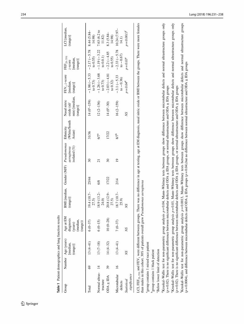
234 Lung (2018) 196:231–238
1 3
Tabl
e 1
Pat
ient
dem
ogra
phic
s and
lung
func
tion
resu
lts
LCI,
FEF 2
5–75
% a
nd F
EV1 w
ere
diffe
rent
bet
wee
n gr
oups
. The
re w
as n
o di
ffere
nce
in a
ge a
t tes
ting,
age
at E
M d
iagn
osis
, nas
al n
itric
oxi
de o
r BM
I bet
wee
n th
e gr
oups
. The
re w
ere
mor
e fe
mal
es
than
mal
es in
this
coh
ort.
30%
of p
atie
nts o
vera
ll gr
ew P
seud
omon
as a
erug
inos
aa gr
oup
cont
ains
1 m
ixed
race
pat
ient
b grou
p co
ntai
ns 1
bla
ck p
atie
ntc B
elow
low
er li
mit
of d
etec
tion
d Kru
skal
–Wal
lis te
st fo
r no
n-pa
ram
etric
gro
up a
naly
sis
p = 0.
04, M
ann–
Whi
tney
tests
bet
wee
n gr
oups
sho
w d
iffer
ence
bet
wee
n m
icro
tubu
lar
defe
cts
and
norm
al u
ltras
truct
ure
grou
ps o
nly
(p =
0.02
). Th
ere
is n
o si
gnifi
cant
diff
eren
ce b
etw
een
mic
rotu
bula
r def
ects
and
OD
A ±
IDA
gro
ups,
or n
orm
al u
ltras
truct
ure
and
OD
A ±
IDA
gro
ups
e Kru
skal
–Wal
lis te
st fo
r no
n-pa
ram
etric
gro
up a
naly
sis
p = 0.
03, M
ann–
Whi
tney
tests
bet
wee
n gr
oups
sho
w d
iffer
ence
bet
wee
n m
icro
tubu
lar
defe
cts
and
norm
al u
ltras
truct
ure
grou
ps o
nly
(p =
0.02
). Th
ere
is n
o si
gnifi
cant
diff
eren
ce b
etw
een
mic
rotu
bula
r def
ects
and
OD
A ±
IDA
gro
ups,
or n
orm
al u
ltras
truct
ure
and
OD
A ±
IDA
gro
ups
f Kru
skal
–Wal
lis t
est
for
non-
para
met
ric g
roup
ana
lysi
s p =
0.00
13,
Man
n–W
hitn
ey t
ests
bet
wee
n gr
oups
sho
w d
iffer
ence
bet
wee
n m
icro
tubu
lar
defe
cts
and
norm
al u
ltras
truct
ure
grou
ps
(p =
0.00
04),
and
diffe
renc
e be
twee
n m
icro
tubu
lar d
efec
ts a
nd O
DA
± ID
A g
roup
s (p =
0.04
),but
no
diffe
renc
e be
twee
n no
rmal
ultr
astru
ctur
e an
d O
DA
± ID
A g
roup
s
Gro
upN
umbe
rA
ge (y
ears
) [m
edia
n,
(ran
ge)]
Age
at E
M
diag
nosi
s (y
ears
) (m
edia
n,
(ran
ge))
BM
I [m
edia
n,
(ran
ge)]
Gen
der (
M/F
)Ps
eudo
mon
as
aeru
gino
sa
isol
ated
(%)
Ethn
icity
(W
hite
/Sou
th
Asi
an)
Nas
al n
itric
ox
ide
(nl/
min
) [m
edia
n,
(ran
ge)]
FEV
1 z-s
core
[m
edia
n,
(ran
ge)]
FEF 2
5–75
%
z-sc
ore
[med
ian,
(r
ange
)]
LCI [
med
ian,
(r
ange
)]
Tota
l69
13 (4
–41)
6 (0
–37)
19.4
(10.
7–27
.5)
25/4
430
31/3
614
(0c –1
59)
− 1.
98 (−
5.33
to
0.7
3)−
2.17
(− 5.
78
to 0
.55)
8.44
(5.8
4–14
.98)
Nor
mal
ultr
as-
truct
ure
1413
(7–1
8)6
(0–1
5)18
.9 (1
3.2–
24.0
)6/
821
6/7a
12 (2
–136
)−
1.26
(− 3.
68
to 0
.73)
− 1.
19 (−
3.22
to
0.0
.1)
7.63
(6.2
to
11.8
2)O
DA
± ID
A39
14 (4
–32)
10 (0
–28)
20.8
(12.
9–27
.5)
17/2
230
17/2
214
(0c –3
0)−
2.03
(− 4.
81
to 0
.32)
− 2.
2 (−
4.85
to
0.5
5)8.
3 (5
.84–
14.9
8)M
icro
tubu
lar
defe
cts
1613
(4–4
1)7
(0–3
7)18
.7 (1
0.7–
25.9
)2/
1419
8/7b
16 (2
–159
)−
3.1
(− 5.
33
to −
0.36
)−
2.33
(− 5.
78
to −
0.67
)10
.24
(7.9
7–14
.1)
Stat
istic
al
sign
ifica
nce
NS
NS
NS
NS
p = 0.
04d
p = 0.
03e
p = 0.
0013
f
235Lung (2018) 196:231–238
1 3
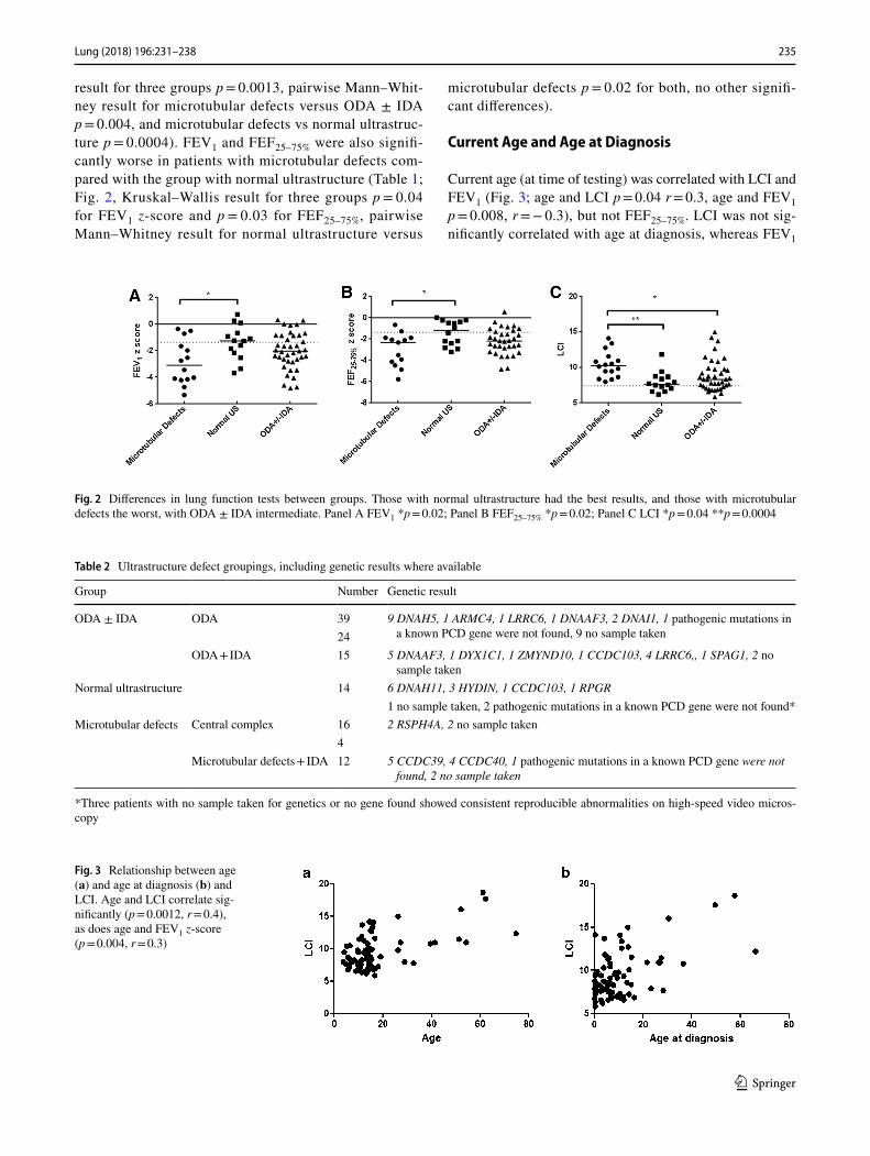
result for three groups p = 0.0013, pairwise Mann–Whit-ney result for microtubular defects versus ODA ± IDA p = 0.004, and microtubular defects vs normal ultrastruc-ture p = 0.0004). FEV1 and FEF25–75% were also signifi-cantly worse in patients with microtubular defects com-pared with the group with normal ultrastructure (Table 1; Fig. 2, Kruskal–Wallis result for three groups p = 0.04 for FEV1 z-score and p = 0.03 for FEF25–75%, pairwise Mann–Whitney result for normal ultrastructure versus
microtubular defects p = 0.02 for both, no other signifi-cant differences).
Current Age and Age at Diagnosis
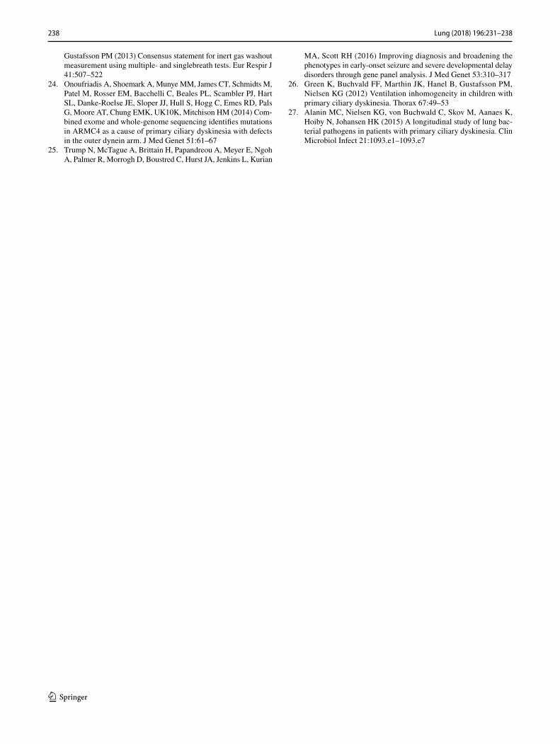
Current age (at time of testing) was correlated with LCI and FEV1 (Fig. 3; age and LCI p = 0.04 r = 0.3, age and FEV1 p = 0.008, r = − 0.3), but not FEF25–75%. LCI was not sig-nificantly correlated with age at diagnosis, whereas FEV1
Fig. 2 Differences in lung function tests between groups. Those with normal ultrastructure had the best results, and those with microtubular defects the worst, with ODA ± IDA intermediate. Panel A FEV1 *p = 0.02; Panel B FEF25–75% *p = 0.02; Panel C LCI *p = 0.04 **p = 0.0004
Table 2 Ultrastructure defect groupings, including genetic results where available
*Three patients with no sample taken for genetics or no gene found showed consistent reproducible abnormalities on high-speed video micros-copy
Group Number Genetic result
ODA ± IDA ODA 39 9 DNAH5, 1 ARMC4, 1 LRRC6, 1 DNAAF3, 2 DNAI1, 1 pathogenic mutations in a known PCD gene were not found, 9 no sample taken24
ODA + IDA 15 5 DNAAF3, 1 DYX1C1, 1 ZMYND10, 1 CCDC103, 4 LRRC6,, 1 SPAG1, 2 no sample taken
Normal ultrastructure 14 6 DNAH11, 3 HYDIN, 1 CCDC103, 1 RPGR1 no sample taken, 2 pathogenic mutations in a known PCD gene were not found*
Microtubular defects Central complex 16 2 RSPH4A, 2 no sample taken4
Microtubular defects + IDA 12 5 CCDC39, 4 CCDC40, 1 pathogenic mutations in a known PCD gene were not found, 2 no sample taken
Fig. 3 Relationship between age (a) and age at diagnosis (b) and LCI. Age and LCI correlate sig-nificantly (p = 0.0012, r = 0.4), as does age and FEV1 z-score (p = 0.004, r = 0.3)

236 Lung (2018) 196:231–238
1 3
was inversely correlated (p = 0.02, r = − 0.4). It was possible that younger patients were diagnosed earlier, as awareness of PCD has improved, thus introducing a bias, but there was no change in age at diagnosis over time (results in OLS).
Ethnicity
Ethnic groups were equally distributed between EM groups (Table 1) and there is no statistically significant difference between LCI, FEV1, FEF25–75%, nasal nitric oxide, age or age at diagnosis between them.
Ciliary Function
45 patients had predominantly static cilia on light micros-copy, and 21 had predominantly dyskinetic. There was no difference in LCI, FEV1 and FEF25–75% between these groups (data in OLS). Where beat frequency was measurable with non-static cilia, there was no relationship between beat fre-quency and lung function (see OLS for full data). There was no relationship between nasal nitric oxide results and lung function (see OLS).
Discussion
We have shown that PCD patients with microtubular defects have worse LCI compared to groups with normal ultrastruc-ture and dynein arm defects. Microtubular defects were first linked to comparatively worse lung function and decline in previous spirometry-based studies [13, 16], but these dif-ferences in LCI are novel, showing the utility of this test as a sensitive tool to investigate lung function differences in PCD. This study supports the hypothesis that there are potential outcome differences between different ultrastruc-tural groups.
Unlike in previous work, there was no significant dif-ference in FEV1 and FEF25–75% between ODA ± IDA and microtubular defects [13]. This may be because the previ-ous article used percent predicted, whereas we have used z-scores. Furthermore, the Wilcoxon signed-rank test for comparison between groups was used, whilst here we used a more stringent approach [16]. However, more data are needed to understand this discrepancy.
The mechanism by which different ultrastructural defects may give rise to differences in lung function in vivo is not clear. Ciliary motility differs between defects which may affect mucociliary transport, but why microtubular defect patients with some ciliary motility (such as the circling pattern seen in transposition defects) should have worse lung function than ODA ± IDA patients with completely static cilia is not known. Cilia also have a role in cell sig-nalling, and we speculate that this could differ in different
ultrastructural groups. In our centre, treatment decisions are based on clinical assessment rather than ultrastructure defect, so it is unlikely any differences arise from treatment differences.
One weakness of this study is the small cohort size. Until very recently, there was no commercially available validated LCI equipment, and measurements were restricted to tertiary centres, and so very large multicentre cohort studies (such as those published in spirometry) were not possible. Recent advances in equipment mean this will change in the com-ing years, and so establishing the role of this test in these patients is important.
Green et al. [26] found no significant correlation between age or age at diagnosis and lung function in their 2012 PCD cohort. In our cohort we found a correlation between age and LCI and FEV1. This could have been due to a number of factors. Our cohort is larger so may have been better pow-ered to detect this difference, and also Green et al’s cohort contained no patients over the age of 18 years, so the effect may be less pronounced in paediatric patients. In this study, we have used age at EM diagnosis as a surrogate measure for age at diagnosis, due to incomplete and varied history of our patients prior to their referral to the PCD centre. This is an acknowledged compromise and may be why no significant association was found.
As many patients do not solely attend our clinic, the microbiological results are incomplete. However, the preva-lence of Pseudomonas aeruginosa seen here (30%) is similar to previous reports; a large multicentre study gives a preva-lence of 37% overall [11], a large adult patient cohort 45% [16] and another showed a varying prevalence in different calendar years of between 15 and 47% [27].
In our study, median BMI for all ultrastructure groups was in the healthy range (Table 1). This differs from the previous study [13], where microtubular defects patients had a lower BMI than the other groups. The exact reason for this discrepancy is unclear and may relate to differences in management protocols between centres, but excludes the possibility that low BMI was the cause of poorer lung func-tion results in our microtubular defects patients.
In summary, in PCD, microtubular defects ultrastruc-ture group and older age are associated with worse LCI. We cannot explain this intriguing finding, and more work is needed to address the pathways whereby different ultrastruc-ture affects patient outcomes. In the future, determining the reasons for these differences could open up new treatment pathways, and delineation of high risk groups may allow more targeted intensive treatment to improve prognosis.
Acknowledgements We are very grateful to the families with PCD who participated in this study and to the UK PCD Family Support Group for their support. We would like to thank Mr Winston Banya for his help with statistical analysis, and the NE Thames Regional Genetics Service at Great Ormond Street Hospital for support in

237Lung (2018) 196:231–238
1 3
the genetic analysis. The research is supported by the COST Action 1407 network (BEAT-PCD: Better Evidence to Advance Therapeutic Options for PCD). A.B. was supported by the NIHR Respiratory Dis-ease Biomedical Research Unit at the Royal Brompton and Harefield NHS Foundation Trust and Imperial College London. Work by A.S. is an independent research funded by a postdoctoral research fellowship from the National Institute of Health Research and Health Education England, and the views expressed in this publication are those of the authors and not necessarily those of the NHS, the National Institute of Health Research or the Department of Health. H.M.M. acknowledges support from the National Institute for Health Research Biomedical Research Centre at Great Ormond Street Hospital for Children NHS Foundation Trust and University College London and was supported by Grants from Action Medical Research (GN2101), Newlife Foundation (10-11/15) and the Great Ormond Street Hospital Children’s Char-ity. M.L received funding from Bayer Pharmaceuticals (Berlin) for an investigator lead research grant in lung clearance index.
Compliance with Ethical Standards
Conflict of interest SI, MD, MRF, EF, JH, KK, SO, AO, MPP, JS, SC, HMM, MRL, CH, AS and AB have no conflicts of interest to declare.
Open Access This article is distributed under the terms of the Creative Commons Attribution 4.0 International License (http://creativecom-mons.org/licenses/by/4.0/), which permits unrestricted use, distribu-tion, and reproduction in any medium, provided you give appropriate credit to the original author(s) and the source, provide a link to the Creative Commons license, and indicate if changes were made.
References
1. Lucas JS, Burgess A, Mitchison HM, Moya E, Williamson M, Hogg C, Service UKNPCD (2014) Diagnosis and management of primary ciliary dyskinesia. Arch Dis Child 99:850–856
2. Noone PG, Leigh MW, Sannuti A, Minnix SL, Carson JL, Hazu-cha M, Zariwala MA, Knowles MR (2004) Primary ciliary dyski-nesia: diagnostic and phenotypic features. Am J Respir Crit Care Med 169:459–467
3. Bush A, Chodhari R, Collins N, Copeland F, Hall P, Harcourt J, Hariri M, Hogg C, Lucas J, Mitchison HM, O’Callaghan C, Phil-lips G (2007) Primary ciliary dyskinesia: current state of the art. Arch Dis Child 92:1136–1140
4. Bush A, Hogg C (2012) Primary ciliary dyskinesia: recent advances in epidemiology, diagnosis, management and relation-ship with the expanding spectrum of ciliopathy. Expert Rev Respir Med 6:663–682
5. Dehlink E, Hogg C, Carr SB, Bush A (2016) Clinical phenotype and current diagnostic criteria for primary ciliary dyskinesia. Expert Rev Respir Med 10:1163–1175
6. Oda T, Yanagisawa H, Kamiya R, Kikkawa M (2014) A molecular ruler determines the repeat length in eukaryotic cilia and flagella. Science 346:857–860
7. Shoemark A, Dixon M, Corrin B, Dewar A (2012) Twenty-year review of quantitative transmission electron microscopy for the diagnosis of primary ciliary dyskinesia. J Clin Pathol 65:267–271
8. Knowles MR, Daniels LA, Davis SD, Zariwala MA, Leigh MW (2013) Primary ciliary dyskinesia. Recent advances in diagnostics, genetics, and characterization of clinical disease. Am J Respir Crit Care Med 188:913–922
9. Lucas JS, Barbato A, Collins SA, Goutaki M, Behan L, Caudri D, Dell S, Eber E, Escudier E, Hirst RA, Hogg C, Jorissen M,
Latzin P, Legendre M, Leigh MW, Midulla F, Nielsen KG, Omran H, Papon J-F, Pohunek P, Redfern B, Rigau D, Rindlisbacher B, Santamaria F, Shoemark A, Snijders D, Tonia T, Titieni A, Walker WT, Werner C et al (2017) European respiratory society guide-lines for the diagnosis of primary ciliary dyskinesia. Eur Respir J 49. https://doi.org/10.1183/13993003.01090-2016
10. Marthin JK, Petersen N, Skovgaard LT, Nielsen KG (2010) Lung function in patients with primary ciliary dyskinesia: a cross-sec-tional and 3-decade longitudinal study. Am J Respir Crit Care Med 181:1262–1268
11. Maglione M, Bush A, Nielsen KG, Hogg C, Montella S, Marthin JK, Di Giorgio A, Santamaria F (2014) Multicenter analysis of body mass index, lung function, and sputum microbiology in pri-mary ciliary dyskinesia. Pediatr Pulmonol 49:1243–1250
12. Ellemunter H, Fuchs SI, Unsinn KM, Freund MC, Waltner-Romen M, Steinkamp G, Gappa M (2010) Sensitivity of lung clearance index and chest computed tomography in early cf lung disease. Respir Med 104:1834–1842
13. Davis SD, Ferkol TW, Rosenfeld M, Lee HS, Dell SD, Sagel SD, Milla C, Zariwala MA, Pittman JE, Shapiro AJ, Carson JL, Krischer JP, Hazucha MJ, Cooper ML, Knowles MR, Leigh MW (2015) Clinical features of childhood primary ciliary dyskinesia by genotype and ultrastructural phenotype. Am J Respir Crit Care Med 191:316–324
14. Ellerman A, Bisgaard H (1997) Longitudinal study of lung function in a cohort of primary ciliary dyskinesia. Eur Respir J 10:2376–2379
15. Vallet C, Escudier E, Roudot-Thoraval F, Blanchon S, Fauroux B, Beydon N, Boule M, Vojtek AM, Amselem S, Clement A, Tama-let A (2013) Primary ciliary dyskinesia presentation in 60 children according to ciliary ultrastructure. Eur J Pediatr 172:1053–1060
16. Shah A, Shoemark A, MacNeill SJ, Bhaludin B, Rogers A, Bilton D, Hansell DM, Wilson R, Loebinger MR (2016) A longitudinal study characterising a large adult primary ciliary dyskinesia popu-lation. Eur Respir J 48:441–450
17. Boon M, Vermeulen FL, Gysemans W, Proesmans M, Jorissen M, De Boeck K (2015) Lung structure-function correlation in patients with primary ciliary dyskinesia. Thorax 70:339–345
18. Horsley AR, Gustafsson PM, Macleod KA, Saunders C, Greening AP, Porteous DJ, Davies JC, Cunningham S, Alton EW, Innes JA (2008) Lung clearance index is a sensitive, repeatable and practical measure of airways disease in adults with cystic fibrosis. Thorax 63:135–140
19. Aurora P, Gustafsson P, Bush A, Lindblad A, Oliver C, Wallis CE, Stocks J (2004) Multiple breath inert gas washout as a measure of ventilation distribution in children with cystic fibrosis. Thorax 59:1068–1073
20. Irving SJ, Ives A, Davies G, Donovan J, Edey AJ, Gill SS, Nair A, Saunders C, Wijesekera NT, Alton EW, Hansell D, Hogg C, Davies JC, Bush A (2013) Lung clearance index and high-resolu-tion computed tomography scores in primary ciliary dyskinesia. Am J Respir Crit Care Med 188:545–549
21. Miller MR, Hankinson J, Brusasco V, Burgos F, Casaburi R, Coates A, Crapo R, Enright P, van der Grinten CP, Gustafsson P, Jensen R, Johnson DC, MacIntyre N, McKay R, Navajas D, Pedersen OF, Pellegrino R, Viegi G, Wanger J, Force AT (2005) Standardisation of spirometry. Eur Respir 26:319–338
22. Quanjer PH, Stanojevic S, Cole TJ, Baur X, Hall GL, Culver BH, Enright PL, Hankinson JL, Ip MS, Zheng J, Stocks J, Initiative ERSGLF (2012) Multi-ethnic reference values for spirometry for the 3-95-yr age range: the global lung function 2012 equations. Eur Respir J 40:1324–1343
23. Robinson PD, Latzin P, Verbanck S, Hall GL, Horsley A, Gappa M, Thamrin C, Arets HGM, Aurora P, Fuchs SI, King GG, Lum S, Macleod K, Paiva M, Pillow JJ, Ranganathan S, Ratjen F, Singer F, Sonnappa S, Stocks J, Subbarao P, Thompson BR,
238 Lung (2018) 196:231–238
1 3
Gustafsson PM (2013) Consensus statement for inert gas washout measurement using multiple- and singlebreath tests. Eur Respir J 41:507–522
24. Onoufriadis A, Shoemark A, Munye MM, James CT, Schmidts M, Patel M, Rosser EM, Bacchelli C, Beales PL, Scambler PJ, Hart SL, Danke-Roelse JE, Sloper JJ, Hull S, Hogg C, Emes RD, Pals G, Moore AT, Chung EMK, UK10K, Mitchison HM (2014) Com-bined exome and whole-genome sequencing identifies mutations in ARMC4 as a cause of primary ciliary dyskinesia with defects in the outer dynein arm. J Med Genet 51:61–67
25. Trump N, McTague A, Brittain H, Papandreou A, Meyer E, Ngoh A, Palmer R, Morrogh D, Boustred C, Hurst JA, Jenkins L, Kurian
MA, Scott RH (2016) Improving diagnosis and broadening the phenotypes in early-onset seizure and severe developmental delay disorders through gene panel analysis. J Med Genet 53:310–317
26. Green K, Buchvald FF, Marthin JK, Hanel B, Gustafsson PM, Nielsen KG (2012) Ventilation inhomogeneity in children with primary ciliary dyskinesia. Thorax 67:49–53
27. Alanin MC, Nielsen KG, von Buchwald C, Skov M, Aanaes K, Hoiby N, Johansen HK (2015) A longitudinal study of lung bac-terial pathogens in patients with primary ciliary dyskinesia. Clin Microbiol Infect 21:1093.e1–1093.e7


![Spirometric indices in primary ciliary dyskinesia ... · Primary ciliary dyskinesia (PCD) is a genetic, heterogeneous disease caused by absence and/or dysfunction of cilia [1]. Its](https://img.pdfslide.us/doc/110x75/602d6adf39b4c9715972d1d7/spirometric-indices-in-primary-ciliary-dyskinesia-primary-ciliary-dyskinesia.jpg)