Embed Size (px)
Citation preview
CYTOBIOLOGIE European Journal of Cell Biology 15, 285-302 (1977) © WissenschJftliche Verlagsges. mbH . Stuttgart
Identification of microtubular structures in diverse plant and animal cells by immunological cross-reaction revealed in
immunofluorescence microscopy using antibody against tubulin from porcine brain
Darstellung von Mikrotubuli-Strukturen in verschiedenen Pflanzen- und Tierzellen durch immunologische Kreuzreaktion
mit Hilfe der Immunfluoreszenzmikroskopie
KLAUS WEBER 1), MARY OSBORN
Max-Planck-Institute for Biophysical Chemistry, Gottingen
WERNER W. FRANKE, ERINITA SEIB, ULRICH SCHEER
Division for Membrane Biology and Biochemistry, Institute of Experimental Pathology, German Cancer Research Center, Heidelberg
and
WERNER HERTH
Lehrstuhl fur Zellenlehre, University of Heidelberg, Heidelberg
Received December 27, 1976
In revised form May 9, 1977
Abstract
Microtubules - immunofluorescence - evolution - antibody - sperm Antibody against tubulin from porcine brain was used to evaluate the immunological cross
reactivity of tubulin from a variety of animal and plant cells. Indirect immunofluorescence microscopy revealed microtubule-containing structures including cytoplasmic microtubules, spindle microtubules, cilia and fIagella. Thus tubulin from diverse species of both mammals and plants show immunological cross-reactivity with tubulin from porcine brain. Results obtained by immunofluorescence microscopy are whenever possible compared with previously known ultrastructural results obtained by electron microscopy.
1) Prof. DR. KLAUS WEBER, Max-Planck-Institut fUr Biophysikalische Chemie, Am FaRberg, D-3400 Goningen/Germany.
286 K. WEBER, M. OSBORN, W. W. FRANKE, E. SEW, U. SCHEER, and W. HERTH
Antibody against tubuJin 287
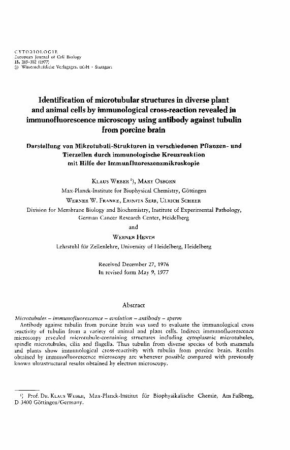
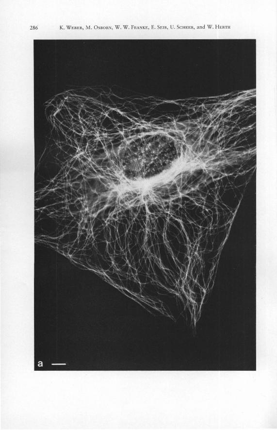
Figs. 1 a to h. Demonstration of tubulin contall1l11g structures in mammalian cells grown in culture (c to f present cells of the rat line RMCD, a, b, g and h show examples of mouse 3 T 3 cell s) by immunofluorescence microscopy with antibody to porcine brain tubulin. a shows cytoplasmic microrubules in an interphase cell. Most microtubules radiate from a juxtanuclear focus in the perinuclear region (cytocenter). The arrows in b denote the margins of the mitotic cell during meta- to early anaphase. Note the absence of cytoplasmic fluorescence outside the spindle. c to f show the concentration of the cellular tubulin in the mitotic apparatus (c, d meta to early anaphase; elate anaphase-to-telophase; f telophase). Note the positive reaction: chromosomal fibers (c, d ), continuous (pole-to-pole; d, e ) spindle fibers; polar Scales 10 f.lm. - a 1500 X . - b 1200 X . - c 1350 X . - d 1600 X . - e, f 1500 X . - g 1100 X . -caps (e), and region of the bridge between the daughter cells, the mid-body (arrows in f to h ). -h 2500 X .
In trod uction
Microtubules are fibrous structures unique to and characteristic of eukaryotic cells. The major protein constituent of microtubular structures is the globular tubulin molecule which consists of two similar but not identical polypeptides of an approximate molecular weight of 55000 [4,9, 18, 19,23,27,32,35 to 38, 42, 43, 48, 62].
Immunological studies have indicated that the tubulin contained in cytoplasmic microtubules as well as in various types of more complex microtubular arrays such as cilia, flagella, axopodia, centrioles, cytasters, mitotic apparatus, mid bodies, and the vinca alkaloid-induced paracrystals share at least one antigenic determinant, resulting in immunological cross-reaction within a species, with antibodies to a given isolated tubulin ([2, 6, 7, 22, 23, 44, 52, 56 to 59]; for earlier work using antibodies to isolated
288 K. WEBER, M. OSBORN, W. W. FRANKE, E. SEIB, U. SCHEER, and W. HERTH
vinblastine-induced paracrystals see [11, 12, 41]). Immunological procedures have also suggested that tubulins from different species also share antigenic determinants ([2, 6 to 8, 14,20, 22, 23, 44, 49, 56 to 59] ; for reports on differences between different echinoderms see [23]). Such immunological similarities have been noted even among evolutionarily quite distant species. This cross-species similarity of tubulin is supported by biochemical studies including the determination of partial amino acid sequences of tubulins from different sources [35,37,38,43,53].
In the present study we confirm and extend the observations on the conservation of antigenic determinants of tubulin from different sources and demonstrate that different microtubular structures from a variety of plant and animal cell s bind an antibody to porcine brain tubulin.
Materials and methods
Cells
(a) M o use 3 T 3 cells were grown as previously described [30, 44] . (b) Cells of an established line originally derived from a n experimenta ll y induced rat mam
mary carcinoma (RMCD cells) were grown and a ll owed to attach to cover slips as o utlined elsewhere [46,51,60].
(c) Bull sperm was obtained from bulls (4, 6 and 12 years o ld ) of two breeds ("Canadian Holstein-Friesian" or "Nordbadisches Fleckvieh" ) as fresh ejaculates which were stored in phosphate buffered saline (PBS; 0.14 M NaCI, 2.7 mM KCl, 8 mM Na2HP04, 1.5 mM KH2P04). Bull testes were obtained from the local slaughterho use within 20 minutes post mortern.
Epididymal or testicul ar sperm was collected with a pipette after dissection of the specific tissue in PBS.
(d ) Human sperm was o bta ined as froze n preparations or fresh ejacul ates (in PBS) from 2.5 to 40 year old volunteers of demonstra ted fertility.
(e) Rat sperm was o bta ined either fro m shock ejaculates (after throttling) or, more frequently, as testicul ar or epididymal sperm aspira ted with a pipette from th e tes ti cu lar or epididymal fluids after di ssection of the ti ssues in PBS.
(f) Di ssociated ciliated epitheli al cells were obtained from scrapings (in PBS) of the nose epithelium of Sprague-Dawley rats of bo th sexes (details in [51]).
(g) Newt sperm was obtained from three species (Tr iturus alpestris, Triturus cristatus, and Pleurodeles waltli; cf. [51]). Testes were dissected in PBS, a nd liberated sperm suspensions were collected with a pipette.
(h) Spermatozoa of the sea urchin , Echinus esculentus, were kindly provided by DR. PETZELT (Germa n Cancer Resea rch Center). Sperm suspensions in sea water were obtained after injection of 0.5 M KCI into the bod y cavity of the a nim als via the " peri stomal membrane" (cf. [24]).
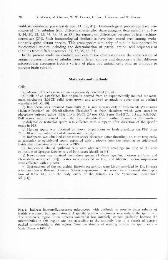
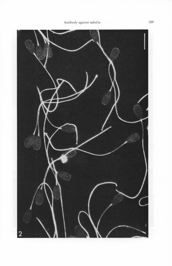
Fig. 2. Indirect immunofluorescence microscopy with an tibody to porcine brain tubulin of freshly ejaculated bull sperma tozoa. A specific positive reaction is seen only in the sperm tail. The mid-piece region often appears somewhat less intensely stained, probably because the microtubules in this region are less accessi ble to the an tibody due to a shea th of densely packed mitochondria in this region. Note the a bsence of sta ining o utside the sperm tails. -Scale 10 ~ml. - 1400 X .
Antibody against tubulin 289
290 K. WEBER, M. OSBORN, W. W. FRANKE, E. SEIR, U. SCHEER, and W. HERTH
(i) Cultures of the ciliate Tetrahymena pyriformis, a micro-nucleate strain GL, were obtained as previously described [15] and were used at concentration of 15 000 cells per m!. Cell suspensions were washed for 5 to 10 minutes in PBS and were collected by centrifugation at approximately 500 g for 5 to 10 minutes prior to fixation in formaldehyde. Crude fractions enriched in cilia fragments were obtained as described in detail elsewhere [51] and were washed after resuspension in PBS by centrifugation at 800 g, prior to the application to cover slips (see below).
Antibodies and antisera
The monospecific antibody against 6 S tubulin from pig brain has been described previously [57, 61]. This antibody preparation was made monospecific by subjecting the IgG fraction of the rabbit serum to affinity chromatography on tubulin covalently bound to Sepharose 4 B (Pharmacia, Uppsala, Sweden; cf. [61]). It has also been shown that expression of structures decorated by this antibody in animal cells is sensitive to mitotic drugs and low temperature [44, 57, 61]. Controls used included (a) the omission of the first antibody, (b) the replacement of the tubulin antibody by a serum containing antibodies to actin, (c) the replacement of the tubulin antibody by an IgG fraction from incompetent rabbit serum at concentrations equal to, or ten times higher, than the concentration of the monospecific tubulin antibody used, (d) the replacement of the tubulin antibody by complete incompetent rabbit serum (cf. [51]).
Indirect immunofluorescence microscopy
The procedures used for indirect immunofluorescence microscopy of cells grown on cover slips have been described in detail elsewhere [51, 56, 57, 58, 59]. Cells in suspension were placed on a round cover slip in a small drop of PBS or sea water, allowed to attach briefly to the glass (avoiding drying), and were then fixed with 3.7 Ofo formaldehyde in PBS. In some instances the objects were treated either in suspension or after attachment to the cover slip with a nonionic detergent (0.33 Ofo Triton X-lOO, in PBS containing 3 mM MgCI2 ) in order to enhance the disintegration of the plasma membrane. After 2 to 3 minutes of treatment the detergent was removed by repeated washing in PBS containing 3 mM MgCb and the specimens fixed with formaldehyde as described above. Antibodies against rabbit globulins labelled with fluorescein were purchased from Miles Co. (Elkhart, Indiana, USA) as 1 Ofo solution. This antibody preparation was diluted tenfold prior to use. Micrographs with epifluorescence and with phase contrast were made with the Zeiss microscope using the appropriate filter arrangements and sensitive films (for details see [51,56 to 59]).
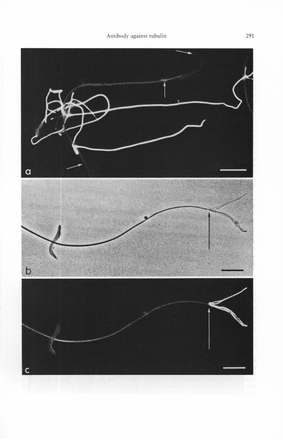
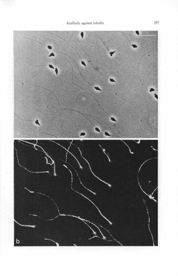
Figs. 3 a to c. Indirect immunofluorescence microscopy of rat sperm cells (epididymal sperm cells) with antibody to porcine brain tubulin as revealed by epifluorescence (a, c) and by phase contrast (b). The preparations shown here have been selected in order to demonstrate the heterogeneity of the reaction even among adjacent sperm tails. While the tubulin antibody is bound strongly by some sperm tails shown in a others react only weakly (for example, the sperm cell denoted by the arrows pointing, from top to bottom, to head, mid-piece, and mainpiece). - band c show the end piece of the same rat spermatozoon in phase contrast (b) and in fluorescence (c), illustrating the enhanced reaction of the sperm tail microtubules at the very end where the plasma membrane is ruptured and the microtubules are fraying and flattening out (transition point is indicated by the arrow). This demonstrates the importance of antigen accessibility in immunofluorescence microscopy of intracellular structures. - Bars 10 ftm. - a 1300 X. - b, c 1100 X.
Antibody against tllblllin 291
292 K. WEBER, M. OSBORN, W. W. FRANKE, E. SEIB, U. SCHEER, and W. HERTH
Results
Mammalian cells grown in culture
Mammalian and avian cells grown as cell lines in vitro have been studied previously by immunofluorescence microscopy using three different preparations of tubulin antibody [6, 7, 22, 44, 56 to 59]. We show here examples of cytoplasmic microtubules in mouse 3 T 3 cells and of mitotic microtubules in 3 T 3 cells and other cells obtained with the mono specific antibody against porcine brain tubulin which is the antibody preparation which was used in our detailed studies on microtubules in a variety of animal and plant cells (see below).
Interphase mouse 3 T 3 cells are characterized by a well developed system of fragile cytoplasmic microtubules (Fig. 1 a). Many of these cytoplasmic microtubules are oriented in such a way that they radiate from a juxtanuclear center, obviously the centrosphere or cytocenter (for details see [44]). This complex system of fibers is rearranged with the onset of mitosis (for details see [6, 7, 56 to 58]) and already during pro-metaphase and metaphase one can clearly note the absence of cytoplasmic microtubules outside the mitotic spindle (Fig. 1 b). Centrioles are often recognizable and the typical perinuclear cytaster arrays located at the nuclear poles can be recognized in prophase (see also [7, 47, 56 to 58]). During the transition from pro-metaphase to telophase the tubulin becomes concentrated in the mitotic apparatus. Both chromosomal (pole-to-kinetochore) and interpolar (pole-to-pole) fibers are well stained and the polar regions also stain strongly (Figs. 1 c to f; see references quoted above). The "stembody" and the narrow cytoplast isthmus between the daughter cells, the "Flemming body", bind the antibodies very strongly (Fig. 1, g and h; see references quoted above). This agrees with electron microscopic studies showing that microtubules are very densely packed in these regions [10, 39]. The center of the cytokinetic bridge usually appears as a dark or very faintly stained band in the midbody (Fig. 1, g to h; see also [6, 7, 57, 58]). It is known from electron microscopical studies that microtubules run through the midbody and that they are cemented in a dense osmiophilic and sudanophilic matrix in this region [39]. The decreased staining of the midbody is probably due to the inability of the antibody to penetrate this dense matrix.
Mammalian sperm
Ejaculated spermatozoa from men, bulls (Fig. 2) and rats (Fig. 3) as well as epididymal and testicular spermatozoa from bovine and murine testes react strongly and specifically with the antibody against porcine brain tubulin. Staining of the main and distal portions of the tails is especially intense. The staining appears somewhat reduced in some sperm cells in the mid-piece (Fig. 2). The intensity of the fluorescence can differ among adjacent spermatozoa (Fig. 3 a), and even in different regions of the same sperm tail (Fig. 3 b, c). This obviously reflects local heterogeneities in the ability of the antibody
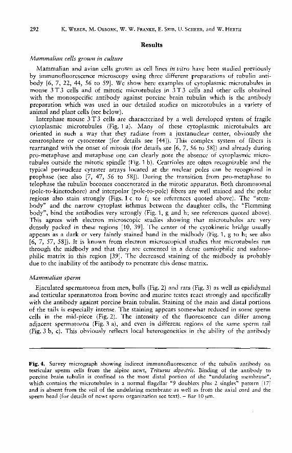
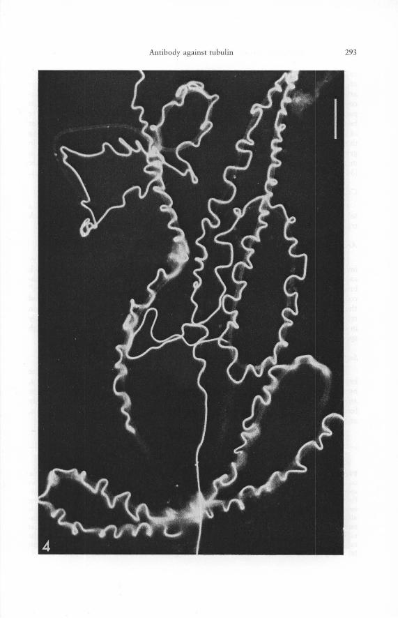
Fig. 4. Survey micrograph showing indirect immunofluorescence of the tubulin antibody on testicular sperm cells from the alpine newt, Triturus alpestris. Binding of the antibody to porcine brain tubulin is confined to the most distal portion of the "undulating membrane", which contains the microtubules in a normal flagellar "9 doublets plus 2 singles" pattern [17] and is absent from the veil of the undulating membrane as well as from the axial cord and the sperm head (for details of newt sperm organization see text). - Bar 10 [lm.
Antibody against tubulin 293
294 K. WEBER, M. OSBORN, W. W. FRANKE, E. SEIB, U. SCHEER, and W. HERTH
to penetrate the sperm tail probably due to differences in the degree of disintegration of the plasma membrane. This interpretation is strengthened by observations on the artificially induced frayed ends of some spermatozoa. These ends (for electron microscopic observations see [45, 63]) in which the plasma membrane has been at least partially solubilized, and in which outer doublets and/or individual microtubules have been liberated, react very strongly, even in tails which otherwise were negative for most of their length (Fig. 3 c). Up to seven individual fibers could be detected, suggesting that individual microtubules (or outer doublets) can be revealed by this procedure. In general, the treatment of the sperm cells with nonionic detergent was quite helpful in many experiments (for the effects of Triton X-lOO on the sperm ultrastructure see [34, 63]) in contrast to the cultured cells in which such pretreatment was not necessary.
Ciliated mammalian epithelia
Ciliated cells from rat nose epithelium (for details of preparation see [51]) showed a selective and positive staining of cilia, similar to that recently reported for cilia in chick tracheal epithelium [2].
Amphibian sperm
Amphibian sperm cells are especially interesting objects for the location of tubulin by immunofluorescence microscopy. They have a complex tail structure involving a thick axial cord and an "undulating membrane" which consists of a fold of the plasma membrane including some cytoplasmic material and a distally located "axonemal complex" containing a flagellar microtubule arrangement of 9 doublets and 2 single tubules and the marginal fiber (a review of the ultrastructure of amphibian sperm is given in ref. [17]). As shown in Figures 4 and 5 antibodies to porcine tubulin are bound only in the microtubule-containing region and not the thick axial cord, illustrating the high specificity of the method.
Sea urchin sperm
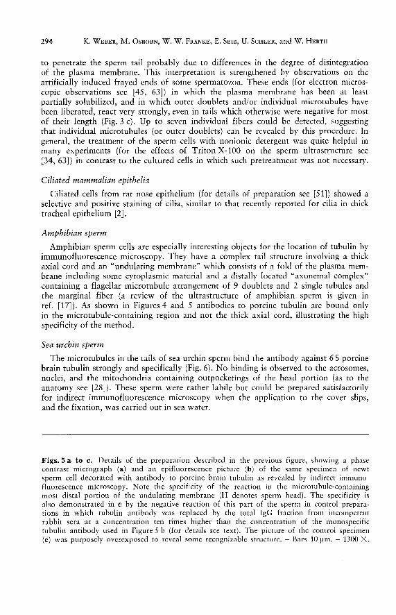
The microtubules in the tails of sea urchin sperm bind the antibody against 6 S porcine brain tubulin strongly and specifically (Fig. 6). No binding is observed to the acrosomes, nuclei, and the mitochondria containing outpocketings of the head portion (as to the anatomy see [28]). These sperm were rather labile but could be prepared satisfactorily for indirect immunofluorescence microscopy when the application to the cover slips, and the fixation, was carried out in sea water.
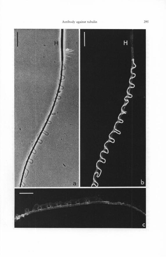
Figs. 5 a to c. Details of the preparation described in the previous figure, showing a phase contrast micrograph (a) and an epifluorescence picture (b) of the same specimen of newt sperm cell decorated with antibody to porcine brain tubulin as revealed by indirect immunofluorescence microscopy. Note the specificity of the reaction in the microtubule-containing most distal portion of the undulating membrane (H denotes sperm head). The specificity is also demonstrated in c by the negative reaction of this part of the sperm in control preparations in which tubulin antibody was replaced by the total IgG fraction from incompetent rabbit sera at a concentration ten times higher than the concentration of the monospecific tubulin antibody used in Figure 5 b (for details see text). The picture of the control specimen (c) was purposely overexposed to reveal some recognizable structure. - Bars 10 [tm. - 1300 X.
Antibody against tubulin 295
296 K. WEBER, M. OSBORN, W. W. FRANKE, E. SEIB, U. SCHEER, and W. HERTH
Tetrahymena pyriformis
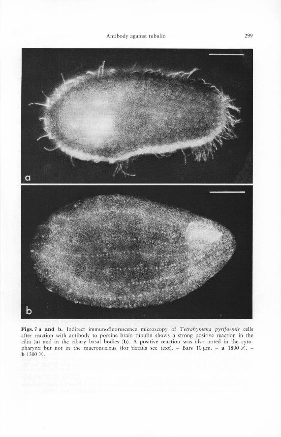
These ciliates are classic objects in the research on microtubules and tubulin. The antibody bound to the cilia (Fig. 7 a), the basal bodies (Fig. 7 b), and the mouthfield of these cells (Fig. 7 b). This correlates well with the abundance of microtubular structures in these cortical structures [1]. The strong positive reaction of the ciliary microtubules was also seen in fractions containing ciliary fragments which indicates the usefulness of this method for identification of specific components in cell fractions. The macronucleus of Tetrahymena pyriformis which has been shown to contain individual microtubules [16] did not stain significantly (cf. [51]). This could be due to the low concentration of microtubules in the macronucleus or to their inaccessibility to the antibody in the presence of the relatively large amount of cytoplasm and the pellicle. Our observation contrasts with an observation of faint and spotty immunofluorescence reaction in isolated macronuclei of Tetrahymena using antibodies made against ciliary tubulin from the same species [54].
Heliozoa and algae
The axopodia of Heliozoa (for review see [55]), including those of the common species Actinophrys sol are prominent microtubule-rich structures. Tu bulin antibody bound to these axopodia as well as to some spot-like regions in the peripheral cytoplasm. The axopodial staining was on occasion interrupted, possibly due to local breakdown of microtubular structures in the course of the preparation (for details see [51]).
As a representative of the green algae we chose the gametes of Acetabularia mediterranea (for details see [51]). We observed an intense staining of the two flagella and some indistinct fluorescence in the anterior portion of the algae. This agrees with the recent report [81 that the flagella and the rootlet system of the quadriflagellate green alga Polytomella agilis (Volvocales) bind antibodies to purified chicken brain tubulin.
The golden-green alga Poteriochromonas (syn. Ochromonas) is an especially well characterized alga both in terms of biochemical and ultrastructural studies [5]. As in the green algae, the tubulin antibody (cf. [51]) decorated the flagella, a portion of the cytoplasm and, in some cases, also the stalk, which is known to contain microtubules [50].
Mitosis of higher plant, Leucojum aestivum
The mitotic apparatus of the endosperm cells of this monocotyledonous plant showed an intense and specific binding of the antibody against porcine brain tubulin, and individual spindle fibers could be visualized by immunofluorescence microscopy (for details see [21]).
Figs. 6 a and b. Suspension of spermatozoa of the sea urchin, Echinus csculentus, stained in indirect immunofluorescence microscopy with antibody to porcine brain tubulin, as revealed with phase contrast optics (a) and in epifluorescence (b). Note that the binding of the tubulin antibody is confined to the sperm tails and is absent from acrosome, nucleus, and the mitochondria-containing portion of the sperm head. For details see text. - Bars 10 ~lm. -1300 X.
Antibody aga inst tllblllin 297
298 K. WEBER, M. OSBORN, W. W. FRANKE, E. SEIB, U. SCHEER, and W. HERTH
Discussion
Our data extend the previous indications of an extensive protein chemical conservation of tubulin(s) and of a cross-reactivity between the tubulin(s) contained in the microtubules of functionally different structures (see Introduction). We did not find significant differences in the cross-reaction between evolutionary distant species as detected by indirect immunofluorescence microscopy with an antibody against porcine brain tubulin. For example, the decoration of the mitotic apparatus of the plant endosperm examined with antibody against porcine brain tubulin was as strong as that of the mammalian mitotic apparatus (cf. also [21]). This antigenic similarity of tubulin(s) in different functional structures, and in different species, is also in agreement with experiments showing that porcine brain tubulin is incorporated into the meiotic spindle apparatus and asters after injection into the oocytes of an annelid worm, Chaetopterus [26]. The conservation of tubulin is also suggested from the experiments of HEIDEMANN and KIRSCHNER [25] who found that purified basal bodies of a green alga, Chlamydomonas reinhardi, and a ciliate, Tetrahymena pyriformis (after injection into the egg of a vertebrate animal, the amphibian Xenopus laevis) induce asters and cleavage furrows.
Differences in the intensity of staining of microtubular structures with antibody against a specific tubulin might not be a priori explained by true differences in antigenic determinants but can also reflect different degrees in the accessibility and preservation of the specific microtubular structure. The problem of accessibility is illustrated in this study by the difficulty in staining the mid-region of the sperm tail (Fig. 2), by the enhanced reaction of the "frayed ends" of the rat spermatozoon (Fig.3) and by the inability to stain the Flemming body in mitotic cells in tissue culture (Fig. 1 hand see [7, 56 to 59]). Electron microscopic examinations of the cell preparations used for indirect immunofluorescence (OSBORN, FRANKE, WEBER, and GRUND, manuscript in preparation) have indeed shown differences in the stability of microtubular structures: Microtubules of sperm tails and cilia, for example, were usually better preserved than cytoplasmic microtubules and microtubules of the spindle apparatus (cf. also [20]). However, even in cases in which the microtubules showed local interruptions and distortions in the electron microscope large parts of their tubulin is still present in situ and recognized by immunofluorescence, most probably due to the association with other, high molecular weight protein components of microtubules or the microtubular sheath [3, 13, 20, 29, 31, 33, 40]. However in general, as shown above, structures which are known to contain microtubules from electron microscopy stain with the antibody to tubulin. These structures include flagella, cilia and mitotic apparatuses. The visualization of individual outer doublets in the frayed ends of spermatozoa illustrates the resolution of the method and strengthens the idea [6, 56, 57, 59] that in tissue culture cells in interphase one can under favorable circumstances reveal the original display of individual cytoplasmic microtubules. Thus the antibody seems to provide a fast and relatively easy way to monitor tubulin containing structures in a wide variety of cells and organisms under different experimental conditions.
Acknowledgement. We thank H. J. KOITZSCH and T. BORN for expert technical assistance.
Antibody against tubulin 299
Figs. 7 a and b. Indirect immunofluorescence microscopy of Tetrahymena pyriformis cells after reaction with antibody to porcine brain tubulin shows a strong positive reaction in the ci lia (a) and in the ci li ary basal bodies (b ). A positive reaction was also noted in the cytopharynx but not in the macronucleus (for 'details see text) . - Bars 10 flm. - a 1800 X. -b 1300 X .
300 K. WEBER, M. OSBORN, W. W. FRANKE, E. SEIB, U. SCHEER, and W. HERTH
References
[1] ALLEN, R. D.: Fine structure, reconstruction and possible functions of components of the cortex of Tetrahymena pyriformis. J. Protozool. 14,553-565 (1967).
[2] AUBIN, J. E., L. SUBRAHMANYAN, V. I. KALNINS, and V. LING: Antisera against electronphoretically purified tubulin stimulate colchicine-binding activity. Proc. Nat. Acad. Sci. (USA) 73, 1246-1249 (1976).
[3] BEHNKE, 0.: Studies on isolated microtubules. Evidence for a clear space component. Cytobiologie 11, 366-381 (1975).
[4] BIBRING, TH., J. BAXANDALL, S. DENSLOW, and B. WALKER: Heterogeneity of the alpha subunit of tubulin within single organism. J. Cell BioI. 69,301-312 (1976).
[5] BOUCK, G. B., and D. L. BROWN: Microtubule biogenesis and cell shape in Ochromonas. I. The distribution of cytoplasmic and mitotic microtubules. J. Cell BioI. 56, 340-359 (1973).
[6] BRINKLEY, B. R., G. M. FULLER, and D. P. HIGHFIELD: Cytoplasmic microtubules in normal and transformed cells in culture: Analysis by tubulin antibody immunofluorescence. Proc. Nat. Acad. Sci. (USA) 72, 4981-4985 (1975).
[7] BRINKLEY, B. R., G. M. FULLER, and D. P. HIGHFIELD: Studies of microtubules in dividing and non-dividing mammalian cells using antibody to 6-S bovine brain tubulin. In: M. BORGERS and M. DE BRABANDER (eds.): Microtubules and Microtubule Inhibitors, pp. 297-312. NorthHolland Publishing Company, Amsterdam 1975.
[8] BROWN, D. L., A. MASSALSKI and R. PATENAUDE: Organization of the flagellar apparatus and associated cytoplasmic microtubules in the quadriflagellate alga Polytomella agilis. J. Cell BioI. 69, 106-125 (1976).
[9] BRYAN, J., and L. WILSON: Are cytoplasmic microtubules heteropolymers? Proc. Nat. Acad. Sci. (USA) 68, 1762-1766 (1971).
[10] BYERS, B., and D. H. ABRAMSON: Cytokinesis in HeLa: Posttelophase delay and mICrotubule-associated motility. Protoplasma 66, 413-435 (1968).
[11] DALES, S.: Concerning the universality of a microtubule antigen 111 animal cells. J. Cell BioI. 52, 748-754 (1972).
[12] DALES, S., K. C. Hsu, and A. NAGAYAMA: The fine structure and immunological labelling of the achromatic mitotic apparatus after disruption of cell membranes. J. Cell BioI. 59, 643-660 (1973).
[13] DENTLER, W. L., S. GRANETT, and J. L. ROSENllAUM: Ultrastructural localization of the high molecular weight proteins associated with in vitro-assembled brain microtubules. J. Cell BioI. 65, 237-241 (1975).
[14] DONGES, K. H., and E. ROTH: Serological similarity of microtubule proteins. Naturwissenschaften 59, 372 (1972).
[15] ECKERT, W. A., W. W. FRANKE, and U. SCHEER: Nucleocytoplasmic translocation of RNA in Tetrahymena pyriformis and its inhibition by actinomycin D and cycloheximide. Exp. Cell Res. 94, 31-46 (1975).
[16] FALK, H., F. WUNDERLICH, and W. W. FRANKE: Microtubular structures in macronuclei of synchronously dividing Tetrahymena pyriformis. J. Protozool. 15, 776-780 (1968).
[17] FAwcETT, D. W.: A comparative view of sperm ultrastructure. In: J. D. BIGGERS (ed.): Biology of Reproduction, pp. 13-28. Academic Press, New York - London 1970.
[18] FElT, H., L. SLUSAREK, and M. L. SHELANSKI: Heterogeneity of tubulin subunits. Proc. Nat. Acad. Sci. (USA) 68,2028-2031 (1971).
[19] FINE, R. E.: Heterogeneity of tubulin. Nature New BioI. 233, 283-284 (1971). [20] FORER, A., V. I. KALNINS, and A. M. ZIMMERMAN!,;: Spindle birefringence of isolated
mitotic apparatus: Further evidence for two birefringent spindle components. J. Cell Sci. 22, 115-131 (1976).
[21] FRANKE, W. W., E. SEIB, M.OSBORN, K. WEBER, W. HERTH, and H. FALK: Tubulincontaining structures in the anastral mitotic apparatus of endosperm cells of the plant Leuco;um aestivum as revealed by immunofluorescence microscopy. Cytobiologie 15, 24-48 (1977).
Antibody against tubulin 301
[22] FULLER, G. M., B. R. BRINKLEY, and J. M. BOUGHTER: Immunofluorescence of mitotic spindles by using monospecific antibody against bovine brain tubulin. Science 187, 948-950 (1975).
[23] FULTON, c., R. E. KANE, and R. E. STEPHENS: Serological similarity of flagellar and mitotic microtubules. J. Cell BioI. 50, 762-773 (1971).
[24] GUIDICE, G.: Developmental biology of the sea urchin embryo. Academic Press, New York 1973.
[25] HEIDEMANN, S. R., and M. W. KIRSCHNER: Aster formation in eggs of Xenopus laevis. Induction by isolated basal bodies. J. Cell BioI. 67, 105-117 (1975).
[26] INouE, S., G. G. BORISY, and D. P. KIEHART: Growth and lability of Chaetopterus oocyte mitotic spindles in the presence of porcine brain tubulin. J. Cell BioI. 62, 175-184 (1974).
[27] JACOBS, M., and MCVITTIE: Identification of the flagellar proteins of Chlamydomonas reinhardi. Exp. Cell Res. 63, 53-61 (1970).
[28] JESSEN, J., O. BEHNKE, K. G. WINGSTRAND, and J. ROSTGAARD: Actin-like filaments in the acrosomal apparatus of spermatozoa of a sea urchin. Exp. Cell Res. 80, 47-54 (1973).
[29] KEATES, R. A. B., and R. H. HALL: Tubulin requires an accessory protein for self assembly into microtubules. Nature 257, 418-420 (1975).
[30] KEENAN, T. W., E. SCHMID, W. W. FRANKE, and H. WIEGANDT: Exogeneous glycosphingolipids suppress growth rate of transformed and untransformed 3 T 3 mouse cells. Exp. Cell Res. 92, 259-270 (1975).
[31] KIRSCHNER, M. W., R. C. WILLIAMS, M. WEINGARTEN, and J. C. GERHARDT: Microtubules from mammalian brain: Some properties of their depolymerization products and a proposed mechanism of assembly and disassembly. Proc. Nat. Acad. Sci. 71, 1159-1163 (1974).
[32] LEE, J. c., R. P. FRIGON, and S. N. TIMASHEFF: The chemical characterization of calf brain microtubule protein subunits. J. BioI. Chem. 248, 7253-7262 (1973).
[33] LINCK, R. W.: Chemical and structural differences between cilia and flagella from the lamellibranch mollusc, Aequipecton irradians. J. Cell Sci. 12,951-981 (1973).
[34] LINDEMANN, C. B., and J. R. GIBBONS: Adenosin triphosphate-induced motility and sliding of filaments in mammalian sperm extracted with Triton X-lOO. J. Cell BioI. 65, 147-162 (1975).
[35] LUDUENA, R. F., T. PFEFFER, and D. MYLES: Comparison of tubulin from phylogenetically distant sources. J. Cell BioI. 70, 129 a (1976).
[36] LUDUENA, R. F., 1.. WILSON, and E. M. SHOOTER: Cross-linked tubulin. In: M. BORGERS and M. DE BRABANDER (eds.): Microtubules and Microtubule Inhibitors, pp. 47-58. NorthHolland Publishing Company, Amsterdam 1975.
[37] LUDUENA, R. F., and D. O. WOODWARD: Isolation and partial characterization of a- and ~-tubulin from outer doublets of sea-urchin sperm and microtubules of chick-embryo brain. Proc. Nat. Acad. Sci. (USA) 70,3594-3598 (1973).
[38] LUDUENA, R. F., and D. O. WOODWARD: a- and ~-tubulin: Separation and partial sequence analysis. Ann. N. Y. Acad. Sci. 253, 272-283 (1975).
[39] McINTOSH, J. R., and S. C. LANDIS: The distribution of spindle microtubules during mitosis in cultured human cells. J. Cell BioI. 49, 468-497 (1971).
[40] MURPHY, D. B., and G. G. BORISY: Association of high-molecular-weight proteins with microtubules and their role in microtubule assembly in vitro. Proc. Nat. Acad. Sci. (USA) 72, 2696-2700 (1975).
[41] NAGAYAMA, A., and S. DALES: Rapid purification and the immunological specificity of mammalian microtubular paracrystals possessing an ATP-ase activity. Proc. Nat. Acad. Sci. (USA) 66,464-471 (1970).
[42] OLMSTED, J. B., and G. G. BORISY: Microtubules. Ann. Rev. Biochem. 42, 507-540 (1973).
[43] OLMSTED, J. B., G. B. WITMAN, K. CARLSON, and J. 1.. ROSENBAUM: Comparison of the
302 K. WEBER, M. OSBORN, W. W. FRANKE, E. SEIB, U. SCIIEEH, and W. HERTH
microtubule proteins of neuroblastoma cells, brain, and Chlamydomonas flagella. Proe. Nat. Acad. Sci. (USA) 68, 2273-2277 (1971).
[44] OSBORN, M., and K. WEBER: Cytoplasmic micro tubules in tissue culture cells appear to grow from an organizing structure towards the plasma membrane. Proe. Nat. Acad. Sci. (USA) 73, 867-871 (1976).
[45] PEASE, D. c.: Histological techniques for electron microscopy. Academic Press, New York - London 1964.
[46] RATHKE, P. c., E. SCHMID, and W. W. FRANKE: The action of the cytochalasins at the subcellular level. I. Effects and binding of cytochalasin B in cells of a line derived from a rat mammary adenocarcinoma and in rat erythrocytes. Cytobiologie 10, 366-396 (1975).
[47] ROBBINS, E., G. JENTSCH, and A. MICALI: The centriole cycle in synchronized HeLa cells. J. Cell BioI. 36,329-339 (1968).
[48] ROBERTS, K.: Cytoplasmic microtubules and their function. In: A. J. V. BUTLER and D. NOBLE (eds.): Progress in Biophysics and Molecular Biology, Vol. 28, pp. 371-419. Pergamon Press, Oxford - New York 1974.
[49] SAMSON JR., F. E., M. F. RUSCHA, and S. L. TWOMEY: Antigenicity of brain microtubule protein. Brain Res. 28, 143-151 (1971).
[50] SCHNEPF, E., G. RODERER, and W. HERTH: The formation of the fibrils in the lorica of Poteriochromonas stipitata: Tip growth, kinetics, site, orientation. Planta 125,45-62 (1975).
[51] SEIB, E.: Lokalisierung von Actin und Tubulin mit der indirekten Immunfluoreszenz in Ausstrich- and Tropfenpraparation. Diploma Thesis, University of Koln, pp. 1-75 (1976).
[52] STARLING, D.: Two ultrastructurally distinct tubulin paracrystals induced in sea urchin eggs by vinblastine suI fate. J. Cell Sci. 20, 79-89 (1976).
[53] STEPHENS, R. E.: On the structural protein of flagellar outer fibers. J. Mol. BioI. 32, 277-283 (1968).
[54] TAMuRA, S.: Properties of microtubule protein in different organelles in Tetrahymena pyriformis. Exp. Cell Res. 68, 169-179 (1971).
[55] TILNEY, L. G.: Origin and continuity of microtubules. In: J. REINEHT and H. URSPRUNG (eds.): Origin and Continuity of Cell Organelles, pp. 222-260. Springer-Verlag, Berlin 1971.
[56] WEBER, K.: Specific visualization of tubulin containing structure by immunofluorescence microscopy: Cytoplasmic microtubules, vinblastine-induced paracrystals and mitotic figures. In: M. BORGERS and M. DE BRABANDER (eds.): Microtubules and Microtubule Inhibitors, pp. 313-325. North-Holland Publishing Company, Amsterdam 1975.
[57] WEBER, K.: Visualization of tubulin contammg structures by immunofluorescence microscopy. In: R. GOLDMAN, T. POLLARD, and J. ROSENBAUM (eds.): Cell Motility, pp. 403-417. Cold Spring Harbor Laboratory 1976.
[58] WEBER, K., TH. BIBRING, and M. OSBORN: Specific visualization of tubulin-containing structures in tissue culture cells by immunofluorescence. Cytoplasmic microtubules, vinblastineinduced para crystals, and mitotic figures. Exp. Cell Res. 95, 111-120 (1975).
[59] WEBER, K., R. POLLACK, and T. BIBRING: Antibody against tubuJin: The specific visualization of cytoplasmic microtubules in tissue culture cells. Proc. Nat. Acad. Sci. (USA) 72, 459-463 (1975).
[60] WEBER, K., P. C. RATHKE, M. OSBORN, and W. W. FRANKE: Distribution of actin and tubulin in cells and in glycerinated cell models after treatment with cytochalasin B. Exp. Cell Res. 102,285-297 (1976).
[61] WEBER, K., J. WEHLAND, and W. HERZOG: Griseofulvin interacts with microtubules both in vivo and in vitro. J. Mol. BioI. 102, 817-829 (1976).
[62] WITMAN, G. B., K. CARLSON, and J. L. ROSENBAUM: ChlamY'domonas flagella. n. The distribution of tubulins 1 and 2 in the outer doublet microtubules. J. Cell BioI. 54, 540-555 (1972).
[63] WOODING, F. B. P.: The effect of Triton X-lOO on the ultrastructure of ejaculated bovine sperm. J. Ultrastruct. Res. 42,502-516 (1973).


![Preferential Identification of Agonistic OX40 Antibodies ... · phage display [6,7]. Recently, we invented a novel method for screening millions-diverse antibody repertoires using](https://img.pdfslide.us/doc/110x75/5ea899f26dff2a7ae712e1bc/preferential-identification-of-agonistic-ox40-antibodies-phage-display-67.jpg)